Embed Size (px)
Citation preview
REVIEW ARTICLE
Skeletal Muscle Pathology in X-Linked MyotubularMyopathy: Review With Cross-Species Comparisons
Michael W. Lawlor, MD, PhD, Alan H. Beggs, PhD, Ana Buj-Bello, MD, PhD,Martin K. Childers, DO, PhD, James J. Dowling, MD, PhD, Emma S. James, PhD,
Hui Meng, MD, PhD, Steven A. Moore, MD, PhD, Suyash Prasad, MBBS, MRCP, MRCPCH FFPM,Benedikt Schoser, MD, and Caroline A. Sewry, PhD
AbstractX-linked myotubular myopathy (XLMTM) is a devastating, rare,
congenital myopathy caused by mutations in the MTM1 gene, result-
ing in a lack of or dysfunction of the enzyme myotubularin. This
leads to severe perinatal weakness and distinctive muscle pathology.
It was originally thought that XLMTM was related to developmental
arrest in myotube maturation; however, the generation and characteri-
zation of several animal models have significantly improved our un-
derstanding of clinical and pathological aspects of this disorder.
Myotubularin is now known to participate in numerous cellular pro-
cesses including endosomal trafficking, excitation-contraction cou-
pling, cytoskeletal organization, neuromuscular junction structure,
autophagy, and satellite cell proliferation and survival. The available
vertebrate models of XLMTM, which vary in severity from complete
absence to reduced functional levels of myotubularin, recapitulate
features of the human disease to a variable extent. Understanding
how pathological endpoints in animals with XLMTM translate to hu-
man patients will be essential to interpret preclinical treatment trials
and translate therapies into human clinical studies. This review sum-
marizes the published animal models of XLMTM, including those of
zebrafish, mice, and dogs, with a focus on their pathological features
as compared to those seen in human XLMTM patients.
Key Words: Congenital, Centronuclear, Hypotrophy, Myopathy,
Myotubular, Myotubularin, Sarcotubular.
INTRODUCTIONX-linked myotubular myopathy (XLMTM) is a rare, dev-
astating, congenital myopathy that mainly affects infants andchildren. XLMTM is caused by mutations in the MTM1 gene,the result of which is a loss of function of the myotubularin pro-tein that leads to severe perinatal weakness and distinctive mus-cle pathology. While “myotubular myopathy” was first thoughtto be related to a developmental arrest of myotube maturation(1, 2), the generation and characterization of several animalmodels over the past 15 years have led to significant advancesin our understanding of this disorder. Myotubularin is known toparticipate in numerous cellular processes including endosomaltrafficking, excitation-contraction coupling (ECC), cytoskeletal
From the Division of Pediatric Pathology, Department of Pathology and Lab-oratory Medicine, Medical College of Wisconsin, Milwaukee, Wisconsin(MWL, HM); Division of Genetics and Genomics, The Manton Centerfor Orphan Disease Research, Boston Children’s Hospital, Harvard Med-ical School, Boston, Massachusetts (AHB); Genethon, INSERM U951,Evry, France (ABB); Department of Rehabilitation Medicine, Universityof Washington, Seattle, Washington (MKC); Division of Neurology,Department of Pediatrics, Hospital for Sick Children, Toronto, Ontario,Canada (JJD); Audentes Therapeutics, San Francisco, California (EJ,SP); The University of Iowa, Carver College of Medicine, Department ofPathology, Iowa City, Iowa (SAM); Friedrich-Baur-Institute, Departmentof Neurology, Ludwig-Maximilians-University Munich, Germany (BS);Dubowitz Neuromuscualar Centre, UCL Institute of Child Health/GreatOrmond Street Hospital for Children, London, UK (CAS); and WolfsonCentre for Inherited Neuromuscular Diseases, RJAH Orthopaedic Hospi-tal, Oswestry, UK (CAS)
Send correspondence to: Michael W. Lawlor, MD, PhD, 9000 W. WisconsinAve., TBRC Building, Room C4490, Milwaukee, WI 53226; E-mail:[email protected]
This study was supported in part by funding from the National Institutes ofHealth grants K08 AR059750, R01 AR044345, R21 AR064503, R01HL115001, and U54 NS053672, the Senator Paul D. Wellstone MuscularDystrophy Cooperative Research Center, Seattle (NIH U54 AR065139),the Association Francaise contre les Myopathies, the Muscular Dystro-phy Association, the Joshua Frase Foundation, Cure CMD, WhereThere’s a Will, There’s a Cure Foundation, A Foundation BuildingStrength, the Peter Khuri Myopathy Research Foundation, and sponsoredresearch agreements with Audentes Therapeutics.
Disclosures: Dr Lawlor is a member of the neuromuscular advisory boardfor Audentes Therapeutics, and has been supported by sponsored re-search agreements by Audentes Therapeutics. He is also a paid consul-tant for Sarepta Therapeutics and a scientific collaborator withAcceleron Pharma and Pfizer. Dr Beggs is an inventor of a patent forgene therapy in X-linked myotubular myopathy and a member of the sci-entific advisory Board of Audentes Therapeutics. Dr Buj-Bello is an in-ventor of a patent for gene therapy in X-linked myotubular myopathyand a scientific advisor for Audentes Therapeutics. Dr Childers is an in-ventor on a patent for gene therapy in X-linked myotubular myopathyand a member of the scientific advisory Board of Audentes Therapeutics.Drs. James and Prasad are employees and shareholders of AudentesTherapeutics. Dr Moore has fee-for-service consulting agreements withSarepta Therapeutics, Audentes Therapeutics, and Flagship Biosciences.Dr Schoser is a member of the neuromuscular advisory board forAudentes Therapeutics. Dr Sewry is a member of the neuromuscular ad-visory board for Audentes Therapeutics. She is also a member of the edi-torial boards of Neuromuscular Disorders, Muscle and Nerve, associateeditor for Neuropathology and Applied Neurobiology, and receives royal-ties for published books. Dr Dowling and Dr Meng have no relevant dis-closures.
Supplementary Data can be found at http://www.jnen.oxfordjournals.org.
102 VC 2016 American Association of Neuropathologists, Inc.
This is an Open Access article distributed under the terms of the Creative Commons Attribution Non-Commercial License (http://creativecom-
mons.org/licenses/by-nc/4.0/), which permits non-commercial re-use, distribution, and reproduction in any medium, provided the original work
is properly cited. For commercial re-use, please contact [email protected]
J Neuropathol Exp NeurolVol. 75, No. 2, February 2016, pp. 102–110doi: 10.1093/jnen/nlv020
by guest on February 17, 2016http://jnen.oxfordjournals.org/
Dow
nloaded from
organization, neuromuscular junction structure, autophagy, andsatellite cell proliferation and survival (3–7). Muscle-specificknockdown of myotubularin in adult mice elicits similar pheno-types, which indicates that myotubularin is essential for theseprocesses throughout life (8). The available vertebrate models ofXLMTM recapitulate features of the human disease to a variableextent. Understanding how pathological endpoints in myotubu-larin-deficient animals translate to human XLMTM patients willbe essential for interpretation of preclinical studies of potentialtreatments and bridging therapies to the human clinical trialstage. This review provides a summary of the published animalmodels of myotubularin deficiency with a focus on comparingthe pathological features of these models with those seen inhuman XLMTM patients. These pathological abnormalities aresummarized in the Table.
KNOWN FUNCTIONS OF MYOTUBULARINMyotubularin belongs to a family of 15 active and inac-
tive phosphatases that act on phosphoinositides, which are lipidmessengers that play important roles in membrane identityand protein recruitment (9, 10). Through a common tyrosinephosphatase-like domain, active myotubularin family membersdephosphorylate phosphatidylinositol 3-phosphate and phospha-tidylinositol 3,5-bisphosphate, key regulators of membrane traf-ficking and endocytosis (10, 11). The specificity of the differentmyotubularins for particular organelles, suborganelle domains,and particular membranes is a function of the unique combina-tion of several shared domain structures that target the enzymeto particular phosphatidylinositol pools (9, 11, 12). In animalmodels, myotubularin appears to localize specifically to the sar-colemma (plasma membrane) and sarcotubular membranes, andregulates a range of functions within skeletal muscle cells (13,14). The characterization of myotubularin localization in hu-mans is not well studied due to the current lack of robust anti-body-based tools. In cardiomyocytes, myotubularin has noreported function, nor any clinical association with cardiac dis-ease, despite its ubiquitous distribution (15). In skeletal muscle,
myotubularin is involved in the formation and maintenance ofthe “triad,” the set of juxtaposed sarcoplasmic reticulum (SR)and T-tubule structures responsible for mediating ECC (4, 5, 16,17). The severely disrupted architecture of the sarcotubularstructures in XLMTM patients and myotubularin-deficient ani-mal models leads to abnormal Ca2þ exchange at the sarcotubu-lar junction and impaired ECC. Studies of contractile function(4, 18) and myostatin inhibition therapy (19) in myotubularin-deficient mice suggest that this impairment of ECC is a majorcause of weakness in XLMTM. Myotubularin also binds to des-min and appears to control assembly of desmin intermediate fil-aments (6); this interaction may contribute to the organelle mis-localization observed in myotubularin-deficient muscle fibers.Additionally, myotubularin deficiency produces abnormalitiesof the neuromuscular junction (3) and satellite cell numbers (8,20, 21), which, in mouse models, likely contributes to progres-sive disease pathology and contractile dysfunction. Abnormali-ties in the apoptotic, AKT/mTOR and ubiquitin-proteasomepathways (8, 22, 23), have also been reported in multiple modelsof myotubularin deficiency; these may further exacerbate thedisease pathology and progression.
PATHOLOGICAL FINDINGS IN HUMAN XLMTMThe diagnosis of XLMTM is often strongly suggested by
the histopathology observed in a muscle biopsy. The character-istic light microscopic changes of XLMTM in humans ofteninclude: (1) myofiber hypotrophy (smallness), particularly oftype 1 fibers, (2) a variable number of large-appearing, centralmyofiber nuclei spaced at regular intervals along the longitudi-nal axis, (3) central aggregations of organelles including mito-chondria, lysosomes, and SR, (4) pale peripheral halos seenwith staining for oxidative enzymes (Fig. 1), and (5) perinu-clear vacuole-like areas (24). The central nuclei occur in bothfiber types, and their frequencies range from as low as 5% (25)to greater than 50% (26). Central nuclei are often more com-mon in myofibers expressing slow myosin and they can occurin fibers without developmental or fetal myosin, indicating
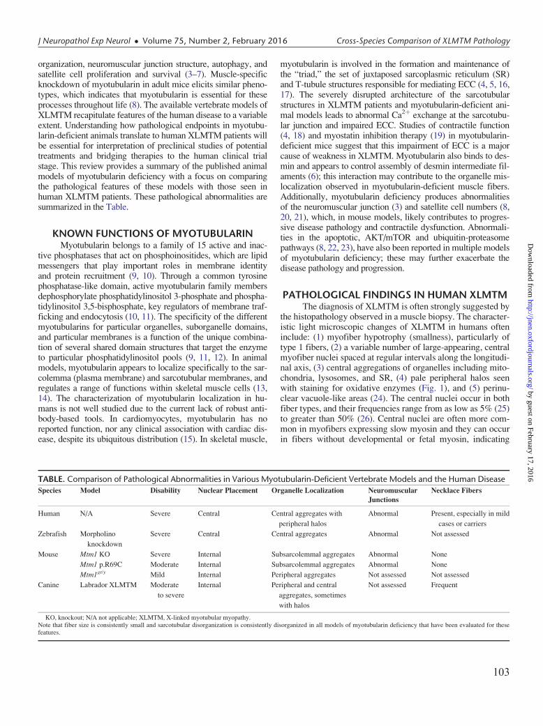
TABLE. Comparison of Pathological Abnormalities in Various Myotubularin-Deficient Vertebrate Models and the Human Disease
Species Model Disability Nuclear Placement Organelle Localization Neuromuscular
Junctions
Necklace Fibers
Human N/A Severe Central Central aggregates with
peripheral halos
Abnormal Present, especially in mild
cases or carriers
Zebrafish Morpholino
knockdown
Severe Central Central aggregates Abnormal Not assessed
Mouse Mtm1 KO Severe Internal Subsarcolemmal aggregates Abnormal None
Mtm1 p.R69C Moderate Internal Subsarcolemmal aggregates Abnormal None
Mtm1gt/y Mild Internal Peripheral aggregates Not assessed Not assessed
Canine Labrador XLMTM Moderate
to severe
Internal Peripheral and central
aggregates, sometimes
with halos
Not assessed Frequent
KO, knockout; N/A not applicable; XLMTM, X-linked myotubular myopathy.Note that fiber size is consistently small and sarcotubular disorganization is consistently disorganized in all models of myotubularin deficiency that have been evaluated for thesefeatures.
J Neuropathol Exp Neurol � Volume 75, Number 2, February 2016 Cross-Species Comparison of XLMTM Pathology
103
by guest on February 17, 2016http://jnen.oxfordjournals.org/
Dow
nloaded from
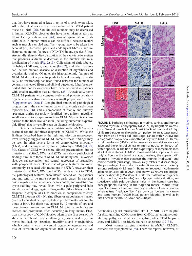
that they have matured at least in terms of myosin expression.All of these features are often seen in human XLMTM patientmuscle at birth (26). Satellite cell numbers may be decreasedin human XLMTM biopsies that have been taken as early as30 weeks of gestational age (26); however, quantitation of sat-ellite cells in human muscle can be difficult because factorssuch as muscle sampled and fiber typing have to be taken intoaccount (26). Necrosis, peri- and endomysial fibrosis, and in-flammation are not features of XLMTM in any species. Ultra-structurally, there is disorganization of the sarcotubular systemthat produces a dramatic decrease in the number and mis-localization of triads (Fig. 2) (5). Collections of dark tubules,probably of SR origin, can occur (Fig. 2); and other featurescan include marked absence or disruption of myofibrils andcytoplasmic bodies. Of note, the histopathologic features ofXLMTM do not appear to predict clinical severity. Specifi-cally, no relationship has been found between the number ofcentrally nucleated fibers and clinical outcomes. It has been re-ported that poorer outcomes have been observed in patientswith smaller myofiber size at biopsy (25). Anecdotally, someXLMTM patients with comparatively mild phenotypes showorganelle mislocalization in only a small proportion of fibers(Supplementary Data 1). Longitudinal studies of pathologicalprogression in the same human patients have only rarely beenreported (27, 28), and a comparison of biopsy and autopsyspecimens during our review identified more uniform myofibersmallness in autopsy specimens from XLMTM patients in com-parison to the fiber size variation (including numerous hypotro-phic fibers) that is typically seen on diagnostic biopsies.
Genetic confirmation of a mutation in the MTM1 gene isessential for the definitive diagnosis of XLMTM. While thefindings described here at the light and electron microscopiclevel strongly suggest XLMTM, some of these findings maybe seen in other severe forms of centronuclear myopathy(CNM) and in congenital myotonic dystrophy (CDM) (24, 29,30). Cases of CNM with severe clinical presentations due tomutations in DMN2, BIN1, and RYR1 may show pathologicalfindings similar to those in XLMTM, including small myofibersize, central nucleation, and central aggregates of organelleswith peripheral halos. These pathological features are morecommonly associated with mutations in MTM1, however, thanmutations in DMN2, BIN1, and RYR1. With respect to CDM,the pathological features encountered depend on the patientsage and tend to be more severe in early cases. In neonatalcases, myofibers are small, nuclei are central, and oxidative en-zyme staining may reveal fibers with a pale peripheral haloand dark central aggregates of organelles. Slow fibers are lessfrequent in congenital CDM biopsies, in comparison to earlyXLMTM biopsies (31). No ring fibers or sarcoplasmic masses(areas of abundant acid-phosphatase positive material) are ob-vious at birth, but these may appear by 12 months of age andthese features are not seen in XLMTM. Internal nuclei are in-creased and prominent, often occurring in long chains. Elec-tron microscopy of CDM biopsies taken in the first year of lifeshow a peripheral zone containing glycogen and myofila-ments but lacking organized myofibrils and mitochondria,which contrasts with the central organelle aggregation andloss of sarcotubular organization that is seen in XLMTM.
Antibodies against muscleblind-like 1 (MNBL1) are helpfulfor distinguishing CDM cases from CNMs, including myotub-ular myopathy, as the latter are negative, while CDM biopsiesshow anti-MBNL1 positively stained nuclear foci (32–34).
Most women carrying mutations in MTM1 (XLMTMcarriers) are asymptomatic (35). There are reports, however, of
FIGURE 1. Pathological findings in murine, canine, and humanX-linked myotubular myopathy (XLMTM) by brightfield micros-copy. Skeletal muscle from an Mtm1 knockout mouse at 43 daysof life (mid-stage) are shown in comparison to an autopsy speci-men from an 18-week-old (end-stage) canine with XLMTM anda diagnostic biopsy of a 2-month-old boy with XLMTM. Hema-toxylin and eosin (H&E) staining reveals abnormal fiber size vari-ation and the extent of central or internal nucleation in each af-fected species. In addition to the hypotrophy of some fibers seenat all disease stages, XLMTM shows marked atrophy of essen-tially all fibers in the terminal stage; therefore, the apparent dif-ference in myofiber size between the murine (mid-stage) andcanine models (end-stage) shown likely relates to disease stage.The percentage of centrally nucleated fibers can vary markedlyamong patients (H&E inset). Stains for reduced nicotinamideadenine dinucleotide (NADH, also known as NADH-TR) and pe-riodic acid-Schiff (PAS) stain illustrate the patterns of organelle(mitochondrial/sarcotubular) and glycogen mislocalization, re-spectively, with pale peripheral halos in the human case butdark peripheral staining in the dog and mouse. Mouse tissuetypically shows subsarcolemmal aggregation of mitochondriawhereas true “necklace fibers” (arrows) can be seen in canineand some human (NADH inset) patient biopsies, as well as inrare fibers in the mouse. Scale bar¼ 40 mm.
Lawlor et al J Neuropathol Exp Neurol � Volume 75, Number 2, February 2016
104
by guest on February 17, 2016http://jnen.oxfordjournals.org/
Dow
nloaded from
XLMTM carriers with myopathic symptoms and pathology con-sistent with CNM. The clinical severity of these symptomaticXLMTM carriers ranges broadly from severe neonatal weakness(extremely rare) to mild/moderate weakness with a normal life-span. This broad range of symptomatic severities probably re-flects skewed inactivation of the X chromosome (lyonization),where symptomatic XLMTM carriers have more lyonization ofthe normal MTM1 allele than those carriers with no symptoms(35–38). The pathology in the reported cases is variable (Supplementary Data 1), but (in contrast to XLMTM male patients) thedegree of XLMTM pathology does tend to correlate with thelevel of clinical severity. Biopsy findings in XLMTM carrierswith symptoms during infancy were very similar to those seenin male patients, including myofiber smallness, numerous cen-trally nucleated fibers, type 1 fiber predominance, central
aggregations of organelles, and peripheral halos (36, 39). In ad-dition, necklace fibers with a basophilic loop a few microns be-neath the sarcolemma, and associated with internal nuclei, canbe a feature of female carriers and milder cases, but may be lesscommon in severe cases (40). XLMTM carriers identified dur-ing adulthood display slightly different muscle pathology tothose identified in infancy. While carriers with infantile onsettend to have severe disease and pathology similar to affectedXLMTM boys, those with later onset display pathological find-ings including increased fiber size variation (due to the presenceof large and small fibers), numerous fibers containing internalnuclei (with a subpopulation of centrally nucleated fibers), andoccasional perinuclear vacuole-like areas (37, 38). As such, thepathology of these adult symptomatic XLMTM carriers is moresimilar to non-XLMTM cases of CNM; this should be a diag-nostic consideration when encountering CNM in adult femalepatients.
ZEBRAFISH MODEL OF XLMTMThe zebrafish knockdown model of myotubularin defi-
ciency was created using antisense morpholinos to generateembryos with reduced myotubularin protein expression (5).Similar to human XLMTM patients, the zebrafish knockdownmodel exhibits severe impairment of motor function, musclefiber hypotrophy, and large-appearing, abnormally locatednuclei (Fig. 3). Phenotypic effects are evident very early inthe development process. By 24 hours postfertilization, em-bryos have abnormal dorsal curvature through the back andtail and significantly fewer spontaneous muscle contractions.By 72 hours postfertilization, embryos typically have thin-ning of the muscle compartment and bent and/or foreshort-ened tails that correlate with markedly decreased ability tohatch from their chorions, poor touch-evoked escape re-sponses, and diminished swimming capacity.
Pathologically, the zebrafish knockdown model displaysfindings similar to those seen in human XLMTM patients, in-cluding myofiber smallness, centrally nucleated fibers, andmislocalization of organelles (5). There is also ultrastructuraldisarray of the sarcotubular system that is similar to thatseen in human myotubularin deficiency (Fig. 3) (5). The neu-romuscular junction in myotubularin-deficient zebrafish, asobserved using a-bungarotoxin staining, shows normal-appear-ing neuronal input but abnormal organization, which is re-flected as sparse staining that is generally located in the middleof the myofiber (41). Treatment with an acetylcholinesteraseinhibitor strongly improved deficits in spontaneous coiling andtouch-evoked escape behaviors, supporting the association be-tween myotubularin support of membrane trafficking andregulation of acetylcholine receptors (42, 43). A similar, albeitless dramatic, response to acetyl cholinesterase inhibitors hasalso been observed in human cases of XLMTM (41).
Ultrastructural analysis of morpholino-treated zebrafishmuscle tissue shows grossly aberrant SR and T-tubule networks(5). SR networks are irregular, disorganized, and often randomlyinterspersed throughout the sarcomere. Abnormalities in T-tubule structures range from mild changes in triad electron den-sity to fibers with nearly unrecognizable SR/triad regions. These
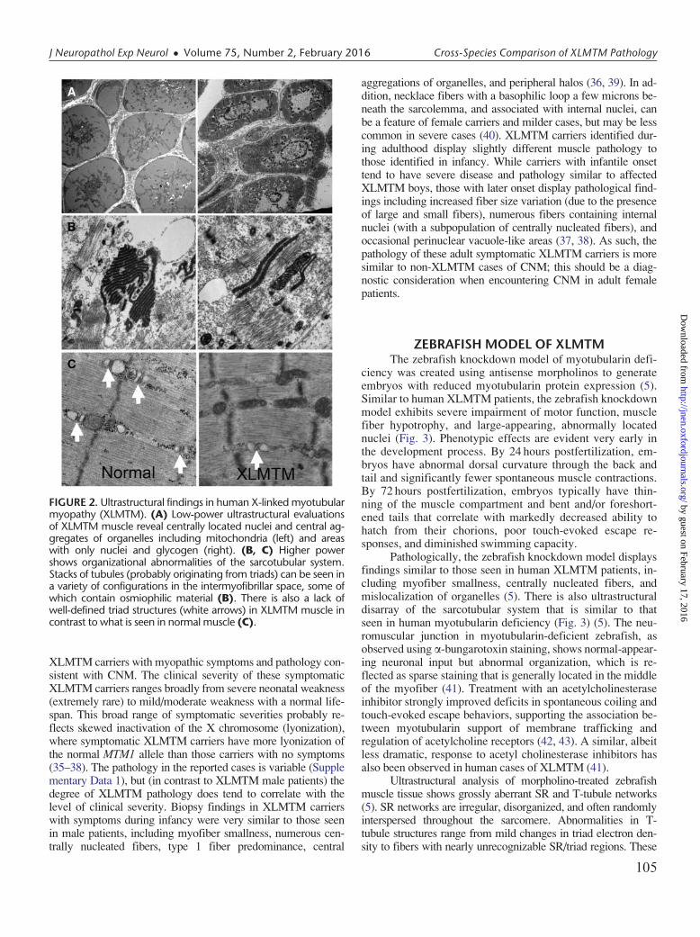
FIGURE 2. Ultrastructural findings in human X-linked myotubularmyopathy (XLMTM). (A) Low-power ultrastructural evaluationsof XLMTM muscle reveal centrally located nuclei and central ag-gregates of organelles including mitochondria (left) and areaswith only nuclei and glycogen (right). (B, C) Higher powershows organizational abnormalities of the sarcotubular system.Stacks of tubules (probably originating from triads) can be seen ina variety of configurations in the intermyofibrillar space, some ofwhich contain osmiophilic material (B). There is also a lack ofwell-defined triad structures (white arrows) in XLMTM muscle incontrast to what is seen in normal muscle (C).
J Neuropathol Exp Neurol � Volume 75, Number 2, February 2016 Cross-Species Comparison of XLMTM Pathology
105
by guest on February 17, 2016http://jnen.oxfordjournals.org/
Dow
nloaded from
alterations in the T-tubule and SR networks, which resemble hu-man pathology, are related to functional impairments in ECC.
MOUSE MODELS OF XLMTMThe first animal model of XLMTM, the Mtm1 knock-
out (KO) mouse (also called the Mtm1d4 mouse), was pro-duced through a large deletion in exon 4 of Mtm1(44). Mtm1KO mice show weakness starting at approximately 3 weeks
of life that progresses to death in a fairly uniform timescale(usually around 5–7 weeks of life, range 4–14 weeks, depend-ing on the background strain) (19, 44). Abnormal nuclearplacement is observed in Mtm1 KO muscle at a lower per-centage of muscle fibers than is usually observed in humanXLMTM patients, and the placement of these nuclei is typi-cally eccentric rather than central (Fig. 1). A similar “inter-nally nucleated fiber” phenotype is observed in other murineand canine models of myotubularin deficiency. The numberof internally nucleated fibers is essentially normal during theperinatal period in Mtm1 KO mice and this graduallyincreases with age to between 5% and 45% of fibers at end-stage disease. Mtm1 KO mice show marked myofiber hypo-trophy in at least a subset of fibers (including both oxidativeand glycolytic fibers), and this is most apparent after 3 weeksof age when fibers are mature. At this stage of disease, fibersmallness is most consistent with a hypotrophic processrather than an atrophic process, based on fiber shape at thelight microscopic level and the absence of redundant basallamina at the ultrastructural level. Organelle mislocalizationoccurs in myofibers of Mtm1 KO mice, with fibers that con-tain perinuclear central accumulation of mitochondria, butthe pattern is most frequently that of a peripheral ring of mito-chondria at the sarcolemma and markedly decreased numbersof mitochondria in the central areas of the fiber (and thussomewhat distinct from the patterns seen in canines and hu-mans) (Fig. 1) (4, 19, 44). Sarcotubular elements are also ag-gregated in the same locations as the mitochondria, whichcan be clearly visualized on immunostaining for the ryano-dine receptor (RYR1) and the dihydropyridine receptor a(DHPR-a) (Fig. 4) (4). By electron microscopy, there is dis-organization of the sarcotubular system, with presence of lon-gitudinal tubules (L-tubules) and a decrease in triad numbersimilar to that seen in human XLMTM (4, 5, 18). Organellemislocalization and triad disorganization are the earliest de-scribed pathological signs of the disease in these mice, andprogress over time (4).
The availability of the Mtm1 KO model has allowed sys-tematic evaluations of disease pathology in the context of dis-ease progression and the spectrum of disease severity amongseveral different muscles. Progression of disease in Mtm1 KOmice generally correlates with increasing numbers of myofib-ers displaying characteristic XLMTM-like pathology: myo-fiber smallness and organelle mislocalization. These featuresare found in nearly all fibers by the time the mice are killeddue to symptomatic severity between 4 and 14 weeks of life(19, 44). Myofiber smallness at late stages of disease is proba-bly caused by a combination of hypotrophy and atrophy, as av-erage fiber size decreases during terminal phases of disease.As noted above, the number of fibers with internal nuclei alsoincreases with age. Approximately 5% to 45% of fibers haveinternal nuclei in most Mtm1 KO mice by the end of the dis-ease course, but this percentage varies depending on the mus-cle in the body and strain background (4, 13). The number ofmuscle satellite cells in Mtm1 KO mice decreases with age toapproximately 30% of wild-type values at late disease stages(20). Neuromuscular junctions are enlarged and less complexthan normal at the ultrastructural level (3). Whole-muscleex vivo contractile function in extensor digitorum longus and
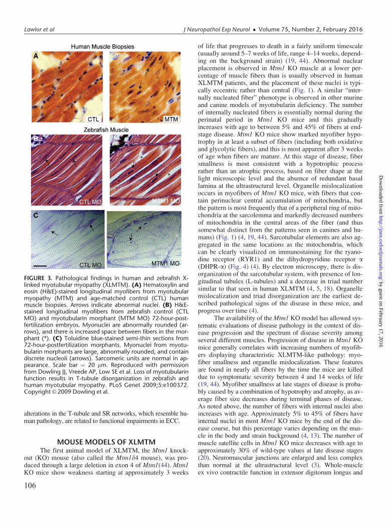
FIGURE 3. Pathological findings in human and zebrafish X-linked myotubular myopathy (XLMTM). (A) Hematoxylin andeosin (H&E)-stained longitudinal myofibers from myotubularmyopathy (MTM) and age-matched control (CTL) humanmuscle biopsies. Arrows indicate abnormal nuclei. (B) H&E-stained longitudinal myofibers from zebrafish control (CTLMO) and myotubularin morphant (MTM MO) 72-hour-post-fertilization embryos. Myonuclei are abnormally rounded (ar-rows), and there is increased space between fibers in the mor-phant (*). (C) Toluidine blue-stained semi-thin sections from72-hour-postfertilization morphants. Myonuclei from myotu-bularin morphants are large, abnormally rounded, and containdiscrete nucleoli (arrows). Sarcomeric units are normal in ap-pearance. Scale bar ¼ 20 mm. Reproduced with permissionfrom Dowling JJ, Vreede AP, Low SE et al. Loss of myotubularinfunction results in T-tubule disorganization in zebrafish andhuman myotubular myopathy. PLoS Genet 2009;5:e100372.Copyright VC 2009 Dowling et al.
Lawlor et al J Neuropathol Exp Neurol � Volume 75, Number 2, February 2016
106
by guest on February 17, 2016http://jnen.oxfordjournals.org/
Dow
nloaded from
soleus muscles of Mtm1 KO mice showed impaired contractileforce, with slightly better performance seen in the predomi-nantly oxidative soleus muscle than in the predominantly gly-colytic extensor digitorum longus muscle (18). In contrast,skinned fiber preparations using Mtm1 KO myofibers showedequivalent function between slow and fast myofibers, suggest-ing that the better performance in oxidative muscles notedabove was due to a factor (perhaps mitochondria) that was re-moved or destroyed during the chemical skinning process.
Mtm1 KO mice are the most frequently used model forpreclinical treatment trials, and a variety of pathological recov-ery patterns have been seen. An early study using intramuscu-lar injection of adeno-associated virus (AAV) carrying theMtm1 coding sequence (AAV8-Mtm1) showed complete cor-rection of XLMTM pathology at the light microscopic level inthe injected limbs (13). Correction of pathology was seenacross all muscle groups, and the lifespan of the mice was ex-tended beyond 6 months after a single dose of the vector at 9weeks of age (45). Concurrently, a targeted enzyme replace-ment agent (3E10Fv-MTM1) was developed that combinedenzymatically active myotubularin with an antibody fragmentthat improved distribution to muscle. Proof-of-concept pilotstudies using local injections of a nonoptimized 3E10Fv-MTM1 dose over 2 weeks showed improvements in single-muscle strength and sarcotubular ultrastructural organization,whereas XLMTM pathology at the light microscopic level wasunchanged (18). In contrast, a trial of the myostatin inhibitor,ActRIIB-mFc, produced dramatic improvement of glycolyticfiber size but with minimal benefits to lifespan or strength(19). Because ActRIIB-mFc provided a means of mitigatingmyofiber hypotrophy without affecting the other pathologicalfeatures of myotubularin deficiency, these studies suggest thatabnormalities of organelle localization and/or ECC comprisethe predominant cause(s) of weakness in these mice. This con-cept is further supported by the comparison of functional stud-ies using whole-muscle versus skinned fiber preparations,which reveal a large component of contractile deficit that isonly apparent when membranous structures are intact (18).While this component of weakness may be due to abnormali-ties in the sarcolemma, mitochondria, or sarcotubular system,the known abnormalities of sarcotubular organization and cal-cium handling suggest that ECC impairment is a major factor.
TISSUE-SPECIFIC MTM1 KO MICEThere have been several studies to induce myotubularin
deficiency to specific tissues and/or time periods. ConditionalMtm1 KO mice that were myotubularin deficient only in skele-tal muscle (using a transgenic line that expresses Cre recombi-nase under the skeletal muscle a-actin promoter) showed aphenotype that was indistinguishable from Mtm1 KO mice(44). In contrast, conditional Mtm1 KO mice that were myotu-bularin deficient in the central nervous system but not skeletalmuscle (using a transgenic line expressing Cre recombinaseunder the neuron-specific enolase promoter) did not show anyobvious clinical or pathological phenotype. To study the im-pact of myotubularin deficiency on adult muscle, a recombi-nant AAV that expresses Cre recombinase under the muscle-
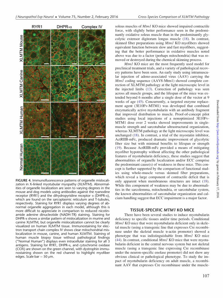
FIGURE 4. Immunofluorescence patterns of organelle mislocali-zation in X-linked myotubular myopathy (XLMTM). Abnormal-ities of organelle localization are seen to varying degrees in themouse and dog models using antibodies against the ryanodinereceptor (RYR1) and the dihydropyridine receptor a (DHPR-a),which are found on the sarcoplasmic reticulum and T-tubules,respectively. Staining for RYR1 displays varying degrees of ab-normal organelle aggregation in each model, although this ismore difficult to appreciate in comparison to reduced nicotin-amide adenine dinucleotide (NADH-TR) staining. Staining forDHPR-a shows a similar pattern of mislocalization in murine andcanine XLMTM, but organelle mislocalization cannot be clearlyvisualized on human XLMTM tissue. Immunostaining for elec-tron transport chain complex IV shows clear mitochondrial mis-localization in mouse, canine, and human XLMTM. Staining ofhuman muscle biopsy tissue without pathological findings(“Normal Human”) displays even intracellular staining for all 3antigens. Staining for RYR1, DHPR-a, and cytochrome oxidase(COX) are shown on the green channel, with dystrophin immu-nostaining shown on the red channel to highlight myofiberedges. Scale bar¼ 50 mm.
J Neuropathol Exp Neurol � Volume 75, Number 2, February 2016 Cross-Species Comparison of XLMTM Pathology
107
by guest on February 17, 2016http://jnen.oxfordjournals.org/
Dow
nloaded from
specific desmin promoter was injected locally into the muscleof adult conditional Mtm1 KO mice (8). Myotubularin defi-ciency in these muscles produced weakness and pathology thatwas strikingly similar to the effects of germline mutations inMtm1, including progressive decreases in myofiber size, mis-localization of organelles, increases in the number of internallynucleated fibers, and abnormal sarcotubular morphology onelectron microscopy. The number of satellite cells progres-sively decreased in myotubularin-deficient adult muscles, andabnormalities in proteins related to Akt signaling, neuromuscu-lar transmission, and autophagy were also observed.
MTM1 P.R69C MICEThe Mtm1 p.R69C mouse was designed to produce a
less severe myotubularin deficiency phenotype by “knockingin” the c.205C>T mutation that had been found in several hu-man XLMTM patients who survived past infancy (46). Aspredicted, Mtm1 p.R69C mice have greater strength thanMtm1 KO mice, and lifespans often exceeding 1 year. Similarto the pathology seen in Mtm1 KO mice, affected myofibersdisplay hypotrophy and mislocalization of organelles (as de-scribed above). As observed in the Mtm1 KO and canineXLMTM models, there is abnormal localization of nuclei,which are usually eccentric rather than central. This pattern isslightly different from that seen in human XLMTM, wherenuclear placement tends to be central rather than eccentric.The number of internally nucleated fibers may only be mildlyincreased (21, 46). Neuromuscular junctions are enlarged andhave less complex junctional folds than are seen in wild-typemice (3). Satellite cell depletion is noted at late stages of dis-ease in these mice (21). It should be noted that the Mtm1p.R69C mutation functions as a splice site mutation leadingto the expression of only a very small amount of full-lengthmutant myotubularin transcript, and a similar impact on splic-ing has been observed in human tissue harboring the MTM1p.R69C mutation (46). This suggests that induction of verysmall amounts of myotubularin may produce substantial clin-ical benefits in the myotubularin-null state.
Mtm1 p.R69C mice have been used in several publishedtrials of potential treatments for XLMTM. Pyridostigmine (anacetylcholinesterase inhibitor that improves neuromusculartransmission) produced improvements in grip strength and en-durance (3) as has been found in some human cases (41), butevaluations of treatment effects on histopathology were notperformed. Localized wild-type myoblast transplantation intothe gastrocnemius muscle of Mtm1 p.R69C mice produced in-creases in muscle mass and evoked muscle action potentials,but increases in fiber size were not observed and other patho-logical features were not assessed (47). Additionally, a studyof myostatin inhibition using ActRIIB-mFc in Mtm1 p.R69Cmice identified some muscle-specific phenotypes in thesemice. Treated mice developed hypertrophy of glycolytic myo-fibers only in the gastrocnemius muscle, without apparent ef-fect in other muscles such as the quadriceps or triceps (21).This was markedly different from the effects observed in theMtm1 KO model using the same agent, which produced signif-icant hypertrophy in all of these major muscles (19). Subse-quent studies identified differences between the Mtm1 p.R69C
quadriceps and gastrocnemius muscles with respect to satellitecell behavior and hypertrophic signaling that may have beenassociated with differential treatment responses in these ani-mals (21). While the precise mechanism(s) responsible forthese different responses remains unclear, the results estab-lished that treatment studies using the mouse models should becautiously designed, and analysis of various muscle groups isadvisable.
MTM1gt/y MICEAn Mtm1 mutant mouse with a truncating mutation in
exon 1 of Mtm1 has been described in a single publication(23). The Mtm1gt/y mice are similar in weight to wild-type an-imals until approximately 4 weeks of age, and, therefore,have a less severe phenotype than Mtm1 KO mice. Musclepathology in Mtm1gt/y appears similar to other myotubularin-deficient mice, including myofiber smallness, subsarcolem-mal organelle localization, mild increases in the number ofinternal nuclei, and ultrastructural sarcotubular disorganiza-tion. A blockage of a late stage of autophagy was detected inthese mice, resulting in protein aggregates that are positivefor ubiquitin and p62. Such protein inclusions have not beenreported in other models of XLMTM, although other studiesin Mtm1 KO mice showed dysregulation of autophagy andubiquitin-proteasome pathways that resulted in ultrastructur-ally abnormal autophagosomes (8, 22). Analysis of Mtm1gt/y
mice also revealed abnormalities of mitochondrial structureincluding mitochondria with fewer decondensed and swollencristae (23), which can be seen to some extent across allXLMTM models studied. The degree to which mitochondrialstructural abnormalities are due to primary mitochondrial dys-function versus secondary effects is currently unclear.
CANINE XLMTMX-linked myotubular myopathy in dogs was discovered
as a naturally occurring disease in several related litters of Lab-rador Retriever (MTM1 p.N155K) puppies (14). This led to theestablishment of a XLMTM canine colony that has been in-strumental for preclinical studies in this canine model.XLMTM Labradors, and subsequent generations bred onto abeagle background, appear phenotypically normal at birth, butby 8 weeks of age, they begin to display a characteristic “slackjaw” appearance. Shortly thereafter, XLMTM puppies displaysubtle hind limb muscle weakness, gait abnormalities, and re-duced physical activity. Ultimately, these clinical impairmentsprogress until death becomes necessary between 15 and 26weeks of age (14). Affected dogs exhibit muscle pathologysimilar to the Mtm1 KO and Mtm1 p.R69C murine models, in-cluding myofiber hypotrophy (of both slow and fast fibers),mild-to-moderate increases in the number of internally nucle-ated fibers, mislocalization of organelles, and disorganizationof the sarcotubular system at the ultrastructural level (14, 45).All of these features are present in a subpopulation of fibersat 10 weeks of life, and they tend to affect greater numbers offibers as the disease progresses. One remarkable pathologicfeature of canine XLMTM is a pattern of organelle mislocali-zation distinctively different from both murine and humanXLMTM. Whereas the pattern of organelle/mitochondrial
Lawlor et al J Neuropathol Exp Neurol � Volume 75, Number 2, February 2016
108
by guest on February 17, 2016http://jnen.oxfordjournals.org/
Dow
nloaded from
mislocalization in humans tends to be a central aggregation,and in murine models tends to be subsarcolemmal (necklace-like), XLMTM Labradors and “Lab-beagles” show a combina-tion of central organelle aggregation, true necklace fibers, andfibers with necklace-like subsarcolemmal organelle aggregates(14, 45). In cases where these aggregates are associated withinternalized nuclei along the same line, they can be consideredtrue “necklace fibers” that have been reported in a range ofCNMs. Similar to that demonstrated in Mtm1 KO mice, theseaggregates of mislocalized organelles stain for markers of mi-tochondria (complex IV), T-tubules (DHPR-a), and SR(RYR1) (Fig. 4) (14). While the varying patterns of organellemislocalization across different myotubularin-deficient modelslikely represent different aspects of a similar pathogenic pro-cess, it is important to recognize this spectrum of patternswhen considering how best to evaluate and quantify pathologi-cal features. A pilot study assessing satellite cell number in thevastus lateralis and gastrocnemius muscles of XLMTM Lab-beagles comparing satellite cell numbers at 10 and 17 weeks ofage did not show satellite cell depletion (unpublished). Itshould also be noted that canine XLMTM carriers do not showweakness or any sign of MTM pathology in any of the studiesperformed thus far, despite extensive evaluation. CanineXLMTM has also recently been reported in Rottweiler dogs(MTM1 p.Q384P) (48), which showed extensive similarities tothe MTM1 p.N155K dogs, both in terms of pathological find-ings and clinical features.
The canine MTM1 p.N155K XLMTM model has beenused to test gene therapy for XLMTM (AAV8-MTM1—arecombinant AAV8 vector, carrying the canine MTM1 cDNAunder control of the desmin promoter) in a pilot study (45).Intramuscular injections of AAV8-MTM1 in hind limb cra-nial tibialis muscles demonstrated dramatic and rapid im-provements in strength and a concomitant reversal ofXLMTM pathology (including resolution of myofiber hypo-trophy, organelle mislocalization, number of centrally nucle-ated fibers, and sarcotubular organization) in the injectedlimb in comparison with contralateral saline-injected limbs(45). Follow-up studies using systemic AAV8-MTM1 admin-istered intravenously produced similar histopathologic andfunctional improvements in all muscles sampled for at least 1year after a single dose (45), and further systemic dosing andlong-term follow-on studies are in progress. Based on theseencouraging results, clinical studies utilizing gene therapy arenow planned.
CONCLUSIONXLMTM is a severe congenital myopathy with charac-
teristic pathological findings in most cases, including myofiberhypotrophy, centrally nucleated myofibers, and mislocalizationof organelles. When comparing myotubularin-deficient fish,mice, and dogs to human patients, there are many similaritiesin pathology, but there are also some differences in how theseabnormalities manifest (Table). Such variation needs to be ac-counted for when designing strategies for the quantitation ofpathological findings in XLMTM, and our group is currentlydeveloping techniques that address these issues. As therapeuticdevelopment progresses from nonclinical to clinical studies in
XLMTM, an understanding of the impact of myotubularin de-ficiency in each species is essential for the appropriate designof pathological endpoints using human muscle tissue, and in-deed for understanding the translatability of findings in an ani-mal model to humans.
ACKNOWLEDGMENTSSome images in this publication were produced with the
resources of the Children’s Hospital of Wisconsin ResearchInstitute’s Imaging Core Facility and the electron microscopycore facility at the Medical College of Wisconsin. We wouldalso like to thank Dr Magda Morton for her editorial assistance.
REFERENCES1. Spiro AJ, Shy GM, Gonatas NK. Myotubular myopathy. Persistence of
fetal muscle in an adolescent boy. Arch Neurol 1966;14:1–142. van Wijngaarden GK, Fleury P, Bethlem J, et al. Familial “myotubular”
myopathy. Neurology 1969;19:901–83. Dowling JJ, Joubert R, Low SE, et al. Myotubular myopathy and the neu-
romuscular junction: A novel therapeutic approach from mouse models.Dis Model Mech 2012;5:852–9
4. Al-Qusairi L, Weiss N, Toussaint A, et al. T-tubule disorganization and de-fective excitation-contraction coupling in muscle fibers lacking myotubu-larin lipid phosphatase. Proc Natl Acad Sci U S A 2009;106:18763–8
5. Dowling JJ, Vreede AP, Low SE, et al. Loss of myotubularin function re-sults in T-tubule disorganization in zebrafish and human myotubular my-opathy. PLoS Genet 2009;5:e1000372
6. Hnia K, Tronchere H, Tomczak KK, et al. Myotubularin controls desminintermediate filament architecture and mitochondrial dynamics in humanand mouse skeletal muscle. J Clin Invest 2011;121:70–85
7. Tsujita K, Itoh T, Ijuin T, et al. Myotubularin regulates the function ofthe late endosome through the gram domain-phosphatidylinositol 3,5-bisphosphate interaction. J Biol Chem 2004;279:13817–24
8. Joubert R, Vignaud A, Le M, et al. Site-specific Mtm1 mutagenesis by anAAV-Cre vector reveals that myotubularin is essential in adult muscle.Hum Mol Genet 2013;22:1856–66
9. Vicinanza M, D’Angelo G, Di Campli A, et al. Function and dysfunctionof the PI system in membrane trafficking. EMBO J 2008;27:2457–70
10. Cowling BS, Toussaint A, Muller J, et al. Defective membrane remodel-ing in neuromuscular diseases: Insights from animal models. PLoS Genet2012;8:e1002595
11. Laporte J, Bedez F, Bolino A, et al. Myotubularins, a large disease-asso-ciated family of cooperating catalytically active and inactive phosphoi-nositides phosphatases. Hum Mol Genet 2003;12 Spec No 2:R285–92
12. Mruk DD, Cheng CY. The myotubularin family of lipid phosphatases indisease and in spermatogenesis. Biochem J 2011;433:253–62
13. Buj-Bello A, Fougerousse F, Schwab Y, et al. AAV-mediated intramus-cular delivery of myotubularin corrects the myotubular myopathy pheno-type in targeted murine muscle and suggests a function in plasmamembrane homeostasis. Hum Mol Genet 2008;17:2132–43
14. Beggs AH, Bohm J, Snead E, et al. MTM1 mutation associated with X-linked myotubular myopathy in Labrador Retrievers. Proc Natl Acad SciU S A 2010;107:14697–702
15. Das S, Dowling J, Pierson CR. X-linked centronuclear myopathy. NCBIBookshelf, GeneReviews 2002.
16. Al-Qusairi L, Laporte J. T-tubule biogenesis and triad formation in skele-tal muscle and implication in human diseases. Skelet Muscle 2011;1:26
17. Cowling BS, Chevremont T, Prokic I, et al. Reducing dynamin 2 expressionrescues X-linked centronuclear myopathy. J Clin Invest 2014;124:1350–63
18. Lawlor MW, Armstrong D, Viola MG, et al. Enzyme replacementtherapy rescues weakness and improves muscle pathology in mice withX-linked myotubular myopathy. Hum Mol Genet 2013;22:1525–38
19. Lawlor MW, Read BP, Edelstein R, et al. Inhibition of activin receptortype IIb increases strength and lifespan in myotubularin-deficient mice.Am J Pathol 2011;178:784–93
20. Lawlor MW, Alexander MS, Viola MG, et al. Myotubularin-deficientmyoblasts display increased apoptosis, delayed proliferation, and poorcell engraftment. Am J Pathol 2012;181:961–8
J Neuropathol Exp Neurol � Volume 75, Number 2, February 2016 Cross-Species Comparison of XLMTM Pathology
109
by guest on February 17, 2016http://jnen.oxfordjournals.org/
Dow
nloaded from
21. Lawlor MW, Viola MG, Meng H, et al. Differential muscle hypertrophyis associated with satellite cell numbers and Akt pathway activation fol-lowing activin Type IIB receptor inhibition in Mtm1 p.R69C mice. Am JPathol 2014;184:1831–42
22. Al-Qusairi L, Prokic I, Amoasii L, et al. Lack of myotubularin (MTM1)leads to muscle hypotrophy through unbalanced regulation of the autoph-agy and ubiquitin-proteasome pathways. FASEB J 2013;27:3384–94
23. Fetalvero KM, Yu Y, Goetschkes M, et al. Defective autophagy andmTORC1 signaling in myotubularin null mice. Mol Cell Biol 2013;33:98–110
24. Dubowitz V, Sewry C, Oldfors A. Congenital myopathies and related dis-orders. In: Muscle Biopsy: A Practical Approach 4th Edn. Oxford: Elsevier2013:358–405
25. Pierson CR, Agrawal PB, Blasko J, et al. Myofiber size correlates withMTM1 mutation type and outcome in X-linked myotubular myopathy.Neuromuscul Disord 2007;17:562–8
26. Shichiji M, Biancalana V, Fardeau M, et al. Extensive morphologicaland immunohistochemical characterization in myotubular myopathy.Brain Behav 2013;3:476–86
27. de Goede CG, Kelsey A, Kingston H, et al. Muscle biopsy without cen-trally located nuclei in a male child with mild X-linked myotubular my-opathy. Dev Med Child Neurol 2005;47:835–7
28. Helliwell TR, Ellis IH, Appleton RE. Myotubular myopathy: Morpholog-ical, immunohistochemical and clinical variation. Neuromuscul Disord1998;8:152–61
29. Dubowitz V, Sewry C, Oldfors A. Muscular dystrophies and allied disor-ders: Facioscapulohumeral, myotonic, and oculopharyngeal musculardystrophies. In: Muscle Biopsy: A Practical Approach 4th Edn. Oxford:Elsevier 2013
30. Jungbluth H, Gautel M. Pathogenic mechanisms in centronuclear myopa-thies. Front Aging Neurosci 2014;6:339
31. Soussi-Yanicostas N, Chevallay M, Laurent-Winter C, et al. Distinctcontractile protein profile in congenital myotonic dystrophy and X-linked myotubular myopathy. Neuromuscul Disord 1991;1:103–11.
32. Schoser B. Muscle diseases with DNA expansions. In: Goebel HH,Sewry C, Weller RO, eds. Muscle Disease: Pathology and Genetics. Ox-ford: Wiley Blackwell 2013:273–83
33. Holt I, Jacquemin V, Fardaei M, et al. Muscleblind-like proteins: Simi-larities and differences in normal and myotonic dystrophy muscle. Am JPathol 2009;174:216–27
34. Sewry CA, Quinlivan RC, Squier W, et al. A rapid immunohistochemicaltest to distinguish congenital myotonic dystrophy from X-linked myotub-ular myopathy. Neuromuscul Disord 2012;22:225–30
35. Kristiansen M, Knudsen GP, Tanner SM, et al. X-inactivation patterns incarriers of X-linked myotubular myopathy. Neuromuscul Disord 2003;13:468–71
36. Jungbluth H, Sewry CA, Buj-Bello A, et al. Early and severe presentationof X-linked myotubular myopathy in a girl with skewed X-inactivation.Neuromuscul Disord 2003;13:55–9
37. Tanner SM, Orstavik KH, Kristiansen M, et al. Skewed X-inactivation ina manifesting carrier of X-linked myotubular myopathy and in her non-manifesting carrier mother. Human Genet 1999;104:249–53
38. Penisson-Besnier I, Biancalana V, Reynier P, et al. Diagnosis of myotub-ular myopathy in the oldest known manifesting female carrier: A clinicaland genetic study. Neuromuscul Disord 2007;17:180–5
39. Dahl N, Hu LJ, Chery M, et al. Myotubular myopathy in a girl with a de-letion at Xq27-q28 and unbalanced X inactivation assigns the MTM1gene to a 600-kb region. Am J Human Genet 1995;56:1108–15
40. Bevilacqua JA, Bitoun M, Biancalana V, et al. “Necklace” fibers, a newhistological marker of late-onset MTM1-related centronuclear myopa-thy. Acta Neuropathol 2009;117:283–91
41. Robb SA, Sewry CA, Dowling JJ, et al. Impaired neuromuscular trans-mission and response to acetylcholinesterase inhibitors in centronuclearmyopathies. Neuromuscul Disord 2011;21:379–86
42. Kumari S, Borroni V, Chaudhry A, et al. Nicotinic acetylcholine receptoris internalized via a Rac-dependent, dynamin-independent endocyticpathway. J Cell Biol 2008;181:1179–93
43. Nicot AS, Laporte J. Endosomal phosphoinositides and human diseases.Traffic 2008;9:1240–9
44. Buj-Bello A, Laugel V, Messaddeq N, et al. The lipid phosphatase myo-tubularin is essential for skeletal muscle maintenance but not for myogenesisin mice. Proc Natl Acad Sci U S A 2002;99:15060–5
45. Childers MK, Joubert R, Poulard K, et al. Gene therapy prolongs survivaland restores function in murine and canine models of myotubular myopa-thy. Sci Transl Med 2014;6:220ra10
46. Pierson CR, Dulin-Smith AN, Durban AN, et al. Modeling the human MTM1p.R69C mutation in murine Mtm1 results in exon 4 skipping and a less severemyotubular myopathy phenotype. Hum Mol Genet 2012;21:811–25
47. Lim HJ, Joo S, Oh SH, et al. Syngeneic myoblast transplantation im-proves muscle function in a murine model of X-linked myotubular myop-athy. Cell Transplant 2015;24:1887–900
48. Shelton GD, Rider BE, Child G, et al. X-linked myotubular myopathy inRottweiler dogs is caused by a missense mutation in exon 11 of theMTM1 gene. Skelet Muscle 2015;5:1
Lawlor et al J Neuropathol Exp Neurol � Volume 75, Number 2, February 2016
110
by guest on February 17, 2016http://jnen.oxfordjournals.org/
Dow
nloaded from