Embed Size (px)
Citation preview

Skeletal Muscle Contraction8th ed 50.5
7th ed 49.6

In order to move all animals require muscle activity in response to nervous system input.
Skeletal muscles responsible for voluntary movement.

Skeletal muscles are attached to bones by tendons and are responsible for their movement.
Bicepscontracts
Tricepsrelaxes
Forearmflexes
Bicepsrelaxes
Tricepscontracts
Forearmextends
Tendons
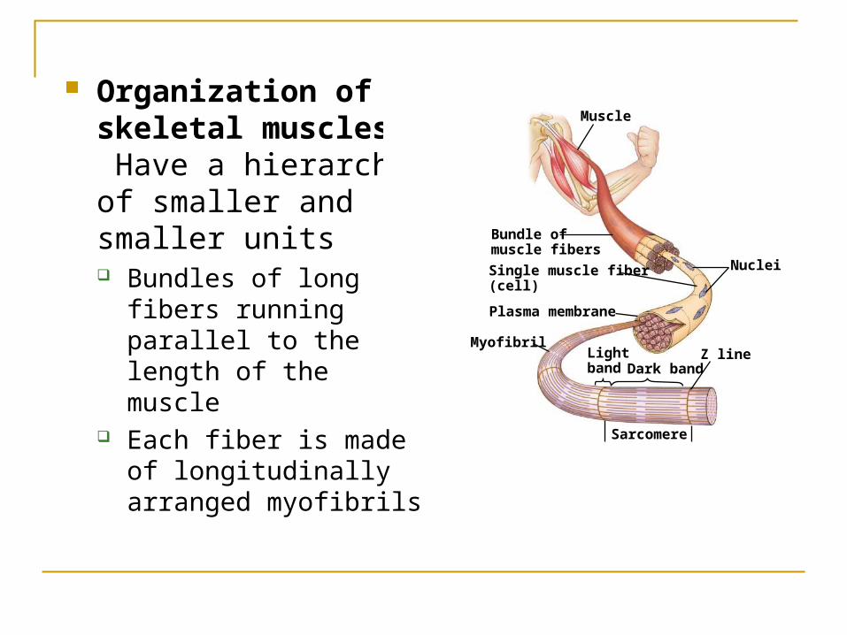
Organization of skeletal muscles: Have a hierarchy of smaller and smaller units Bundles of long fibers
running parallel to the length of the muscle
Each fiber is made of longitudinally arranged myofibrils
Bundle ofmuscle fibers
Single muscle fiber(cell)
Plasma membrane
Nuclei
Muscle
Myofibril
Dark band
Sarcomere
Z lineLightband

Bundle ofmuscle fibers
Single muscle fiber(cell)
Plasma membrane
Nuclei
Muscle
Myofibril
Dark band
Sarcomere
Z lineLightband
I band
TEM
A band I band0.5 µm
M lineThick filaments(myosin)
SarcomereH zoneZ line
Thin filaments(actin)
Z line
Myofibrils are composed of thin and thick filaments
Thin filaments are made of two strands of regulatory proteins and two stands of actin
Thick filaments are made of staggered myosin molecules

Skeletal muscles are striated – arrangement of filaments create dark and light bands
Sarcomere
0.5 µm
ZHA I
Skeletal muscle tissue under a light microscope Skeletal muscle tissue under a electron microscope

Sarcomere – repeating contractile unit of a muscle Thin filaments are attached at the Z line and proceed to the center of the
sarcomere Thick filaments are attached at the M line in the center of the sarcomere Area near edge of sarcomere with only thin filament is the I band Region corresponding to the length of the thick filament is the A band Center of A band containing only thick filaments is called the H zone At a relaxed state thin and thick filaments partially overlap This overlapping arrangement is key to the contraction mechanism This regular arrangement produces dark and light bands and hence make
the fibers look striatedSarcomere
I band
TEM
A band I band0.5 µm
M lineThick filaments(myosin)
SarcomereH zoneZ line
Thin filaments(actin)
Z line

Sliding-Filament model of muscle contraction (focus on a single sarcomere) During contractions
the thin and thick filaments do not change in length but increase the overlap.
This shortens the length of the sarcomere
LE 49-29
Sarcomere
0.5 µm
ZHA
Relaxed muscle fiber
I
Contracting muscle fiber
Fully contracted muscle fiber

Myosin molecule (thick filament) has a globular “head” and a long “tail”. Tail adheres to other tails.
Thin filaments are actin molecules along with regulatory proteins
Thin filaments
Thick filament
Thin filament
Thick filament
Myosin head

Steps in muscle contraction: Myosin head is bound to ATP in a low-
energy configuration
Thin filaments
Thick filament
Thin filament
Thick filament
Myosin head (low-energyconfiguration)

ATP is hydrolyzed to ADP and Pi and the head is now in high-energy configuration
Thin filaments
Thick filament
Thin filament
Thick filament
Myosin head (low-energyconfiguration)
Cross-bridgebinding site
Myosin head (high-energy configuration)
Actin

Head binds with actin filament at the myosin binding sites; forms cross bridge
Thin filaments
Thick filament
Thin filament
Thick filament
Myosin head (low-energyconfiguration)
Cross-bridgebinding site
Myosin head (high-energy configuration)
Actin
Cross-bridge

Thin filaments
Thick filament
Thin filament
Thick filament
Myosin head (low-energyconfiguration)
Cross-bridgebinding site
Myosin head (high-energy configuration)
Actin
Cross-bridge
Myosin head (low-energy configuration)
Thin filament movestoward center of sacomere.
Head releases the ADP and Pi and returns to low-energy configuration; Actin (thin) filament moves towards the center of the sarcomere

Myosin binds to new ATP molecule and resumes the cycle
Thin filaments
Thick filament
Thin filament
Thick filament
Myosin head (low-energyconfiguration)

Nerves conduct signals by changing the voltage on the membranes (action potential)
Sensory neuron: nerve cell that receives information from the internal or external environments and transmits the signal to the central nervous system (brain and spinal cord)
Motor neuron: transmits signals from brain or spinal cord to muscles or glands.


Synapse: junction where one neuron communicates with another neuron or with muscle/gland etc.
Synaptic terminal: A bulb at the end of the axon in which neurotransmitter molecules are stored and released.
Synaptic cleft: narrow gap separating synaptic knob of a transmitting neuron or its effector cell.
Synaptic cleft
Synaptic terminalof motor neuron

Motor unit: A single neuron and all the muscle fibers it controls
When motor neuron produces action potential all the muscle fibers in its motor unit contract as a group.
Motorunit 1
Motorunit 2
Nerve
Synaptic terminals
Motor neuroncell body
Spinal cord
Motor neuronaxon
Muscle
Tendon
Muscle fibers

Regulation of skeletal muscle contraction:
Synaptic terminal receives action potential and releases neurotransmitter Acetylcholine (ACh)
ACh binds to receptors in the muscle and triggers action potential in the muscle fiber.
Action potential is propagated along the plasma membrane and down the T-tubule.
PLASMAMEMBRANET TUBULESynaptic cleft
Synaptic terminalof motor neuron
ACh

Ca2+CYTOSOL
Ca2+
SR
PLASMAMEMBRANET TUBULESynaptic cleft
Synaptic terminalof motor neuron
ACh
Action potential triggers Ca2+ release from sarcoplasmic reticulum

Ca2+CYTOSOL
Myosin cross-bridges attach and detach, powered by ATP pulling the actin filament towards center of the sarcomere
When action potential ends Ca2+ is absorbed back into the sarcoplasmic reticulum
Muscle contraction ends, muscle fibers relax

Ca2+CYTOSOL
Ca2+
SR
PLASMAMEMBRANET TUBULESynaptic cleft
Synaptic terminalof motor neuron
ACh

Ca2+ releasedfrom sarcoplasmicreticulum
MitochondrionMotorneuron axon
Synapticterminal
T tubule
Sarcoplasmicreticulum
Myofibril
Plasma membraneof muscle fiber
Sarcomere

Ca+ and regulatory proteins and their role in muscle fiber contraction Actin filaments have regulatory proteins on them. Tropomyosin, trponin complex and Ca2+ regulate
muscle contraction At rest tropomyosin covers the actin binding sites
preventing actin and myosin from interacting
Myosin-binding sites blocked.
Tropomyosin Ca2+-binding sites
Actin Troponin complex

Myosin-binding sites exposed.
Myosin-binding site
Ca2+
When Ca2+ is released into the cytosol from the sarcoplasmic reticulum it binds to troponin complex.
This changes the alignment of the troponin That in turn shifts the position of the tropomyosin, exposing
the myosin binding sites on the actin filament When Ca2+ concentration drops the binding sites are
covered and contraction stops.

Myosin-binding sites blocked.
Myosin-binding sites exposed.
Tropomyosin Ca2+-binding sites
Actin Troponin complex
Myosin-binding site
Ca2+