Embed Size (px)
Citation preview
1
Size Matters!: Impact of Age, Gender, Height, and Weight on the Normal Heart Size
Pfaffenberger et al: Size Matters
Stefan Pfaffenberger, MD; Philipp Bartko, MD; Alexandra Graf, PhD;
Elisabeth Pernicka, PhD; Jamil Babayev, MD; Emina Lolic, MD; Diana Bonderman, MD;
Helmut Baumgartner, MD; Gerald Maurer, MD; Julia Mascherbauer, MD
From the Departments of Cardiology (S.P., P.B., J.B., E.L., D.B., G.M., J.M.) and Medical
Statistics (A.G., E.P.), Medical University of Vienna, Vienna, Austria and the Adult
Congenital and Valvular Heart Disease Center (H.B.), Department of Cardiology and
Angiology, University Hospital Muenster, Muenster, Germany.
Correspondence to Julia MascherbauerDepartment of Internal Medicine II, Division of Cardiology Medical University Vienna, Waehringer Guertel 18-20, 1090 Vienna, Austria Tel.: +43-1-40400-4614 Fax: +43-1-40400-4216 Email: [email protected] DOI: 10.1161/CIRCIMAGING.113.000690 Journal Subject Codes: [31] diagnostic testing: echocardiography
enennnnnnt t t t ttt ofofofofofofof CCCCCCCararararararardididididididiololololololologogogogogogogyyyyyyy
eutity Vienna Waehringer Guertel 18 20 1090 Vienna Austria
e to uerteteternrnrnalalal MMMMedededicicicininineee IIIIII, ,, DiDiDivivivisisisiononon ooofff CaCaCardrdrdioioiooololologygygyygy iiitytyty VVVieieiennnnnnaaa WWWaeaeaehrhrhrininingegegerrr GuGuGuererertetetelll 181818 2-22000 111090909000 ViViVienenennanana AuAuAustststriririaaa
by guest on July 12, 2018http://circim
aging.ahajournals.org/D
ownloaded from
2
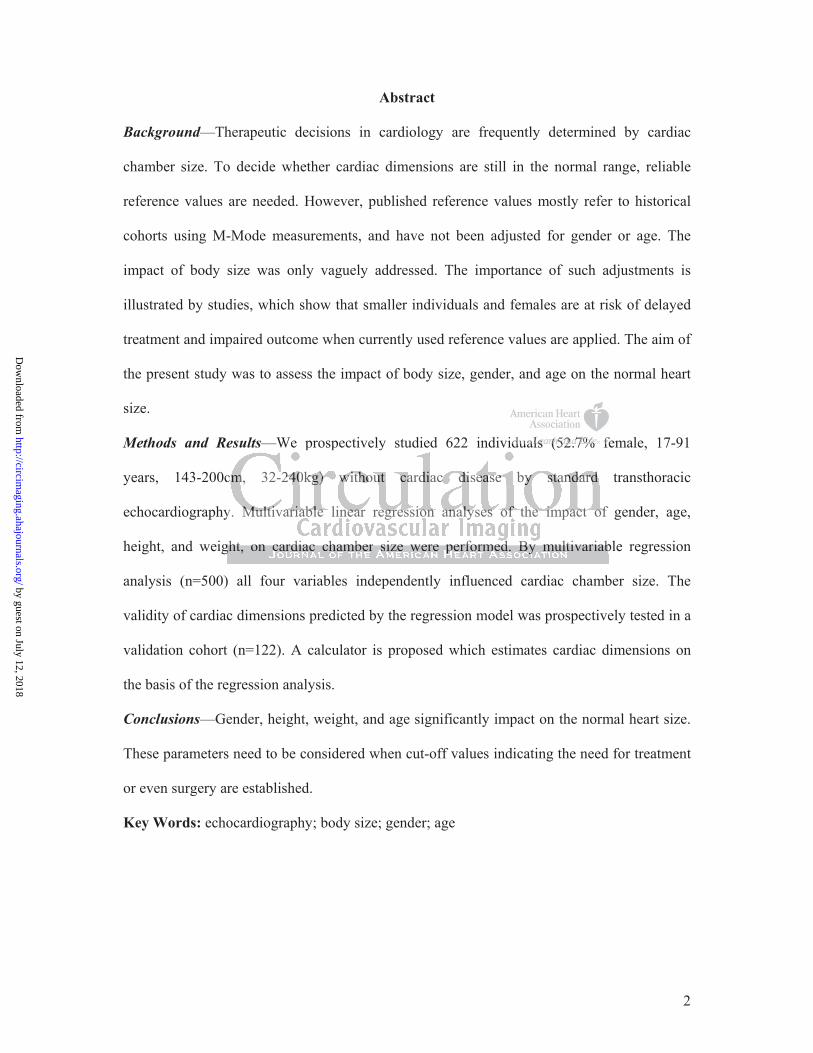
Abstract
Background—Therapeutic decisions in cardiology are frequently determined by cardiac
chamber size. To decide whether cardiac dimensions are still in the normal range, reliable
reference values are needed. However, published reference values mostly refer to historical
cohorts using M-Mode measurements, and have not been adjusted for gender or age. The
impact of body size was only vaguely addressed. The importance of such adjustments is
illustrated by studies, which show that smaller individuals and females are at risk of delayed
treatment and impaired outcome when currently used reference values are applied. The aim of
the present study was to assess the impact of body size, gender, and age on the normal heart
size.
Methods and Results—We prospectively studied 622 individuals (52.7% female, 17-91
years, 143-200cm, 32-240kg) without cardiac disease by standard transthoracic
echocardiography. Multivariable linear regression analyses of the impact of gender, age,
height, and weight, on cardiac chamber size were performed. By multivariable regression
analysis (n=500) all four variables independently influenced cardiac chamber size. The
validity of cardiac dimensions predicted by the regression model was prospectively tested in a
validation cohort (n=122). A calculator is proposed which estimates cardiac dimensions on
the basis of the regression analysis.
Conclusions—Gender, height, weight, and age significantly impact on the normal heart size.
These parameters need to be considered when cut-off values indicating the need for treatment
or even surgery are established.
Key Words: echocardiography; body size; gender; age
idualalalllllsssssss (5(5(5(5(5(5(52222222 7%7%7%7%7%7%7% fe
cm, 32-240kg) without cardiac disease by standard
y
ght, on cardiac chamber size were erformed. multivari b
cmmmmm, 32-2222240404404 kgkgkgkgkg))))) wiwiwiwiwithththththououoouout t caacarddiaccccc dididididiseeasaaseeeee byyyyy ssssstatatatatandndndndndararararard dd dd
y. MuMuMuMuMultltltltltiiviii ararariaiablblbbb eee linenenenen ararararar regegegegegrerererer sssssiiiiiononon aaaaannnnan lylylylylyseeeeess s offofff thehehehe iiiiimpmpmpacacact tt offofff
ggghththt, ononon ccccararardididiacacac ccchahahambmbmbererer sssizizizeee wewewererere ppppperererfofoformrmrmededed. ByByByyy mmmululultititivavavariririababab
by guest on July 12, 2018http://circim
aging.ahajournals.org/D
ownloaded from
3
The judgment whether a heart is normally sized or enlarged is of enormous importance,
particularly when patient management is determined by such estimates. Although it is
generally accepted that cardiac cavity dimensions are determined by body size, this has not
resulted in the widespread use of indexed values. The reason for this is the lack of reliable
reference data, which could resolve the uncertainties related to the exclusive use of body
surface area as an indexing tool. Obese, but small individuals and slim, tall ones may present
with similar body surface areas but may presumably have different expected “normal values”
concerning the normal size of their heart. Such questions have so far not been addressed
systematically. Furthermore, the independent impact of gender and age on cardiac size is not
well defined.
This fact is surprising but has recently been highlighted in a review in Circulation 1 and has
also triggered the design of a multicenter trial to provide such values 2.
Published reference values of the normal sized heart are scarce 3-11. Most of these papers
include small patient numbers and go back to the 1980s and 1990s, when spatial resolution of
two-dimensional (2D) echocardiography was limited and mainly Motion (M)-Mode imaging
was used (Table 1). While the majority of these studies were focused on left ventricular cavity
size 3-5, 7-10, 12, reference values for atrial and right ventricular dimensions barely exist 6, 11.
Particularly in valvular heart disease, accurate assessment of cardiac cavity enlargement is
crucial. Several studies in the past have reported cut-off values of left ventricular dimensions,
which indicate the necessity of a surgical intervention in severe aortic 13-20 or mitral 21-26
regurgitation. These cut-off values have entered the current guidelines 27, 28 for the
management of valvular heart disease. It was consistently shown that besides symptomatic
status and ventricular systolic function, left ventricular dimensions are predictive of outcome
15, 16.
However, it has not been assessed whether cut-off values of cardiac cavity dimensions would
be of even higher predictive value if they were adjusted for body size, gender, and age.
viewwwwwww ininininininin CiCiCiCiCiCiCircrcrcrcrcrcrculululululululataaaa
e
n f
ient numbers and back to the 1980s and 1990s, when s ti l
e dddddeesee ign ofofofff aaaaa mmmmmululululultititititicececececentnntnntererererer tttttriririrr alllll tooo pprovovovvvidididididee e ee susususs chchchh vvvvvalalalaa ueeeees ss ss 22222.
nce vavavavavalululululuesss ooof ffff ththhhheee nonononoormrmrmrmrmalllll sssssizizizizizedededdd hhhheaeaeaeaartrtrtrtrt aaaaarrrerr ssscacacacc rcrcrcee e 3-13-13-13-13-111111. MoMoMoMM stststst ooof ffff
iiienenenttt nununumbmbmbm ererersss anananddd gogogogg bbbacacack kk tototo ttthehehe 1119898980s0s0s00 aaandndnd 1119999990s0s0s, ,,,, whwhwhenenen ssspapapatititialalal
by guest on July 12, 2018http://circim
aging.ahajournals.org/D
ownloaded from

4
Furthermore, several of these studies were limited by an underrepresentation of females,
particularly those focused on aortic regurgitation (76% to 87% males 13-19). This fact has been
shown to result in under-treatment of women with aortic regurgitation, and higher mortality
rates 20.
The present prospective study was intended to assess the impact of body size, gender, and age
on the normal heart size. We furthermore provide a calculation tool, which facilitates the
application of the results of our statistical analysis in the individual patient.
by guest on July 12, 2018http://circim
aging.ahajournals.org/D
ownloaded from
5
Methods
Between November 2008 and June 2012 we prospectively included 622 consecutive
individuals (52.7% female) who were referred to our outpatient clinic for a standard
transthoracic echocardiogram. Age was limited to 17 years.
Exclusion criteria comprised a cardiac murmur at auscultation, a history of cardiac disease
like coronary artery disease, cardiomyopathy, rheumatic disease with cardiac involvement,
valvular or congenital heart disease, and hypertension. If more than mild valvular heart
disease was present, participants were also excluded from this protocol.
Study participants were weighted on a calibrated scale without shoes, jacket or coat and
height was determined.
The ethical committee of the Medical University of Vienna approved the study protocol. All
patients gave written informed consent.
Echocardiographic measurements
Study participants underwent a comprehensive echocardiographic examination by board
certified physicians in the echocardiographic laboratory of the Medical University of Vienna
using high-end scanners, such as Siemens Acuson Sequoia C512 and GE (General Electric)
Vivid 7. All measurements were obtained according to current recommendations for cardiac
chamber quantification 29.
Left ventricle. Left ventricular end-diastolic diameters (LVEDD-2D) were measured by 2D
echo from the apical 4-chamber view. Left ventricular end-diastolic and end-systolic volumes
(LVEDD-Vol, LVES-Vol) were calculated using the biplane method of discs (modified Simpsons´s
rule) from the apical 4-chamber and apical 2-chamber views. Left ventricular end-diastolic
and end-systolic diameters (LVEDD-MM, LVESD-MM) by M-Mode were determined from the
parasternal short axis view.
Right ventricle. End-diastolic diameters (RVEDD-2D) and areas (RVArea) of the right ventricle
were measured by 2D echo from the apical 4-chamber view.
proveveeeveveeddddddd thththththththeeeeeee stststststststudududududududyy
t
t i
ttetetet n informmmmmedee cccconononononsesesesesentnntnnt.
hic memememm aaaasurururemememenenents
tststs uuundndndererere wewewentntnt aaa cccomomomprprprp ehehehenenensisisivvveee ececechohohohh cacacardrdrdioioiogrgrgrgg apapappphihihiccc exexexamamaminininatatatiii
by guest on July 12, 2018http://circim
aging.ahajournals.org/D
ownloaded from
6
Atria. End-systolic longitudinal atrial diameters (LADiam, RADiam) and areas (LAArea, RAArea)
were measured by 2D echo from the apical 4-chamber view.
Atrial volumes (LAVol, RAVol) were determined with the area-length method using the 2D
apical 4-chamber and apical 2-chamber views.
Left ventricular wall thickness. End-diastolic diameters of the interventricular septum were
measured by 2D echo from the apical 4-chamber view (IVS2D) and by M-Mode from the
parasternal short axis view (IVSMM). End-diastolic diameters of the posterior wall (PWMM)
were also measured by M-Mode from the parasternal short axis view.
Statistical Analysis
To identify influence factors on echocardiographic measurements, multivariable linear
regressions with stepwise selection were performed with data of 500 consecutive study
participants, accounting for height, weight, gender, and age. Only variables with p-values
below 0.01 were left in the linear regression models with stepwise selection. 95% confidence
intervals and p-values of the corresponding test were calculated for the regression
coefficients. Model assumptions were checked using residual plots. Baseline characteristics
were compared between men and women using unpaired t-tests. M-Mode values and 2D
measurements were compared using paired t-tests. To evaluate an additional potential impact
of body surface area (BSA) and body mass index (BMI) on top of the parameters of height
and weight, similar analyses were performed including these two parameters. Analyses were
repeated by replacing height and weight by BSA.
Additionally, bootstrap samples were drawn 1000 times. A linear regression with stepwise
selection was performed for each bootstrap sample. A local significance level of 0.01 was
applied as a selection criterion to keep overfitting low. Variables, which were included in
more than 70% of the samples, were selected. For each bootstrap sample, a linear regression
with the chosen independent variables were performed.
To test the validity of the regression model obtained from the data of the 500 consecutive
rereeeeeememememememementntntntntntnts,s,s,s,s,s,s mmmmmmmululululululultititititititivavavavavavava
ata ofofofofofofof 555555500000000000000 ccccccconononononononse
ounti for height, weight, ender, and a . Only variables w
left in the linear regression models with stepwise selection. 95
- h
ounununnting fffororororor hheieieieieighghghghght,t,t,t,t, wwwweieieieieighghghghghtt,t gggggeeendedeer,,,,, andndndndnd aagegeg . OnOnOnOnnlylylylyly vvvvvarararara iaiaiaiaiablblblblbleseseseses w
leftftftftft iiiinnnn n thththththeee lilililineneneararar regegegeggreerereessioooonnnn n mommmm dededdd lslslsll wwwwwititititithh hhh stttttepepepeppwiiwiiisses sssseleleleleleeece tititititionononn. 9595955
-vavavalululueseses ooofff thththeee cococorrrrrresesespopopondndndinininggggg teteteststst wwwererereee cacacalclclculululatatatededed fffororor ttthhh
by guest on July 12, 2018http://circim
aging.ahajournals.org/D
ownloaded from
7
study participants (test data set), data of another 122 individuals (validation data set) were
used.
In this cohort, mean and standard deviations of original measurements as well as of predicted
measurements, using the regression models from the test data set, were calculated.
Furthermore, mean bias (mean difference between original and predicted value), mean
absolute bias (mean absolute difference between original and predicted value) and root mean
squared error (square root of the mean squared difference between original and predicted
value) were assessed.
Statistical analyses were performed using SAS 9.2 for Windows (SAS statistical software,
SAS Institute, Cary, NC, USA).
by guest on July 12, 2018http://circim
aging.ahajournals.org/D
ownloaded from
8
Results
Table 2 shows the baseline characteristics of the study population. Study participants were
between 17 and 91 years old (mean 42±16), 52.7% were females. Height ranged from 143cm
to 183cm in women (mean 164±7cm) and from 156cm to 200cm (mean 179±8cm) in males.
Body weight ranged from 32kg to 128kg in women (mean 66±14kg) and from 54kg to 240kg
(mean 83±20kg) in men.
Average cardiac dimensions as determined in the test cohort (n=500) are given below. Tables
3 and 4 show the results of the multivariable regression analysis.
Left ventricle (LV)
2-D echo (Table 3): LVEDD-2D 43.2±4.3mm
LVEDD-Vol 96.8±28.9ml
M-Mode (Table 4): LVEDD-MM 46.4±5.2mm
LVESD-MM 28.9±4.4mm
M-Mode estimates significantly overestimated 2D measurements of the LVEDD by 3.2±4.5mm
(p<0.001).
Right ventricle (RV)
2-D echo (Table 3): RVEDD-2D 29.6±3.9mm
RVArea 19.4±4.7cm²
Left atrium (LA)
2-D echo (Table 3): LADiam 45.4±5.2mm,
LAArea 15.0±3.5cm²
LAVol 38.9±14.7ml
Right atrium (RA)
2-D echo (Table 3): RADiam 45.7±5.1mm
RAArea 13.6±3.6cm²
RAVol 33.7±14.1ml
4
e b
4))))): LVLVLVLVVEDDEDDEDDEDDEDD-MMMM-MMMM-MM 4444466.666 4±4±4±4±±55.5 2mmmm mm mm
LVLVLVVVESDDD-MMMM 28.9±9±9±9±9±44.444 4m4m4m44 mmm m
eseses sssigigignininifififif cacacantntntlylylyyy oooveveverereressstititimamamateteteddd 2D2D2D mmmeaeaeaaasususurererememementntntsss ofofof ttthehehe LLLVVVEDD bbb
by guest on July 12, 2018http://circim
aging.ahajournals.org/D
ownloaded from

9
Left ventricular wall thickness
2-D echo (Table 3): IVS2D 9.8±1.2mm
M-Mode (Table 4): IVSMM 9.4±1.3mm
PWMM 9.2±1.3mm
Septal wall thickness estimates by 2-D echo overestimated M-Mode measurements by
0.4±1.9mm on average (p<0.001).
BMI and BSA
In a further linear regression analysis with stepwise selection, the influence of BMI and BSA
was evaluated in addition to weight, height, gender and age. BSA influenced all analyzed
cardiac cavity dimensions, whereas BMI revealed no statistically significant influence (data
not shown). Thus, in a third linear regression analysis, weight and height were replaced by
BSA. However, using BSA instead of height and weight did not increase the statistical
stability and accuracy of the model as reflected by similar coefficients of determination (data
not shown).
Bootstrap estimates
Bootstrap estimates (N=1000) were performed for all measurements and confirmed the high
stability of the regression models. Bootstrap means for the corresponding regression
coefficients were close to the corresponding regression coefficients of the original model
(data not shown).
Validation data
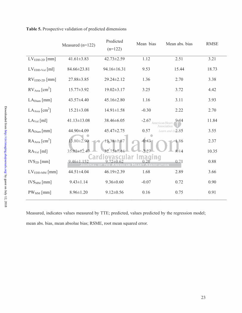
Table 5 describes the accuracy of measures predicted by the regression model (test data set,
n=500) in 122 study participants. Best agreement was found for 2-D measurements (root
mean squared error (RMSE) 0.88 – 3.93) and M-Mode measurements (RMSE 0.90 – 3.66).
Larger deviations, as expected, were observed for areas (RMSE 2.37 – 4.42) and volume
estimates (RMSE 10.35 - 18.73).
Calculator
llllllllylylylylylyly sssssssigigigigigigignininininininififififififificacacacacacacantntntntntntnt iiiiiiinnnnnnn
anddddddd heheheheheheheigigigigigigighththththththt wwwwwweeeere e, g y , g g
t
ura of the model as reflected b similar coefficients of dete m
, g y , g g
usususu ing BSSSSAAA AA ininini ststststs eaeaeaeaead d ddd ofofofofof hhhheeeighghght anannd dddd weweweweweigigigii hthtt dddddididid nnnnnoootoo iiiincncncncn rerereereasasssseee e t
uracy offfff thhhe mommmm ddel asaaaa reflectedddd bbbbbyy iisiiimimimimilalallalar coeffffififificienttts of ff ddeterm
by guest on July 12, 2018http://circim
aging.ahajournals.org/D
ownloaded from

10
Based on the results of the multivariable regression analysis we designed a calculator, which
estimates cardiac dimensions and accounts for gender, age, height, and weight.
by guest on July 12, 2018http://circim
aging.ahajournals.org/D
ownloaded from
11
Discussion
Whether the heart of an individual patient is enlarged or not bears important information in
clinical cardiology. Surprisingly, though, the evidence concerning reference values of the
normal sized heart is scarce. Most publications in the field were solely focused on the left
ventricle 3-5, 7-10 (Table 1), not taking the right ventricle or atria into account. Moreover,
almost all of these papers go back to the 1980s and 1990s when M-Mode was the primary
echocardiographic modality. Only Triulzi et al. and Lauer et al. examined the influence of
body size on adult left ventricular dimension variability, and also described gender specific
differences 6, 9, 12.
The acquisition of reproducible M-Mode measurements of the left ventricular cavity size,
however, is challenging. Today, 2D imaging of the LV from the apical 4-chamber-view is the
mainstay. Nevertheless, to our knowledge no systematic reference values of the normal sized
heart on the basis of 2D measurements exist.
Although the prognostic importance of left atrial area and volume has repeatedly been pointed
out, data regarding the normal size of the left atrial area or volume are very limited 6, 30. Data
on the normal sized right ventricle and right atrium are even more limited. Recent guidelines
on echocardiographic assessment of the right heart in adults 11 lack information concerning a
potential impact of body size, gender, or age.
Due to the lack of published indexing recommendations, clinical cardiologists at present may
use the same normal range of cardiac cavity dimensions for everybody, including competitive
athletes, and old, frail patients. This fact frequently causes diagnostic ambiguity. The stakes
for resolving such diagnostic ambiguity are high as consequences for the individual patient
may be considerable. Particularly in patients with small body dimensions the diagnosis of
cardiac enlargement may not be made because of an erroneous underestimation, leading to
delayed treatment and poor prognosis.
eeeeeee lllllllefefefefefefeft t t t tt t veveveveveveventntntntntntntririrrrrr cucucucucucuculalalalalalala
e apiciciciciciccalalalalalalal 4444444-c-c-cc-c-c-chahahahahahahambmbmmmmm
h
s
g ostic im rtance of left atrial area and volume has re ated y
hehehehh lllell ss, to oooooururur kkkkknononononowlwlwlwlwledededededgegeggg nnno syysttemememmmatatatataticicicc reefefferere enenenenenccce vvvvvalalalalalueueueueues sss ofofofofof ttttthehhhh
s of ffff 2D2D2D2D2D mmeaeaeasususurereremmem ntntntntntss s eexee issssttt.tt
gngngnososostititiccc imimimpopopop rtrtrtananancecece ooofff leleleftftft aaatrtrtriaiaialll ararareaeaea aaaaandndnd vvvolololumumumeee hahahasss rererepepepepp atatatedededlylyly
by guest on July 12, 2018http://circim
aging.ahajournals.org/D
ownloaded from
12
Such a causal relationship has been described several years ago 20. Cut-off values of left
ventricular size indicating the need for surgery in aortic regurgitation 27, 28 are based on 7
studies published between 1991 and 2006 13-19. The proportion of female patients in these
studies was low with an average of 19.1% (range 13%-24%). The work of Klodas and
coworkers 20 demonstrated an excess late mortality of women with aortic regurgitation after
valve surgery when these cut-off values were applied. This observation was explained by
delayed surgery in women, who never reached the cut-off values presumably due to smaller
hearts at baseline. The European guidelines for the management of valvular heart disease 28
state that the patient’s stature should be considered and that indexing is helpful for decision
making. Aortic valve replacement for aortic regurgitation, for instance, is recommended when
the LV end-systolic diameter exceeds 25mm/m² BSA. However, this cut-off value again was
derived from those studies, which comprised less than 20% women 13-19.
Current scaling methods mostly normalize cardiovascular structures to BSA by simply using
the form y=x/BSA, where x is the cardiovascular parameter, and y is the scaled cardiovascular
parameter 1. It is noteworthy, that BSA quantifies both muscle, adipose tissue, and
extravascular fluid volumes. The contribution of adipose tissue and extravascular fluid to
BSA, however, may vary widely between males and females and in dependence of age.
The aim of the present prospective study was to collect data of normal sized hearts, analyse
them with respect of body size, gender, and age. We furthermore provide a calculation tool to
estimate reference values for the normal sized heart.
Our study revealed considerable differences in cardiac size depending on gender, body size,
and age. The average male person in our cohort (1.79m, 85kg, 37years, BMI 25.9kg/m², BSA
2.03m²) had a left ventricular end-diastolic diameter (2D) of 46.0±3.8mm compared with the
average female (1.64m, 66kg, 43years, BMI 24.5kg/m², BSA 1.73m²) whose left ventricular
end-diastolic diameter was significantly smaller (41.4±3.5 mm, p<0.001). Furthermore, when
we compared a young tall man (1.95m, 115kg, 34years, BMI 30.2kg/m², BSA2.50m²) with an
nsnsssssstatatatatatatancncncncncncnce,e,e,e,e,e,e, iiiiiiisssssss rererererererecococococococommmmmmmmmmmmmm
r thhisisisisisisis cccccccututututututut-o-o-o-o-o-ooffffffffffffff vvvvvvvala
s
m
A c
seeeee studies, whwhwhww icicicicich hh hh cococococompmpmpmpmprirririr sseses d d lelelll sss tthhhan n n n n 202002020% %%% % wowowooomemmmm n n nn n 13-13-13-13-13-1919191919.
methththththodododododssss momomostststlylylylyl nnnormamamamamalllill ze cccccarararara dididididiovovovaaasascucucucuculalalalalarrr rr ststststs rurururr ctctctuuru essss tttooo o BSBSBSB AAAAA bybbyb
AAA, whwhwhererereee xxx isisis ttthehehe cccararardididiovovovasasascucuculalalarrr papapapp rararamemememm teteter,r,r, aaandndnd yyyyy iiisss thththeee scscscalalalededed ccc
by guest on July 12, 2018http://circim
aging.ahajournals.org/D
ownloaded from
13
elderly small lady (1.51m, 48kg, 83years, BMI 21.1kg/m², BSA 1.42m²), left ventricular end-
diastolic diameters were 50.4mm (95%CI, 44.1; 56.7) versus 37.4mm (95%CI, 31.1; 43.7),
respectively.
This demonstrates that tall and rather heavy individuals consistently meet accepted thresholds
for normal left ventricular end-diastolic diameters 3-10. However, a small (female) person may
suffer from a significantly dilated left ventricle although the end-diastolic left ventricular
diameter is still far below 50mm, but still may be classified as normal.
The present study clearly shows that besides height and weight, gender, and age are
significant predictors of cardiac cavity dimensions. Thus, a thorough assessment of an
individuals heart should include the consideration of all four variables.
Limitations
Our data have been collected in a single center in central Europe, thus the ethnic background
of our study population was mainly Caucasian. No Asian or African individuals were studied.
Thus, conclusions concerning other ethnic populations are limited.
Conclusions
The present work shows that gender, age, and body size affect the normal heart size. These
parameters need to be considered when cut-off values indicating the need for treatment or
even surgery are applied.
Sources of Funding
This study received support from the Austrian Society of Cardiology (to JM), the
Österreichischer Herzfonds (to JM) and the Austrian fellowship grant KLI 245 (to JM).
Disclosures
None.
iaiaiaiaiaiaablblblblblblb eseseseseseses.......
een collected in a si le center in central Eur e, thus the ethni
ulation was main Caucasian. No Asian or African individuals
s
eenenenne collectctcttededededed iiiiinn nnn a a a aa sisisisisingnngnn lelelelele cccceenteteteteerrr inn ccenenenenentrtrttrtralalalalal EEEEEururu opopopopopeee, ttttthuhuhuhuhus sss s thththththe eee etetetetethnhnhnhnhni
ulatttttioioioioionnnnn wawawasss mamamaininlylyly CCCCCauauauauaucasisisisianananana . NoNoNoNN AAAAAsisiisiananananan or rrrr AfAfAfAfAf iririiiccac n nn inininininddiddd vividudududd allalllss s
sss cococoncncncerererrnininingngng ooothththererer eeethththnininiccc popopopp pupupupp lalalatititionononsss ararareee lililimimimiteteteddd.
by guest on July 12, 2018http://circim
aging.ahajournals.org/D
ownloaded from

14
References
1. Dewey FE, Rosenthal D, Murphy DJ, Jr., Froelicher VF, Ashley EA. Does size matter? Clinical applications of scaling cardiac size and function for body size. Circulation. 2008;117:2279-2287.
2. Lancellotti P, Badano LP, Lang RM, Akhaladze N, Athanassopoulos GD, Barone D, Baroni M, Cardim N, Gomez de Diego JJ, Derumeaux G, Dulgheru R, Edvardsen T, Galderisi M, Goncalves A, Habib G, Hagendorff A, Hristova K, Kou S, Lopez T, Magne J, de la Morena G, Popescu BA, Penicka M, Rasit T, Rodrigo Carbonero JD, Salustri A, Van de Veire N, von Bardeleben RS, Vinereanu D, Voigt JU, Voilliot D, Zamorano JL, Donal E, Maurer G. Normal reference ranges for echocardiography: Rationale, study design, and methodology (norre study). European heart journal cardiovascular Imaging. 2013;14:303-308.
3. Gardin JM, Henry WL, Savage DD, Ware JH, Burn C, Borer JS. Echocardiographic measurements in normal subjects: Evaluation of an adult population without clinically apparent heart disease. J Clin Ultrasound. 1979;7:439-447.
4. Henry WL, Gardin JM, Ware JH. Echocardiographic measurements in normal subjects from infancy to old age. Circulation. 1980;62:1054-1061.
5. Devereux RB, Lutas EM, Casale PN, Kligfield P, Eisenberg RR, Hammond IW, Miller DH, Reis G, Alderman MH, Laragh JH. Standardization of m-mode echocardiographic left ventricular anatomic measurements. J Am Coll Cardiol. 1984;4:1222-1230.
6. Triulzi M, Gilliam LD, Gentile R, Newell JB, Weyman AE. Normal adult cross-sectional echocardiographic values: Linear dimensions and chamber areas. Echcardiography. 1984;1:403-426.
7. Hammond IW, Devereux RB, Alderman MH, Laragh JH. Relation of blood pressure and body build to left ventricular mass in normotensive and hypertensive employed adults. J Am Coll Cardiol. 1988;12:996-1004.
8. Lauer MS, Anderson KM, Kannel WB, Levy D. The impact of obesity on left ventricular mass and geometry. The framingham heart study. Jama. 1991;266:231-236.
9. Lauer MS, Larson MG, Levy D. Gender-specific reference m-mode values in adults: Population-derived values with consideration of the impact of height. J Am Coll Cardiol. 1995;26:1039-1046.
10. George KP, Batterham AM, Jones B. The impact of scalar variable and process on athlete-control comparisons of cardiac dimensions. Med Sci Sports Exerc. 1998;30:824-830.
11. Rudski LG, Lai WW, Afilalo J, Hua L, Handschumacher MD, Chandrasekaran K, Solomon SD, Louie EK, Schiller NB. Guidelines for the echocardiographic assessment of the right heart in adults: A report from the american society of echocardiography endorsed by the european association of echocardiography, a registered branch of the european society of cardiology, and the canadian society of echocardiography. J Am Soc Echocardiogr. 2010;23:685-713; quiz 786-688.
12. Triulzi MO, Wilkins GT, Gilliam LD, Gentile F, Weyman AE. Normal adult cross-sectional echocardiographic values: Left ventricular volumes. Echcardiography. 1985;2:153-169.
13. Dujardin KS, Enriquez-Sarano M, Schaff HV, Bailey KR, Seward JB, Tajik AJ. Mortality and morbidity of aortic regurgitation in clinical practice. A long-term follow-up study. Circulation. 1999;99:1851-1857.
14. Tarasoutchi F, Grinberg M, Spina GS, Sampaio RO, Cardoso LF, Rossi EG, Pomerantzeff P, Laurindo F, da Luz PL, Ramires JA. Ten-year clinical laboratory
erererererererggggggg RRRRRRRRRRRRRR,, HaHaHaHaHaHaHammmmmmmmmmmmmmoooooooizatitiiiiiiononononononon ooooooof f fffff mm-m-m-m-mm momomomomomomoddddddd
nts JJJJJJJ AmAmAmAmAmAmAm CCCCCCCololololololollllllll CaCCaCCCC2, Gilliam LD, Gentile R, Newell JB, We an AE. Normal adulte sod IW, Devereux RB, Alderman MH, Lar h JH. Relation of bl o
Am Coll Cardiol 1988;12:996 1004
222222-2-2-2-2-12121212123030303030. , GGGiGG lliam LDLDLDLDLD, GeGeGeGeGentntntntntililililileeeee RRRR,R NNNNNewwwelee l JBBB,,, WeWeWeWeWeymymymyy ananann AAAAAE..... NNNNNororororormamamamam l ll l adadadadaduluuuu tececccchhohhh cardddioi grgrrappphhhic vvvavv lueseseee : Linenearr dddimmmmmenennssiononns anannd cchccc ammmmmbebebbb rr arreeeasograaaaaphphphphphyyy. 1919199848484;1;1;1:4:4:4033333 44-444226222 .d IW, Deverrrrreueueueueuxxxxx RBRBRBRBRB, , , AlAlAlAlAldededededermrmrmrmrmananananan MMMMMH,H,H,H,H LLLaraaragagagagh h h hh JHJHJHJHJH. ReReReReRelalalallation of bloobbbuiuiuildldld ttto o o o leleleftftft vvvenenentrtrtriciciculululararar mmmasasassss ininin nnnororormomomoootetetensnsnsiviviveee anananddd hyhyhyyypepepepp rtrtrtenenensisisiveveve
AmAm CColollllll CaCaCC rdrddddioioiolll 191919888888;1;1;12:2:2:9999996-66 101010040404
by guest on July 12, 2018http://circim
aging.ahajournals.org/D
ownloaded from
15
follow-up after application of a symptom-based therapeutic strategy to patients with severe chronic aortic regurgitation of predominant rheumatic etiology. J Am Coll Cardiol. 2003;41:1316-1324.
15. Corti R, Binggeli C, Turina M, Jenni R, Luscher TF, Turina J. Predictors of long-term survival after valve replacement for chronic aortic regurgitation; is m-mode echocardiography sufficient? Eur Heart J. 2001;22:866-873.
16. Tornos P, Sambola A, Permanyer-Miralda G, Evangelista A, Gomez Z, Soler-Soler J. Long-term outcome of surgically treated aortic regurgitation: Influence of guideline adherence toward early surgery. J Am Coll Cardiol. 2006;47:1012-1017.
17. Klodas E, Enriquez-Sarano M, Tajik AJ, Mullany CJ, Bailey KR, Seward JB. Aortic regurgitation complicated by extreme left ventricular dilation: Long-term outcome after surgical correction. J Am Coll Cardiol. 1996;27:670-677.
18. Bonow RO, Lakatos E, Maron BJ, Epstein SE. Serial long-term assessment of the natural history of asymptomatic patients with chronic aortic regurgitation and normal left ventricular systolic function. Circulation. 1991;84:1625-1635.
19. Gaasch WH, Schick EC. Symptoms and left ventricular size and function in patients with chronic aortic regurgitation. J Am Coll Cardiol. 2003;41:1325-1328.
20. Klodas E, Enriquez-Sarano M, Tajik AJ, Mullany CJ, Bailey KR, Seward JB. Surgery for aortic regurgitation in women. Contrasting indications and outcomes compared with men. Circulation. 1996;94:2472-2478.
21. Rosenhek R, Rader F, Klaar U, Gabriel H, Krejc M, Kalbeck D, Schemper M, Maurer G, Baumgartner H. Outcome of watchful waiting in asymptomatic severe mitral regurgitation. Circulation. 2006;113:2238-2244.
22. Wisenbaugh T, Skudicky D, Sareli P. Prediction of outcome after valve replacement for rheumatic mitral regurgitation in the era of chordal preservation. Circulation. 1994;89:191-197.
23. Zile MR, Gaasch WH, Carroll JD, Levine HJ. Chronic mitral regurgitation: Predictive value of preoperative echocardiographic indexes of left ventricular function and wall stress. J Am Coll Cardiol. 1984;3:235-242.
24. Enriquez-Sarano M, Tajik AJ, Schaff HV, Orszulak TA, McGoon MD, Bailey KR, Frye RL. Echocardiographic prediction of left ventricular function after correction of mitral regurgitation: Results and clinical implications. J Am Coll Cardiol. 1994;24:1536-1543.
25. Schuler G, Peterson KL, Johnson A, Francis G, Dennish G, Utley J, Daily PO, Ashburn W, Ross J, Jr. Temporal response of left ventricular performance to mitral valve surgery. Circulation. 1979;59:1218-1231.
26. Gaasch WH, John RM, Aurigemma GP. Managing asymptomatic patients with chronic mitral regurgitation. Chest. 1995;108:842-847.
27. Bonow RO, Carabello BA, Kanu C, de Leon AC, Jr., Faxon DP, Freed MD, Gaasch WH, Lytle BW, Nishimura RA, O'Gara PT, O'Rourke RA, Otto CM, Shah PM, Shanewise JS, Smith SC, Jr., Jacobs AK, Adams CD, Anderson JL, Antman EM, Fuster V, Halperin JL, Hiratzka LF, Hunt SA, Nishimura R, Page RL, Riegel B. Acc/aha 2006 guidelines for the management of patients with valvular heart disease: A report of the american college of cardiology/american heart association task force on practice guidelines (writing committee to revise the 1998 guidelines for the management of patients with valvular heart disease): Developed in collaboration with the society of cardiovascular anesthesiologists: Endorsed by the society for cardiovascular angiography and interventions and the society of thoracic surgeons. Circulation. 2006;114:e84-231.
28. Vahanian A, Alfieri O, Andreotti F, Antunes MJ, Baron-Esquivias G, Baumgartner H, Borger MA, Carrel TP, De Bonis M, Evangelista A, Falk V, Iung B, Lancellotti P,
sssssss aaaaaaandndndndndndnd oooooooutututututututcococococococomememememememes s ss s s s ccccccc
beckkkkkkk DDDDDDD SSSSSSSchchchchchchchememememememempeppppgartner H. Outcome of watchful waiting in asymptomatic seve e
ugh T, Skudicky D, Sareli P. Prediction of outcome after valve em u1Gaasch WH, Carroll JD, Levine HJ. Chronic mitral re itat o
preoperative echocardiographic indexes of left ventricular functio
gararararrtntntntntner HHHHH. OuOuOuOuOutcome of watchful wawawaw iting in aaaaasympmmmm tomatic severeioooonn.nn Circululullulataaaa iooooonnnnn. 20202020200606060606;1;1;1;1;111313131 :2222232323232 8--22224444444444.
ughghghghgh T, Skkududuuu icckyyy D, , ,, SSSaSS reeeeelill PPP. PrPrreddiccctiiiiiononoono oof ououutcomommmmeeeee afteeeeerr rrr vavalvee rematiiiic cccc mimimimimitrtrtrtrt alllll rrregegegurururgig taaatitititiiooonoo in nnnn thththhtheeeee erereraaa ofofofofof ccccchohohohohordrdrdrdrdalalalalal ppprereresses rvrvrvrvvatatatattioioii nnn. CiCiCiCC rcrcrcu191-197. GGGaaaaaascscschhh WHWHWH,,, CaCaCarrrrrrololollll JDJDJD, LeLeLevivivinenene HHHJJJ. CCCCChrhrhronononicicic mmmitititrararall l rereregugugugg rgrgrgitititatatatioioio
ppprerereopopoperereratatativiviveee ececechohohocacacardrdrdioioiogrgrgrapapaphihihiccc ininindededexexexesss ofofof lllefefefttt veveventntntririricucuculalalarrr fufufuncncnctititiooo
by guest on July 12, 2018http://circim
aging.ahajournals.org/D
ownloaded from

16
Pierard L, Price S, Schafers HJ, Schuler G, Stepinska J, Swedberg K, Takkenberg J, Von Oppell UO, Windecker S, Zamorano JL, Zembala M. Guidelines on the management of valvular heart disease (version 2012). European heart journal. 2012;33:2451-2496.
29. Lang RM, Bierig M, Devereux RB, Flachskampf FA, Foster E, Pellikka PA, Picard MH, Roman MJ, Seward J, Shanewise J, Solomon S, Spencer KT, St John Sutton M, Stewart W. Recommendations for chamber quantification. Eur J Echocardiogr. 2006;7:79-108.
30. Pearlman JD, Triulzi MO, King ME, Abascal VM, Newell J, Weyman AE. Left atrial dimensions in growth and development: Normal limits for two-dimensional echocardiography. Journal of the American College of Cardiology. 1990;16:1168-1174.
by guest on July 12, 2018http://circim
aging.ahajournals.org/D
ownloaded from

17
Table 1. Publications on normal values of left ventricular cavity dimensions by
echocardiography
Author, year of publication, reference
n n women
n men
Age LVEDD women
LVEDDmen
Mode
Henry, 1980 [4]
136 58 (34%) 78 (57%) 20-97 Regression Equations and Graphs
MM
Devereux, 1984 [5]
133 55 (41%) 78 (59%) 44±12 45±4 50±5 MM
Triulzi, 1984 [6]
72 34 (47%) 38 (53 %) 15-76 Regression Equations 2D
Hammond, 1988 [7]
162 44±13 50±5 MM
Lauer, 1991 [8]
2922 1666 (57%) 1256 (43%) 30-62 48±3 51±4 MM
Lauer, 1995 [9]
812 524 (65%) 288 (35%) 20-45 46.1±3.0 50.8±3.6 MM 914 503 (55%) 411 (45%) 20-45 47.5±3.6 51.1±3.7
George, 1998 [10]
45 45 (100%) 22±2 52.4±3.3 MM
LVEDD indicates left ventricular end-diastolic diameter; and MM, motion mode and 2D, two-
dimensional.
448±8±±±±33
812 524 (65%) 288 (35%) 20-45 46.1±3.0 501
2
812222 52222244444 (65%) 288 (35%%%%)))) 20-45 464444 .1±3.0 5091919191914 44 503333 (55%5%5%%) )))) 411 (4((( 5%) 20200-45 555 5 4744 .5±333.6 51
4545454545 455555 (((10101010100%0%0%0%0%))) 2222222222±2±2±2±2±2 525555
by guest on July 12, 2018http://circim
aging.ahajournals.org/D
ownloaded from
18
Table 2. Baseline characteristics
All
n=622
Range
Women
n=328
Range
Men
n=294
Range
p-value
Age [years] 41.8±15.5 17-91 43.8±14.8 17-91 38.3±16.0 17-82 <0.0001
Height [cm] 169.6±10.0 143-200 164.2±6.9 143-183 178.6±7.8 156-200 <0.0001
Weight [kg] 72.6±18.6 32-240 66.1±14.0 32-128 83.4±20.4 54-240 <0.0001
BMI [kg/m²] 25.1±5.6 12.8-74.1 24.6±5.4 12.8-50.0 26.1±5.8 16.1-74.1 0.001
BSA [m²] 1.8±0.3 1.2-3.5 1.7±0.2 1.2-2.4 2.0±0.2 1.6-3.5 <0.0001
BMI indicates body mass index; BSA, body surface area.
by guest on July 12, 2018http://circim
aging.ahajournals.org/D
ownloaded from
19
Table 3. Results of the multivariable regression model, 2D measurements
Variable Regression
Coefficients 95% Confidence Intervall Limits p-value
LVEDD-2D
Intercept 20.98637 13.82007 28.15268 <.0001
Weight 0.07404 0.05687 0.09121 <.0001
Height 0.11429 0.07359 0.15499 <.0001
Gender -1.45756 -2.25856 -0.65657 0.0004
Age -0.04141 -0.06023 -0.02260 <.0001
LVEDD-Vol
Intercept -39.37731 -91.07454 12.31991 0.1351
Weight 0.42808 0.30268 0.55349 <.0001
Height 0.73703 0.44358 1.03049 <.0001
Gender -9.47838 -15.24652 -3.71023 0.0013
Age -0.33895 -0.47457 -0.20332 <.0001
RVEDD-2D
Intercept 15.94580 9.13081 22.76078 <.0001
Weight 0.04375 0.02731 0.06020 <.0001
Height 0.07014 0.02731 0.06020 0.0005
Gender -2.35620 -3.15617 -1.55622 <.0001
RVArea
Intercept -0.97314 -10.35089 8.40461 0.8384
Weight 0.09270 0.06347 0.12193 <.0001
Height 0.10040 0.04619 0.15460 0.0003
Gender -2.62433 -3.69133 -1.55733 <.0001
12 33333331919191919191991919191919191
0.42808 0.30268 0.55349
0.73703 0.44358 1.03049
-9.47838 -15.24652 -3.71023
0.4444422280808080808 00.302022688888 00000.5.555553535353534949494949
0.77773737370303003 00000 44.44343434343585858588 1.00000303003030499999
-9.9.9.9.4747474 838383838 8 8 1-1-1155.5.5.24242446565656 2 2 2 22 3-3-33 7.7.777101010101 23232323
by guest on July 12, 2018http://circim
aging.ahajournals.org/D
ownloaded from

20
Age -0.03906 -0.06443 -0.01369 0.0027
LADiam
Intercept 34.23014 32.30756 36.15272 <.0001
Weight 0.11356 0.09181 0.13530 <.0001
Gender -1.73000 -2.59145 -0.86855 <.0001
Age 0.09453 0.07041 0.11866 <.0001
LAArea
Intercept -4.07280 -9.31691 1.17130 0.1277
Weight 0.05673 0.04028 0.07317 <.0001
Height 0.08135 0.04926 0.11344 <.0001
Age 0.02779 0.00956 0.04602 0.0029
LAVol
Intercept -34.73810 -57.37315 -12.10305 0.0027
Weight 0.21533 0.14446 0.28620 <.0001
Height 0.31554 0.17710 0.45397 <.0001
Age 0.10538 0.02674 0.18403 0.0087
RADiam
Intercept 35.52012 33.62341 37.41683 <.0001
Weight 0.10712 0.08567 0.12857 <.0001
Gender -2.03817 -2.88803 -1.18831 <.0001
Age 0.08743 0.06364 0.11123 <.0001
RAArea
Intercept 9.15398 7.54409 10.76388 <.0001
Weight 0.07818 0.05971 0.09665 <.0001
Gender -1.85918 -2.55769 -1.16067 <.0001
0000000.1.1.1.1.11.11313131313131344444444444444
0 04040404040404606060606060602222222
-34.73810 -57.37315 -12.10305
0.21533 0.14446 0.28620
3-334.44 7373738181818 00 000 555-55777.7 37373737373131313115555 -12.2222 101010010300000555
0000.2.2.2215151553333333 0000.1.1.1.144444444646464 0000.2.2.2. 868686868 20202020
by guest on July 12, 2018http://circim
aging.ahajournals.org/D
ownloaded from

21
RAVol
Intercept 18.97912 12.61130 25.34693 <.0001
Weight 0.27999 0.20694 0.35304 <.0001
Gender -8.55635 -11.31925 -5.79344 <.0001
IVS2D
Intercept 7.66249 7.19746 8.12753 <.0001
Weight 0.02031 0.01505 0.02557 <.0001
Gender -0.56512 -0.77349 -0.35675 <.0001
Age 0.02344 0.01761 0.02928 <.0001
LV indicates left ventricle; EDD, end-diastolic diameter; Vol, volume; 2D, two dimensional;
RV, right ventricle; LA, left atrium; Diam, diameter; RA, right atrium; and IVS,
interventricular septum.
volummmmmmme;e;e;e;e;e;e; 2222222D,D,D,D,D,D,D tttttttwowowwwww
r m
e
riclclclclcleee;ee LLLLLAAAA,A left atrium; Diam, diameterrrrr;;;; RARARARARA, right atrium
epepepepeptttutt m.
by guest on July 12, 2018http://circim
aging.ahajournals.org/D
ownloaded from
22
Table 4. Results of the multivariable regression model, M Mode measurements
Variable Regression
Coefficients 95% Confidence Intervall Limits p-value
LVEDD-MM
Intercept 15.38196 8.54423 22.21970 <.0001
Weight 0.07658 0.05280 0.10036 <.0001
Height 0.15007 0.10616 0.19397 <.0001
LVESD-MM
Intercept 26.76878 24.90119 28.63637 <.0001
Weight 0.04765 0.02621 0.06909 <.0001
Gender -2.11032 -2.93269 -1.28795 <.0001
IVSMM
Intercept 7.34011 6.80535 7.87488 <.0001
Weight 0.01872 0.01266 0.02477 <.0001
Gender -0.53678 -0.77686 -0.29671 <.0001
Age 0.02469 0.01797 0.03141 <.0001
PWMM
Intercept 7.49088 6.95555 8.02622 <.0001
Weight 0.01964 0.01357 0.02570 <.0001
Gender -0.57818 -0.81862 -0.33775 <.0001
Age 0.01489 0.00816 0.02162 <.0001
LV indicates left ventricle; EDD, end-diastolic diameter; MM, motion mode; ESD, end-
systolic diameter; IVS, interventricular septum; and PW, posterior wall.
0.06909
--1.1.1.1.1.1.1.28282828282828797979797979795 5 5 5555
7.34011 6.80535 7.87488
0.01872 0.01266 0.02477
77.777 34343434340101010 11111 6.6666 808080808053535353535 5555 7.7.7.77.87878787874848848888 888
000.0.0.0181818727272 000.0.0.0121212666666 00000.0.0.0242424777777
by guest on July 12, 2018http://circim
aging.ahajournals.org/D
ownloaded from
23
Table 5. Prospective validation of predicted dimensions
Measured (n=122) Predicted
(n=122) Mean bias Mean abs. bias RMSE
LVEDD-2D [mm] 41.61±3.83 42.73±2.59 1.12 2.51 3.21
LVEDD-Vol [ml] 84.66±23.81 94.16±16.31 9.53 15.44 18.73
RVEDD-2D [mm] 27.88±3.85 29.24±2.12 1.36 2.70 3.38
RVArea [cm2] 15.77±3.92 19.02±3.17 3.25 3.72 4.42
LADiam [mm] 43.57±4.40 45.16±2.80 1.16 3.11 3.93
LAArea [cm2] 15.21±3.08 14.91±1.58 -0.30 2.22 2.70
LAVol [ml] 41.13±13.08 38.46±6.05 -2.67 9.44 11.84
RADiam [mm] 44.90±4.09 45.47±2.75 0.57 2.85 3.55
RAArea [cm2] 13.80±2.90 13.38±1.87 -0.43 1.86 2.37
RAVol [ml] 35.02±12.47 32.75±7.44 -2.27 8.14 10.35
IVS2D [mm] 9.46±1.152 9.72±0.62 0.26 0.71 0.88
LVEDD-MM [mm] 44.51±4.04 46.19±2.39 1.68 2.89 3.66
IVSMM [mm] 9.43±1.14 9.36±0.60 -0.07 0.72 0.90
PWMM [mm] 8.96±1.20 9.12±0.56 0.16 0.75 0.91
Measured, indicates values measured by TTE; predicted, values predicted by the regression model;
mean abs. bias, mean absolue bias; RSME, root mean squared error.
9.9.9.9.9.9.9.444444444
2.2.2.2.2.22 858
13.80±2.90 13.38±1.87 -0.43 1. 6
3 4
1313131313.80±2.222 9090909090 111113.3.3.33 38338388±1±1±1±1.8.88.87 -0-0-00.44433333 1.1.1.1.1 86
35555 00.0002±2±2±2±2±1212121212.4444477777 3232323232.75±5±5±5±5±7.7777 4444444444 -2.2222 2727272727 8.888 1411
99.4646±1±1 1.15252 99 7.72±2±00.6262 00 2.266666 00.77
by guest on July 12, 2018http://circim
aging.ahajournals.org/D
ownloaded from
Lolic, Diana Bonderman, Helmut Baumgartner, Gerald Maurer and Julia MascherbauerStefan Pfaffenberger, Philipp Bartko, Alexandra Graf, Elisabeth Pernicka, Jamil Babayev, Emina
Size Matters!: Impact of Age, Gender, Height, and Weight on the Normal Heart Size
Print ISSN: 1941-9651. Online ISSN: 1942-0080 Copyright © 2013 American Heart Association, Inc. All rights reserved.
TX 75231is published by the American Heart Association, 7272 Greenville Avenue, Dallas,Circulation: Cardiovascular Imaging
published online September 6, 2013;Circ Cardiovasc Imaging.
http://circimaging.ahajournals.org/content/early/2013/09/06/CIRCIMAGING.113.000690World Wide Web at:
The online version of this article, along with updated information and services, is located on the
http://circimaging.ahajournals.org/content/suppl/2013/09/06/CIRCIMAGING.113.000690.DC1Data Supplement (unedited) at:
http://circimaging.ahajournals.org//subscriptions/
is online at: Circulation: Cardiovascular Imaging Information about subscribing to Subscriptions:
http://www.lww.com/reprints Information about reprints can be found online at: Reprints:
document. Permissions and Rights Question and Answer this process is available in the
located, click Request Permissions in the middle column of the Web page under Services. Further information aboutnot the Editorial Office. Once the online version of the published article for which permission is being requested is
can be obtained via RightsLink, a service of the Copyright Clearance Center,Circulation: Cardiovascular Imaging Requests for permissions to reproduce figures, tables, or portions of articles originally published inPermissions:
by guest on July 12, 2018http://circim
aging.ahajournals.org/D
ownloaded from