Embed Size (px)
Citation preview
SIXTH GRADE HUMAN BIOLOGY
2 WEEKS LESSON PLANS AND
ACTIVITIES
Math/Science Nucleus ©1990,2000 2
LIFE CYCLEOVERVIEW OF SIXTH GRADE
ORGANISMSWEEK 1.PRE: Defining classification. LAB: Exploring characteristics of the lower kingdoms. POST: Comparing classification and taxonomy. WEEK 2.PRE: Exploring how food gets rotten. LAB: Discovering why food rots. POST: Defining the parameters of the kingdom system.
HUMAN BIOLOGYWEEK 3.PRE: Exploring the endocrine system. LAB: Analyzing the different stages of human growth.POST: Comparing mitosis and meiosis.WEEK 4.PRE: Distinguishing bacteria, protozoa, and viruses.LAB: Distinguishing bacteria and viruses.POST: Comparing genetic disorders with diseases.
PLANT LIFEWEEK 5. PRE: Distinguishing land from aquatic plants. LAB: Comparing growth factors of plants. POST: Exploring uses of auxins. WEEK 6.PRE: Exploring the history of genetics. LAB: Testing heredity models. POST: Developing a mutation theory.
NATURAL ENVIRONMENTWEEK 7.PRE: Exploring ecosystem requirements. LAB: Comparing the pH of different soils. POST: Interpreting the results of soil pH. WEEK 8.PRE: Adapting to the local environment. LAB: Researching factors on adaptation. POST: Comparing different theories on evolution.
Math/Science Nucleus ©1990,2000 3
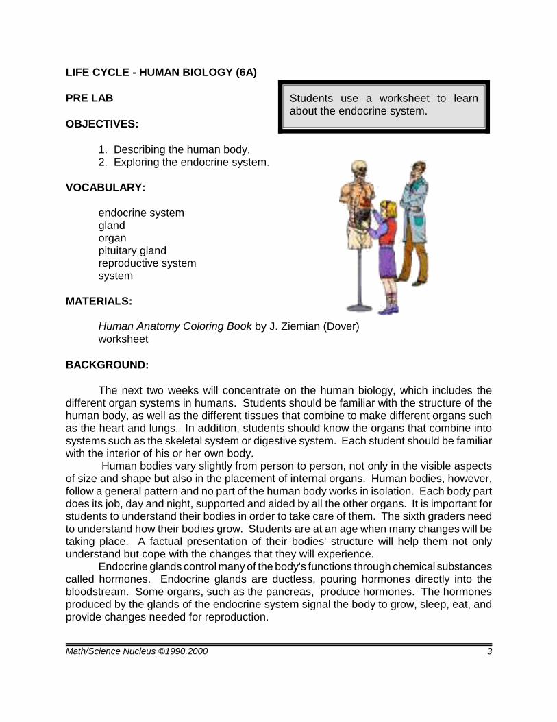
Students use a worksheet to learnabout the endocrine system.
LIFE CYCLE - HUMAN BIOLOGY (6A)
PRE LAB
OBJECTIVES:
1. Describing the human body.2. Exploring the endocrine system.
VOCABULARY:
endocrine systemgland organpituitary glandreproductive systemsystem
MATERIALS:
Human Anatomy Coloring Book by J. Ziemian (Dover)worksheet
BACKGROUND:
The next two weeks will concentrate on the human biology, which includes thedifferent organ systems in humans. Students should be familiar with the structure of thehuman body, as well as the different tissues that combine to make different organs suchas the heart and lungs. In addition, students should know the organs that combine intosystems such as the skeletal system or digestive system. Each student should be familiarwith the interior of his or her own body. Human bodies vary slightly from person to person, not only in the visible aspectsof size and shape but also in the placement of internal organs. Human bodies, however,follow a general pattern and no part of the human body works in isolation. Each body partdoes its job, day and night, supported and aided by all the other organs. It is important forstudents to understand their bodies in order to take care of them. The sixth graders needto understand how their bodies grow. Students are at an age when many changes will betaking place. A factual presentation of their bodies' structure will help them not onlyunderstand but cope with the changes that they will experience.
Endocrine glands control many of the body's functions through chemical substancescalled hormones. Endocrine glands are ductless, pouring hormones directly into thebloodstream. Some organs, such as the pancreas, produce hormones. The hormonesproduced by the glands of the endocrine system signal the body to grow, sleep, eat, andprovide changes needed for reproduction.
Math/Science Nucleus ©1990,2000 4
The glands of the endocrine system include the pituitary, thymus, thyroid, andadrenal. On the worksheet the students can locate these glands. The thymus glandcontrols activities of the spleen and the lymph glands which are important in the immunesystem. The thyroid gland produces a hormone that regulates the metabolic rate. Eachof the adrenal glands provide hormones for emotions such as fright or anger. This reactionis responsible for the extraordinary feats of strength that people sometimes perform inemergencies.
The pituitary gland is important because it produces many growth hormones that areused throughout life. The hormones in the pituitary signal to the male and femalereproductive parts to start developing the adult male and female characteristics. Thehypothalamus gland regulates the output of the pituitary gland. It is the pituitary thatsignals the testes (male gland) to produce testosterone which tells a male body to growhair, increase bone growth, have greater muscle strength, and a deeper voice. Thehormones, estrogen and progesterone from the pituitary gland, regulate the work of thefemale's ovaries. Estrogens are important in the development of the adult female.Progesterone along with estrogen is needed to prepare a female body for pregnancy.
PROCEDURE:
1. If you feel the students lack an understanding, we suggest you do some of theexercises for the lower grades. You can also use Human Anatomy Coloring Book by J.Ziemian as a coloring/review of the human body.
2. Go over the worksheet with the student. See what parts they know already beforeshowing them the location of each of the organ. Please note, that students at this age will“giggle,” but if taught in a straight forward scientific way, the giggles will turn into curiosity.
3. Students will undoubtedly have questions. We suggest that you have availablebooks on the human body. You may want students to do a research paper. It is soimportant for them to realize that their bodies are human machines with all parts workingtogether to produce an effective and efficient product.
Math/Science Nucleus ©1990,2000 5
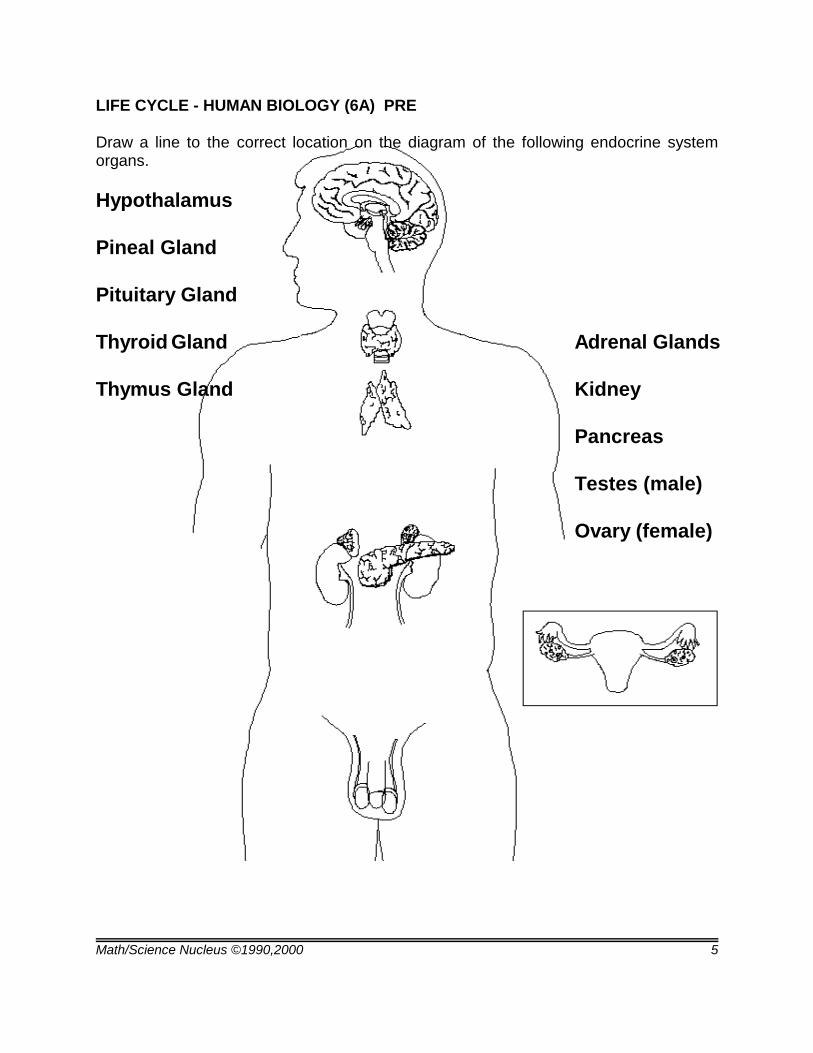
LIFE CYCLE - HUMAN BIOLOGY (6A) PRE
Draw a line to the correct location on the diagram of the following endocrine systemorgans.
Hypothalamus
Pineal Gland
Pituitary Gland
Thyroid Gland Adrenal Glands
Thymus Gland Kidney
Pancreas
Testes (male)
Ovary (female)
Math/Science Nucleus ©1990,2000 6
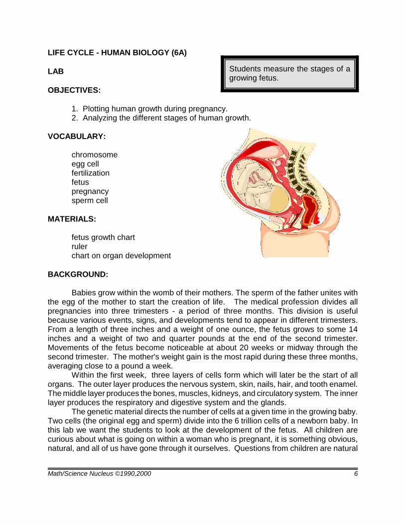
Students measure the stages of agrowing fetus.
LIFE CYCLE - HUMAN BIOLOGY (6A)
LAB
OBJECTIVES:
1. Plotting human growth during pregnancy.2. Analyzing the different stages of human growth.
VOCABULARY:
chromosomeegg cellfertilizationfetuspregnancysperm cell
MATERIALS:
fetus growth chartrulerchart on organ development
BACKGROUND:
Babies grow within the womb of their mothers. The sperm of the father unites withthe egg of the mother to start the creation of life. The medical profession divides allpregnancies into three trimesters - a period of three months. This division is usefulbecause various events, signs, and developments tend to appear in different trimesters.From a length of three inches and a weight of one ounce, the fetus grows to some 14inches and a weight of two and quarter pounds at the end of the second trimester.Movements of the fetus become noticeable at about 20 weeks or midway through thesecond trimester. The mother's weight gain is the most rapid during these three months,averaging close to a pound a week.
Within the first week, three layers of cells form which will later be the start of allorgans. The outer layer produces the nervous system, skin, nails, hair, and tooth enamel.The middle layer produces the bones, muscles, kidneys, and circulatory system. The innerlayer produces the respiratory and digestive system and the glands.
The genetic material directs the number of cells at a given time in the growing baby.Two cells (the original egg and sperm) divide into the 6 trillion cells of a newborn baby. Inthis lab we want the students to look at the development of the fetus. All children arecurious about what is going on within a woman who is pregnant, it is something obvious,natural, and all of us have gone through it ourselves. Questions from children are natural
Math/Science Nucleus ©1990,2000 7
and remember that this is not a morality issue, it is a fact of life. Describing and explainingpregnancy in a factual method, will make these students confident of what is going on.
PROCEDURE:
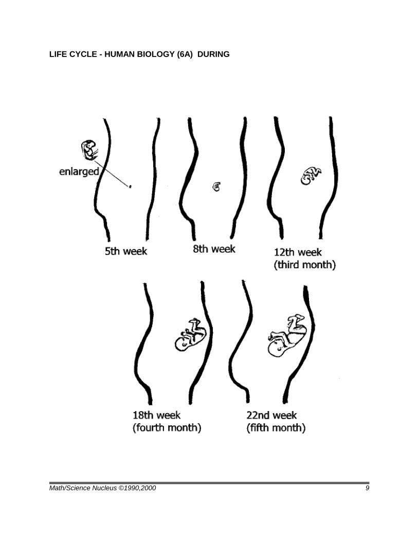
1.This lab looks at the development of the baby in the mother's womb. Studentsneed to measure the fetus during selected periods of growth. On the lab sheet graph,students need to obtain the following information: number of weeks of growth; the size ofthe fetus at that time; the difference between the last growth and the new growth; the rateof growth (growth/time); and the percent of growth.
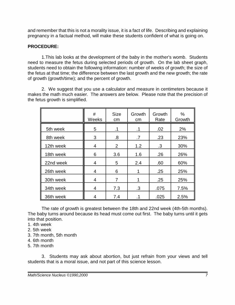
2. We suggest that you use a calculator and measure in centimeters because itmakes the math much easier. The answers are below. Please note that the precision ofthe fetus growth is simplified.
# Weeks
Sizecm
Growthcm
GrowthRate
%Growth
5th week 5 .1 .1 .02 2%
8th week 3 .8 .7 .23 23%
12th week 4 2 1.2 .3 30%
18th week 6 3.6 1.6 .26 26%
22nd week 4 5 2.4 .60 60%
26th week 4 6 1 .25 25%
30th week 4 7 1 .25 25%
34th week 4 7.3 .3 .075 7.5%
36th week 4 7.4 .1 .025 2.5%
The rate of growth is greatest between the 18th and 22nd week (4th-5th months).The baby turns around because its head must come out first. The baby turns until it getsinto that position.1. 4th week2. 5th week3. 7th month, 5th month4. 6th month5. 7th month
3. Students may ask about abortion, but just refrain from your views and tellstudents that is a moral issue, and not part of this science lesson.
Math/Science Nucleus ©1990,2000 8
LIFE CYCLE - HUMAN BIOLOGY (6A)
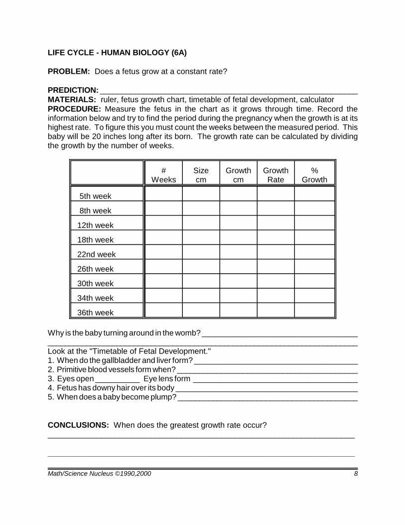
PROBLEM: Does a fetus grow at a constant rate?
PREDICTION: ___________________________________________________________MATERIALS: ruler, fetus growth chart, timetable of fetal development, calculatorPROCEDURE: Measure the fetus in the chart as it grows through time. Record theinformation below and try to find the period during the pregnancy when the growth is at itshighest rate. To figure this you must count the weeks between the measured period. Thisbaby will be 20 inches long after its born. The growth rate can be calculated by dividingthe growth by the number of weeks.
#Weeks
Sizecm
Growthcm
GrowthRate
%Growth
5th week
8th week
12th week
18th week
22nd week
26th week
30th week
34th week
36th week
Why is the baby turning around in the womb? ___________________________________________________________________________________________________________Look at the "Timetable of Fetal Development." 1. When do the gallbladder and liver form? _____________________________________2. Primitive blood vessels form when? _________________________________________3. Eyes open __________ Eye lens form _____________________________________4. Fetus has downy hair over its body _________________________________________5. When does a baby become plump? _________________________________________
CONCLUSIONS: When does the greatest growth rate occur? _____________________________________________________________________
_____________________________________________________________________
Math/Science Nucleus ©1990,2000 9
LIFE CYCLE - HUMAN BIOLOGY (6A) DURING
Math/Science Nucleus ©1990,2000 10
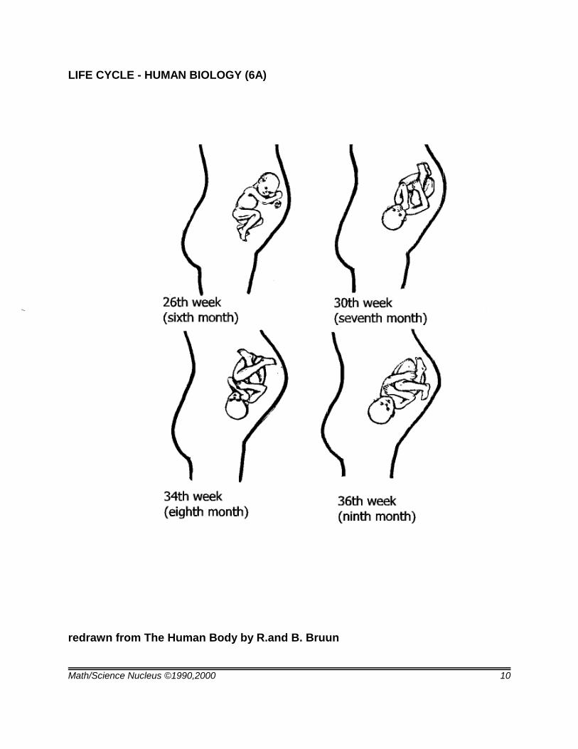
LIFE CYCLE - HUMAN BIOLOGY (6A)
redrawn from The Human Body by R.and B. Bruun
Math/Science Nucleus ©1990,2000 11
LIFE CYCLE - HUMAN BIOLOGY (6A) LAB
GROWTH OF A FETUS
5-9 days - Remains free in uterine cavity.
10-11 days - Attaches to and begins to become imbedded in the prepared lining of theuterus. The different tissue layers are developing.
14 days - Irregular, blob-like oval body with a longitudinal depression from which cells arepushed into an enlarging body.
18-21 days - Thickening of the neural plate, which is the first sign of the central nervoussystem. The primitive heart is a simple tube. There are primitive lung buds. Two faintdepressions are the sites of eyes. The embryo begins to curve head-to-tail to fit itsenvironment.
4th week - Beginning of gallbladder and liver tubules. Parts of brain begin differentiation.Local dilations indicates beginning of stomach. Heart tube becomes slightly bend. Noseparts suggested. Tiny liver and belly stalk. Primitive head parts, mount, brain, eyes, ears,are forming. Opening from mouth to gut breaks through; a little later, the anus. Primitivethyroid cells. Windpipe and larynx beginning. The heart is under the chin. First heartbeatoccurs. Blood corpuscles form, circulation begins.
5th week - Nasal pit, buds that will be arms and legs, cells that will develop into pancreasgland, tiny thickening that will be tongue appear. The gut elongates. Primitive bloodvessels function. Beginnings of eye lens, cranial nerves, retinal layer.
6th week - Arm and leg buds lengthen, faint grooves suggest toes and fingers. Brainrecognizable. Lung buds bifurcate. Primitive kidney established. Eyes far to either sideof head. Epithelium and primitive ear parts begin to form. Nasal pits recognizable asnostrils. Stomach suggests adult form. Salivary glands identifiable. Skeleton systembegins.
7th week - Distinct beginnings of fingers, toes, eyelids, delicate fibrils that will be muscles,autonomic nervous system. Nasal opening break through, optic nerve fibers extend,gallbladder elongates.
8th week - Centers of bone growth established. Thumb and big toe begin to diverge.Local buds destined to be teeth. Rapid growth of nose and upper jaw. Ears are very lowon head. Recognizable human form.
3rd month - Eyelids meet and fuse, eyes remain closed until seventh month. Bony partsof skull develop from base of skull upward. Inner ear structure almost complete. Hair
Math/Science Nucleus ©1990,2000 12
follicles appear. External genitals evidenced by swellings; sex not obvious.
4th month - Brain a recognizable miniature of adult brain; large bulge of forebraindistinguishable from cerebellum and brain stem. Sweat glands appear. Outer skinthickens into distinctive layer.
5th month - Structures of testes and windpipe are well established. Branching structureswill become kidney tubules.
6th month - Eyebrows, eyelashes are visible. Fetus is coated with downy hair. Skinridges form on palms and soles, the lifelong basis of fingerprints and sole prints. Thebronchial tree branches out actively, continues to do so after birth.
7th month - Eyelids unfuse, Testes begin to descent. Fat begins to be deposited undertranslucent skin layers and fetus becomes plumper.
Math/Science Nucleus ©1990,2000 13
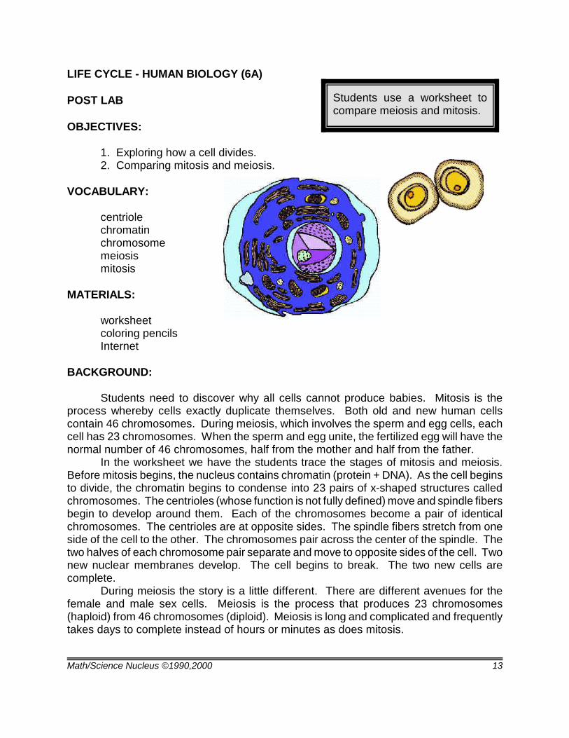
Students use a worksheet tocompare meiosis and mitosis.
LIFE CYCLE - HUMAN BIOLOGY (6A)
POST LAB
OBJECTIVES:
1. Exploring how a cell divides. 2. Comparing mitosis and meiosis.
VOCABULARY:
centriolechromatinchromosomemeiosismitosis
MATERIALS:
worksheetcoloring pencilsInternet
BACKGROUND:
Students need to discover why all cells cannot produce babies. Mitosis is theprocess whereby cells exactly duplicate themselves. Both old and new human cellscontain 46 chromosomes. During meiosis, which involves the sperm and egg cells, eachcell has 23 chromosomes. When the sperm and egg unite, the fertilized egg will have thenormal number of 46 chromosomes, half from the mother and half from the father.
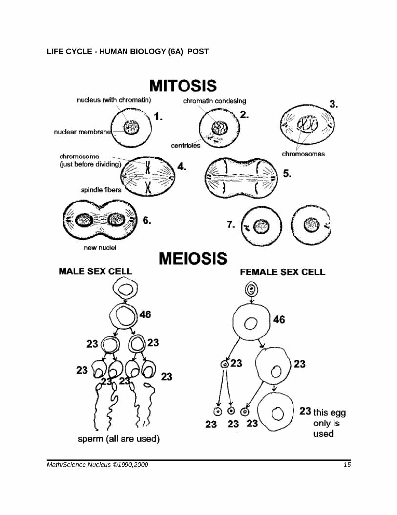
In the worksheet we have the students trace the stages of mitosis and meiosis.Before mitosis begins, the nucleus contains chromatin (protein + DNA). As the cell beginsto divide, the chromatin begins to condense into 23 pairs of x-shaped structures calledchromosomes. The centrioles (whose function is not fully defined) move and spindle fibersbegin to develop around them. Each of the chromosomes become a pair of identicalchromosomes. The centrioles are at opposite sides. The spindle fibers stretch from oneside of the cell to the other. The chromosomes pair across the center of the spindle. Thetwo halves of each chromosome pair separate and move to opposite sides of the cell. Twonew nuclear membranes develop. The cell begins to break. The two new cells arecomplete.
During meiosis the story is a little different. There are different avenues for thefemale and male sex cells. Meiosis is the process that produces 23 chromosomes(haploid) from 46 chromosomes (diploid). Meiosis is long and complicated and frequentlytakes days to complete instead of hours or minutes as does mitosis.
Math/Science Nucleus ©1990,2000 14
PROCEDURE:
1. Go over the information on the worksheet emphasizing mitosis and meiosis.Mitosis is just the dividing of a cell into equal parts. The two types of meiosis one involvingthe sperm and one involving the egg. Meiosis is characterized by going through twophases of cell division. The first phase closely resembles mitosis. One sex cell producestwo sex cells in the first phase. The two sex cells will then divide again to produce 4 sexcells. In females, however, only one egg is produced (the remaining 3 cells are called"polar bodies" and their function is not positively known.) The resulting sex cell will haveone half of the normal number of chromosomes (23). Meiosis only occurs in sex cells, notin other cells.
2. Instruct students to using coloring pencils to make the worksheet more readable.
3. Use the Internet if students are unfamiliar with cell structure. Search under“cells” or “genes” to look for information.
Math/Science Nucleus ©1990,2000 15
LIFE CYCLE - HUMAN BIOLOGY (6A) POST
Math/Science Nucleus ©1990,2000 16
Students uses a worksheet to comparebacteria, virus, and protozoa.
LIFE CYCLE - HUMAN BIOLOGY (6B)
PRE LAB
OBJECTIVES:
1. Distinguishing bacteria, viruses, and protozoa.2. Understanding an infection.
VOCABULARY:
bacteriadiseasegeneticpathogenvirus
MATERIALS:
worksheet culture demonstration (optional)
BACKGROUND:
A disease is the abnormal functioning of the body that prevents the body organs orprocesses from functioning as they should. There are many causes of disease. Diseasecan be caused by fungi (i.e. athlete's foot), bacteria (i.e. tuberculosis), viruses (i.e. commoncold) and protozoa (amoebic dysentery). Not all types of fungi, bacteria, viruses andprotozoa are disease-causing agents. The causes of disease are sometimes referred toas germs. The definition of the term germ is sometimes ambiguous. Germs can be carriedthrough food, water, air, animals, and people. "Bad" germs are pathogenic or diseasecausing organisms and are the ones that make people sick.
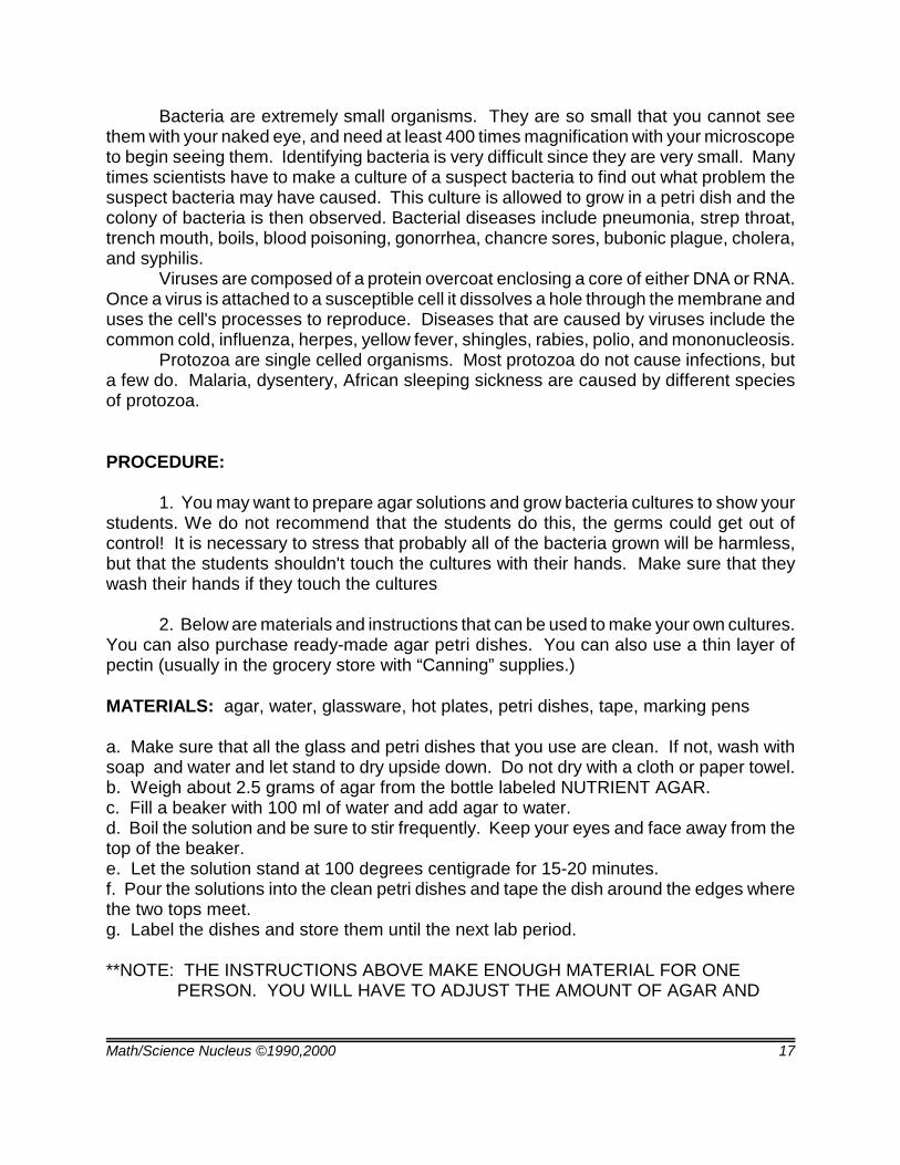
Bacteria, viruses, and protozoa cause different infections. The worksheet is just away for students to look at these "germs" without a microscope. They should look like thefollowing:
streptococcus rhinovirus entamoeba
Math/Science Nucleus ©1990,2000 17
Bacteria are extremely small organisms. They are so small that you cannot seethem with your naked eye, and need at least 400 times magnification with your microscopeto begin seeing them. Identifying bacteria is very difficult since they are very small. Manytimes scientists have to make a culture of a suspect bacteria to find out what problem thesuspect bacteria may have caused. This culture is allowed to grow in a petri dish and thecolony of bacteria is then observed. Bacterial diseases include pneumonia, strep throat,trench mouth, boils, blood poisoning, gonorrhea, chancre sores, bubonic plague, cholera,and syphilis.
Viruses are composed of a protein overcoat enclosing a core of either DNA or RNA.Once a virus is attached to a susceptible cell it dissolves a hole through the membrane anduses the cell's processes to reproduce. Diseases that are caused by viruses include thecommon cold, influenza, herpes, yellow fever, shingles, rabies, polio, and mononucleosis.
Protozoa are single celled organisms. Most protozoa do not cause infections, buta few do. Malaria, dysentery, African sleeping sickness are caused by different speciesof protozoa.
PROCEDURE:
1. You may want to prepare agar solutions and grow bacteria cultures to show yourstudents. We do not recommend that the students do this, the germs could get out ofcontrol! It is necessary to stress that probably all of the bacteria grown will be harmless,but that the students shouldn't touch the cultures with their hands. Make sure that theywash their hands if they touch the cultures
2. Below are materials and instructions that can be used to make your own cultures.You can also purchase ready-made agar petri dishes. You can also use a thin layer ofpectin (usually in the grocery store with “Canning” supplies.)
MATERIALS: agar, water, glassware, hot plates, petri dishes, tape, marking pens
a. Make sure that all the glass and petri dishes that you use are clean. If not, wash withsoap and water and let stand to dry upside down. Do not dry with a cloth or paper towel.b. Weigh about 2.5 grams of agar from the bottle labeled NUTRIENT AGAR.c. Fill a beaker with 100 ml of water and add agar to water.d. Boil the solution and be sure to stir frequently. Keep your eyes and face away from thetop of the beaker.e. Let the solution stand at 100 degrees centigrade for 15-20 minutes.f. Pour the solutions into the clean petri dishes and tape the dish around the edges wherethe two tops meet.g. Label the dishes and store them until the next lab period.
**NOTE: THE INSTRUCTIONS ABOVE MAKE ENOUGH MATERIAL FOR ONE PERSON. YOU WILL HAVE TO ADJUST THE AMOUNT OF AGAR AND
Math/Science Nucleus ©1990,2000 18
WATER DEPENDING ON YOUR CLASS SIZE.
2. You can then prepare your bacteria colonies by inoculating the agar. Removethe tape from the petri dish and take the top cover off. With a clean toothpick make twolines so that there are 4 sections in the dish. With a clean toothpick scrape the inside ofyour mouth. Do not poke your skin, just scrape your inside of the cheek. Rub the toothpickon the agar, very gently. DO NOT poke or scratch the agar, just rub it. On the othersection, place a piece of hair, spit, scab, or anything else to see if bacteria will grow.
3. After you have inoculated all 4 sections tape the petri dish. Store in a place thatis warm (not hot). After you see growth, we recommend that you use a Microbiology bookif you want to identify your colony of bacteria. Discard the germs with care under sanitaryconditions.
4. Use the worksheet and have the students create a picture of each of the differenttypes of diseases. You may also want to do an Internet search to find out more informationon diseases.
Math/Science Nucleus ©1990,2000 19
LIFE CYCLE - HUMAN BIOLOGY (6B) PRE
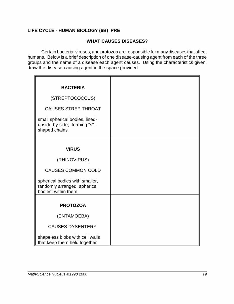
WHAT CAUSES DISEASES?
Certain bacteria, viruses, and protozoa are responsible for many diseases that affecthumans. Below is a brief description of one disease-causing agent from each of the threegroups and the name of a disease each agent causes. Using the characteristics given,draw the disease-causing agent in the space provided.
BACTERIA
(STREPTOCOCCUS)
CAUSES STREP THROAT
small spherical bodies, lined-upside-by-side, forming "s"-shaped chains
VIRUS
(RHINOVIRUS)
CAUSES COMMON COLD
spherical bodies with smaller, randomly arranged sphericalbodies within them
PROTOZOA
(ENTAMOEBA)
CAUSES DYSENTERY
shapeless blobs with cell wallsthat keep them held together
Math/Science Nucleus ©1990,2000 20
Students sort bacteria and viruses.
LIFE CYCLE - HUMAN BIOLOGY (6B)
LAB
OBJECTIVES:
1. Distinguishing viruses and bacteria.2. Distinguishing inherited characteristics.
VOCABULARY:
bacteriavaccinevirus
MATERIALS:
worksheet on viruses/bacteria
BACKGROUND:
Bacteria come in three different shapes: (1) coccus or sphere shaped; (2) bacillusor rod shaped; (3) spirillum or spiral, corkscrew shape. Bacteria have a tough outer coatingwhich gives them a cell-like shape.
Viruses are non-living and cannot reproduce without using the mechanisms of ahost cell. Once a virus enters a host it can cause great damage. Viruses come in differentshapes. Viruses are smaller than bacteria, have no nucleus, no cytoplasm, and nosurrounding cell membrane. There are, however, some viruses, that produce a "fake" cellmembrane that is used in tricking the immune system. Actually, a virus can almost beconsidered a chemical crystal. Bacteria and viruses that cause disease are called pathogens. Pathogens can entera body through the air, water, or through contact with an infected body. Diseases causedby bacteria can usually be cured with medication. Viral diseases on the other hand cannotbe cured because there is no medication that will stop viruses. The immune system of thehuman body has to fight the viruses, sometimes the body wins but many times it loses.Vaccines can help prevent the virus in the first place. Viruses are very difficult to study.
PROCEDURE:
1. Go over the characteristics of bacteria and viruses using the student's lab sheet.
2. Instruct the students to cut out the viruses and bacteria and try and sort them intotheir respective groups. Tell students to write their answer on the back site of each picturewith the reason for the student putting it in its group. This is not an easy lab, because the
Math/Science Nucleus ©1990,2000 21
weird shapes that both bacteria and viruses take.
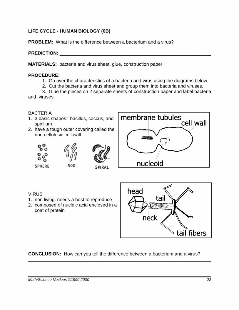
3. The bacteria and viruses on the chart are: 1. phage, a virus that invades thecells of bacteria; 2. Influenza virus, causes influenza; 3. generalized mycoplasma(bacteria); 4. tobacco mosaic virus, results in disease of tobacco plants; 5. smallertobacco mosaic virus; 6. micrococcus radiodurans (bacteria); 7. spirillum bacteria; 8.crystal of adenovirus particles within the nucleus of a human cell; 9. polio virus; 10.bacillus bacteria; 11. tumor virus; 12. pseudomonad bacteria; 13. prochloron bacteria; 14.spirillum bacteria; 15. cell invaded by bacillus bacteria; 16. influenza virus.
Math/Science Nucleus ©1990,2000 22
LIFE CYCLE - HUMAN BIOLOGY (6B)
PROBLEM: What is the difference between a bacterium and a virus?
PREDICTION: __________________________________________________________
MATERIALS: bacteria and virus sheet, glue, construction paper
PROCEDURE: 1. Go over the characteristics of a bacteria and virus using the diagrams below.2. Cut the bacteria and virus sheet and group them into bacteria and viruses.3. Glue the pieces on 2 separate sheets of construction paper and label bacteria
and viruses.
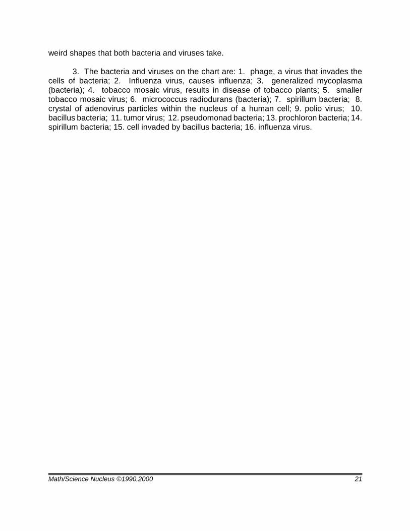
BACTERIA 1. 3 basic shapes: bacillus, coccus, and spirillum2. have a tough outer covering called the non-cellulosic cell wall
VIRUS1. non living, needs a host to reproduce2. composed of nucleic acid enclosed in a coat of protein
CONCLUSION: How can you tell the difference between a bacterium and a virus?______________________________________________________________________________
Math/Science Nucleus ©1990,2000 23
LIFE CYCLE - HUMAN BIOLOGY (6B)
Math/Science Nucleus ©1990,2000 24
Students discuss geneticdisorder versus other diseases.
LIFE CYCLE - HUMAN BIOLOGY (6B)
POST LAB
OBJECTIVES:
1. Distinguishing illnesses.2. Comparing genetic disorders with diseases.
VOCABULARY:
bacteriadiseasedisordergenesinheritedvirus
MATERIALS:
Germs Make me Sick! by Melvin Berger (optional)
BACKGROUND:
Many students cannot distinguish the difference between a genetic disorder and adisease. Cavities, earaches, and skin boils are caused by bacteria. The common cold,AIDS, measles, and cancer are all caused by viruses. Sickle cell anemia and hemophiliaare genetic disorders. If students are not clear or unaware of what heredity and geneticsare, you may want to discuss that certain characteristics of humans are genetic, or carriedon from one family generation to another generation.
How do you determine which characteristics are genetic? Ask the students to lookat their neighbor's ear lobe and observe if the ear is attached to his face or if it is free. Thisis a genetic trait within families. If you can roll your tongue, if your thumb bends back, youreye color, and your hairline type are all genetic traits that are inherited. The direction ofyour hair growth, or hair whorl is also genetic. These are characteristics that do notadversely affect the human body, but some genetic traits can cause severe damage to ahuman body. The chances of getting a genetically impairing trait are statistically linked torecessive and dominant gene pools.
PROCEDURE:
1. To illustrate this to your students, get a piece of 8 1/2" x 11 paper and randomlyplace one or two colored lines on the paper (short direction). This represents a "geneticallytransmitted" trait that will result in a disorder. Cut out 4 paper dolls, by folding the paper
Math/Science Nucleus ©1990,2000 25
in half. (This represents how many children were born by parents with the trait.) Somegenetic mishaps are not inherited, but represent a departure from the average humangenetic make up. In other words, not all the children will have the disorder.
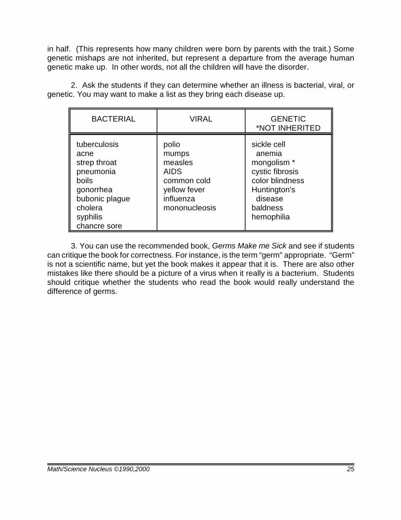
2. Ask the students if they can determine whether an illness is bacterial, viral, orgenetic. You may want to make a list as they bring each disease up.
BACTERIAL VIRAL GENETIC*NOT INHERITED
tuberculosis acne strep throat pneumonia boils gonorrhea bubonic plague cholera syphilis chancre sore
polio mumps measles AIDS common cold yellow fever influenza mononucleosis
sickle cell anemia mongolism * cystic fibrosis color blindness Huntington's disease baldness hemophilia
3. You can use the recommended book, Germs Make me Sick and see if studentscan critique the book for correctness. For instance, is the term “germ" appropriate. “Germ”is not a scientific name, but yet the book makes it appear that it is. There are also othermistakes like there should be a picture of a virus when it really is a bacterium. Studentsshould critique whether the students who read the book would really understand thedifference of germs.