Embed Size (px)
Citation preview
MOLECULAR AND CELLULAR BIOLOGY,0270-7306/01/$04.0010 DOI: 10.1128/MCB.21.10.3343–3350.2001
May 2001, p. 3343–3350 Vol. 21, No. 10
Copyright © 2001, American Society for Microbiology. All Rights Reserved.
Six4, a Putative myogenin Gene Regulator, Is Not Essential forMouse Embryonal Development
HIDENORI OZAKI,1 YOKO WATANABE,1 KATSUMASA TAKAHASHI,2 KEN KITAMURA,2†AKIRA TANAKA,3 KOKO URASE,4 TAKASHI MOMOI,4 KATSUKO SUDO,5 JUNKO SAKAGAMI,5
MASAHIDE ASANO,5‡ YOICHIRO IWAKURA,5 AND KIYOSHI KAWAKAMI1*
Departments of Biology,1 Otolaryngology,2 and Pathology,3 Jichi Medical School, Tochigi 329-0498, Division ofDevelopment and Differentiation, National Institute of Neuroscience, NCNP, Kodaira, Tokyo 187-8502,4
and Division of Cell Biology, Center for Experimental Medicine, Institute of MedicalScience, University of Tokyo, Tokyo 108-8639,5 Japan
Received 27 November 2000/Returned for modification 9 January 2001/Accepted 21 February 2001
Six4 is a member of the Six family genes, homologues of Drosophila melanogaster sine oculis. The gene isthought to be involved in neurogenesis, myogenesis, and development of other organs, based on its specificexpression in certain neuronal cells of the developing embryo and in adult skeletal muscles. To elucidate thebiological roles of Six4, we generated Six4-deficient mice by replacing the Six homologous region and homeoboxby the b-galactosidase gene. 5-Bromo-4-chloro-3-indolyl-b-D-galactopyranoside staining of the heterozygousmutant embryos revealed expression of Six4 in cranial and dorsal root ganglia, somites, otic and nasalplacodes, branchial arches, Rathke’s pouch, apical ectodermal ridges of limb buds, and mesonephros. Theexpression pattern was similar to that of Six1 except at the early stage of embryonic day 8.5. Six4-deficient micewere born according to the Mendelian rule with normal gross appearance and were fertile. No hearing defectswere detected. Six4-deficient embryos showed no morphological abnormalities, and the expression patterns ofseveral molecular markers, e.g., myogenin and NeuroD3 (neurogenin1), were normal. Our results indicate thatSix4 is not essential for mouse embryogenesis and suggest that other members of the Six family seem tocompensate for the loss of Six4.
Six family genes are homologues of Drosophila melanogastersine oculis, one of the homeobox genes essential for compoundeye formation (5). They are characterized by the presence ofthe Six domain and Six-type homeodomain in the encodedproteins, which confer specific DNA binding activity and func-tion as transcription factors (12, 28). In mammals, six membersof the family have so far been identified (2–4, 7, 8, 10, 12, 13,31, 32, 34, 41; for a review, see reference 14). Each member ofthe family is expressed in a spatiotemporally regulated mannerduring embryogenesis. In mice, Six3 and Six6 are exclusivelyexpressed in the developing forebrain and eyes (10, 22, 31, 40).In contrast, Six1, Six2, and Six5 show relatively broader expres-sion patterns (15, 32). Six1 is expressed in the cranial anddorsal root ganglia, somites, otic and nasal placodes, branchialarches, limbs, Rathke’s pouch, and nephrogenic cords (32).Six2 is expressed in head mesenchyme, branchial arches, limbs,and some mesenchymal regions surrounding the gastrointesti-nal tract (32). Six5 shows a broad expression in branchialarches, limb buds, telencephalon, eye, sclerotomes, and carti-lages (15). Such distribution suggests that these genes playspecific roles in embryogenesis.
Overexpression and misexpression experiments showed thatSix3 and Six6 genes are involved in forebrain and eye organo-
genesis (18, 21, 30, 48). Consistently, SIX3 mutations causeholoprosencephaly, a severe malformation of the brain in hu-mans (42). SIX5 is located immediately downstream of theCTG trinucleotide repeats whose expansion causes myotonicdystrophy (3). Downregulation of the gene was observed inmyotonic dystrophy patients and is thought to be responsiblefor some of the symptoms of the disease such as cataracts (16,44). In agreement with this, Six5-heterozygous and -homozy-gous mutant mice develop cataracts (15, 35).
Six4 was originally identified as a binding factor to the pos-itive regulatory element of the Na1,K1-ATPase a1 subunitgene (Atp1a1) (12, 38). Immunostaining showed the presenceof Six4 protein in some populations of neuronal cells in devel-oping mouse embryos and developing retina (27, 29). In adultmice, Six4 protein is localized in skeletal muscles, as demon-strated by gel retardation assay (37). These observations sug-gest that this gene is involved in neurogenesis, myogenesis, andprobably the development of other organs. In myogenesis,myogenin plays an important role along with other myogenicgenes such as MyoD, Myf5, and MRF4 (33). Promoter analysiswith transgenic mice demonstrated that the MEF3 site in thepromoter region is essential for myogenin expression (37).Mouse Six1 and Six4 proteins are present in the developingsomites in which myogenin expression is activated and bind tothe MEF3 site in the myogenin promoter, as shown by gelretardation assay (37). Furthermore, Six4 can activate the myo-genin gene promoter alone or in synergy with the specificcofactor, Eya, through direct binding to the MEF3 site incultured cells (28). These results support the notion that Six4 isone of the genes that control myogenesis through activation ofmyogenin. In addition, Eya1, one of the specific cofactors of
* Corresponding author. Mailing address: Department of Biology,Jichi Medical School, 3311-1 Yakushiji, Minamikawachi, Kawachi,Tochigi 329-0498, Japan. Phone: 81 (285) 58-7311. Fax: 81 (285) 44-5476. E-mail: [email protected].
† Present address: Department of Otolaryngology, Tokyo Medicaland Dental University, Tokyo 113-8519, Japan.
‡ Present address: Institute for Experimental Animals, Faculty ofMedicine, Kanazawa University, Kanazawa 920-8640, Japan.
3343
on February 2, 2018 by guest
http://mcb.asm
.org/D
ownloaded from
Six4, is implicated in branchio-oto-renal syndrome, a domi-nantly inherited disorder characterized by hearing loss andbranchial arch and renal anomalies in humans (1). Eya1-defi-cient mice lack ears and kidneys, and heterozygous mutantmice show hearing loss and renal anomalies, as seen in humanbranchio-oto-renal syndrome (46). Because immunostainingshowed the presence of Six4 protein in acoustic ganglia andotic vesicles (29), it is possible that Six4 is involved in thedevelopment of the ear in association with Eya1. Nevertheless,the biological function of Six4 in development is not clear, dueto the lack of natural mutants, knockout models, and overex-pression experiments. To access the biological function of Six4,we generated Six4-deficient mice and analyzed phenotypicchanges both in adults and in embryos. We found no apparentchanges in morphology and expression patterns of somemarker genes in Six4-deficient mice. Functionally, hearing abil-ity was normal. The reason for the lack of phenotype in Six4-deficient mice and the possible compensation among Six familygenes is discussed.
MATERIALS AND METHODS
Construction of Six4 gene targeting vector. The complete murine Six4 locuswas cloned from a 129/SvJ genomic library (Stratagene, La Jolla, Calif.) andpartially sequenced, and the exon-intron organization was determined (34). PCRmutagenesis using AmpliTaq Gold DNA polymerase (Perkin Elmer-Cetus, Fos-ter City, Calif.) was performed to introduce a KpnI site immediately downstreamof the initiation codon to allow the insertion of an in-frame lacZ gene, with theforward primer (9705; 59-CAA AAG GAG GAG TCA CGT T-39) and reverseprimer (9706; 59-CGG GGT ACC CTT TCC ATC CCA TTC TC-39). The PCRproducts were sequenced with Sequencing Pro kits (Toyobo, Osaka, Japan). Thetargeting vector was constructed as follows. The lacZ fragment (KpnI-BamHI)from pCH110 (Amersham Pharmacia Biotech, Buckinghamshire, United King-dom) was ligated to the 39 end of a 59 homology region (XbaI-KpnI; 5.1 kb), thePGKneobpA cassette (XhoI-PvuII) from pPGKneobpA (36) was ligated to the 59end of a 39 homology region (SmaI-SalI; 2.5 kb), and the resulting two insertswere ligated together. Finally, the diphtheria toxin A cassette (XhoI-NotI) frompMC1DTpA (47) was ligated to the 39 end of the 39 homology region. Theplasmid was linearized with SalI at the 59 end. In this construct, the homeoboxand the Six homologous region, which together encode a specific DNA bindingdomain, were completely removed.
ES cell screening and chimeric mouse production. The linearized targetingvector (60 mg) was electroporated (250 V; 500 mF) into 107 E14-1 embryonicstem (ES) cells (19), and transformants were selected with 250 mg (active form)G418 (Gibco/BRL) per ml for 7 to 10 days. Homologous recombinants werescreened by PCR as follows. The forward primer in the PGKneobpA cassette was59-CTC TAT GGC TTC TGA GGC GGA AAG-39, and the reverse primer was59-GGC AAG GTC TGC TAG AAA CGG TAC-39. PCR was carried out withLA Taq DNA polymerase (TaKaRa, Kyoto, Japan) for 35 cycles at 94°C for 1min, 60°C for 2 min, and 72°C for 3 min in a volume of 36 ml. Homologousrecombination was further confirmed by Southern blot hybridization as follows:15 mg of DNA from PCR-positive clones was digested with BamHI (to confirm59 homologous recombination) or SacI (to confirm 39 homologous recombina-tion), electrophoresed through a 0.7% agarose gel, and transferred to Hy-bond-N1 membranes (Amersham Pharmacia Biotech). Hybridization was car-ried out with a buffer containing 53 SSC (13 SSC is 0.15 M NaCl plus 0.015 Msodium citrate), 2.53 Denhardt’s solution, 0.5% sodium dodecyl sulfate, 0.1 mgof heat-denatured herring testis DNA per ml, and radiolabeled probes specific toeither the 59 or 39 restriction fragment (see below). Two ES clones that yieldedhybridization bands of the correct size gave germ line chimeras by the aggrega-tion method (26). Once homologous recombination and germ line transmissionwere confirmed, mouse genotyping was carried out by PCR as follows. Theforward primer in exon 1 was 59-ACA TCA AGC AGG AGA ATG GGATGG-39. The reverse primer specific to lacZ in the mutant allele was 59-CCGTAA TGG GAT AGG TTA CGT TGG-39. The reverse primer for the wild-typeallele was 59-AGA AGT TCC GAG TGG AGT TGT ACC-39. PCR was carriedout with AmpliTaq Gold DNA polymerase for 35 cycles at 95°C for 58 s, 63°C for28 s, and 72°C for 55 s in a volume of 9 ml. The germ line chimeras werebackcrossed to C57BL/6 mice. F2 or F3 mice were backcrossed to C57BL/6 mice
or intercrossed, and the resulting founder mice were used in the followingexperiments. Mice were maintained under specific-pathogen-free conditions inenvironmentally controlled clean rooms at the Laboratory Animal ResearchCenter, Institute of Medical Science, University of Tokyo, and the Center forExperimental Medicine, Jichi Medical School. The experiments were conductedaccording to the institutional ethical guidelines for animal experiments andsafety guidelines for gene manipulation experiments.
Northern blot analysis. Total RNA was extracted with Isogen (Nippon Gene,Tokyo, Japan) from adult tissues or embryos, electrophoresed through a 1.2%denatured agarose gel containing 2.2 M formaldehyde, and transferred to Hy-bond-N1 membranes (Amersham Pharmacia Biotech). Hybridization was car-ried out under the same conditions used for Southern blot analysis describedabove.
Preparation of probes. For Southern and Northern blot analyses, 25 ng of thefollowing DNA fragments was used to synthesize 32P-labeled probes with aMegaprime DNA labeling kit (Amersham Pharmacia Biotech): 0.6-kbBamHI-HindIII fragment 1.8 kb upstream of the 59 end of the 59 homologyregion (for the 59 probe in Southern analysis), a 0.8-kb KpnI-XbaI fragmentimmediately downstream of the 39 end of the 39 homology region (for the 39probe in Southern analysis), a 2.3-kb XhoI-XbaI fragment of mouse Six4 cDNA(SM type, for Six4) (12), a 0.7-kb PstI-PstI fragment of Six1-LZ8 (for Six1) (32),a 1.1-kb EcoRI-Sau3AI fragment of pfSix2 (for Six2) (28), a 2.4-kb NotI-BglIIfragment of pfSix5 (for Six5) (28), a 2.2-kb NcoI-NcoI fragment of rat Atp1a1cDNA (for Atp1a1) (9), a 1.5-kb EcoRI-EcoRI fragment of pEMSV2a-MGN(for myogenin) (45), and a 0.8-kb fragment amplified from mRNAs extractedfrom HeLa cells by reverse transcription-PCR using primers 59-TGGTGGGAATGGGTCAGA-39 and 59-AGGGAGGAAGAGGATGCG-39 (for b-actin).
X-Gal staining of mouse embryo. Embryos were removed from the uterus inice-cold phosphate-buffered saline (PBS). Genotyping was carried out by PCRusing DNA extracted from yolk sac. For whole-mount staining, embryos werefixed in the fixing solution (1% formaldehyde, 0.2% glutaraldehyde, and 0.02%Nonidet P-40 in PBS) on ice for 30 min, washed twice in PBS at room temper-ature for 30 min, and then stained in the 5-bromo-4-chloro-3-indolyl-b-D-galac-topyranoside (X-Gal) staining solution [1 mg/ml X-Gal, 5 mM K3Fe(CN)6, 5 mMK4Fe(CN)6, and 2 mM MgCl2 in PBS] at 30°C overnight. After being stained,embryos were washed and stored in PBS at 4°C. For sections, embryos wereembedded in freezing medium and frozen on dry ice. The embedded embryoswere sectioned at a 30-mm thickness at 215°C. Each section was transferred ontoa silanized slide, allowed to dry, and fixed in a fixing solution (0.2% glutaralde-hyde, 2 mM MgCl2, and 5 mM EGTA in PBS). After being washed three timesin a washing solution (2 mM MgCl2, 0.01% sodium deoxycholate, and 0.02%Nonidet P-40 in PBS), each section was stained in the X-Gal staining solution asabove.
Hematoxylin-eosin staining. Skeletal muscles of the hindlimb of wild-type andSix4-deficient mice were fixed in 10% formalin. After being washed with water,fixed samples were dehydrated by sequentially increasing concentrations of eth-anol, cleared in xylene, and then embedded in paraffin. The embedded sampleswere sectioned at a 5-mm thickness, and each section was transferred onto a slide,dewaxed in xylene, rehydrated by sequentially decreasing concentrations of eth-anol, stained in hematoxylin solution [0.1% hematoxylin, 5% K2Al2(SO4)4,0.02% NaIO3, 5% chloral hydrate, 0.1% citric acid, and 20% glycerol] for 15 min,and differentiated in water. Then, the sections were counterstained in eosinsolution (0.25% eosin Y, 0.55% acetic acid, and 60% ethanol) for 30 min;differentiated sequentially in 70, 80, and 90% ethanol solutions; dehydrated inabsolute ethanol; cleared in xylene; and coverslipped.
In situ hybridization. Embryos were removed from the uterus in ice-cold PBS.Genotyping was carried out by PCR using DNA extracted from yolk sac. Forwhole-amount in situ hybridization, embryos were fixed in a fixing solution (4%paraformaldehyde in PBS) at 4°C overnight. After being washed twice in PBT(0.1% Tween 20 in PBS), embryos were dehydrated by being washed sequentiallyin 25, 50, and 75% methanol solutions in PBT and 100% methanol and thenrehydrated by being washed sequentially in 75, 50, and 25% methanol solutionsin PBT and then in PBT alone. After being bleached in 6% H2O2 in PBT for 1 h,embryos were treated in 10 mg of proteinase K per ml in PBT for 15 min, washedwith 2 mg of glycine per ml in PBT and in PBT alone, and refixed in 0.2%glutaraldehyde and 4% formaldehyde in PBT for 20 min. After being washed inPBT, embryos were prehybridized in a prehybridization buffer (50% formamide,53 SSC [pH 5.0], 50 mg of yeast tRNA per ml, 1% sodium dodecyl sulfate, and50 mg of heparin per ml) at 70°C for 1 h and then hybridized in a hybridizationbuffer containing 1 mg of digoxigenin (DIG)-labeled antisense RNA probe (seebelow) at 70°C overnight. After being washed, embryos were treated twice in 100mg of RNaseA per ml in 0.5 M Tris-HCl (pH 7.5) and 0.1% Tween 20 at 37°C for30 min, followed by blocking in 10% fetal calf serum in TBST (0.14 M NaCl, 2.7
3344 OZAKI ET AL. MOL. CELL. BIOL.
on February 2, 2018 by guest
http://mcb.asm
.org/D
ownloaded from
mM KCl, 25 mM Tris-HCl [pH 7.5], and 0.1% Tween 20) for 90 min. Then,embryos were treated in TBST containing 1% fetal calf serum and alkalinephosphatase-labeled anti-DIG antibody (Boehringer GmbH, Mannheim, Ger-many) at 4°C overnight. After being washed in TBST and NTMT (100 mM NaCl,100 mM Tris-HCl [pH 9.5], 50 mM MgCl2, and 0.1% Tween 20), embryos werestained in NTMT containing nitroblue tetrazolium and X-phosphate (Boehr-inger). Frozen sections (10-mm thick) were cut on a cryostat and attached toslides coated with Vectabond reagent (Vector Laboratories, Burlingame, Calif.).Samples were treated with proteinase K (1 mg/ml) at 37°C for 10 min, refixed in4% paraformaldehyde, and hybridized overnight with the DIG-labeled RNAprobe. The hybridized mRNA was detected by alkaline phosphatase-conjugatedanti-DIG Fab fragments (Boehringer) according to the procedure described byWilkinson (43).
DIG (Boehringer)-labeled antisense RNA probes were prepared from thefollowing linearized plasmids with DIG RNA Labeling mixture (Boehringer) andT3 or T7 RNA polymerase according to the instructions provided by the manu-facturer: pKSMGN1, which contains a 1.5-kb EcoRI-EcoRI fragment ofpEMSV2a-MGN in pBulescript KS(1) (for myogenin) (45); pKS-ngn1/E2 (forNeuroD3) (23); mouse NeuroD1 pSK P/P350#5 (for NeuroD1) (25); and pSK,which contains a 630-bp PstI(1545)-PstI(2175) fragment from Six4 cDNA SMtype (for Six4) (12).
ABR. Hearing was assessed by recording auditory brainstem response (ABR)as described previously (39). Acoustic stimuli, consisting of tone bursts at fre-quencies of 10, 20, 30, and 40 kHz, with a rise-and-fall time of 1 ms, a 5-msduration, and repetition every 70 ms, were delivered to each mouse with a soundstimulator (DPS-725; Diamedical System) and a speaker (PT-RIII; Pioneer) inan open field. A microcomputer (Synapac 1100; NEC Sanei) was used to analyzethe response. For each time point, 500 responses for each mouse were recordedand filtered for bandwidths of 100 to 3,000 Hz.
RESULTS AND DISCUSSION
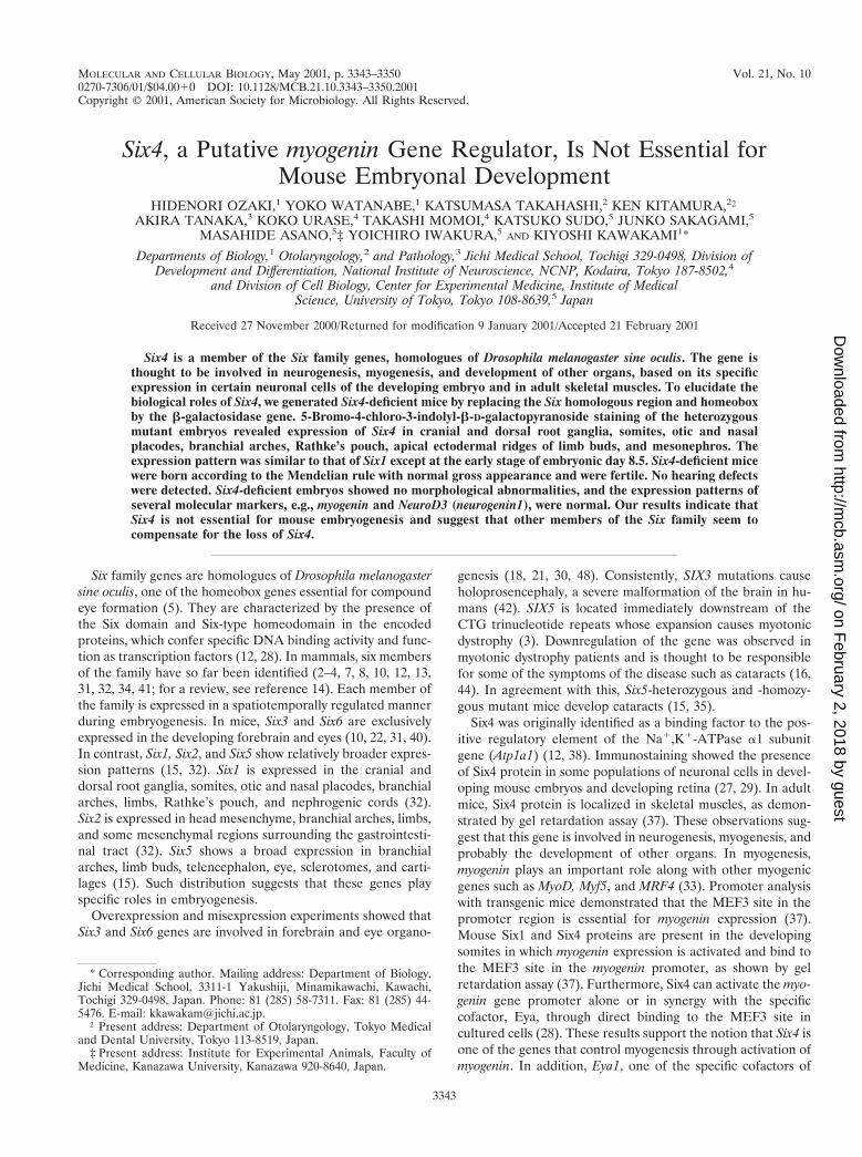
Generation of Six4-deficient mice. To inactivate Six4, anin-frame b-galactosidase (lacZ) reporter and a neomycin-re-sistant cassette (neo) were introduced for monitoring the ex-pression of endogenous Six4 and for positive selection, respec-
tively, which replaced the Six homologous region and thehomeobox in exon 1 (Fig. 1).
No obvious phenotype was apparent in heterozygous mu-tants. When heterozygous mutants were intercrossed, wild-type offspring, heterozygotes, and homozygotes were born ac-cording to the Mendelian rule (Table 1). The heterozygotesand homozygotes had a normal appearance, and both male andfemale homozygotes were fertile.
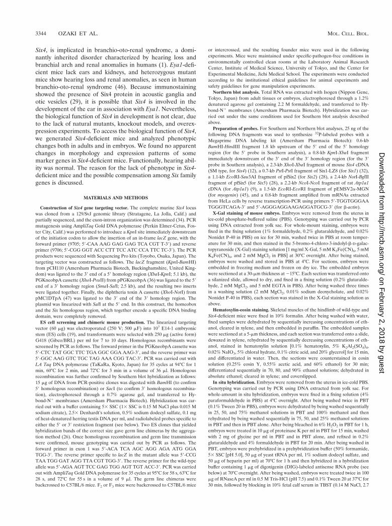
In our targeting strategy, exon 3, which encodes a transcrip-tional activation domain (12), was left intact. To confirm thatexon 3 was not transcribed irregularly to produce aberrant Six4molecules with some activity, Northern blot analysis of thetotal RNA from embryonic day 11.5 (E11.5) whole embryos(Fig. 2) and from adult skeletal muscle (data not shown) wasperformed, using a probe that covered the 39 part of exon 1,exon 2, and the coding region of exon 3. The amount of Six4transcripts was proportional to the gene dosage, and in ho-mozygous mutants, no Six4 transcripts of correct size or irreg-ular transcripts were detected. Thus, we concluded that theSix4 gene was functionally inactivated in the targeted allele.
FIG. 1. Targeted disruption of mouse Six4. (A) Structures of the wild-type allele, targeting vector, and targeted allele. Boxes represent exons.Gray shading indicates coding regions, and black shading indicates the Six homologous region and homeobox. The hatched region marks the regionencoding the transactivation domain. The targeting vector consisted of the 59 homology region, lacZ, neo, 39 homology region, and at the 39 endthe diphtheria toxin A gene (dt) for negative selection. Arrows beneath the target allele represent PCR primers (9705 and 9706) for screening.Restriction fragments detected by Southern blot analysis are shown by horizontal arrows with their sizes in kilobases. B, BamHI; S, SacI. (B) South-ern blot analysis of mouse tail DNA isolated from the founder mice from a mating of heterozygous parents. DNAs were digested with BamHI orSacI and hybridized with the probes indicated in panel A. 1/1, wild-type mouse; 1/2, heterozygous mutant mouse; 2/2, homozygous mutant mouse.
TABLE 1. Genotypic analysis of founder mice at 3 or 4 weeks ofage from heterozygous 3 heterozygous intercross
SexNo. (%) of genotype
Wild type Heterozygous Homozygous
Male 33 (31) 56 (53) 17 (16)Female 31 (26) 59 (49) 31 (26)
Combined 64 (28) 115 (51) 48 (21)
VOL. 21, 2001 Six4-DEFICIENT MICE 3345
on February 2, 2018 by guest
http://mcb.asm
.org/D
ownloaded from
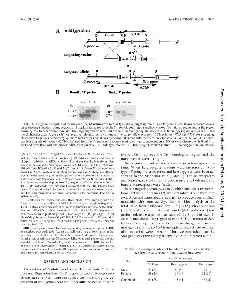
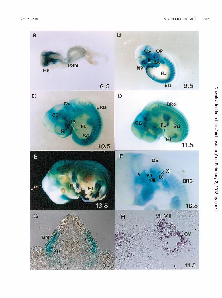
Expression pattern of Six4lacZ in heterozygous mutant em-bryos. So far, Six4 expression has been analyzed by immuno-staining, gel retardation assay, and Northern analysis in re-stricted areas of embryonic tissue and at restricted stages ofdevelopment (12, 27, 29, 37). To analyze the expression patternof Six4 during the entire developmental process, X-Gal stain-ing was performed on heterozygous embryos (Fig. 3). Six4lacZ
expression was detected in various ganglia, somites, nasal andotic placodes, branchial arches, and several other tissues, assummarized in Table 2. To our knowledge, this is the firstcomprehensive analysis of the Six4 expression pattern in mice.In a previous report, Six4 protein was detected mainly in thecranial and dorsal root ganglia by immunostaining (29). This isprobably because of the higher level of expression of Six4and/or a higher number of Six4-expressing cells in these gangliathan in other sites in which X-Gal staining was confirmed inour heterozygous mutant embryos.
In chickens, Six4 is expressed in a pattern similar to that ofmouse Six4, although chick Six4 is expressed in additionaltissues such as the optic placodes and motoneurons in thespinal cord (6). Moreover, in zebra fish two orthologues of
mammalian Six4, Six4.1 and Six4.2, exhibit essentially the sameexpression pattern with mouse Six4 in combination (17). Thus,the Six4 expression pattern is essentially conserved throughvertebrate evolution. In addition, the expression pattern ofSix4 in the mouse was strikingly similar to that of Six1, exceptin the head region at E8.5, immediately after the onset of theirexpression (Six4 in surface ectoderm outside the neural folds;Six1 in head mesenchyme) (Fig. 3A) (32). Because Six1 andSix4 are located in tandem on mouse chromosome 12 (H.Ozaki, unpublished data), these two genes might share com-mon cis-regulatory elements. Alternatively, cis-regulatory ele-ments that control the expression of each gene might be wellconserved between these two genes, although their proteinstructure itself was different in that Six4 protein, but not Six1protein, has a large C-terminus region containing a transacti-vation domain (12, 32).
Eya1 and Eya2, the putative coactivators of Six4 and otherSix family genes, are expressed in an extensively overlappingpattern with Six4, for example, in cranial ganglia, cranial pla-codes, and somites, indicating the possible interaction of Six4with Eya1 and/or Eya2 in these organs, as shown by transienttransfection assays (28).
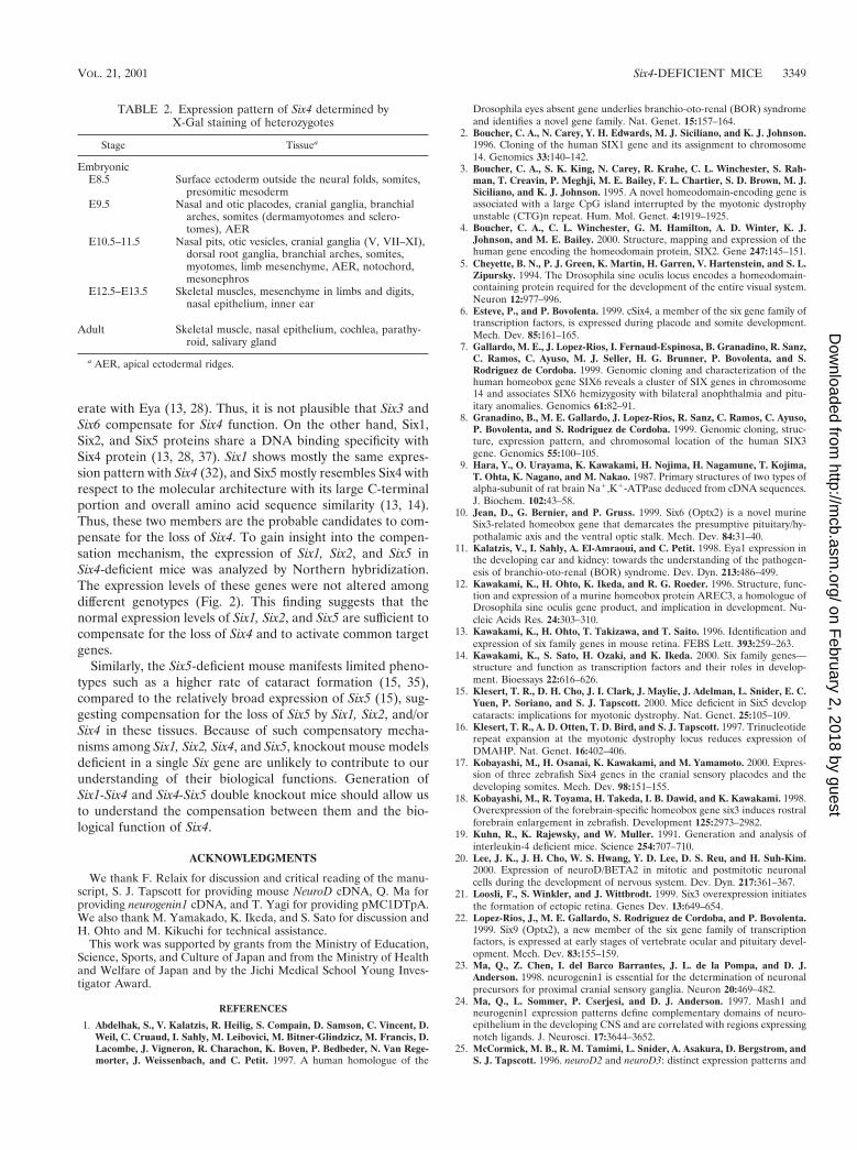
Hearing ability in Six4-deficient mice. Six4 showed overlap-ping expression with Eya1 in the otic vesicle and in the acousticganglion (11). Considering the functional cooperativity be-tween Six4 and Eya1 in target gene activation (28), we suspect-ed that Six4-deficient mice might have hearing defects. How-ever, hearing ability was normal in Six4-deficient mice as testedby ABR (Fig. 4).
Morphological analysis and X-Gal staining of Six4 homozy-gous mutant embryos. Six4-deficient mice seemed normal inappearance and in anatomical aspects after birth. We thenassessed the morphological abnormalities in Six4 homozygousmutant embryos, focusing on the sites of Six4lacZ expression.We compared the expression pattern of Six4 in heterozygousand homozygous mutant embryos by X-Gal staining. The over-all expression pattern was the same except that staining wasstronger in homozygotes than in heterozygotes, probably re-flecting the gene dosage (data not shown).
Expression of putative targets of Six4 and markers forsomites, muscle, and cranial and dorsal root ganglia. BecauseSix4 was previously reported to activate the myogenin promoterthrough the MEF3 site (28, 37), we analyzed the expression ofmyogenin by in situ hybridization. The staining pattern wasexactly the same in wild-type and Six4-deficient embryos (Fig.5A to D). Furthermore, Northern analysis revealed that themyogenin expression level was similar in wild-type mice, het-erozygotes, and homozygotes (Fig. 2). In accordance with thesefindings, skeletal muscles of adult mice (Fig. 5E and F) and of
FIG. 2. Analysis of gene expression in wild-type and mutant mouseembryos. Ten micrograms of total RNA from E11.5 embryos of theindicated genotypes was analyzed by Northern hybridization with theindicated probes. Three independent experiments yielded essentiallythe same results, and two representative hybridization patterns areshown here. In spite of complete lack of Six4 mRNA in Six42/2 em-bryos, the expression levels of the genes analyzed except Six4 were notaltered.
FIG. 3. X-Gal staining of Six4 heterozygous mutant embryos showing spatiotemporally regulated expression of Six4 in somites and myotomes(SO), cranial and dorsal root ganglia (V to XI) (DRG), sensory placodes (otic [OP] and nasal [NP]), and some other restricted areas. (A) At E8.5,Six4 expression commences in the surface ectoderm of the head region (HE) and presomitic mesoderm (PSM). (B) At E9.5, note the expressionof Six4 in OP, NP, SO, branchial arches (BA), and cranial ganglia (CG). (C) At E10.5, Six4 expression is evident also in DRG. (D) At E11.5, Six4is expressed also in mesenchymal tissues of fore- (FL) and hindlimb (HL) buds at the posterior margin. (E) At E13.5, Six4 expression in digitsbecomes evident. (F) The embryo at E10.5 was cleared with benzylbenzoate-benzylalcohol after staining. Note the staining of cranial ganglia Vand VII-XI, DRG, and otic vesicles (OV). (G) X-Gal staining of a transverse section of an embryo at E9.5 at the hindlimb level shows strongstaining in dermamyotomes (DM) and weak staining at sclerotomes (SC) of somites. (H) In situ hybridization of a sagittal section of an embryo(Jcl:ICR strain) at E11.5, showing Six4 expression at cranial ganglia (VII and VIII) and OV. As analyzed, in situ hybridization to Six4 transcriptsand X-Gal staining of heterozygotes showed essentially the same pattern. One of the typical hybridization results are shown.
3346 OZAKI ET AL. MOL. CELL. BIOL.
on February 2, 2018 by guest
http://mcb.asm
.org/D
ownloaded from
VOL. 21, 2001 Six4-DEFICIENT MICE 3347
on February 2, 2018 by guest
http://mcb.asm
.org/D
ownloaded from
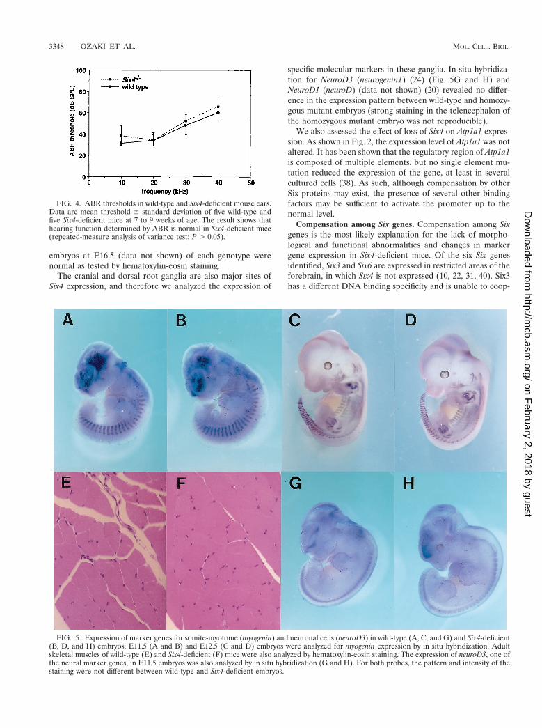
embryos at E16.5 (data not shown) of each genotype werenormal as tested by hematoxylin-eosin staining.
The cranial and dorsal root ganglia are also major sites ofSix4 expression, and therefore we analyzed the expression of
specific molecular markers in these ganglia. In situ hybridiza-tion for NeuroD3 (neurogenin1) (24) (Fig. 5G and H) andNeuroD1 (neuroD) (data not shown) (20) revealed no differ-ence in the expression pattern between wild-type and homozy-gous mutant embryos (strong staining in the telencephalon ofthe homozygous mutant embryo was not reproducible).
We also assessed the effect of loss of Six4 on Atp1a1 expres-sion. As shown in Fig. 2, the expression level of Atp1a1 was notaltered. It has been shown that the regulatory region of Atp1a1is composed of multiple elements, but no single element mu-tation reduced the expression of the gene, at least in severalcultured cells (38). As such, although compensation by otherSix proteins may exist, the presence of several other bindingfactors may be sufficient to activate the promoter up to thenormal level.
Compensation among Six genes. Compensation among Sixgenes is the most likely explanation for the lack of morpho-logical and functional abnormalities and changes in markergene expression in Six4-deficient mice. Of the six Six genesidentified, Six3 and Six6 are expressed in restricted areas of theforebrain, in which Six4 is not expressed (10, 22, 31, 40). Six3has a different DNA binding specificity and is unable to coop-
FIG. 4. ABR thresholds in wild-type and Six4-deficient mouse ears.Data are mean threshold 6 standard deviation of five wild-type andfive Six4-deficient mice at 7 to 9 weeks of age. The result shows thathearing function determined by ABR is normal in Six4-deficient mice(repeated-measure analysis of variance test; P . 0.05).
FIG. 5. Expression of marker genes for somite-myotome (myogenin) and neuronal cells (neuroD3) in wild-type (A, C, and G) and Six4-deficient(B, D, and H) embryos. E11.5 (A and B) and E12.5 (C and D) embryos were analyzed for myogenin expression by in situ hybridization. Adultskeletal muscles of wild-type (E) and Six4-deficient (F) mice were also analyzed by hematoxylin-eosin staining. The expression of neuroD3, one ofthe neural marker genes, in E11.5 embryos was also analyzed by in situ hybridization (G and H). For both probes, the pattern and intensity of thestaining were not different between wild-type and Six4-deficient embryos.
3348 OZAKI ET AL. MOL. CELL. BIOL.
on February 2, 2018 by guest
http://mcb.asm
.org/D
ownloaded from
erate with Eya (13, 28). Thus, it is not plausible that Six3 andSix6 compensate for Six4 function. On the other hand, Six1,Six2, and Six5 proteins share a DNA binding specificity withSix4 protein (13, 28, 37). Six1 shows mostly the same expres-sion pattern with Six4 (32), and Six5 mostly resembles Six4 withrespect to the molecular architecture with its large C-terminalportion and overall amino acid sequence similarity (13, 14).Thus, these two members are the probable candidates to com-pensate for the loss of Six4. To gain insight into the compen-sation mechanism, the expression of Six1, Six2, and Six5 inSix4-deficient mice was analyzed by Northern hybridization.The expression levels of these genes were not altered amongdifferent genotypes (Fig. 2). This finding suggests that thenormal expression levels of Six1, Six2, and Six5 are sufficient tocompensate for the loss of Six4 and to activate common targetgenes.
Similarly, the Six5-deficient mouse manifests limited pheno-types such as a higher rate of cataract formation (15, 35),compared to the relatively broad expression of Six5 (15), sug-gesting compensation for the loss of Six5 by Six1, Six2, and/orSix4 in these tissues. Because of such compensatory mecha-nisms among Six1, Six2, Six4, and Six5, knockout mouse modelsdeficient in a single Six gene are unlikely to contribute to ourunderstanding of their biological functions. Generation ofSix1-Six4 and Six4-Six5 double knockout mice should allow usto understand the compensation between them and the bio-logical function of Six4.
ACKNOWLEDGMENTS
We thank F. Relaix for discussion and critical reading of the manu-script, S. J. Tapscott for providing mouse NeuroD cDNA, Q. Ma forproviding neurogenin1 cDNA, and T. Yagi for providing pMC1DTpA.We also thank M. Yamakado, K. Ikeda, and S. Sato for discussion andH. Ohto and M. Kikuchi for technical assistance.
This work was supported by grants from the Ministry of Education,Science, Sports, and Culture of Japan and from the Ministry of Healthand Welfare of Japan and by the Jichi Medical School Young Inves-tigator Award.
REFERENCES
1. Abdelhak, S., V. Kalatzis, R. Heilig, S. Compain, D. Samson, C. Vincent, D.Weil, C. Cruaud, I. Sahly, M. Leibovici, M. Bitner-Glindzicz, M. Francis, D.Lacombe, J. Vigneron, R. Charachon, K. Boven, P. Bedbeder, N. Van Rege-morter, J. Weissenbach, and C. Petit. 1997. A human homologue of the
Drosophila eyes absent gene underlies branchio-oto-renal (BOR) syndromeand identifies a novel gene family. Nat. Genet. 15:157–164.
2. Boucher, C. A., N. Carey, Y. H. Edwards, M. J. Siciliano, and K. J. Johnson.1996. Cloning of the human SIX1 gene and its assignment to chromosome14. Genomics 33:140–142.
3. Boucher, C. A., S. K. King, N. Carey, R. Krahe, C. L. Winchester, S. Rah-man, T. Creavin, P. Meghji, M. E. Bailey, F. L. Chartier, S. D. Brown, M. J.Siciliano, and K. J. Johnson. 1995. A novel homeodomain-encoding gene isassociated with a large CpG island interrupted by the myotonic dystrophyunstable (CTG)n repeat. Hum. Mol. Genet. 4:1919–1925.
4. Boucher, C. A., C. L. Winchester, G. M. Hamilton, A. D. Winter, K. J.Johnson, and M. E. Bailey. 2000. Structure, mapping and expression of thehuman gene encoding the homeodomain protein, SIX2. Gene 247:145–151.
5. Cheyette, B. N., P. J. Green, K. Martin, H. Garren, V. Hartenstein, and S. L.Zipursky. 1994. The Drosophila sine oculis locus encodes a homeodomain-containing protein required for the development of the entire visual system.Neuron 12:977–996.
6. Esteve, P., and P. Bovolenta. 1999. cSix4, a member of the six gene family oftranscription factors, is expressed during placode and somite development.Mech. Dev. 85:161–165.
7. Gallardo, M. E., J. Lopez-Rios, I. Fernaud-Espinosa, B. Granadino, R. Sanz,C. Ramos, C. Ayuso, M. J. Seller, H. G. Brunner, P. Bovolenta, and S.Rodriguez de Cordoba. 1999. Genomic cloning and characterization of thehuman homeobox gene SIX6 reveals a cluster of SIX genes in chromosome14 and associates SIX6 hemizygosity with bilateral anophthalmia and pitu-itary anomalies. Genomics 61:82–91.
8. Granadino, B., M. E. Gallardo, J. Lopez-Rios, R. Sanz, C. Ramos, C. Ayuso,P. Bovolenta, and S. Rodriguez de Cordoba. 1999. Genomic cloning, struc-ture, expression pattern, and chromosomal location of the human SIX3gene. Genomics 55:100–105.
9. Hara, Y., O. Urayama, K. Kawakami, H. Nojima, H. Nagamune, T. Kojima,T. Ohta, K. Nagano, and M. Nakao. 1987. Primary structures of two types ofalpha-subunit of rat brain Na1,K1-ATPase deduced from cDNA sequences.J. Biochem. 102:43–58.
10. Jean, D., G. Bernier, and P. Gruss. 1999. Six6 (Optx2) is a novel murineSix3-related homeobox gene that demarcates the presumptive pituitary/hy-pothalamic axis and the ventral optic stalk. Mech. Dev. 84:31–40.
11. Kalatzis, V., I. Sahly, A. El-Amraoui, and C. Petit. 1998. Eya1 expression inthe developing ear and kidney: towards the understanding of the pathogen-esis of branchio-oto-renal (BOR) syndrome. Dev. Dyn. 213:486–499.
12. Kawakami, K., H. Ohto, K. Ikeda, and R. G. Roeder. 1996. Structure, func-tion and expression of a murine homeobox protein AREC3, a homologue ofDrosophila sine oculis gene product, and implication in development. Nu-cleic Acids Res. 24:303–310.
13. Kawakami, K., H. Ohto, T. Takizawa, and T. Saito. 1996. Identification andexpression of six family genes in mouse retina. FEBS Lett. 393:259–263.
14. Kawakami, K., S. Sato, H. Ozaki, and K. Ikeda. 2000. Six family genes—structure and function as transcription factors and their roles in develop-ment. Bioessays 22:616–626.
15. Klesert, T. R., D. H. Cho, J. I. Clark, J. Maylie, J. Adelman, L. Snider, E. C.Yuen, P. Soriano, and S. J. Tapscott. 2000. Mice deficient in Six5 developcataracts: implications for myotonic dystrophy. Nat. Genet. 25:105–109.
16. Klesert, T. R., A. D. Otten, T. D. Bird, and S. J. Tapscott. 1997. Trinucleotiderepeat expansion at the myotonic dystrophy locus reduces expression ofDMAHP. Nat. Genet. 16:402–406.
17. Kobayashi, M., H. Osanai, K. Kawakami, and M. Yamamoto. 2000. Expres-sion of three zebrafish Six4 genes in the cranial sensory placodes and thedeveloping somites. Mech. Dev. 98:151–155.
18. Kobayashi, M., R. Toyama, H. Takeda, I. B. Dawid, and K. Kawakami. 1998.Overexpression of the forebrain-specific homeobox gene six3 induces rostralforebrain enlargement in zebrafish. Development 125:2973–2982.
19. Kuhn, R., K. Rajewsky, and W. Muller. 1991. Generation and analysis ofinterleukin-4 deficient mice. Science 254:707–710.
20. Lee, J. K., J. H. Cho, W. S. Hwang, Y. D. Lee, D. S. Reu, and H. Suh-Kim.2000. Expression of neuroD/BETA2 in mitotic and postmitotic neuronalcells during the development of nervous system. Dev. Dyn. 217:361–367.
21. Loosli, F., S. Winkler, and J. Wittbrodt. 1999. Six3 overexpression initiatesthe formation of ectopic retina. Genes Dev. 13:649–654.
22. Lopez-Rios, J., M. E. Gallardo, S. Rodriguez de Cordoba, and P. Bovolenta.1999. Six9 (Optx2), a new member of the six gene family of transcriptionfactors, is expressed at early stages of vertebrate ocular and pituitary devel-opment. Mech. Dev. 83:155–159.
23. Ma, Q., Z. Chen, I. del Barco Barrantes, J. L. de la Pompa, and D. J.Anderson. 1998. neurogenin1 is essential for the determination of neuronalprecursors for proximal cranial sensory ganglia. Neuron 20:469–482.
24. Ma, Q., L. Sommer, P. Cserjesi, and D. J. Anderson. 1997. Mash1 andneurogenin1 expression patterns define complementary domains of neuro-epithelium in the developing CNS and are correlated with regions expressingnotch ligands. J. Neurosci. 17:3644–3652.
25. McCormick, M. B., R. M. Tamimi, L. Snider, A. Asakura, D. Bergstrom, andS. J. Tapscott. 1996. neuroD2 and neuroD3: distinct expression patterns and
TABLE 2. Expression pattern of Six4 determined byX-Gal staining of heterozygotes
Stage Tissuea
EmbryonicE8.5 Surface ectoderm outside the neural folds, somites,
presomitic mesodermE9.5 Nasal and otic placodes, cranial ganglia, branchial
arches, somites (dermamyotomes and sclero-tomes), AER
E10.5–11.5 Nasal pits, otic vesicles, cranial ganglia (V, VII–XI),dorsal root ganglia, branchial arches, somites,myotomes, limb mesenchyme, AER, notochord,mesonephros
E12.5–E13.5 Skeletal muscles, mesenchyme in limbs and digits,nasal epithelium, inner ear
Adult Skeletal muscle, nasal epithelium, cochlea, parathy-roid, salivary gland
a AER, apical ectodermal ridges.
VOL. 21, 2001 Six4-DEFICIENT MICE 3349
on February 2, 2018 by guest
http://mcb.asm
.org/D
ownloaded from
transcriptional activation potentials within the neuroD gene family. Mol.Cell. Biol. 16:5792–5800.
26. Nagy, A., J. Rossant, R. Nagy, W. Abramow-Newerly, and J. C. Roder. 1993.Derivation of completely cell culture-derived mice from early-passage em-bryonic stem cells. Proc. Natl. Acad. Sci. USA 90:8424–8428.
27. Niiya, A., H. Ohto, K. Kawakami, and M. Araki. 1998. Localization ofSix4/AREC3 in the developing mouse retina; implications in mammalianretinal development. Exp. Eye Res. 67:699–707.
28. Ohto, H., S. Kamada, K. Tago, S. Tominaga, H. Ozaki, S. Sato, and K.Kawakami. 1999. Cooperation of Six and Eya in activation of their targetgenes through nuclear translocation of Eya. Mol. Cell. Biol. 19:6815–6824.
29. Ohto, H., T. Takizawa, T. Saito, M. Kobayashi, K. Ikeda, and K. Kawakami.1998. Tissue and developmental distribution of Six family gene products. Int.J. Dev. Biol. 42:141–148.
30. Oliver, G., F. Loosli, R. Koster, J. Wittbrodt, and P. Gruss. 1996. Ectopiclens induction in fish in response to the murine homeobox gene Six3. Mech.Dev. 60:233–239.
31. Oliver, G., A. Mailhos, R. Wehr, N. G. Copeland, N. A. Jenkins, and P.Gruss. 1995. Six3, a murine homologue of the sine oculis gene, demarcatesthe most anterior border of the developing neural plate and is expressedduring eye development. Development 121:4045–4055.
32. Oliver, G., R. Wehr, N. A. Jenkins, N. G. Copeland, B. N. Cheyette, V.Hartenstein, S. L. Zipursky, and P. Gruss. 1995. Homeobox genes andconnective tissue patterning. Development 121:693–705.
33. Olson, E. N., and W. H. Klein. 1994. bHLH factors in muscle development:dead lines and commitments, what to leave in and what to leave out. GenesDev. 8:1–8.
34. Ozaki, H., K. Yamada, M. Kobayashi, S. Asakawa, S. Minoshima, N.Shimizu, M. Kajitani, and K. Kawakami. 1999. Structure and chromosomalmapping of human SIX4 and mouse Six4 genes. Cytogenet. Cell Genet.87:108–112.
35. Sarkar, P. S., B. Appukuttan, J. Han, Y. Ito, C. Ai, W. Tsai, Y. Chai, J. T.Stout, and S. Reddy. 2000. Heterozygous loss of Six5 in mice is sufficient tocause ocular cataracts. Nat. Genet. 25:110–114.
36. Soriano, P., C. Montgomery, R. Geske, and A. Bradley. 1991. Targeteddisruption of the c-src proto-oncogene leads to osteopetrosis in mice. Cell64:693–702.
37. Spitz, F., J. Demignon, A. Porteu, A. Kahn, J. P. Concordet, D. Daegelen,and P. Maire. 1998. Expression of myogenin during embryogenesis is con-trolled by Six/sine oculis homeoproteins through a conserved MEF3 bindingsite. Proc. Natl. Acad. Sci. USA 95:14220–14225.
38. Suzuki-Yagawa, Y., K. Kawakami, and K. Nagano. 1992. HousekeepingNa,K-ATPase a1 subunit gene promoter is composed of multiple cis ele-ments to which common and cell type-specific factors bind. Mol. Cell. Biol.12:4046–4055.
39. Takahashi, K., N. Osawa, M. Ohmura, and K. Kitamura. 1999. Evaluationof inner ear histology and auditory brainstem response in Wriggle MouseSagami. Acta Otolaryngol. 119:767–772.
40. Toy, J., and O. H. Sundin. 1999. Expression of the optx2 homeobox geneduring mouse development. Mech. Dev. 83:183–186.
41. Toy, J., J. M. Yang, G. S. Leppert, and O. H. Sundin. 1998. The optx2homeobox gene is expressed in early precursors of the eye and activatesretina-specific genes. Proc. Natl. Acad. Sci. USA 95:10643–10648.
42. Wallis, D. E., E. Roessler, U. Hehr, L. Nanni, T. Wiltshire, A. Richieri-Costa,G. Gillessen-Kaesbach, E. H. Zackai, J. Rommens, and M. Muenke. 1999.Mutations in the homeodomain of the human SIX3 gene cause holoprosen-cephaly. Nat. Genet. 22:196–198.
43. Wilkinson, D. G. 1992. Whole mount in situ hybridization of vertebrateembryos, p. 75–83. In D. G. Wilkinson (ed.), In situ hybridization: a practicalapproach. Oxford IRL Press, New York, N.Y.
44. Winchester, C. L., R. K. Ferrier, A. Sermoni, B. J. Clark, and K. J. Johnson.1999. Characterization of the expression of DMPK and SIX5 in the humaneye and implications for pathogenesis in myotonic dystrophy. Hum. Mol.Genet. 8:481–492.
45. Wright, W. E., D. A. Sassoon, and V. K. Lin. 1989. Myogenin, a factorregulating myogenesis, has a domain homologous to MyoD. Cell 56:607–617.
46. Xu, P. X., J. Adams, H. Peters, M. C. Brown, S. Heaney, and R. Maas. 1999.Eya1-deficient mice lack ears and kidneys and show abnormal apoptosis oforgan primordia. Nat. Genet. 23:113–117.
47. Yagi, T., S. Nada, N. Watanabe, H. Tamemoto, N. Kohmura, Y. Ikawa, andS. Aizawa. 1993. A novel negative selection for homologous recombinantsusing diphtheria toxin A fragment gene. Anal. Biochem. 214:77–86.
48. Zuber, M. E., M. Perron, A. Philpott, A. Bang, and W. A. Harris. 1999. Gianteyes in Xenopus laevis by overexpression of XOptx2. Cell 98:341–352.
3350 OZAKI ET AL. MOL. CELL. BIOL.
on February 2, 2018 by guest
http://mcb.asm
.org/D
ownloaded from


![BIOCHIMICA ET BIOPHYSICA ACTA ii!i,, · 18 D.M. Mazzuca, T. C. Y. Lo / Biochimica et Biophysica A cta 1414 (1998) 16-30 the myogenin promoter [36]. The PGK-myogenin con- struct and](https://img.pdfslide.us/doc/110x75/5ebe218ebd2e88479e3be038/biochimica-et-biophysica-acta-iii-18-dm-mazzuca-t-c-y-lo-biochimica-et.jpg)