Embed Size (px)
Citation preview

RIGHT:
URL:
CITATION:
AUTHOR(S):
ISSUE DATE:
TITLE:
Siphonaxanthin, a carotenoid from greenalgae Codium cylindricum, protects Ob/Obmice fed on a high-fat diet againstlipotoxicity by ameliorating somatic stressesand restoring anti-oxidative capacity
Zheng, Jiawen; Manabe, Yuki; Sugawara, Tatsuya
Zheng, Jiawen ...[et al]. Siphonaxanthin, a carotenoid from green algae Codium cylindricum, protects Ob/Ob mice fedon a high-fat diet against lipotoxicity by ameliorating somatic stresses and restoring anti-oxidative capacity. NutritionResearch 2020, 77: 29-42
2020-05
http://hdl.handle.net/2433/252354
© 2020. This manuscript version is made available under the CC-BY-NC-ND 4.0 licensehttp://creativecommons.org/licenses/by-nc-nd/4.0/.; The full-text file will be made open to the public on 1 May 2021 inaccordance with publisher's 'Terms and Conditions for Self-Archiving'.; この論文は出版社版でありません。引用の際には出版社版をご確認ご利用ください。; This is not the published version. Please cite only the published version.

1
Title Siphonaxanthin, a carotenoid from green algae Codium cylindricum, protects ob/ob mice
fed on a high-fat diet against lipotoxicity by ameliorating somatic stresses and restoring anti-
oxidative capacity
Author: Jiawen Zhenga, Yuki Manabea, Tatsuya Sugawaraa
aDivision of Applied Biosciences, Graduate School of Agriculture, Kyoto University, Kyoto
606-8502, Japan
Corresponding author: Tatsuya Sugawara; Tel: +81-75 753 6212; Fax: +81- 75 753 6212. E-
mail: [email protected]
First author: Jiawen Zheng [email protected]
Co-author: Yuki Manabe [email protected]
A Self-archived copy inKyoto University Research Information Repository
https://repository.kulib.kyoto-u.ac.jp

2
Abbreviations
HPLC, high performance liquid chromatography
PDA, photodiode array detector
HFD, high fat diet
TAG, triacylglycerol
HDL, high density lipoprotein
NEFA, non-esterified fatty acids
AST, aspartate aminotransferase
ALT, alanine aminotransferase
H&E, hematoxylin and eosin
TBARS, thiobarbituric acid reactive substances
TBA, thiobarbituric acid
TCA, trichloroacetic acid
GSSG, glutathione disulfide
GSH, glutathione
DMSO, dimethyl sulfoxide
qRT-PCR, quantitative reverse transcription-polymerase chain reaction
SDS-PAGE, sodium dodecyl sulfate polyacrylamide gel electrophoresis
ER, endoplasmic reticulum
ERAD, endoplasmic-reticulum-associated protein degradation
NAFLD, non-alcoholic fatty liver disease
ROS, reactive oxygen species
A Self-archived copy inKyoto University Research Information Repository
https://repository.kulib.kyoto-u.ac.jp

3
NASH, non-alcoholic steatohepatitis
UPR, unfolded protein response
A Self-archived copy inKyoto University Research Information Repository
https://repository.kulib.kyoto-u.ac.jp

4
Abstract
Oxidative stress is implicated in the pathogenesis of many diseases including obesity, non-
alcoholic fatty liver disease, and diabetes mellitus. Previously, we reported that siphonaxanthin, a
carotenoid from green algae, elicited a potent inhibitory effect on hepatic de novo lipogenesis, and
an anti-obesity effect in both 3T3L1 cells and KKAy mice. Thus, we hypothesized that
consumption of siphonaxanthin could improve metabolic disorders including hepatic steatosis and
systemic adiposity, as well as ameliorate somatic stress under obese conditions. Both the
hepatocyte cell line HepG2 and a mouse model of severe obesity, produced by feeding ob/ob mice
on a high-fat diet (HFD), were used to test this hypothesis. In obese mice, siphonaxanthin intake
did not improve liver steatosis or systemic adiposity. However, intake did lower plasma glucose
and alanine aminotransferase (ALT) levels and diminished hepatic lipid peroxidation products and
antioxidant gene expression, which increased significantly in control group obese mice. Renal
protein carbonyl content decreased significantly in the siphonaxanthin group, which might also
indicate an ameliorated oxidative stress. Relevantly, siphonaxanthin intake restored gene
expression related to antioxidant signaling, lipid β-oxidation, and endoplasmic-reticulum-
associated protein degradation in the kidney, which decreased significantly in obese mice. We
found that the liver and kidney respond to obesity-induced somatic stress in a divergent pattern. In
addition, we confirmed that siphonaxanthin potently induced Nrf2-regulated antioxidant signaling
in HepG2 cells. In conclusion, our results indicated that siphonaxanthin might protect obesity-
leading somatic stress through restoration of Nrf2-regulated antioxidant signaling, and might
therefore be a promising nutritional supplement.
A Self-archived copy inKyoto University Research Information Repository
https://repository.kulib.kyoto-u.ac.jp

5
Keywords
Obesity; Non-alcoholic fatty liver diseases; Oxidative stress; Endothelium reticulum stress;
Carotenoid.
A Self-archived copy inKyoto University Research Information Repository
https://repository.kulib.kyoto-u.ac.jp

6
1. Introduction
Obesity is considered a leading risk factor for many diseases which is accompanied by
increasing circulating fatty acids and insulin resistance. In obesity, substantial increases in
intracellular pro-oxidant influx, electrophilic stress, and mitochondrial burden occur, leading to
generation of reactive oxygen species (ROS) and oxidative stress. ROS can denaturize or modify
structural and functional molecules such as proteins and DNA, thus inducing dysregulation in
molecular events and biological processes. ROS and oxidative stress are also intimately related
to endoplasmic reticulum (ER) stress, which can act in a highly coordinated manner to induce
cell apoptosis and tissue damage, as well as to exacerbate local inflammatory response [1-3].
Obesity-induced oxidative stress and ER stress can therefore further increase the risks of
developing diseases such as diabetes mellitus, non-alcoholic fatty liver disease (NAFLD), renal
diseases, and cardiovascular diseases in obese individuals [1, 4-6].
Nrf2 is a primary transcription factor in counteracting oxidative stress. It regulates a
variety of antioxidant genes, phase II detoxifying enzymes, biotransformation enzymes,
xenobiotic efflux transporters, and inflammatory factors, which form the integral antioxidant
defense system [5]. This system protects tissues and organs from oxidative injury and maintains
endogenous homeostasis by scavenging ROS, highly reactive intermediates or toxic substrates.
Nrf2-regulated pathways have been observed to play a role in various diseases [7]. Meakin et al.
reported that Nrf2-/- mice developed more severe nonalcoholic steatohepatitis (NASH) with
cirrhosis, than wild-type mice, when fed on a high-fat diet (HFD) [8]. Moreover, in Nrf2-/- mice,
a rapid onset and progression of nutritional steatohepatitis was induced by a methionine- and
A Self-archived copy inKyoto University Research Information Repository
https://repository.kulib.kyoto-u.ac.jp

7
choline-deficient diet [9]. In addition, ablation of Nrf2 in experimental animals was found to
cause lupus-like autoimmune nephritis and to exacerbate diabetes-induced oxidative stress,
inflammation, and nephropathy [10, 11]. These studies also indicate that oxidative stress is a
shared etiological factor in different diseases.
To reduce somatic oxidative stress, a sustained healthy lifestyle, consisting of dietary
management and routine exercise, is generally recommended. Additionally, novel functional
compounds that can boost the anti-oxidative capacity of the body with improved efficacy and
prolonged action could be promising in establishing therapeutic strategies for different diseases.
In light of the role of Nrf2 in detoxification and the defense system, its enhancers have more
recently been proposed as a new therapeutic class in combating diseases involving oxidative
stresses from divergent stimuli [7]. In fact, several natural Nrf2 enhancers such as protandim
(containing herbal ingredients), sulforaphane, and curcumin have been found out to be quite
effective [12]. Some nutritional compounds such as flavonoids and catechins have also been
reported as potent natural Nrf2 activators [13, 14].
Siphonaxanthin is a carotenoid specifically derived from green algae such as Codium
cylindricum. It shares a common structure with other carotenoids, containing 8 isoprene
molecules, and is distinguished by a C-8 carbonyl and C-19 hydroxyl groups on its main bond
[15]. Previously, we have discovered that siphonaxanthin possesses moderate anti-obesity
activity by inhibiting the expression of Pparg and Cebpa, both in the 3T3L1 cell line and in the
obese and diabetic murine model, KK-Ay [16]. Additionally, our previous study also showed
that siphonaxanthin inhibits de novo synthesis of triacylglycerol in hepatocytes by exerting an
A Self-archived copy inKyoto University Research Information Repository
https://repository.kulib.kyoto-u.ac.jp

8
antagonistic effect on the nuclear receptor LXRα, which is a master regulator of de novo
lipogenesis [17]. Based on a previous study, we hypothesized that siphonaxanthin might
ameliorate hepatic steatosis and systemic adiposity by inhibiting the expression of lipogenic
genes, and could prevent oxidative stress and ER stress by inducing antioxidant signaling. To test
this hypothesis, we used the leptin deficient ob/ob mice, well documented as a murine model of
spontaneous obesity, and fed them a HFD to manifest both nature and nurture factors in the
pathogenesis of obesity and hepatic steatosis. The HepG2 cell line was used to investigate the
effect of siphonaxanthin on Nrf2-regulated antioxidant signaling. Above all, we aimed to
demonstrate the potential of siphonaxanthin as a nutritional compound targeting metabolic
diseases.
2. Methods and materials
2.1. Preparation of siphonaxanthin rich fraction
Siphonaxanthin was extracted from the green algae Codium cylindricum Holmes [18]. For the
animal study, a crude lipid fraction was first obtained by extracting with acetone from freeze
dried C. cylindricum H. powder. The crude lipid extract was then dissolved in hexane/acetone
(6:4) and subjected to silica gel column chromatography. Next, the siphonaxanthin-rich fraction
was prepared through a gradient elution with hexane/acetone (9:1, 8:2, 7:3, 6:4; v/v). The final
siphonaxanthin-rich fraction used in the animal study was composed of 68% siphonaxanthin and
32% other lipids of which the major component was monogalactosyldiacylglycerol (Fig. 1). For
cellular study, the siphonaxanthin-rich fraction was re-dissolved in methanol and further purified
A Self-archived copy inKyoto University Research Information Repository
https://repository.kulib.kyoto-u.ac.jp

9
by high performance liquid chromatography (HPLC) (LC-6; Shimadzu, Japan) connected to a
photodiode array detector (PDA) (SPD-M20A; Shimadzu, Japan) with a purity of above 99%.
All the samples were stored at −80°C, until further use.
2.2. Animals and diets
All experimental animal protocols were approved by the Animal Experimentation Committee of
Kyoto University for the care and use of experimental animals (Approve No. 29-80). Male
C57BL/6JHamSlc-ob/ob mice (6 weeks) and C57BL/6JJmsSlc mice were obtained from Japan
SLC. All mice were housed individually and maintained on an alternating 12-h light/dark cycle
at 23±1°C. After an acclimatization period of 5 days, the ob/ob mice were randomly divided into
control and siphonaxanthin (SPX) groups (n = 6 per group), with ad libitum access to drinking
water. The control group was fed a modified 45% HFD (D12451, Research Diets, NJ, USA)
supplemented with 2.2% soybean oil (Table 1). The siphonaxanthin group was fed a modified
45% HFD supplemented with siphonaxanthin at a dosage of 0.016% (w/w) (calculated by
siphonaxanthin weight equivalent) dissolved in soybean oil (2.2% of HFD weight) (Table 1).
C57BL/6J mice were designated as the normal group, fed on a basal AIN93G diet (Table 1) [19].
Body weight and food intake were monitored throughout the study. After 43 days of feeding, the
mice were euthanized by exsanguination under anesthesia with isoflurane, after a 12-hour fast,
and blood was collected in heparinized syringes from the inferior vena cava. Organs were rapidly
removed, weighed, and immediately frozen in liquid nitrogen. Liver and kidney tissues were
partially stored in RNA laterTM solution (Ambion, CA, USA) at −80°C until further analyses.
A Self-archived copy inKyoto University Research Information Repository
https://repository.kulib.kyoto-u.ac.jp

10
2.3. Biochemical analyses
Blood plasma was separated by centrifugation at 1000 × g for 15 min at 4°C and stored at −80°C
until use. Plasma concentrations of glucose, triacylglycerol (TAG), free cholesterol, high density
lipoprotein (HDL) cholesterol, total cholesterol, non-esterified fatty acid (NEFA), aspartate
aminotransferase (AST), and alanine aminotransferase (ALT) were measured using
commercially available kits (Glu C II, TG E, F-Cho E, HDL-C E, T-Cho E, NEFA, and GOT
GPT C II, respectively; Wako Pure Chemical Industries, Osaka, Japan) according to the
manufacturer’s instructions. Plasma creatinine was measured using the commercially available
kit (Creatinine Colorimetric Assay Kit, Cayman Chemical, MI, USA). TAG, total cholesterol,
and NEFA concentrations in the lipid fraction prepared from the liver tissue were measured
using the commercial kits mentioned above.
2.4. Histomorphology analyses
Liver tissues were fixed in 4% paraformaldehyde and then embedded in paraffin to form blocks.
Slices were stained with hematoxylin and eosin (H&E) or Sirius Red to observe the lipid droplets
and fibrosis in liver tissues. Liver tissue morphology was observed, and photos were taken using
the fluorescence microscope BZ-9000 (Keyence, Osaka, Japan).
2.5. Hepatic and renal oxidative stress marker
A Self-archived copy inKyoto University Research Information Repository
https://repository.kulib.kyoto-u.ac.jp

11
Levels of malondialdehyde in liver and kidney tissues were measured by using the TBARS assay
[20]. Briefly, 40 mg tissues were homogenized in 1.15% KCl aqueous solution (400 μL) with 5%
butylated hydroxytoluene methanol solution (16 uL). Next, 400 μL of 0.375% thiobarbituric acid
(TBA)–0.25 M HCl solution and 15% trichloroacetic acid (TCA) solution were added into the
tissue homogenate, respectively, and boiled in a water bath at 95°C for 15 min. The solution was
then cooled and centrifuged at 10,000 × g for 5 min under room temperature. Absorption of the
supernatant at a wavelength of 535 nm was measured with a microplate reader (Molecular
Devices Co., Sunnyvale, CA). Glutathione and glutathione disulfide contents in tissue
homogenates were measured using a commercial kit (GSSG/GSH Quantification Kit, Dojindo
Molecular Technologies, Kumamoto, Japan), according to the manufacturer’s instructions.
Protein carbonyl content in kidney homogenate was measured using a commercial kit (Protein
Carbonyl Content Assay Kit, Sigma-Aldrich, MO, USA), per manufacturer’s instructions.
2.6. Cell culture and treatment
HepG2 cells (JCRB 1054; Health Science Research Resources Bank, Osaka, Japan) were
cultured in Dulbecco’s modified essential medium (DMEM) containing 10% fetal bovine serum
(Invitrogen, CA, USA) and antibiotics (100 unit/mL penicillin and 100 μg/mL streptomycin, Life
Technologies Corporation, NY, USA) at 37°C in a humidified atmosphere with 5% CO2. Cells
were seeded in 12-well plates at 2.5 × 10⁵ cells/mL for real-time quantitative reverse
transcription-polymerase chain reaction (qRT-PCR) analysis or in 6-well plates at 5 × 10⁵
cells/mL for western blot. After confluence, cells were treated with vehicle or siphonaxanthin
alone for a designated time period. Siphonaxanthin was dissolved in dimethyl sulfoxide (DMSO)
A Self-archived copy inKyoto University Research Information Repository
https://repository.kulib.kyoto-u.ac.jp

12
before adding to the culture medium, with a final DMSO concentration of 0.2%. DMSO was
used as vehicle in the experiment.
2.7. Gene expression analysis using real-time quantitative reverse transcription-polymerase chain
reaction
Total RNA was extracted from HepG2 cells or tissues using the sepasol reagent (Nacalai Tesque,
Kyoto, Japan) and cDNA was synthesized from RNA by using ReverTra Ace qPCR RT Master
Mix (TOYOBO, Osaka, Japan) according to the manufacturer’s instructions. To perform the
qRT-PCR, cDNA was diluted and mixed with iQ SYBR Green Supermix (Bio-Rad Laboratories,
CA, USA) containing 1 μmol/L PCR primer (primer sequences are shown in Table 3, 4). Real-
time qRT-PCR was performed using a DNA Engine Option system (Bio-Rad Laboratories) and
the expression level of each gene was normalized using β-actin as an internal control.
2.8. Western blot analysis
Cells or tissue samples were homogenized in lysis buffer [20 mmol/L Tris-HCl, pH 8; 150
mmol/L NaCl, 1% Triton-X 100, protease inhibitor (cOmplete Tablets, mini EASYpack; Roche,
Mannheim, Germany)]. The homogenate was centrifuged at 12,000 × g at 4°C for 15 min to
collect the supernatant. Protein concentration was determined using the DC protein assay kit
(Bio-Rad Laboratories). Next, the proteins were separated by 12.5% SDS-PAGE and transferred
to a polyvinylidene difluoride membrane. Target proteins were probed with HMOX1 or β-actin
primary antibody (1:1000; Cell Signaling, MA, USA) at 4°C overnight, and then incubated with
A Self-archived copy inKyoto University Research Information Repository
https://repository.kulib.kyoto-u.ac.jp

13
HRP-conjugated anti-rabbit IgG secondary antibody (1:2000, Cell signaling) at room
temperature for 1 h. Signals were visualized with the substrate Chemi-lumi One (Nacalai
Tesque) using a LAS-3000 visualizer (Fujifilm, Tokyo, Japan). Protein expression level was
normalized using β-actin as an internal control.
2.9. Quantification of liver siphonaxanthin accumulation by high performance liquid
chromatography
Siphonaxanthin was extracted from the liver tissues and subjected to HPLC analysis as
previously described [16]. The lipid extracts were loaded onto Sep-Pak Plus silica cartridges
(Waters, MA, USA) to remove the TAG fraction, and dissolved in methanol for HPLC analysis.
The peak of siphonaxanthin was further confirmed from its characteristic UV spectrum.
2.10. Statistical analyses
Data analyses were performed using the statistical program SPSS 23 for Mac. Significance was
verified between groups of normally distributed data using a 1-factor ANOVA, followed by a
Tukey’s post hoc analysis for animal experiments and Scheffe’s post hoc analysis for cultured
cell experiments. Variance homogeneity was examined using Levene’s test. When the variances
between groups were unequal, the data were transformed to logarithms before analysis by 1-
factor ANOVA. Data are represented as means ± SEMs. Significance was defined as P < 0.05.
3. Results
A Self-archived copy inKyoto University Research Information Repository
https://repository.kulib.kyoto-u.ac.jp

14
3.1. Physiological parameters
During the experimental period, 2 mice in the control group died and 1 exhibited an obvious
open wound from severe stress, and were therefore excluded from the final statistical analyses of
the control group. Therefore, all the results were presented as a sample size of 6 for the normal
and siphonaxanthin groups and 3 for the control group. As shown in Table 5, there was no
difference in food intake between three groups while ob/ob mice had a significant increase in
body, liver and adipose tissue weight compared to wild type mice (Table 5). Plasma glucose, free
cholesterol, HDL-cholesterol, total cholesterol, ALT and AST increased significantly in the
control group compared to the normal group. The siphonaxanthin group exhibited a decreasing
tendency in plasma glucose and a significant decline in ALT level (Table 6). Moreover, both
liver triacylglycerol (TAG) and cholesterol increased significantly in ob/ob mice, while no
significant difference between the control and siphonaxanthin groups was observed (Table 7).
3.2. Liver histological analyses
To evaluate progression of liver pathology, liver sections from three groups were analyzed using
H&E and Sirius Red staining. As shown in Fig. 2, severe hepatic steatosis without obvious
fibrosis or inflammatory cell infiltration was observed in livers of ob/ob mice compared to
normal mice. No difference between the control and siphonaxanthin groups was observed in
relation to hepatic steatosis.
A Self-archived copy inKyoto University Research Information Repository
https://repository.kulib.kyoto-u.ac.jp

15
3.3. Hepatic TBARS and gene expression related to oxidative stress, ER stress, and lipid
metabolism
Liver TBARS level increased significantly in control group ob/ob mice compared to the normal
group, and was recovered to a normal level in the siphonaxanthin group (Fig. 3A). However, no
significant changes in hepatic GSH, GSSG, and GSH/GSSG ratio were observed (Fig. 3B-D). To
determine the redox state of the liver, genes related to oxidative stress (Fig. 3E), ER stress (Fig.
3F), and lipid metabolism (Fig. 3G) were evaluated. Increases in the expression of antioxidant
genes including Gsta4, Nqo1, and Gpx4 were observed in the control group, while the
siphonaxanthin group showed a significant decline in Gsta4 and Gpx4 expressions, as well as a
decreasing trend in Nqo1 expression (Fig. 3E). Furthermore, the expression of Atf3, which was
related to ER stress, increased significantly in the control group and was downregulated in the
siphonaxanthin group (Fig. 3F). However, expression of the ER stress marker genes, Atf6 and
Hspa5, decreased significantly in the control group, and were not restored by siphonaxanthin
intake (Fig. 3F). Expression of Ppara and Ppard, which were related to lipid β-oxidative
capacity, declined significantly in both the control and siphonaxanthin groups (Fig. 3G). A
significant elevation of Srebf and Cd36 was also observed in the control group, whereas Srebf
tended to decrease in the siphonaxanthin group (Fig. 3G).
3.4. Renal TBARS, protein carbonyl content, and gene expression related to oxidative stress, ER
stress, and lipid metabolism
A Self-archived copy inKyoto University Research Information Repository
https://repository.kulib.kyoto-u.ac.jp

16
In the kidney, TBARS level showed no significant change between the three groups (Fig. 4A),
however, protein carbonyl content increased significantly in the control group and was restored
to a normal level in the siphonaxanthin group (Fig. 4B). No significant change was confirmed in
renal GSH, GSSG, and GSH/GSSG ratio (Fig. 4C-E). To evaluate the redox state of the kidney,
gene expression related to oxidative stress (Fig. 4F), ER stress (Fig. 4G), and lipid metabolism
(Fig. 4H) were evaluated. Gene expression related to antioxidant signaling, including Hmox1,
Gclm, and Gclc, displayed a significant decline in the control group compared to the normal
group. Siphonaxanthin intake tended to restore the expression of Hmox1, Gclm and Gclc and
elevated the expression of Nqo1 significantly (Fig. 4F). Meanwhile, Gsta4 expression tended to
increase in the control group and significantly increased in the siphonaxanthin group. The
expression of Atf3 and Hspa5, genes related to ER stress, significantly decreased in the control
group, which tended towards recovery in the siphonaxanthin group (Fig. 4G). The expression of
Ppara and its target gene Cpt1b, two critical genes involved in lipid β-oxidation, was
significantly elevated by siphonaxanthin intake, compared to the control group (Fig. 4H).
3.5. Hepatic and renal HMOX1 protein expression
Protein expression of HMOX1, an important target gene of Nrf2, increased in the liver, and
contrastingly, significantly decreased in the kidney of the control group, compared to the normal
group (Fig. 5A-D). No significant change between the control and siphonaxanthin groups was
confirmed (Fig. 5A-D).
A Self-archived copy inKyoto University Research Information Repository
https://repository.kulib.kyoto-u.ac.jp
17
3.6. Siphonaxanthin enhanced Nrf2 protein expression and target gene expression in the HepG2
cell line
Treatment with 1.0 or 2.0 μM siphonaxanthin alone for 24 h significantly induced Nrf2 protein
expression (Fig. 6A). Concomitantly, expression of HMOX1 and SOD2 tended to increase and
GCLC increased significantly with 1.0 μM siphonaxanthin treatment for 6 h (Fig. 6B).
Expression of GSTA4 and GCLC tended to increase with 1.0 μM siphonaxanthin treatment and
increased significantly with 2.0 μM siphonaxanthin treatment for 16 h (Fig. 6C). Expression of
NQO1 also increased significantly following 2.0 μM siphonaxanthin treatment for 16 h (Fig.
6C). A similar tendency was observed in GPX4 and SOD2 at 2.0 μM for 16 h (Fig. 6C).
3.7. Siphonaxanthin accumulation in liver tissue
Siphonaxanthin accumulation in liver was measured by HPLC-PDA (Fig. 7). Peaks 1-3 refer to
the metabolites of siphonaxanthin, while peak 4 refers to siphonaxanthin. In the liver, 277 ± 7 ng
of siphonaxanthin and 3428 ± 210 ng of metabolites per gram were detected.
4. Discussion
In the present study, we investigated the effect of siphonaxanthin on metabolic disorders
and systemic stress under obese conditions in murine mouse model manifesting of both obesity
and NAFLD. Siphonaxanthin mitigated liver damage and hepatic oxidative stress in ob/ob mice,
as seen by the significant decline in plasma ALT level and TBARS content respectively.
A Self-archived copy inKyoto University Research Information Repository
https://repository.kulib.kyoto-u.ac.jp

18
Previously, Feng et al. reported that long chain fatty acids could induce antioxidant signaling in
the Hepa1-6 cell line [21]. Moreover, Malaguarnera et al. reported that the induction of hepatic
HMOX1 protein was an adaptive response against oxidative damage elicited by lipid
peroxidation in human NASH progression [22]. In light of the above reports, the significant
increase of antioxidant gene expression and HMOX1 protein expression in the liver of the
control group mice in this study might indicate an antioxidant response stimulated by increased
lipid peroxidation, which was mitigated in the siphonaxanthin group. Therefore, siphonaxanthin
probably relieved hepatic oxidative stress and elicited the hepatoprotective effect through
scavenging of reactive intermediates in the liver, rather than reinforcing antioxidant signaling.
Besides, hepatic content of GSSG and GSH did not show significant changes between 3 groups,
which was consistent with the gene expression results of Gclc and Gclm. Siphonaxanthin did not
restore the expression of Ppara and Ppard in the liver, both of which are associated with lipid β-
oxidative capacity, and this might indicate an overwhelmed oxidative capacity resulted from
lipid overload in the liver.
The protein carbonylation content in the kidney, a marker for oxidative stress, exhibited a
significant increase in the control group compared to the normal group and was restored by
siphonaxanthin. Meanwhile, siphonaxanthin rescued the expression of some antioxidant genes
which decreased in the control group, and might indicate a recovery of Nrf2 signaling in
siphonaxanthin group. The decline of HMOX1 protein expression in control group agreed with
the decrease of antioxidant gene expressions. In addition, unfolded protein response (UPR)
signaling was dysregulated in the control group with significant decreases of Atf3 and Hspa5
expression, and was restored in the siphonaxanthin group. Given that Hspa5 encodes the main
A Self-archived copy inKyoto University Research Information Repository
https://repository.kulib.kyoto-u.ac.jp

19
protein chaperone GRP78, which helps assemble proteins and degrade misfolded proteins in the
ER, and Atf3 encodes a transcriptional factor, which promotes cell survival under ER stress, their
normal expression is indispensable for cells to survive ER stress and cell apoptosis [1, 2, 23-25].
Collectively, despite prevailing oxidative stress under hyperlipidemic and hyperglycemic
conditions, the downregulation of Nrf2 and UPR signaling in the control group suggested a
hyporesponsive defense system in the kidney of ob/ob mice, which was similar to the results
observed in some chronic kidney disease models reporting a failed response of the Nrf2 and UPR
pathway even under strong oxidative stress [32, 33]. The elevated expression of Ppara and
Cpt1b gene in the siphonaxanthin group compared to control group might suggest the recovery
of renal lipid β-oxidative capacity conjugated to restored antioxidant signaling.
Notably, we observed that the expression of antioxidant genes, ER stress-related genes and
the HMOX1 protein exhibited discrepancies between the liver and kidney, and the two organs
responded to obesity-induced somatic stress in disparate manners. While liver had an inducible
Nrf2 signaling, kidney seemed to have a more sever insult and defect in the redox signaling.
Such severe decline in both mRNA and protein levels of antioxidant gene expression was also
reported in murine models of slowly progressive polycystic kidney disease by Maser et al. [26].
This result might reflect a possibility that tissue-specific signaling pathways might exist under
chronic somatic stress. Regardless of these discrepancies, siphonaxanthin seemed to exert a
favorable effect on restoring the redox homeostasis in both the liver and kidney, and this
protective effect might lie in its ROS scavenging property and Nrf2 inducing capacity. Indeed,
we confirmed the potency of purified siphonaxanthin on inducing Nrf2 signaling in cultured
HepG2 cells [7, 27]. Intriguingly, by the end of the experiment, 2 mice in the control group had
A Self-archived copy inKyoto University Research Information Repository
https://repository.kulib.kyoto-u.ac.jp

20
died and 1 exhibited an obvious open wound, while no obvious abnormality was observed in the
siphonaxanthin group. This result suggested that siphonaxanthin might be able to extend life
expectancy by alleviating systemic stress.
Nevertheless, several contradictions between the results and our hypothesis were observed
in this study. Firstly, we expected to observe the development of steatohepatitis by feeding ob/ob
mice with HFD based on some previous study [28-30]. However, liver tissue analysis showed
only simple fatty liver, absent in fibrosis and immune cell infiltration. As Imajo et al. reported
previously that the deficiency of leptin signaling could hamper the progression to steatohepatitis,
this might explain the absence of steatohepatitis in ob/ob mice in our study [31]. Secondly,
siphonaxanthin failed to improve the physiological lipid profile, systemic adiposity, and hepatic
steatosis, regardless of its inhibitory effect on lipogenesis or adipogenesis under the regulation of
LXRα, PPARγ and CEBPα in our previous reports [16, 17]. However, the strong suppressive
effect of siphonaxanthin on either LXRα activation or PPARγ and CEBPα expressions was
confined to cell line study, and showed dosage-dependent efficacy. As the concentration of
siphonaxanthin used in cell line study was much higher than in vivo study, its suppressive effect
on the animal model of obesity was quite limited [16]. In addition, siphonaxanthin was supposed
to compete with excessive body of endogenous ligands to downregulate the lipogenic program in
the present study. Also, no direct evidence had shown that siphonaxanthin was effective in
blocking the uptake of dietary lipids, which was the main source where the body fat in ob/ob
mice derived from. Therefore, the failed rescue of systemic adiposity in ob/ob mice fed a HFD
might be due to the low supplementary dosage of siphonaxanthin and the severe obese state.
Thirdly, although the renal Gclc and Gclm expressions decreased significantly in control group
A Self-archived copy inKyoto University Research Information Repository
https://repository.kulib.kyoto-u.ac.jp

21
and tended to increase in siphonaxanthin group, the GSH and GSSG concentration did not show
any difference among the three groups in the kidney. This result could be possible as the
intrarenal glutathione was determined by three independent processes including GSH uptake,
degradation and resynthesis. Since hepatic GSH content remained intact in this study, kidney
might uptake the circulating GSH, mainly secreted by liver in a large amount, to compensate its
declined synthesis ability and to retain the intracellular GSH/GSSG concentration [32, 33].
However, the mRNA expression might chronically receive a negative modulation signal under
somatic stress.
Overall, the results suggested that siphonaxanthin could protect against liver damage,
ameliorate oxidative stress, consolidate the antioxidant defense system and restore ER
homeostasis at a low dosage. Still, there were several limitations about this study that need to be
addressed. Firstly, we confirmed that siphonaxanthin could directly induce Nrf2 expression in
hepatocyte cell line and that Nrf2 pathway activation was involved in the attenuation of somatic
stress under obese state in vivo. However, how siphonaxanthin induced Nrf2 expression
remained unknown. In addition, we could not exclude the possibility that other pathways were
also involved in the restoration of systemic redox and ER homeostasis by siphonaxanthin. Given
that oxidative stress and ER stress were considered to be multifaceted and multifactorial,
transcriptomics analysis might be a powerful approach to help revealing alternative signaling
receptors, transducers, and regulators that siphonaxanthin might act on to exert its function
beyond Nrf2 pathway [34-36]. Secondly, we concluded in this study that liver and kidney
responded to oxidative stress in different patterns in ob/ob mice fed a HFD. But whether such
tissue-dependent response to somatic stress share any universality across different animal models
A Self-archived copy inKyoto University Research Information Repository
https://repository.kulib.kyoto-u.ac.jp

22
remained unknown. Moreover, the exact factors that contributed to the divergent responses in
liver and kidney were not identified in this study. To elucidate the cross-tissue molecular
mechanisms, metabolomics in combination with transcriptomics study might be helpful in
defining the causative cues related to somatic stress under obese state, and in depicting the gene
expression patterns linked to the regulation of stress responding pathways [37-39]. Finally, to
evaluate the effect of siphonaxanthin on the development of steatohepatitis, other well-
established in vivo models outreaching the frame of obese model might be suitable to set up a
new investigation plan. The purity of siphonaxanthin sample could be another improving point
that should be taken into consideration in the future experiment plan. Nevertheless, together with
our previous research, we have shown the multifunctional properties of siphonaxanthin including
antioxidation, anti-obesity, anti-inflammation and anti-angiogenesis, and thus propose it to be a
promising candidate targeting chronic metabolic diseases [16, 40-43].
Acknowledgment
This research did not receive any specific grants from funding agencies in the public,
commercial, or not-for-profit sectors. The authors declare no conflict of interest.
A Self-archived copy inKyoto University Research Information Repository
https://repository.kulib.kyoto-u.ac.jp

23
References
[1] Kaufman RJ. Orchestrating the unfolded protein response in health and disease. J Clin Invest
2002;110:1389-98.
[2] Hotamisligil GS. Endoplasmic reticulum stress and the inflammatory basis of metabolic
disease. Cell 2010;140:900-17.
[3] Cullinan SB, Diehl JA. Coordination of ER and oxidative stress signaling: the PERK/Nrf2
signaling pathway. Int J Biochem Cell Biol 2006;38:317-32.
[4] Bonora E, Targher G. Increased risk of cardiovascular disease and chronic kidney disease in
NAFLD. Nat Rev Gastroenterol Hepatol 2012;9:372-81.
[5] Singh S, Vrishni S, Singh BK, Rahman I, Kakkar P. Nrf2-ARE stress response mechanism: a
control point in oxidative stress-mediated dysfunctions and chronic inflammatory diseases. Free
Radic Res 2010;44:1267-88.
[6] Forbes JM, Coughlan MT, Cooper ME. Oxidative stress as a major culprit in kidney disease
in diabetes. Diabetes 2008;57:1446-54.
[7] Bataille AM, Manautou JE. Nrf2: a potential target for new therapeutics in liver disease. Clin
Pharmacol Ther 2012;92:340-8.
[8] Meakin PJ, Chowdhry S, Sharma RS, Ashford FB, Walsh SV, McCrimmon RJ, et al.
Susceptibility of Nrf2-null mice to steatohepatitis and cirrhosis upon consumption of a high-fat
diet is associated with oxidative stress, perturbation of the unfolded protein response, and
disturbance in the expression of metabolic enzymes but not with insulin resistance. Mol Cell Biol
2014;34:3305-20.
A Self-archived copy inKyoto University Research Information Repository
https://repository.kulib.kyoto-u.ac.jp

24
[9] Sugimoto H, Okada K, Shoda J, Warabi E, Ishige K, Ueda T, et al. Deletion of nuclear
factor-E2-related factor-2 leads to rapid onset and progression of nutritional steatohepatitis in
mice. Am J Physiol Gastrointest Liver Physiol 2010;298:G283-94.
[10] Yoh K, Itoh K, Enomoto A, Hirayama A, Yamaguchi N, Kobayashi M, et al. Nrf2-deficient
female mice develop lupus-like autoimmune nephritis. Kidney Int 2001;60:1343-53.
[11] Yoh K, Hirayama A, Ishizaki K, Yamada A, Takeuchi M, Yamagishi S, et al.
Hyperglycemia induces oxidative and nitrosative stress and increases renal functional
impairment in Nrf2-deficient mice. Genes Cells 2008;13:1159-70.
[12] Hybertson BM, Gao B, Bose SK, McCord JMJMaom. Oxidative stress in health and
disease: the therapeutic potential of Nrf2 activation. Mol Aspects Med 2011;32:234-46.
[13] Zhu W, Jia Q, Wang Y, Zhang Y, Xia M. The anthocyanin cyanidin-3-O-beta-glucoside, a
flavonoid, increases hepatic glutathione synthesis and protects hepatocytes against reactive
oxygen species during hyperglycemia: Involvement of a cAMP-PKA-dependent signaling
pathway. Free Radic Biol Med 2012;52:314-27.
[14] Park HJ, DiNatale DA, Chung MY, Park YK, Lee JY, Koo SI, et al. Green tea extract
attenuates hepatic steatosis by decreasing adipose lipogenesis and enhancing hepatic antioxidant
defenses in ob/ob mice. J Nutr Biochem 2011;22:393-400.
[15] Sugawara T, Ganesan P, Li Z, Manabe Y, Hirata T. Siphonaxanthin, a green algal
carotenoid, as a novel functional compound. Mar Drugs 2014;12:3660-8.
[16] Li Z, Noda K, Fujita E, Manabe Y, Hirata T, Sugawara T. The green algal carotenoid
siphonaxanthin inhibits adipogenesis in 3T3-L1 preadipocytes and the accumulation of lipids in
white adipose tissue of KK-Ay mice. J Nutr 2015;145:490-8.
A Self-archived copy inKyoto University Research Information Repository
https://repository.kulib.kyoto-u.ac.jp

25
[17] Zheng J, Li Z, Manabe Y, Kim M, Goto T, Kawada T, et al. Siphonaxanthin, a carotenoid
from green algae, inhibits lipogenesis in hepatocytes via the suppression of liver X receptor
alpha activity. Lipids 2018;53:41-52.
[18] Ganesan P, Noda K, Manabe Y, Ohkubo T, Tanaka Y, Maoka T, et al. Siphonaxanthin, a
marine carotenoid from green algae, effectively induces apoptosis in human leukemia (HL-60)
cells. Biochim Biophys Acta 2011;1810:497-503.
[19] Sundaram S, Yan L. Time-restricted feeding reduces adiposity in mice fed a high-fat diet.
Nutr Res 2016;36:603-11.
[20] Corongiu FP, Banni S. [30] Detection of conjugated dienes by second derivative ultraviolet
spectrophotometry. Methods enzymol 1994;233:303-10.
[21] Feng X, Yu W, Li X, Zhou F, Zhang W, Shen Q, et al. Apigenin, a modulator of
PPARgamma, attenuates HFD-induced NAFLD by regulating hepatocyte lipid metabolism and
oxidative stress via Nrf2 activation. Biochem Pharmacol 2017;136:136-49.
[22] Malaguarnera L, Madeddu R, Palio E, Arena N, Malaguarnera M. Heme oxygenase-1 levels
and oxidative stress-related parameters in non-alcoholic fatty liver disease patients. J Hepatol
2005;42:585-91.
[23] WalterP RJS. The unfolded protein response: from stress pathway to homeostatic regulation.
Science 2011;334:1081-6.
[24] Zhu Q, Wang H, Jiang B, Ni X, Jiang L, Li C, et al. Loss of ATF3 exacerbates liver damage
through the activation of mTOR/p70S6K/ HIF-1alpha signaling pathway in liver inflammatory
injury. Cell Death Dis 2018;9:910-23.
[25] Li H, Cheng C, Liao W, Lin H, Yang R. ATF3-mediated epigenetic regulation protects
against acute kidney injury. J Am Soc Nephrol 2010;21:1003-13.
A Self-archived copy inKyoto University Research Information Repository
https://repository.kulib.kyoto-u.ac.jp

26
[26] Maser RL, Vassmer D, Magenheimer BS, Calvet JP. Oxidant stress and reduced antioxidant
enzyme protection in polycystic kidney disease. J Am Soc Nephrol 2002;13:991-9.
[27] Ruiz S, Pergola PE, Zager RA, Vaziri ND. Targeting the transcription factor Nrf2 to
ameliorate oxidative stress and inflammation in chronic kidney disease. Kidney Int
2013;83:1029-41.
[28] Carmiel-Haggai M, Cederbaum AI, Nieto NJTFJ. A high-fat diet leads to the progression of
non-alcoholic fatty liver disease in obese rats. FASEB J 2005;19:136-8.
[29] de Lima VM, de Oliveira CP, Sawada LY, Barbeiro HV, de Mello ES, Soriano FG, et al. Yo
jyo hen shi ko, a novel Chinese herbal, prevents nonalcoholic steatohepatitis in ob/ob mice fed a
high fat or methionine-choline-deficient diet. Liver Int 2007;27:227-34.
[30] Koteish A, Mae Diehl A. Animal models of steatohepatitis. Best Pract Res Clin
Gastroenterol 2002;16:679-90.
[31] Imajo K, Fujita K, Yoneda M, Nozaki Y, Ogawa Y, Shinohara Y, et al. Hyperresponsivity
to low-dose endotoxin during progression to nonalcoholic steatohepatitis is regulated by leptin-
mediated signaling. Cell Metab 2012;16:44-54.
[32] Bartoli G, Häberle D, Sies H. Glutathione efflux from perfused rat liver and its relation to
glutathione uptake by the kidney. Functions of glutathione in liver and kidney: Functions of
Glutathione in Liver and Kidney; Berlin: Heidelberg, 1978.
[33] Hagen TM, Aw TY, Jones DP. Glutathione uptake and protection against oxidative injury in
isolated kidney cells. Kidney Int 1988;34:74-81.
[34] Arkan MC, Hevener AL, Greten FR, Maeda S, Li Z-W, Long JM, et al. IKK-β links
inflammation to obesity-induced insulin resistance. Nat Med 2005;11:191-8.
A Self-archived copy inKyoto University Research Information Repository
https://repository.kulib.kyoto-u.ac.jp

27
[35] Ilan Y, Maron R, Tukpah AM, Maioli TU, Murugaiyan G, Yang K, et al. Induction of
regulatory T cells decreases adipose inflammation and alleviates insulin resistance in ob/ob mice.
Proc Natl Acad Sci U S A 2010;107:9765-70
[36] Item F, Wueest S, Lemos V, Stein S, Lucchini FC, Denzler R, et al. Fas cell surface death
receptor controls hepatic lipid metabolism by regulating mitochondrial function. Nat Commun
2017;8:480.
[37] Andrisic L, Dudzik D, Barbas C, Milkovic L, Grune T, Zarkovic N. Short overview on
metabolomics approach to study pathophysiology of oxidative stress in cancer. Redox Biol
2018;14:47-58.
[38] Whitfield PD, German AJ, Noble PJ. Metabolomics: an emerging post-genomic tool for
nutrition. Br J Nutr 2004;92:549-55.
[39] Hu Y, Li M, Lu Q, Weng H, Wang J, Zekavat SM, et al. A statistical framework for cross-
tissue transcriptome-wide association analysis. Nat Genet 2019;51:568-576.
[40] Manabe Y, Hirata T, Sugawara T. Suppressive effects of carotenoids on the antigen-induced
degranulation in RBL-2H3 rat basophilic leukemia cells. J Oleo Sci 2014;63:291-4.
[41] Manabe Y, Hirata T, Sugawara T. Inhibitory Effect of Carotenoids on Ligand-induced Lipid
Raft Translocation of Immunoreceptors. J Oleo Sci 2019;68:149-58.
[42] Ganesan P, Matsubara K, Sugawara T, Hirata T. Marine algal carotenoids inhibit
angiogenesis by down-regulating FGF-2-mediated intracellular signals in vascular endothelial
cells. Mol Cell Biochem 2013;380:1-9.
[43] Ganesan P, Matsubara K, Ohkubo T, Tanaka Y, Noda K, Sugawara T, et al. Anti-angiogenic
effect of siphonaxanthin from green alga, Codium fragile. Phytomedicine 2010;17:1140-4.
A Self-archived copy inKyoto University Research Information Repository
https://repository.kulib.kyoto-u.ac.jp

28
Figure legends
Fig. 1 Siphonaxanthin rich fraction compositional analysis. (A) Chromatogram of SPX rich
fraction for the in vivo experiment and (B) UV spectrum of SPX. (C) Thin layer chromatography
analysis of SPX rich fraction for in vivo experiment. PC, phosphatidylcholine; DGDG,
digalactosyldiacylglycerol; MGDG, monogalactosyldiacylglycerol; OA, oleic acid; TA, triolein;
C, cholesterol; SPX, siphonaxanthin.
Fig. 2 Liver histological assessment. (A-C) Representative photomicrographs of liver tissue
sections stained with hematoxylin and eosin (H&E) and (D-F) Sirius Red from normal, control
and SPX groups (n=6 for normal and SPX group, n=3 for control group). Original magnification,
×40. SPX, siphonaxanthin.
Fig. 3 Effects of siphonaxanthin on hepatic TBARS, GSH, GSSG and gene expression
involved in oxidative stress, ER stress, and lipid metabolism. (A) TBARS, (B) GSH, (C) GSSG
and (D) GSH/GSSG ratio, (E) hepatic gene expression concerning oxidative stress, (F) ER stress
and (G) lipid metabolism in liver samples from the normal, control and SPX group. Values are
mean ± SEM (n=6 for normal and SPX group, n=3 for control group). Values not sharing a
common letter differ significantly (p<0.05). SPX, siphonaxanthin; GSH, glutathione; GSSG,
glutathione disulfide; ER, endoplasmic reticulum.
Fig. 4 Effects of siphonaxanthin on renal TBARS, protein carbonyl content, GSH, GSSG and
gene expression involved in oxidative stress, ER stress, and lipid metabolism. (A) TBARS, (B)
A Self-archived copy inKyoto University Research Information Repository
https://repository.kulib.kyoto-u.ac.jp

29
protein carbonyl content, (C) GSH, (D) GSSG and (E) GSH/GSSG ratio, (F) renal gene expression
concerning oxidative stress, (G) ER stress and (H) lipid metabolism in kidney samples from normal,
control and SPX groups. Values are mean ± SEM (n=6 for normal and SPX group, n=3 for control
group). Values not sharing a common letter differ significantly (p<0.05). SPX, siphonaxanthin;
GSH, glutathione; GSSG, glutathione disulfide; ER, endoplasmic reticulum.
Fig. 5 Effects of siphonaxanthin on hepatic and renal HMOX1 protein expression. (A)
HMOX1 protein levels in the liver and (C) kidney from normal, control and SPX groups. The
corresponding quantification results are displayed as graphs (B) and (D). Values are mean ± SEM
(n=6 for normal and SPX group, n=3 for control group). Values not sharing a common letter differ
significantly (p<0.05). SPX, siphonaxanthin; HMOX1, heme oxygenase1.
Fig. 6 Effects of siphonaxanthin on Nrf2 activation and target gene expression in HepG2 cells.
(A) Nrf2 protein level in HepG2 cells treated with vehicle, 1.0 or 2.0 μM siphonaxanthin alone
and the corresponding quantification results. Gene expression concerning antioxidation in HepG2
cells treated with vehicle, 1.0 or 2.0 μM siphonaxanthin alone for (B) 6 h and (C) 16 h. Values are
mean ± SEM (n=3~4). Values not sharing a common letter differ significantly (p<0.05). p value
shown in graph is compared to the normal group.
Fig. 7 Hepatic accumulation of siphonaxanthin. Chromatogram of lipid extractions from liver
samples of obese mice supplemented with siphonaxanthin. Peaks 1-3 refers to metabolites and
peak 4 to siphonaxanthin. SPX, siphonaxanthin.
A Self-archived copy inKyoto University Research Information Repository
https://repository.kulib.kyoto-u.ac.jp

30
Fig. 8 Schematic of proposed mechanism underlying the protective effect of SPX on
obesity-leading somatic stress in liver and kidney.
A Self-archived copy inKyoto University Research Information Repository
https://repository.kulib.kyoto-u.ac.jp

Figure legends
Fig. 1 Siphonaxanthin rich fraction compositional analysis. (A) Chromatogram of SPX rich
fraction for the in vivo experiment and (B) UV spectrum of SPX. (C) Thin layer
chromatography analysis of SPX rich fraction for in vivo experiment. PC, phosphatidylcholine;
DGDG, digalactosyldiacylglycerol; MGDG, monogalactosyldiacylglycerol; OA, oleic acid;
TA, triolein; C, cholesterol; SPX, siphonaxanthin.
Fig. 2 Liver histological assessment. (A-C) Representative photomicrographs of liver tissue
sections stained with hematoxylin and eosin (H&E) and (D-F) Sirius Red from normal, control
and SPX groups (n=6 for normal and SPX group, n=3 for control group). Original
magnification, ×40. SPX, siphonaxanthin.
Fig. 3 Effects of siphonaxanthin on hepatic TBARS, GSH, GSSG and gene expression
involved in oxidative stress, ER stress, and lipid metabolism. (A) TBARS, (B) GSH, (C)
GSSG and (D) GSH/GSSG ratio, (E) hepatic gene expression concerning oxidative stress, (F)
ER stress and (G) lipid metabolism in liver samples from the normal, control and SPX group.
Values are mean ± SEM (n=6 for normal and SPX group, n=3 for control group). Values not
sharing a common letter differ significantly (p<0.05). SPX, siphonaxanthin; GSH, glutathione;
GSSG, glutathione disulfide; ER, endoplasmic reticulum.
Fig. 4 Effects of siphonaxanthin on renal TBARS, protein carbonyl content, GSH, GSSG
and gene expression involved in oxidative stress, ER stress, and lipid metabolism. (A)
TBARS, (B) protein carbonyl content, (C) GSH, (D) GSSG and (E) GSH/GSSG ratio, (F) renal
gene expression concerning oxidative stress, (G) ER stress and (H) lipid metabolism in kidney
A Self-archived copy inKyoto University Research Information Repository
https://repository.kulib.kyoto-u.ac.jp

samples from normal, control and SPX groups. Values are mean ± SEM (n=6 for normal and
SPX group, n=3 for control group). Values not sharing a common letter differ significantly
(p<0.05). SPX, siphonaxanthin; GSH, glutathione; GSSG, glutathione disulfide; ER,
endoplasmic reticulum.
Fig. 5 Effects of siphonaxanthin on hepatic and renal HMOX1 protein expression. (A)
HMOX1 protein levels in the liver and (C) kidney from normal, control and SPX groups. The
corresponding quantification results are displayed as graphs (B) and (D). Values are mean ±
SEM (n=6 for normal and SPX group, n=3 for control group). Values not sharing a common
letter differ significantly (p<0.05). SPX, siphonaxanthin; HMOX1, heme oxygenase1.
Fig. 6 Effects of siphonaxanthin on Nrf2 activation and target gene expression in HepG2
cells. (A) Nrf2 protein level in HepG2 cells treated with vehicle, 1.0 or 2.0 μM siphonaxanthin
alone and the corresponding quantification results. Gene expression concerning antioxidation
in HepG2 cells treated with vehicle, 1.0 or 2.0 μM siphonaxanthin alone for (B) 6 h and (C) 16
h. Values are mean ± SEM (n=3~4). Values not sharing a common letter differ significantly
(p<0.05). p value shown in graph is compared to the normal group.
Fig. 7 Hepatic accumulation of siphonaxanthin. Chromatogram of lipid extractions from
liver samples of obese mice supplemented with siphonaxanthin. Peaks 1-3 refers to metabolites
and peak 4 to siphonaxanthin. SPX, siphonaxanthin.
Fig. 8 Schematic of proposed mechanism underlying the protective effect of SPX on
obesity-leading somatic stress in liver and kidney.
A Self-archived copy inKyoto University Research Information Repository
https://repository.kulib.kyoto-u.ac.jp

OA TA C SPX standard
SPX rich fraction
Fig. 1
C
A B
PC DGDG MGDG SPX standard
SPX rich fraction
SPX
70 m
AU
0 10 20 30 40 min
449
nm
266
nm70 m
AU
250 300 350 400 450 nm500 550
A Self-archived copy inKyoto University Research Information Repository
https://repository.kulib.kyoto-u.ac.jp

H&E
Control
SPX
Normal
Sirius Red
Fig. 2
Control
SPX
Normal
A
B
C
D
E
F
A Self-archived copy inKyoto University Research Information Repository
https://repository.kulib.kyoto-u.ac.jp

0
1
2
3
Atf3 Atf4 Atf6 Xbp1 Hspa5
Fold
cha
nge
Normal Control SPX
b
a
a
a
b b
a
bb
0
5
10
15
20
25
Normal Control SPX
GSH
/GSS
G R
atio
0
100
200
300
400
Normal Control SPX
GSS
G (n
mol
/gtis
sue)
0
2000
4000
6000
Normal Control SPX
GSH
(nm
ol/g
tissu
e)
Fig. 3
0
0.04
0.08
0.12
Normal Control SPX
TBA
RS
(μm
ol/g
tissu
e)
aa
bB
C D
E
F
0
2
4
6
8
Ppara Ppard Cpt1a Cpt1b Acox1 Acacb Srebf Cd36 Slc2a4
Fold
cha
nge
Normal Control SPX
a
ba,b
ab b bb
a a
b
b
0
1
2
3
4
5
Gsta4 Hmox1 Nqo1 Sod1 Sod2 Sod3 Gpx1 Gpx4 Gclm Gclc
Fold
cha
nge
Normal Control SPXb
a
a,b
a,b ba a
c
b
G
A
A Self-archived copy inKyoto University Research Information Repository
https://repository.kulib.kyoto-u.ac.jp

0
0.02
0.04
0.06
0.08
Normal Control SPX
TBA
RS
(um
ol/g
tiss
ue)
D E
A B C
0.0
0.2
0.4
0.6
0.8
1.0
Normal Control SPXPr
otei
n ca
rbon
yl c
onte
nt
(mm
ol c
arbo
nyl c
onte
nt/g
pro
tein
)
a
b
a
F
0
1
2
3
4
5
Gsta4 Hmox1 Nqo1 Sod1 Sod2 Sod3 Gpx1 Gpx4 Gclm Gclc
Fold
cha
nge
Normal Control SPX
a
a,b
b
a
b
a,ba
b
aa
b
a,ba
b
a,b
G
0
1
2
3
Ppara Cpt1a Cpt1b Acacb Acox1
Fold
cha
nge
Normal Control SPX
a a
b
a,b
a
b
H
0
0.5
1
1.5
Atf3 Atf4 Atf6 Xbp1 Hspa5
Fold
cha
nge
Normal Control SPX
a
b
a,b
b
a,ba
Fig. 4
0
50
100
150
200
250
Normal Control SPX
GSH
(nm
ol/g
tissu
e)
0
5
10
15
20
Normal Control SPX
GSH
/GSS
G R
atio
0
4
8
12
16
Normal Control SPX
GSS
G (n
mol
/gtis
sue)
A Self-archived copy inKyoto University Research Information Repository
https://repository.kulib.kyoto-u.ac.jp

0
0.4
0.8
1.2
Normal Control SPX
Rel
ativ
e ra
tio
HMOX1/β-actin
a
b b
C
D
Fig. 5
A
B
Normal Control SPX
HMOX1
β-actin
0
1
2
3
4
Normal Control SPX
Rel
ativ
e ra
tio
HMOX1/ β-actin
a
b b
Normal
HMOX1
β-actin
SPXControl
A Self-archived copy inKyoto University Research Information Repository
https://repository.kulib.kyoto-u.ac.jp

0
0.5
1
1.5
2
GSTA4 HMOX1 NQO1 GCLC GCLM GPX4 SOD2
Fold
cha
nge
Normal SPX1.0 SPX2.0
0
1
2
3
4
5
Normal SPX1.0 SPX2.0
Rel
ativ
e ra
tio
Nrf2/β-actin
a
bb
0
1
2
3
GSTA4 HMOX1 NQO1 GCLC GCLM GPX4 SOD2
Fold
cha
nge
Normal SPX1.0 SPX2.0
aa,b
b
a
b
aa
ba,b
b
a
a,bb
a a
b
a
a,b P=0.065 P=0.054
SPX (μM) - 1.0 2.0
Nrf2
Fig. 6
β-actin
C
A
B
A Self-archived copy inKyoto University Research Information Repository
https://repository.kulib.kyoto-u.ac.jp

Fig. 7
1
2
3
4
100
mA
U
Metabolites
SPX
10 20 30 40 50 60 70 min
A Self-archived copy inKyoto University Research Information Repository
https://repository.kulib.kyoto-u.ac.jp

Fig. 8
A Self-archived copy inKyoto University Research Information Repository
https://repository.kulib.kyoto-u.ac.jp

Table 1 Diet Ingredients of the diets (g/Kg)
Basal AIN 93 G diet HFD SPX diet
Milk Casein
L-Cystine
Corn Starch
α-Corn Starch
Maltodextrin 10
Sucrose
Cellulose
Soybean Oil
Lard
Mineral Mix
DiCalcium Phosphate
Calcium Carbonate
Potassium Citrate, 1 H2O
Vitamin Mix
Choline Bitartrate
Tert-Butylhydroquinone
Siphonaxanthin
Total
200
3
397.486
132
100
50
70
35
10
2.5
0.014
0
1000
228.07
3.42
83.01
114.03
197.04
57.01
50.04
202.40
11.40
14.82
6.27
18.81
11.40
2.28
0
1000
228.07
3.42
83.01
114.03
197.04
57.01
49.88
202.40
11.4
14.82
6.27
18.81
11.40
2.28
0.16
1000
HFD, high-fat diet; SPX, high-fat diet + siphonaxanthin (0.016%, w/w).
A Self-archived copy inKyoto University Research Information Repository
https://repository.kulib.kyoto-u.ac.jp

Table 2 Human primers for quantitative real-time RT-PCR
Gene name Primer sequence (5′ > 3′) Primer sequence (3’>5′)
β-ACTIN
HMOX1
NQO1
GSTA4
GCLM
GCLC
GPX4
SOD2
CATGTACGTTGCTATCCAGGC
AAGACTGCGTTCCTGCTCAAC
ATGTATGACAAAGGACCCTTCC
TTGGTACAGACCCGAAGCATT
CATTTACAGCCTTACTGGGAGG
GGAGACCAGAGTATGGGAGTT
GAGGCAAGACCGAAGTAAACTAC
GCTCCGGTTTTGGGGTATCTG
CTCCTTAATGTCACGCACGAT
AAAGCCCTACAGCAACTGTCG
TCCCTTGCAGAGAGTACATTGG
CAGGGTTCTCTCCTTGAGGTT
ATGCAGTCAAATCTGGTGGCA
CCGGCGTTTTCGCATGTTG
CCGAACTGGTTACACGGGAA
GCGTTGATGTGAGGTTCCAG
A Self-archived copy inKyoto University Research Information Repository
https://repository.kulib.kyoto-u.ac.jp
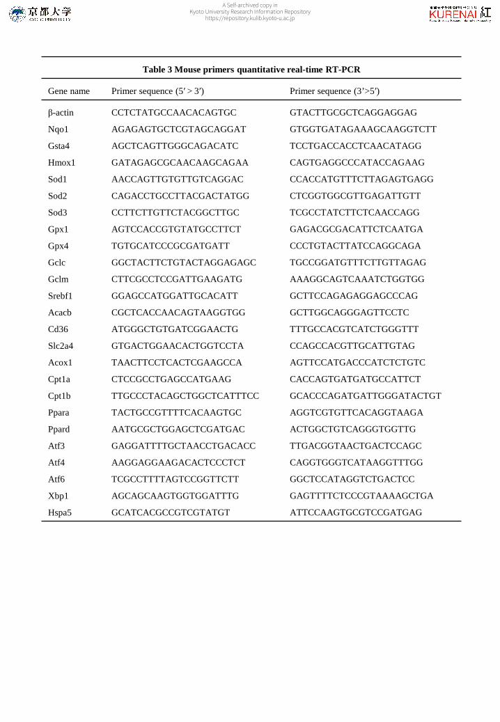
Table 3 Mouse primers quantitative real-time RT-PCR
Gene name Primer sequence (5′ > 3′) Primer sequence (3’>5′)
β-actin
Nqo1
Gsta4
Hmox1
Sod1
Sod2
Sod3
Gpx1
Gpx4
Gclc
Gclm
Srebf1
Acacb
Cd36
Slc2a4
Acox1
Cpt1a
Cpt1b
Ppara
Ppard
Atf3
Atf4
Atf6
Xbp1
Hspa5
CCTCTATGCCAACACAGTGC
AGAGAGTGCTCGTAGCAGGAT
AGCTCAGTTGGGCAGACATC
GATAGAGCGCAACAAGCAGAA
AACCAGTTGTGTTGTCAGGAC
CAGACCTGCCTTACGACTATGG
CCTTCTTGTTCTACGGCTTGC
AGTCCACCGTGTATGCCTTCT
TGTGCATCCCGCGATGATT
GGCTACTTCTGTACTAGGAGAGC
CTTCGCCTCCGATTGAAGATG
GGAGCCATGGATTGCACATT
CGCTCACCAACAGTAAGGTGG
ATGGGCTGTGATCGGAACTG
GTGACTGGAACACTGGTCCTA
TAACTTCCTCACTCGAAGCCA
CTCCGCCTGAGCCATGAAG
TTGCCCTACAGCTGGCTCATTTCC
TACTGCCGTTTTCACAAGTGC
AATGCGCTGGAGCTCGATGAC
GAGGATTTTGCTAACCTGACACC
AAGGAGGAAGACACTCCCTCT
TCGCCTTTTAGTCCGGTTCTT
AGCAGCAAGTGGTGGATTTG
GCATCACGCCGTCGTATGT
GTACTTGCGCTCAGGAGGAG
GTGGTGATAGAAAGCAAGGTCTT
TCCTGACCACCTCAACATAGG
CAGTGAGGCCCATACCAGAAG
CCACCATGTTTCTTAGAGTGAGG
CTCGGTGGCGTTGAGATTGTT
TCGCCTATCTTCTCAACCAGG
GAGACGCGACATTCTCAATGA
CCCTGTACTTATCCAGGCAGA
TGCCGGATGTTTCTTGTTAGAG
AAAGGCAGTCAAATCTGGTGG
GCTTCCAGAGAGGAGCCCAG
GCTTGGCAGGGAGTTCCTC
TTTGCCACGTCATCTGGGTTT
CCAGCCACGTTGCATTGTAG
AGTTCCATGACCCATCTCTGTC
CACCAGTGATGATGCCATTCT
GCACCCAGATGATTGGGATACTGT
AGGTCGTGTTCACAGGTAAGA
ACTGGCTGTCAGGGTGGTTG
TTGACGGTAACTGACTCCAGC
CAGGTGGGTCATAAGGTTTGG
GGCTCCATAGGTCTGACTCC
GAGTTTTCTCCCGTAAAAGCTGA
ATTCCAAGTGCGTCCGATGAG
A Self-archived copy inKyoto University Research Information Repository
https://repository.kulib.kyoto-u.ac.jp

Table 4 Body and tissue weight, food intake
Normal Control SPX
Final body weight (g)
Δ body weight (g)
Food intake (g/day/mouse)
Liver weight (mg/g body weight)
BAT (mg/g body weight)
Mesenteric WAT (mg/g body weight)
Perirenal WAT (mg/g body weight)
Epididymal WAT (mg/g body weight)
32.0±1.09a
9.28±1.15a
3.04±0.20
3.78±0.11a
0.18±0.05
1.90±0.13a
1.62±0.19a
3.70±0.38a
55.3±1.36b
16.0±0.30b
3.30±0.05
7.08±0.42b
0.06±0.01
3.90±0.05b
4.70±0.37b
7.43±0.58b
54.7±1.07b
16.5±0.78b
3.33±0.07
6.38±0.21b
0.06±0.01
3.70±0.09b
5.07±0.20b
7.08±0.43b
Values are mean ± SEM (n=6 for normal and SPX group, n=3 for control group). Values in a row not
sharing a common letter differ significantly (p<0.05). Normal, lean mice fed on a basal AIN 93 G diet;
Control, obese mice fed on a HFD; SPX, obese mice fed on a HFD supplemented with siphonaxanthin;
BAT, brown adipose tissue; WAT, white adipose tissue.
A Self-archived copy inKyoto University Research Information Repository
https://repository.kulib.kyoto-u.ac.jp

Table 5 Plasma physiological measurements
Normal Control SPX
Glucose (mg/dL)
Triacylglycerol (mg/dL)
Free cholesterol ((mg/dL)
HDL cholesterol (mg/dL)
Total cholesterol (mg/dL)
NEFA (mEq/L)
ALT (UI/L)
AST (UI/L)
Creatinine (mg/dl)
273±10.1a
73.5±12.3
29.8±0.52a
85.0±2.14a
123±3.27a
0.54±0.09
7.47±0.46a
11.9±0.43a
0.76±0.03
407±8.58b
61.0±6.87
82.9±8.30b
156±6.57b
264±17.1b
0.74±0.03
157±16.0c
228±40.8b
0.82±0.09
331±22.2a,b
68.4±7.51
87.6±5.82b
166±7.84b
273±13.9b
0.73±0.02
89.8±16.5b
164±35.2b
0.89±0.06
Values are mean ± SEM (n=6 for normal and SPX group, n=3 for control group). Values in a row not
sharing a common letter differ significantly (p<0.05). Normal, lean mice fed on basal AIN 93 G diet;
Control, obese mice fed on a HFD; SPX, obese mice fed on a HFD supplemented with siphonaxanthin;
NEFA, non-esterified fatty acid; ALT, alanine transaminase; AST, aspartate transaminase.
A Self-archived copy inKyoto University Research Information Repository
https://repository.kulib.kyoto-u.ac.jp

Table 6 Liver lipid measurements
Normal Control SPX
Triacylglycerol (mg/g tissue)
Total cholesterol (mg/g tissue)
NEFA (μEq/g)
33.1±5.26a
1.98±0.09a
7.50±1.20
185±4.20b
6.32±0.24b
7.60±0.70
190±5.41b
5.41±0.34b
8.90±1.00
Values are mean ± SEM (n=6 for normal and SPX group, n=3 for control group). Values in a row not sharing a
common letter differ significantly (p<0.05). Normal, lean mice fed on basal AIN93G diet; Control, obese mice
fed on a HFD; SPX, obese mice fed on a HFD supplemented with siphonaxanthin; NEFA, non-esterified fatty
acid;
A Self-archived copy inKyoto University Research Information Repository
https://repository.kulib.kyoto-u.ac.jp

![Simultaneous Detection of Multiple Carotenoid Pigments in Algae … · 2020. 8. 28. · and food colour enhancers [1,2,5]. The global market for carotenoids reached $1.5 billion in](https://img.pdfslide.us/doc/110x75/5fe9e5eec8dfc60e085f2cf4/simultaneous-detection-of-multiple-carotenoid-pigments-in-algae-2020-8-28-and.jpg)