Embed Size (px)
Citation preview
150 Korean J Neurotrauma 2012;8:149-152
SSFS after Craniectomy in a Patient Who Previously Underwent V-P Shunt
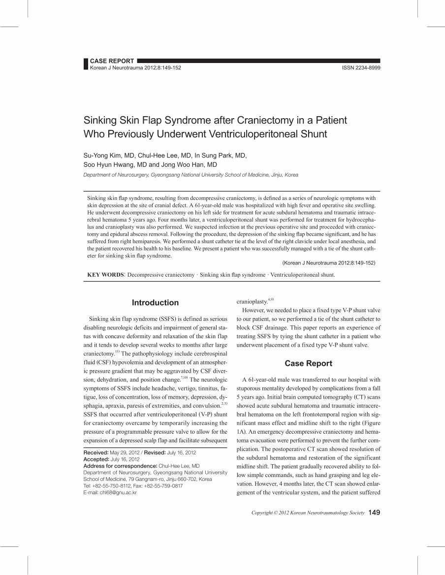
from urinary incontinence. A V-P shunt via right Kocher’s point with MEDTRONIC StrataⓇ valve at 2.0 setting and cranioplasty were performed simultaneously (Figure 1B). After surgery, the patient was discharged after the gradual improvement of his mentation.
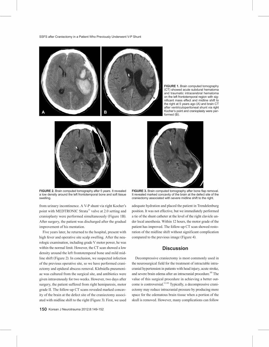
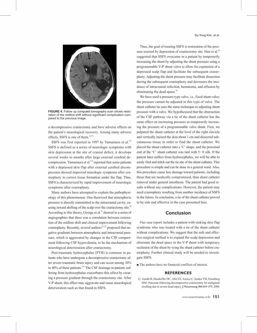
Five years later, he returned to the hospital, present with high fever and operative site scalp swelling. After the neu-rologic examination, including grade V motor power, he was within the normal limit. However, the CT scan showed a low density around the left frontotemporal bone and mild mid-line shift (Figure 2). In conclusion, we suspected infection of the previous operative site, so we have performed crani-ectomy and epidural abscess removal. Klebsiella pneumoni-ae was cultured from the surgical site, and antibiotics were given intravenously for two weeks. However, two days after surgery, the patient suffered from right hemiparesis, motor grade II. The follow-up CT scans revealed marked concav-ity of the brain at the defect site of the craniectomy associ-ated with midline shift to the right (Figure 3). First, we used
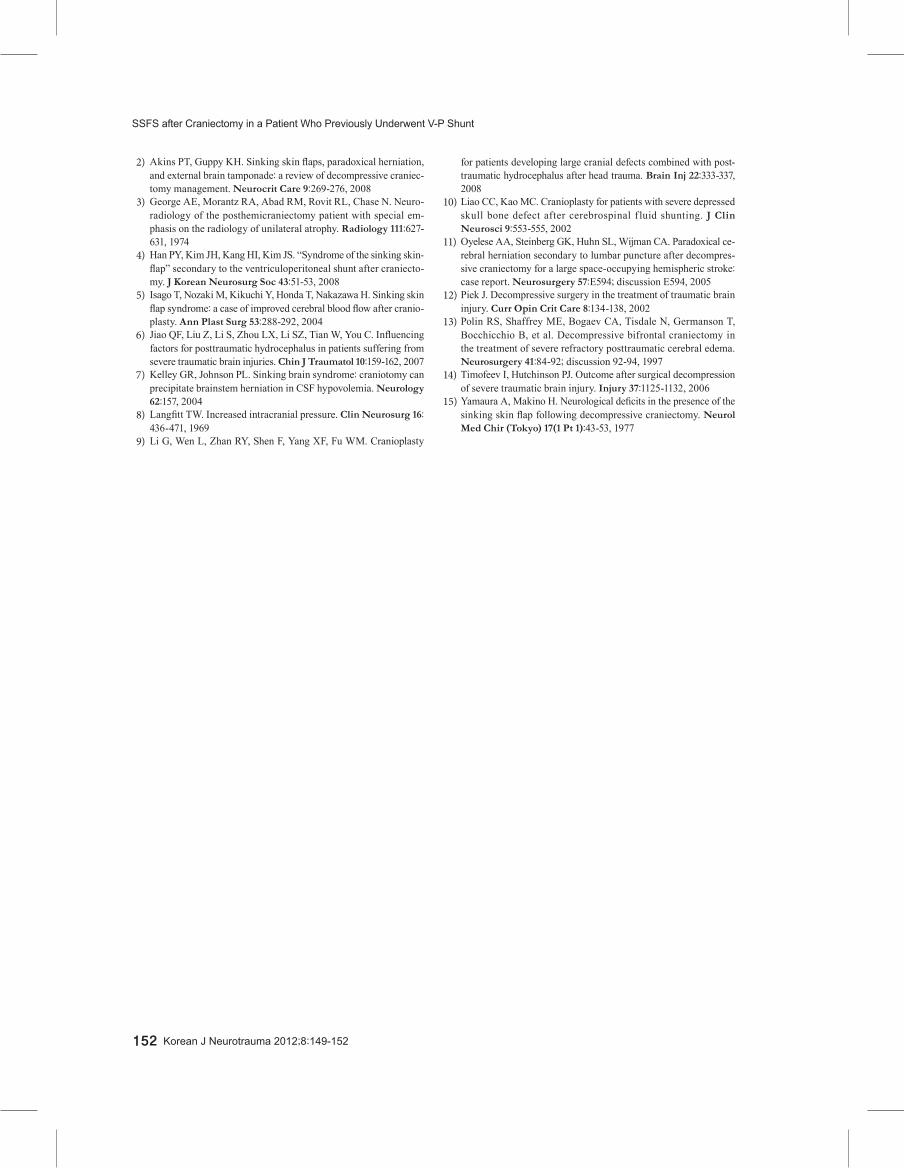
adequate hydration and placed the patient in Trendelenburg position. It was not effective, but we immediately performed a tie of the shunt catheter at the level of the right clavicle un-der local anesthesia. Within 12 hours, the motor grade of the patient has improved. The follow-up CT scan showed resto-ration of the midline shift without significant complication compared to the previous image (Figure 4).
Discussion
Decompressive craniectomy is most commonly used in the neurosurgical field for the treatment of intractable intra-cranial hypertension in patients with head injury, acute stroke, and severe brain edema after an intracranial procedure.10) The value of this surgical procedure in achieving a better out-come is controversial.12,14) Typically, a decompressive crani-ectomy may reduce intracranial pressure by producing more space for the edematous brain tissue when a portion of the skull is removed. However, many complications can follow
FIGURE 1. Brain computed tomography (CT) showed acute subdural hematoma and traumatic intracerebral hematoma on the left frontotemporal region with sig-nificant mass effect and midline shift to the right at 5 years ago (A) and brain CT after ventriculoperitoneal shunt via right Kocher’s point and cranioplasty were per-formed (B).A B
FIGURE 2. Brain computed tomography after 5 years. It revealed a low density around the left frontotemporal bone and soft tissue swelling.
FIGURE 3. Brain computed tomography after bone flap removal. It revealed marked concavity of the brain at the defect site of the craniectomy associated with severe midline shift to the right.
www.neurotrauma.or.kr 151
Su-Yong Kim, et al.
a decompressive craniectomy and have adverse effects on the patient’s neurological recovery. Among many adverse effects, SSFS is one of them.2,13)
SSFS was first reported in 1997 by Yamamura et al.15) SSFS is defined as a series of neurologic symptoms with skin depression at the site of cranial defect; it develops several weeks to months after large external cerebral de-compression. Yamamura et al.15) reported that some patients with a depressed skin flap after external cerebral decom-pression showed improved neurologic symptoms after cra-nioplasty to correct tissue formation under the flap. Thus, SSFS is characterized by rapid improvement of neurologic symptoms after cranioplasty.
Many authors have attempted to explain the pathophysi-ology of this phenomenon. One theorized that atmospheric pressure is directly transmitted to the intracranial cavity, ca-using inward shifting of the scalp over the craniectomy site.8) According to this theory, George et al.3) showed in a series of angiographies that there was a correlation between restora-tion of the midline shift and clinical improvement following cranioplasty. Recently, several authors11,13) proposed that ne-gative gradients between atmospheric and intracranial pres-sure, which is aggravated by changes in the CSF compart-ment following CSF hypovolemia, to be the mechanism of neurological deterioration after craniectomy.
Post-traumatic hydrocephalus (PTH) is common in pa-tients who have undergone a decompressive craniectomy af-ter severe traumatic brain injury and can occur among 10% to 40% of these patients.1,6) The CSF drainage in patients suf-fering from hydrocephalus exacerbates this effect by creat-ing a pressure gradient through the craniectomy site. After V-P shunt, this effect may aggravate and cause neurological deterioration such as that found in SSFS.
Thus, the goal of treating SSFS is restoration of the pres-sure exerted by depression of craniectomy site. Han et al.4) suggested that SSFS overcame in a patient by temporarily increasing the shunt by adjusting the shunt pressure using a programmable V-P shunt valve to allow for expansion of a depressed scalp flap and facilitate the subsequent cranio-plasty. Adjusting the shunt pressure may facilitate dissection during the subsequent cranioplasty and decreases the inci-dence of intracranial infection, hematoma, and effusion by eliminating the dead space.9)
We have used a pressure type valve, i.e., fixed shunt valve; the pressure cannot be adjusted in this type of valve. The shunt catheter tie uses the same technique as adjusting shunt pressure with a valve. We hypothesized that the obstruction of the CSF pathway via a tie of the shunt catheter has the same effect on increasing pressure as temporarily increas-ing the pressure of a programmable valve shunt. First, we palpated the shunt catheter at the level of the right clavicle and vertically incised the skin about 1 cm and dissected sub-cutaneous tissue in order to find the shunt catheter. We placed the shunt catheter into a ‘C’ shape, and the proximal end of the ‘C’ shunt catheter was tied with 5-0 silk. If the patient later suffers from hydrocephalus, we will be able to easily find and stitch out the tie site of the shunt catheter. This procedure is simple and can be done in a general ward. Also, this procedure cause less damage toward patients, including those that are medically compromised, than shunt catheter removal under general anesthesia. The patient had good re-sults without any complications. However, the patient may need cranioplasty resulting from another incidence of SSFS in the future. In conclusion, a tie of the shunt catheter proved to be safe and effective in the case presented here.
Conclusion
Our case report includes a patient with sinking skin flap syndrome who was treated with a tie of the shunt catheter without complications. We suggest that the safe and effec-tive surgical method is to expand the scalp depression and eliminate the dead space in the V-P shunt with temporary occlusion of the shunt by tying the shunt catheter before cra-nioplasty. Further clinical study will be needed to investi-gate SSFS.
■ The authors have no financial conflicts of interest.
REFERENCES1) Aarabi B, Hesdorffer DC, Ahn ES, Aresco C, Scalea TM, Eisenberg
HM. Outcome following decompressive craniectomy for malignant swelling due to severe head injury. J Neurosurg 104:469-479, 2006
FIGURE 4. Follow up computed tomography scan shows resto-ration of the midline shift without significant complication com-pared to the previous image.
152 Korean J Neurotrauma 2012;8:149-152
SSFS after Craniectomy in a Patient Who Previously Underwent V-P Shunt
2) Akins PT, Guppy KH. Sinking skin flaps, paradoxical herniation, and external brain tamponade: a review of decompressive craniec-tomy management. Neurocrit Care 9:269-276, 2008
3) George AE, Morantz RA, Abad RM, Rovit RL, Chase N. Neuro-radiology of the posthemicraniectomy patient with special em-phasis on the radiology of unilateral atrophy. Radiology 111:627-631, 1974
4) Han PY, Kim JH, Kang HI, Kim JS. “Syndrome of the sinking skin-flap” secondary to the ventriculoperitoneal shunt after craniecto-my. J Korean Neurosurg Soc 43:51-53, 2008
5) Isago T, Nozaki M, Kikuchi Y, Honda T, Nakazawa H. Sinking skin flap syndrome: a case of improved cerebral blood flow after cranio-plasty. Ann Plast Surg 53:288-292, 2004
6) Jiao QF, Liu Z, Li S, Zhou LX, Li SZ, Tian W, You C. Influencing factors for posttraumatic hydrocephalus in patients suffering from severe traumatic brain injuries. Chin J Traumatol 10:159-162, 2007
7) Kelley GR, Johnson PL. Sinking brain syndrome: craniotomy can precipitate brainstem herniation in CSF hypovolemia. Neurology 62:157, 2004
8) Langfitt TW. Increased intracranial pressure. Clin Neurosurg 16: 436-471, 1969
9) Li G, Wen L, Zhan RY, Shen F, Yang XF, Fu WM. Cranioplasty
for patients developing large cranial defects combined with post-traumatic hydrocephalus after head trauma. Brain Inj 22:333-337, 2008
10)Liao CC, Kao MC. Cranioplasty for patients with severe depressed skull bone defect after cerebrospinal f luid shunting. J Clin Neurosci 9:553-555, 2002
11) Oyelese AA, Steinberg GK, Huhn SL, Wijman CA. Paradoxical ce-rebral herniation secondary to lumbar puncture after decompres-sive craniectomy for a large space-occupying hemispheric stroke: case report. Neurosurgery 57:E594; discussion E594, 2005
12)Piek J. Decompressive surgery in the treatment of traumatic brain injury. Curr Opin Crit Care 8:134-138, 2002
13)Polin RS, Shaffrey ME, Bogaev CA, Tisdale N, Germanson T, Bocchicchio B, et al. Decompressive bifrontal craniectomy in the treatment of severe refractory posttraumatic cerebral edema. Neurosurgery 41:84-92; discussion 92-94, 1997
14)Timofeev I, Hutchinson PJ. Outcome after surgical decompression of severe traumatic brain injury. Injury 37:1125-1132, 2006
15)Yamaura A, Makino H. Neurological deficits in the presence of the sinking skin flap following decompressive craniectomy. Neurol Med Chir (Tokyo) 17(1 Pt 1):43-53, 1977