Embed Size (px)
Citation preview

VOL. 98-B, No. 9, SEPTEMBER 2016 1289
GENERAL ORTHOPAEDICS
Single-stage treatment of chronic osteomyelitis with a new absorbable, gentamicin-loaded, calcium sulphate/hydroxyapatite biocompositeA PROSPECTIVE SERIES OF 100 CASESM. A. McNally,
J. Y. Ferguson,A. C. K. Lau,M. Diefenbeck,M. Scarborough,A. J. Ramsden,B. L. Atkins
From Nuffield Orthopaedic Centre, Oxford University Hospitals NHS Foundation Trust, Oxford, United Kingdom
M. A. McNally, MD, FRCSEd, FRCS(Orth), Consultant in Limb Reconstruction Surgery, Honorary Senior Lecturer in Orthopaedic Surgery J. Y. Ferguson, MB, BCh(Hons), MEd, FRCS(Orth), Specialty Trainee in Orthopaedics A. C. K. Lau, MBBS, FRCSEd(Orth), Clinical Fellow in Limb Reconstruction M. Diefenbeck, MD, PhD, Honorary Consultant in Orthopaedic Surgery M. Scarborough, PhD, MRCP, FRCPath, Consultant in Infectious Diseases B. L. Atkins, MBBS, MA, MSc,FRCPath, FRCP, Consultant in Microbiology and Infectious Diseases A. J. Ramsden , MB, ChB, MD, FRCS (Plast), Consultant Plastic and Reconstructive MicrosurgeonNuffield Orthopaedic Centre, The Bone Infection Unit, Oxford University Hospitals Foundation NHS Trust, Oxford OX3 7HE, UK.
Correspondence should be sent to Mr M. A. McNally; e-mail: [email protected]
©2016 The British Editorial Society of Bone & Joint Surgerydoi:10.1302/0301-620X.98B9. 38057 $2.00
Bone Joint J 2016;98-B:1289–96.Received 16 February 2016; Accepted after revision 10 May 2016
AimsChronic osteomyelitis may recur if dead space management, after excision of infected bone, is inadequate. This study describes the results of a strategy for the management of deep bone infection and evaluates a new antibiotic-loaded biocomposite in the eradication of infection from bone defects.
Patients and MethodsWe report a prospective study of 100 patients with chronic osteomyelitis, in 105 bones. Osteomyelitis followed injury or surgery in 81 patients. Nine had concomitant septic arthritis. 80 patients had comorbidities (Cierny-Mader (C-M) Class B hosts). Ten had infected nonunions.
All patients were treated by a multidisciplinary team with a single-stage protocol including debridement, multiple sampling, culture-specific systemic antibiotics, stabilisation, dead space filling with the biocomposite and primary skin closure.
ResultsPatients were followed up for a mean of 19.5 months (12 to 34). Infection was eradicated in 96 patients with a single procedure and all four recurrences were successfully managed with repeat surgery. Adverse events were uncommon, with three fractures, six wound leaks and three unrelated deaths. Outcome was not dependant on C-M host class, microbial culture, wound leakage or presence of nonunion.
ConclusionThis single-stage protocol, facilitated by the absorbable local antibiotic, is effective in the treatment of chronic osteomyelitis. It offers a more patient-friendly treatment compared with other published treatment options.
Cite this article: Bone Joint J 2016;98-B:1289–96.
Chronic osteomyelitis is a devastating compli-cation after trauma or orthopaedic surgery.1
Patients may present with multiple comorbidi-ties, poor soft tissues and multi-resistantorganisms, having endured prolonged unsuc-cessful treatment over many years.2-5
Staged surgical treatment is common, withrepeated debridement and delayed skin clo-sure.2,6-14 Further operations may be requiredto remove polymethylmethacrylate (PMMA)antibiotic-loaded beads or to reconstruct bonedefects.15,16 Recently, negative pressure woundtherapy (NPWT), has been combined withmultiple debridement, increasing the numberof revision procedures but not improving therate of resolution of osteomyelitis.17,18
There is now increasing interest in develop-ing a one-stage approach, which might be
more patient friendly. This requires effectivedead space management, to eradicate the infec-tion and prevent the need for secondary bonegrafting.
In animal studies, Branstetter et al19 showedthat local antibiotics in calcium sulphate erad-icated bacteria better after debridement whencompared with calcium sulphate alone. Rand,Penn-Barwell and Wencke20 confirmed thatlocal delivery of antibiotics into an infectedbone defect, with or without an implant, wassuperior to systemic antibiotics alone at 14days after surgery. Xie et al21 compareddebridement alone with bioglass, antibiotic-loaded calcium sulphate (CS) and antibiotic-loaded bioglass in a rabbit methicillin resistantstaphylococcus aureus (MRSA) osteomyelitismodel. Bioglass alone was no better than
1290 M. A. MCNALLY, J. Y. FERGUSON, A. C. K. LAU, M. DIEFENBECK, M. SCARBOROUGH, A. J. RAMSDEN, B. L. ATKINS
THE BONE & JOINT JOURNAL
debridement, but both antibiotic-loaded materials werehighly effective. CS-based materials are effective for eradi-cating infection in humans,22-28 but bone formation in thedefects caused after excision of infected bone is unreliable,with pathological fractures occurring in 5% to 8% ofpatients.24,26,27
The combination of CS and hydroxyapatite (HA) in asynthetic, injectable composite is, however, more promis-ing.29,30 The biphasic resorption of CS/HA forms an osteo-conductive scaffold and leads to an early biologicalresponse.31 This combination may be both osteoinductiveand osteoconductive32 and is now available loaded withgentamicin (175 mg gentamicin in 10 mls CS/HA: CERA-MENT G; Bonesupport, Lund, Sweden). It has been shownthat CERAMENT G injected into osteomyelitic bone voids,decreased the rate of infection and increased bone forma-tion in a rat osteomyelitis model.33
We present the first series of patients with chronic osteo-myelitis treated with a one-stage surgical protocol, usingCERAMENT G in dead space management.
Patients and MethodsInclusion criteria. All patients presenting with possibleCierny-Mader (C-M) Type III (Localised) and Type IV (Dif-fuse) chronic osteomyelitis were recruited into this study.34
The patients were assessed by both orthopaedic and plasticsurgeons specialising in bone infections, as well as by amicrobiologist/infectious disease physician.
Chronic osteomyelitis was defined as having symptomsfor a minimum of six months with clinical and radiologicalfeatures accompanied by at least one of the following: thepresence of a sinus, an abscess or intra-operative pus, sup-portive histology, or two or more microbiological cultures
with indistinguishable organisms.7 When cultures werenegative, a patient was only included in the study whenthere was positive histology, with the presence of a drainingsinus or intra-operative pus. Infected nonunions wereincluded if the bone loss was < 1cm at presentation. Exclu-sion criteria included diabetic foot infections, patients withcalcium metabolism disorders or a known allergy to CS,HA or aminoglycosides and patients with renal failure.
A total of 106 consecutive patients were recruitedbetween March 2013 and February 2015. However, sixpatients were excluded because intra-operative microbio-logical and histological samples did not meet the diagnosticcriteria. The remaining 100 patients (65 male, 35 female)with infection involving 105 bones, were included. Nopatient was excluded due to known gentamicin allergy orrenal compromise. Their mean age was 51.6 years (23 to88). All patients were reviewed at a mean of 19.5 months(12 to 34), except one patient who died of a drug overdosefive months into the study.Data collection. Demographics, comorbidities, aetiology ofinfection, clinical features, diagnostic tests, antibiotic treat-ment choice and duration, were collected prospectively.
The extent of bony involvement (C-M anatomical type)was determined at operation. The host’s physiological sta-tus was recorded at presentation as either Class A (nocomorbidities), Class BL (local compromise in the affectedlimb), Class BS (systemic compromise) or Class BLS (localand systemic comorbidities).34 Radiological and microbio-logical outcomes were confirmed by two assessors whowere not involved in the management of the patients.Surgical management. All patients were treated accordingto a single-stage protocol for chronic osteomyelitis.2,35
Where possible, a tourniquet was used. Multiple deep intra-
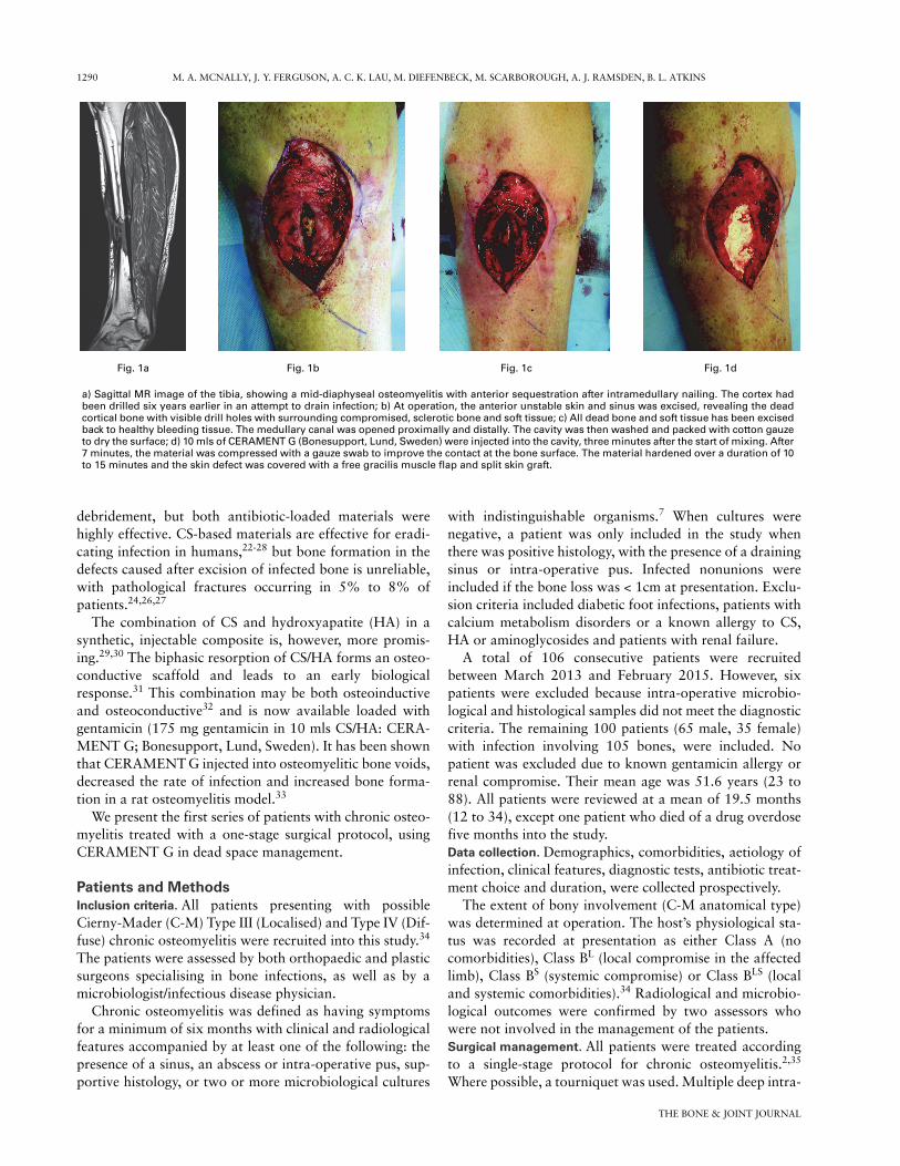
Fig. 1a
a) Sagittal MR image of the tibia, showing a mid-diaphyseal osteomyelitis with anterior sequestration after intramedullary nailing. The cortex hadbeen drilled six years earlier in an attempt to drain infection; b) At operation, the anterior unstable skin and sinus was excised, revealing the deadcortical bone with visible drill holes with surrounding compromised, sclerotic bone and soft tissue; c) All dead bone and soft tissue has been excisedback to healthy bleeding tissue. The medullary canal was opened proximally and distally. The cavity was then washed and packed with cotton gauzeto dry the surface; d) 10 mls of CERAMENT G (Bonesupport, Lund, Sweden) were injected into the cavity, three minutes after the start of mixing. After7 minutes, the material was compressed with a gauze swab to improve the contact at the bone surface. The material hardened over a duration of 10to 15 minutes and the skin defect was covered with a free gracilis muscle flap and split skin graft.
Fig. 1b Fig. 1dFig. 1c

SINGLE-STAGE TREATMENT OF CHRONIC OSTEOMYELITIS 1291
VOL. 98-B, No. 9, SEPTEMBER 2016
operative samples were taken, using an established proto-col.10,36,37 Sinus tracts were excised and infected implantsremoved with excision continuing until healthy, bleedingbone was exposed. After excision, the area was irrigatedwith 0.05% aqueous chlorhexidine solution and the cavitydried by packing with gauze. If instability was present, sta-bilisation was provided by external or internal fixation.After changing of drapes and gloves, the dry dead spacewas filled with CERAMENT G (Fig. 1). No additionalmaterial or antibiotic was added and primary skin closurewas achieved either directly or by local or free microvascu-lar muscle flaps.Antibiotic management. Antibiotic therapy was stopped inall patients at least two weeks prior to surgery, provided itwas safe to do so.27,38 During surgery, patients were givenintravenous vancomycin (continued as 1 g every 12 hoursinitially) and meropenem (500 mg every eight hours ini-tially) after samples had been taken. This empirical antibi-otic regime (a glycopeptide and a carbapenem) has beenshown to be effective against 96% of the isolated organismsin a similar heterogeneous cohort of patients with osteomy-elitis.37 Antibiotic treatment continued for six to 12 weeksbased on the results of the final culture.Outcome parameters. The primary outcome was eradica-tion of infection at a minimum of one year after surgery.Failure of treatment was defined as recurrent infection withpositive cultures from further radiologically guided aspira-tion or biopsy; recurrent sinus formation; further surgeryperformed for infection; or any patient requiring antibiotictreatment for persisting symptoms.27
Secondary outcomes were death, need for re-operation,pathological fracture at the site of surgery and disturbanceof wound healing. Statistical analysis. Data were collated using MicrosoftExcel (Redmond, Washington) and analysed with SPSS v20
(SSPS Inc., Chicago, Illinois). Patient data were regarded asnon-parametric and groups were compared using the chi-squared test for low frequency variables. A p-value < 0.05was considered significant.
ResultsPatients. The C-M classification defined 78 patients asType III and 22 as Type IV. A total of 80 patients (80%)were Class B hosts (Table I). Nine patients had septic arthri-tis of an adjacent joint (three shoulders, two elbows, threeankles, one wrist) and ten had infected nonunion (four tib-iae, three femurs, three humeri). Chronic osteomyelitismost commonly followed a history of an open fracture orfollowing fixation of closed fractures (71 patients). A totalof 19 patients had haematogenous infection, six had infec-tion after elective surgery (osteotomy, ligament repair,arthroscopy or fusion) and four followed a soft-tissueinjury without a fracture.
The tibia, femur and humerus were the most commonlyinvolved bones, with five patients having two bonesinvolved (Table I).
A total of 21 patients required stabilisation and fivepatients were treated with primary joint fusion. Direct skinclosure was possible in 77 patients, but five required a localflap and 18 needed free muscle flap reconstruction (Table I).All patients had primary skin closure. No wounds were leftopen after initial surgery and no second debridement wasplanned in any patient.Microbiology. Staphylococci were the most common organism(41 cultures; 41.8%), with MRSA in six patients (Table II).Proteus mirabilis and Pseudomonas spp were more com-mon in polymicrobial infection, often with a gram-positiveorganism, usually Staphylococcus aureus. A total of 16patients cultured organisms which were shown to be gen-tamicin resistant using EUCAST breakpoints.38 Of these,
Table I. Location of osteomyelitis by Cierny and Mader (C-M) stage, soft-tissue management and stabilisation method
Total C-M stage* Soft-tissue cover Fixation
Bone III A III BL III BS III BLS IV A IV BL IV BS IV BLSDirect closure
Localflap
Freeflap None
Externalfixation
Internalfixation
Tibia 38 7 14 4 9 1 1 1 1 21 4 13 32 6†
Femur 24 5 7 3 4 1 1 3 23 1 19 4 Mono§ 1 IMN‡
Humerus 16 2 2 4 2 1 1 4 15 1 11 3 Mono§ 2 PlatesRadius/ulna 10 4 4 1 1 10 10Calcaneum 3 2 1 1 2 1 3Clavicle 2 2 2 2Fibula 1 1 1 1Sacrum 1 1 1 1Scapula +humerus
1 1 1 1 Ilizarov
Femur+tibia 1 1 1 1 IlizarovTibia+talus 3 1 1 1 1 2 2 Ilizarov 1 IMN‡
Total 100 18 25 19 16 2 4 5 11 77 5 18 79 17 4
* A, Normal host; BL, Host with local compromise; BS, Host with systemic compromise; BLS, Host with local and systemic compromise † 4 monolateral and 2 Ilizarov fixators §Monolateral fixator ‡ Intramedullary nail
1292 M. A. MCNALLY, J. Y. FERGUSON, A. C. K. LAU, M. DIEFENBECK, M. SCARBOROUGH, A. J. RAMSDEN, B. L. ATKINS
THE BONE & JOINT JOURNAL
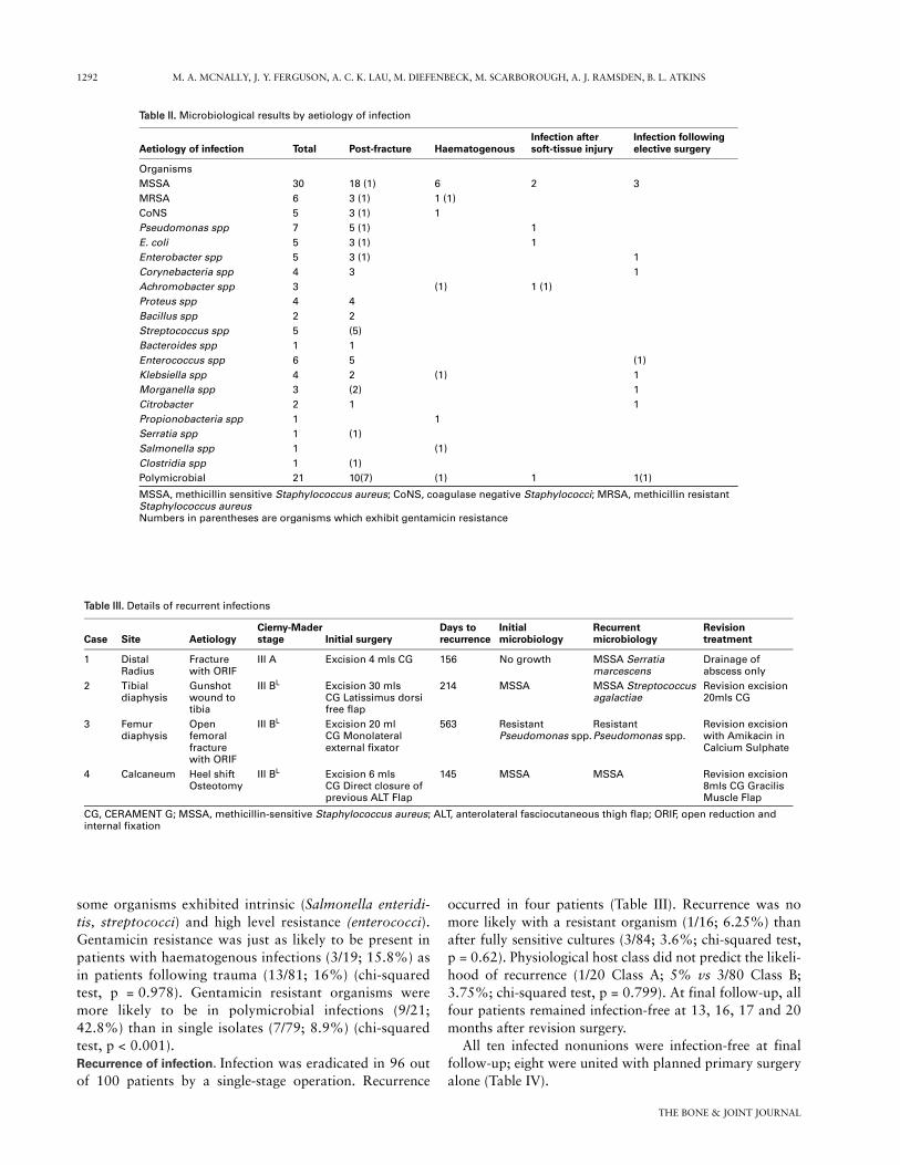
some organisms exhibited intrinsic (Salmonella enteridi-tis, streptococci) and high level resistance (enterococci).Gentamicin resistance was just as likely to be present inpatients with haematogenous infections (3/19; 15.8%) asin patients following trauma (13/81; 16%) (chi-squaredtest, p = 0.978). Gentamicin resistant organisms weremore likely to be in polymicrobial infections (9/21;42.8%) than in single isolates (7/79; 8.9%) (chi-squaredtest, p < 0.001).Recurrence of infection. Infection was eradicated in 96 outof 100 patients by a single-stage operation. Recurrence
occurred in four patients (Table III). Recurrence was nomore likely with a resistant organism (1/16; 6.25%) thanafter fully sensitive cultures (3/84; 3.6%; chi-squared test,p = 0.62). Physiological host class did not predict the likeli-hood of recurrence (1/20 Class A; 5% vs 3/80 Class B;3.75%; chi-squared test, p = 0.799). At final follow-up, allfour patients remained infection-free at 13, 16, 17 and 20months after revision surgery.
All ten infected nonunions were infection-free at finalfollow-up; eight were united with planned primary surgeryalone (Table IV).
Table II. Microbiological results by aetiology of infection
Aetiology of infection Total Post-fracture HaematogenousInfection after soft-tissue injury
Infection following elective surgery
OrganismsMSSA 30 18 (1) 6 2 3MRSA 6 3 (1) 1 (1)CoNS 5 3 (1) 1Pseudomonas spp 7 5 (1) 1E. coli 5 3 (1) 1Enterobacter spp 5 3 (1) 1Corynebacteria spp 4 3 1Achromobacter spp 3 (1) 1 (1)Proteus spp 4 4Bacillus spp 2 2Streptococcus spp 5 (5)Bacteroides spp 1 1Enterococcus spp 6 5 (1)Klebsiella spp 4 2 (1) 1Morganella spp 3 (2) 1Citrobacter 2 1 1Propionobacteria spp 1 1Serratia spp 1 (1)Salmonella spp 1 (1)Clostridia spp 1 (1)Polymicrobial 21 10(7) (1) 1 1(1)
MSSA, methicillin sensitive Staphylococcus aureus; CoNS, coagulase negative Staphylococci; MRSA, methicillin resistant Staphylococcus aureusNumbers in parentheses are organisms which exhibit gentamicin resistance
Table III. Details of recurrent infections
Case Site AetiologyCierny-Mader stage Initial surgery
Days to recurrence
Initial microbiology
Recurrentmicrobiology
Revision treatment
1 Distal Radius
Fracture with ORIF
III A Excision 4 mls CG 156 No growth MSSA Serratia marcescens
Drainage of abscess only
2 Tibial diaphysis
Gunshot wound to tibia
III BL Excision 30 mls CG Latissimus dorsi free flap
214 MSSA MSSA Streptococcus agalactiae
Revision excision 20mls CG
3 Femur diaphysis
Open femoral fracture with ORIF
III BL Excision 20 ml CG Monolateral external fixator
563 Resistant Pseudomonas spp.
ResistantPseudomonas spp.
Revision excision with Amikacin in Calcium Sulphate
4 Calcaneum Heel shift Osteotomy
III BL Excision 6 mls CG Direct closure of previous ALT Flap
145 MSSA MSSA Revision excision 8mls CG Gracilis Muscle Flap
CG, CERAMENT G; MSSA, methicillin-sensitive Staphylococcus aureus; ALT, anterolateral fasciocutaneous thigh flap; ORIF, open reduction and internal fixation

SINGLE-STAGE TREATMENT OF CHRONIC OSTEOMYELITIS 1293
VOL. 98-B, No. 9, SEPTEMBER 2016
All five joint fusions healed with no recurrence of infec-tion at final follow-up.Complications. Three patients died of causes unrelated tothe osteomyelitis or surgery. Table IV summarises the maincomplications and unplanned re-operations.
A total of 94 patients had normal wound healing. Sixpatients had white wound drainage which had the appear-ance of liquefied CS residue. It was managed expectantlywith dressings and required no other intervention. Leakagewas most common in distal tibial cases (4/6) when nomuscle flap was used. No patient with early wound drain-age developed a recurrence of infection.
A total of 11 patients had signs of minor extraosseousleakage of CERAMENT G into the surrounding soft tis-sues which was visible on the radiograph. This was notrelated to the volume of the material used, the site ofinfection or the soft-tissue cover. In ten of the 11patients, this material was fully resorbed and all 11patients remained asymptomatic. Extraosseus leaksoccurred nine times in the first 50 patients, but onlytwice in the second 50, which may represent a learningexperience in the handling of the material and meticu-lous closure over the bone.
Of note, there was no statistically significant differencein recurrence rate, fracture rate, wound leak or extra-osseous collection between those with a history of a frac-ture and the remaining patients. Nor was there anydifference between patients treated with flaps and thosetreated with direct wound closure, or with those with dif-fering C-M stage, although the numbers with complica-tions were small.
DiscussionIn 1931, Kulowski, showed that Orr’s method, (radicalexcision, immobilisation and open wound dressings), wassuccessful in 74% of 130 patients with chronic osteomyeli-tis, at one year after surgery.39 This was without antibioticsor skin closure.
Since then, principles of treatment have evolved whichhave confirmed the place of adequate excision and stabili-sation but have also added careful diagnostic sampling,antimicrobial therapy, dead space management and soft-tis-sue closure.2,6-11,13,14,27,34,35,37,40 Despite these advances,there remains a significant risk of recurrence.
In recent years, staged surgery, involving repeated exci-sions, placement of PMMA-gentamicin loaded beads orrods, or irrigation systems and delayed skin closure, hasbeen used to improve outcomes.8,9,12,14,15,18,41,42 However,PMMA carriers have to be removed, as they prevent boneingrowth8 and can lead to antibiotic resistance.43 Multi-stage surgery often requires prolonged hospital stay withhigh costs.5 Also outcomes have been variable, with infec-tion recurrence rates commonly reported to be above10%8,12,14,18,41,42 and sometimes as high as 45%.15 It isreported that immunocompromised patients with poly-microbial cultures tend to have worse outcomes with thistreatment.5,14,35,44,45
On the other hand, bioabsorbable antibiotic-impreg-nated materials offer the possibility of single-stage surgery,with reduced hospital stay and a more patient friendlyapproach to treatment. In this study, we were able to treatchronic osteomyelitis in all patients with a single operation.The low recurrence rate of 4% at a minimum of one year
Table IV. Summary of secondary complications (excluding recurrence of infection)
Case BoneCierny-Mader stage Complication Timing Treatment Outcome
1 Humerus III A Fracture after high energy fall, with major rib fractures
4 wks Conservative Healed/good function
2 Tibia IV BL Undisplaced fracture after a fall while intoxicated
11 mths Conservative Healed/good function
3 Radius IV BLS Fracture after low energy fall 4 wks Conservative (declined fixation)
Not united. Asympto-matic in frail patient
4 Tibia III A Sterile wound leakage 1 to 6 wks Dressings Healed/no recurrence5 Radius III BS Sterile wound leakage 2 to 9 days Dressings Healed/no recurrence6 Femur III BL Sterile wound drainage 1 to 11 wks Dressings Healed/no recurrence7 Tibia III BLS Sterile wound drainage 1 to 4 mths Dressings Healed/no recurrence8 Tibia III A Sterile wound drainage 1 to 4 wks Dressings Healed/no recurrence9 Tibia III BLS Sterile wound drainage 1 to 8 wks Dressings Healed/no recurrence10 Humerus
Non-unionIV BLS Persistent nonunion after
monolateral fixationConversion to internal fixation with a plate
United/Infection-free at 19 mths
11 Intra-articular proximal tibia nonunion
IV BLS Persistent nonunion None Patient considering endo-prosthetic replacement. Infection-free at 31 mths
12 Calcaneum III BL Bulky fasciocutaneous flap interfering with shoe fitting
Liposuction at 1 yr after surgery
Healed/Infection-free at 23 mths
13 Tibia amputation stump
III BLS Ulceration above tibia from inappropriate prosthetic use
Conversion of below-knee amputation to above-knee amputation
Healed/Infection-free at 15 mths

1294 M. A. MCNALLY, J. Y. FERGUSON, A. C. K. LAU, M. DIEFENBECK, M. SCARBOROUGH, A. J. RAMSDEN, B. L. ATKINS
THE BONE & JOINT JOURNAL
follow-up, is encouraging as the series included manypatients with medical comorbidities, polymicrobial culturesand segmental involvement. In previous studies, these fea-tures were associated with higher recurrence rates of upto 27%.7,14 In this study, we found no correlation betweenC-M physiological grade or polymicrobial infection andrecurrence. This suggests that the single-stage approachwith high bioavailability local antibiotics is a robust man-agement strategy, applicable across a wide range ofpatients.
Recent papers describing similar cohorts using variousmethods of dead space management (CS, bioglass, PMMArods, biocomposite granules, antibiotic-loaded demineral-ised bone matrix), have reported recurrence rates of 9% to37.5% at a similar follow-up period.21,23,25-27,35,41,46 Ourlower rate with CERAMENT G may reflect the increasedability of an injectable delivery system to coat the bone overareas which may harbour residual bacteria, or small frag-ments of biofilm. The high level of a bacteriocidal antibioticfrom CERAMENT G acts at an important time, when most
Fig. 2a
a) Pre-operative anteroposterior (AP) radiograph of the tibia with haematogenous osteomyelitis, pre-sent for more than ten years. There was a draining sinus andStaphylococcus aureus was cultured. Thedead bone and surrounding cavity are visible in the lateral metaphysis; b) Coronal MR image demon-strating the lateral cortical defect, communicating with the skin sinus and central active infection; c) Atsurgery, the infected bone was removed and the cavity filled with 20 mls of CERAMENT G (Bonesup-port, Lund, Sweden). This AP radiograph, taken three days after operation, shows the bone filling andgood contact between the material and the bone surface. CERAMENT G is radio-opaque and initial radi-ographs will show a discreet margin around the material (arrows); d) At six weeks after surgery, themargin of the material is more diffuse and there is a ‘reactive zone’ seen around the CERAMENT G inthe cancellous bone and on the surface of the material, medially; ‘Halo Sign’ (arrows); e) At six monthsafter operation, there is residual, unremodelled material distally; ‘Puddle Sign’ (black arrow) and fur-ther peripheral bone changes. There is a reappearance of trabecular markings in the central zone (whitearrow); f) At 18 months, the material has undergone further remodelling. There is increased density inthe peripheral zone and the lateral cortex is more defined, compared with the six-week and six-monthradiographs (arrow). Distally, the residual ‘Puddle Sign’ remains unremodelled.
Fig. 2b Fig. 2c
Fig. 2d Fig. 2e Fig. 2f

SINGLE-STAGE TREATMENT OF CHRONIC OSTEOMYELITIS 1295
VOL. 98-B, No. 9, SEPTEMBER 2016
residual bacteria will be in planktonic form, following ade-quate debridement.
The use of implanted local antibiotics removes concernsabout compliance with antibiotic therapy and systemic tox-icity. CERAMENT G delivers a burst of gentamicin withhigh local concentrations, around 100 times above the min-imal inhibitory concentration (MIC) for gentamicin sensi-tive Staphylococcus aureus or Pseudomonas species in invitro testing.47 Rapid dissolution prevents a prolongedperiod of low-level antibiotic release, thus reducing the riskof gentamicin resistance.43,48,49
In this study we successfully treated bacteria which weregentamicin resistant on laboratory testing. Resistance report-ing is based on exposing bacteria to levels of antibiotic, whichcan be given systemically without toxicity. The survivability ofbacteria when exposed to very high levels of antibiotics, up to100 times above serum concentrations, is unknown.
Previous studies on antibiotic-loaded CS, have reportedprolonged wound drainage affecting 15% to 32% ofpatients.26,27,35,50,51 In our series, wound leakage was infre-quent and was mainly related to poor skin cover around thedistal tibia. In all patients, we managed wound or extraos-seous leaks expectantly, providing the patient was well. Wedo not believe that these leaks are an indication for re-oper-ation and we found that it had no relationship with laterrecurrence of infection. This has also been described forother absorbable antibiotic carriers.27,35
The low fracture rate in our study may reflect the higherstability achieved with an injectable in situ hardening mate-rial. The compressive strength of CERAMENT G is similarto that of cancellous bone in in vitro biomechanical test-ing.31 The inclusion of HA, which is not passively dis-solved, may provide a longer-lasting scaffold for boneformation. Serial radiographs over the follow-up perioddemonstrated bone remodelling in the majority of patients.Bone formation appears to progress from the periphery ofthe material (Fig. 2). Progressive remodelling is governed byWolff’s law52 and will stop once sufficient bone has beenformed to cope with daily load bearing. It is unlikely thatlarge central medullary zones will fill with new bone aftercortical integrity has been restored.
This study is limited by the relatively short follow-up andby patient selection. We have not investigated this materialin large segmental defects and would not recommend its usefor this indication without further study. Our limited clini-cal experience suggests that CERAMENT G would nothave adequate mechanical strength to support a segmentaldefect without supplementary fixation.
There are many important principles of treating chronicosteomyelitis, including: obtaining multiple bacteriologicalsamples, performing a thorough debridement with metal-work removal, ensuring adequate osseous stabilisation,eliminating the dead space and providing sufficiently vascu-larised soft-tissue cover. We accept that the low recurrencerate of this study is not solely due to the CERAMENT G. Inour Institution, we have previously used different types of
biodegradable antibiotic carriers as adjuncts in the treat-ment of chronic osteomyelitis in the same surgical protocol.We have previously described our experience of using cal-cium sulphate pellets with tobramycin in 195 patients, withan infection recurrence rate of 9.3% and an associatedwound leak rate of 15.4%, which is higher than the recur-rence rate and wound leak rate in this series.27
In conclusion, we report a large series of patients, man-aged with a single-stage protocol, facilitated by the use ofCERAMENT G as a bioabsorbable dead space filler. Thisprotocol delivered low recurrence rates with few re-opera-tions or complications over a one- to three-year follow-upperiod. Our initial experience shows that this offers apatient-friendly treatment which merits further study.
Take home message: This single-stage protocol, facilitated by the absorbable local
antibiotic, is effective in the treatment of chronic osteomyeli-
tis. It offers a more patient-friendly treatment compared with other
published treatment options.
Author contributions:M. A. McNally: Study design, Data collection and analysis, Writing the paper,Performing surgery.J. Y. Ferguson: Data collection and analysis, Writing the paper, Performing sur-gery.M. Diefenbeck: Data collection and analysis, Writing the paper.A. Lau: Data collection.M. Scarborough: Study design, Editing the paper.A. J. Ramsden: Performed surgery, Editing the paper.B. L. Atkins: Study design, Data analysis, Editing the paper.
One author of this study (JYF) was partly supported by an unrestricted researchgrant from Bonesupport AB, Lund, Sweden.
The author or one or more of the authors have received or will receive benefitsfor personal or professional use from a commercial party related directly orindirectly to the subject of this article.
This article was primary edited by S. P. F. Hughes and first proof edited by G.Scott.
References1. Salvana J, Rodner C, Browner BD, et al. Chronic osteomyelitis: results obtained
by an integrated team approach to management. Conn Med 2005;69:195–202.
2. McNally M, Nagarajah K. Osteomyelitis. Orthop Trauma 2010;24:416–429.
3. Romanò CL, Romanò D, Logoluso N, Drago L. Bone and joint infections in adults:a comprehensive classification proposal. Eur Orthop Traumatol 2011;1:207–217.
4. Berendt AR, McNally M. Osteomyelitis. In: Warrell DA, Cox TM, Firth JD, eds.Oxford Textbook of Medicine. Oxford: Oxford University Press, 2010:3788–3795.
5. McNally M, Sendi P. Implant-Associated osteomyelitis of long bones. In: ZimmerliW, ed. Bone and Joint Infections: From Microbiology to Diagnostics and Treatment.Chichester, West Sussex: John Wiley & Son, 2014:303–323.
6. Cierny G III. Chronic osteomyelitis: results of treatment. Instr Course Lect1990;39:495–508.
7. Cierny G III, DiPasquale D. Treatment of chronic infection. J Am Acad Orthop Surg2006;14:S105–S110.
8. Walenkamp GH, Kleijn LL, de Leeuw M. Osteomyelitis treated with gentamicin-PMMA beads: 100 patients followed for 1-12 years. Acta Orthop Scand 1998;69:518–522.
9. McNally MA, Small JO, Tofighi HG, Mollan RA. Two-stage management ofchronic osteomyelitis of the long bones. The Belfast technique. J Bone Joint Surg [Br]1993;75-B:375–380.
10. Spangehl MJ, Masri BA, O’Connell JX, Duncan CP. Prospective analysis of pre-operative and intraoperative investigations for the diagnosis of infection at the sitesof two hundred and two revision total hip arthroplasties. J Bone Joint Surg [Am]1999;81-A:672–683.
11. Krenn V, Morawietz L, Perino G, et al. Revised histopathological consensus clas-sification of joint implant related pathology. Pathol Res Pract 2014;210:779–786.

1296 M. A. MCNALLY, J. Y. FERGUSON, A. C. K. LAU, M. DIEFENBECK, M. SCARBOROUGH, A. J. RAMSDEN, B. L. ATKINS
THE BONE & JOINT JOURNAL
12. Evans RP, Nelson CL. Gentamicin-impregnated polymethylmethacrylate beadscompared with systemic antibiotic therapy in the treatment of chronic osteomyelitis.Clin Orthop Relat Res 1993;295:37–42.
13. Ilizarov GA. The principles of the Ilizarov method. Bull Hosp Jt Dis Orthop Inst1988;48:1–11.
14. Lazzarini L, Mader JT, Calhoun JH. Osteomyelitis in long bones. J Bone Joint Surg[Am] 2004;86-A:2305–2318.
15. Cho SH, Song HR, Koo KH, Jeong ST, Park YJ. Antibiotic-impregnated cementbeads in the treatment of chronic osteomyelitis. Bull Hosp Jt Dis 1997;56:140–144.
16. Tiemann AH, Schmidt HGK, Braunschweig R, Hofmann GO. Strategies for theanalysis of osteitic bone defects at the diaphysis of long bones. Strategies TraumaLimb Reconstr 2009;4:13–18.
17. Diefenbeck M, Mennenga U, Gückel P, et al. Vacuum-assisted closure therapyfor the treatment of acute postoperative osteomyelitis. Z Orthop Unfall2011;149:336–341. (In German).
18. Timmers MS, Graafland N, Bernards AT, et al. Negative pressure wound treat-ment with polyvinyl alcohol foam and polyhexanide antiseptic solution instillation inposttraumatic osteomyelitis. Wound Repair Regen 2009;17:278–286.
19. Branstetter JG, Jackson SR, Haggard WO, Richelsoph KC, Wenke JC. Locally-administered antibiotics in wounds in a limb. J Bone Joint Surg [Br] 2009;91-B:1106–1109.
20. Rand BC, Penn-Barwell JG, Wenke JC. Combined local and systemic antibioticdelivery improves eradication of wound contamination: an animal experimentalmodel of contaminated fracture. Bone Joint J 2015;97-B:1423–1427.
21. Xie Z, Liu X, Jia W, et al. Treatment of osteomyelitis and repair of bone defect bydegradable bioactive borate glass releasing vancomycin. J Control Release2009;139:118–126.
22. Chang W, Colangeli M, Colangeli S, et al. Adult osteomyelitis: debridement ver-sus debridement plus Osteoset T pellets. Acta Orthop Belg 2007;73:238–243.
23. Turner TM, Urban RM, Hall DJ, et al. Local and systemic levels of tobramycindelivered from calcium sulfate bone graft substitute pellets. Clin Orthop Relat Res2005;437:97–104.
24. Gitelis S, Brebach GT. The treatment of chronic osteomyelitis with a biodegradableantibiotic-impregnated implant. J Orthop Surg (Hong Kong) 2002;10:53–60.
25. McKee MD, Wild LM, Schemitsch EH, Waddell JP. The use of an antibiotic-impregnated, osteoconductive, bioabsorbable bone substitute in the treatment ofinfected long bone defects: early results of a prospective trial. J Orthop Trauma2002;16:622–627.
26. McKee MD, Li-Bland EA, Wild LM, Schemitsch EH. A prospective, randomizedclinical trial comparing an antibiotic-impregnated bioabsorbable bone substitute withstandard antibiotic-impregnated cement beads in the treatment of chronic osteomy-elitis and infected nonunion. J Orthop Trauma 2010;24:483–490.
27. Ferguson JY, Dudareva M, Riley ND, et al. The use of a biodegradable antibiotic-loaded calcium sulphate carrier containing tobramycin for the treatment of chronicosteomyelitis: a series of 195 cases. Bone Joint J 2014;96-B:829–836.
28. Nelson CL, McLaren SG, Skinner RA, et al. The treatment of experimental osteo-myelitis by surgical debridement and the implantation of calcium sulfate tobramycinpellets. J Orthop Res 2002;20:643–647.
29. Nilsson M, Zheng MH, Tägil M. The composite of hydroxyapatite and calcium sul-phate: a review of preclinical evaluation and clinical applications. Expert Rev MedDevices 2013;10:675–684.
30. Iundusi R, Gasbarra E, D’Arienzo M, Piccioli A, Tarantino U. Augmentation oftibial plateau fractures with an injectable bone substitute: CERAMENT™. Three yearfollow-up from a prospective study. BMC Musculoskelet Disord 2015;16:115.
31. Nilsson M, Wang J- S, Wielanek L, Tanner KE, Lidgren L. Biodegradation andbiocompatibility of a calcium sulphate-hydroxyapatite bone substitute. J Bone JointSurg [Br] 2004;86-B:120–125.
32. Raina D, Gupta A, Petersen M, et al. A biphasic bone substitute with gentamycinregenerates bone in osteomyelitis with muscle acting as an osteoinductive niche.Bone Joint J Orthopaedic Proceedings Supplement 2015;97(Suppl 16):24.
33. Dvorzhinskiy A, Perino G, Chojnowski R, et al. Cerament bone void filler withgentamicin increases bone formation and decreases detectable infection in a ratmodel of debrided osteomyelitis. Bone Joint J Proc Suppl 2015;97(Suppl 16):9.
34. Cierny G III, Mader JT, Penninck JJ. A clinical staging system for adult osteomy-elitis. Clin Orthop Relat Res 2003;414:7–24.
35. Romano CL, Logoluso N, Meani E, et al. A comparative study of the use of bioac-tive glass S53P4 and antibiotic-loaded calcium-based bone substitutes in the treat-ment of chronic osteomyelitis: a retrospective comparative study. Bone Joint J2014;96-B:845–850.
36. Atkins BL, Athanasou N, Deeks JJ, et al. Prospective evaluation of criteria formicrobiological diagnosis of prosthetic-joint infection at revision arthroplasty. TheOSIRIS Collaborative Study Group. J Clin Microbiol 1998;36:2932–2939.
37. Sheehy SH, Atkins BA, Bejon P, et al. The microbiology of chronic osteomyelitis:prevalence of resistance to common empirical anti-microbial regimens. J Infect2010;60:338–343.
38. No authors listed. Clinical Breakpoints. http://www.eucast.org/clinical_breakpoints/ (datelast accessed 18 May 2016).
39. Kulowski J. The Orr treatment of osteomyelitis and allied suppurative processes.J Bone Joint Surg [Am] 1931;13-A:538–562.
40. Bose D, Kugan R, Stubbs D, McNally M. Management of infected nonunion of thelong bones by a multidisciplinary team. Bone Joint J 2015;97-B:814–817.
41. Kanakaris N, Gudipati S, Tosounidis T, et al. The treatment of intramedullaryosteomyelitis of the femur and tibia using the Reamer-Irrigator-Aspirator system andantibiotic cement rods. Bone Joint J 2014;96-B:783–788.
42. Caesar BC, Morgan-Jones RL, Warren RE, et al. Closed double-lumen suctionirrigation in the management of chronic diaphyseal osteomyelitis: long-term follow-up. J Bone Joint Surg [Br] 2009;91-B:1243–1248.
43. Neut D, van de Belt H, van Horn JR, van der Mei HC, Busscher HJ. Residualgentamicin-release from antibiotic-loaded polymethylmethacrylate beads after 5years of implantation. Biomaterials 2003;24:1829–1831.
44. Zimmerli W, Sendi P. Pathogenesis of implant-associated infection: the role of thehost. Semin Immunopathol 2011;33:295–306.
45. Lew DP, Waldvogel FA. Osteomyelitis. Lancet 2004;364:369–379.
46. Fleiter N, Walter G, Bösebeck H, et al. Clinical use and safety of a novel gen-tamicin-releasing resorbable bone graft substitute in the treatment of osteomyelitis/osteitis. Bone Joint Res 2014;3:223–229.
47. No authors listed. Bonesupport. Gentamicin release in vitro from setting CERA-MENT™|G paste. Data on file BONESUPPORT; Sweden, report S009/2012.
48. Anagnostakos K, Hitzler P, Pape D, Kohn D, Kelm J. Persistence of bacterialgrowth on antibiotic-loaded beads: is it actually a problem? Acta Orthop2008;79:302–307.
49. Walsh C. Molecular mechanisms that confer antibacterial drug resistance. Nature2000;406:775–781.
50. Borrelli J Jr, Prickett WD, Ricci WM. Treatment of nonunions and osseousdefects with bone graft and calcium sulfate. Clin Orthop Relat Res 2003;411:245–254.
51. McNally M, Ferguson J, Kendall J, et al. A comparative study of three bioabsorb-able antibiotic carriers in chronic osteomyelitis: 313 patients with minimum one-yearfollow-up. Bone Joint J Proc Suppl 2015;97(Suppl 16):21–22.
52. Wolff J. Ueber die innere Architectur der Knochen und ihre Bedeutung für die Fragevom Knochenwachsthum. Virchows Arch Pathol Anat Physiol 1870;50:389–450.