Embed Size (px)
Citation preview
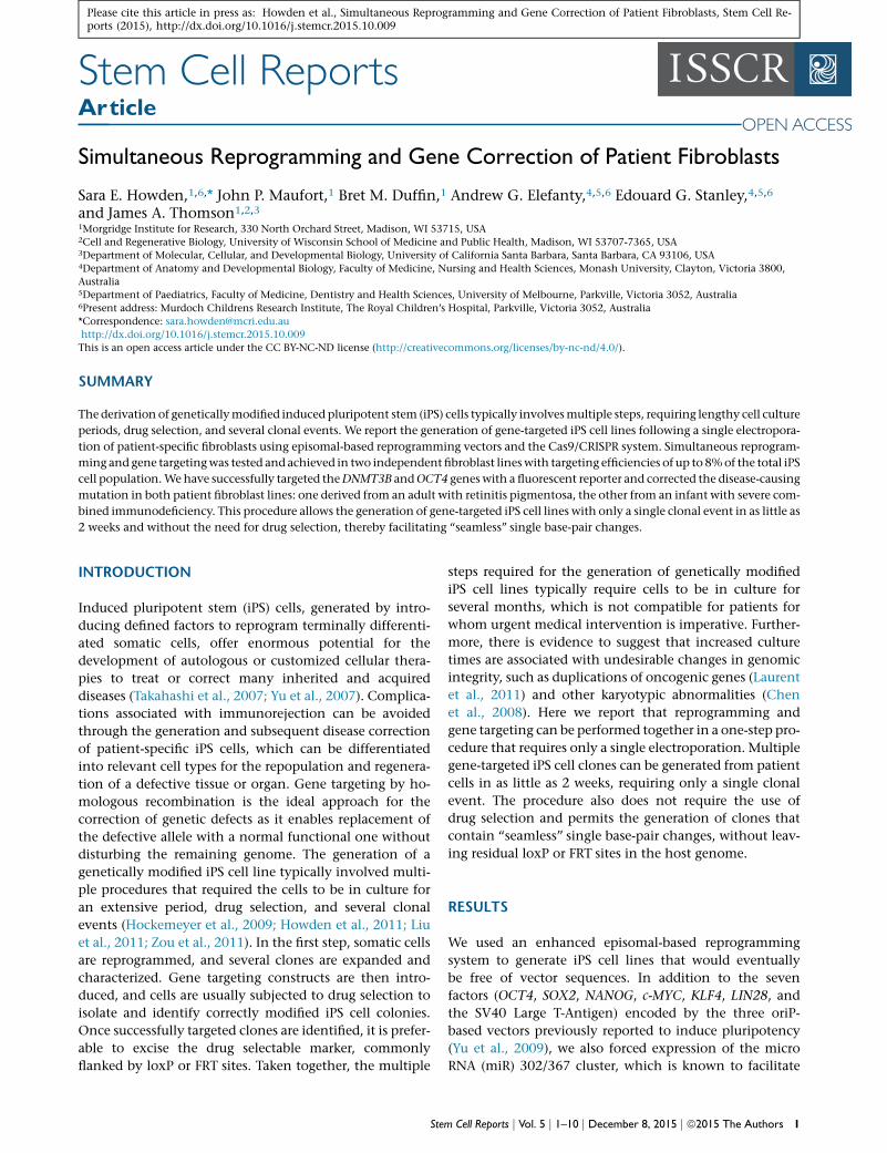
Please cite this article in press as: Howden et al., Simultaneous Reprogramming and Gene Correction of Patient Fibroblasts, Stem Cell Re-ports (2015), http://dx.doi.org/10.1016/j.stemcr.2015.10.009
Stem Cell Reports
ArticleSimultaneous Reprogramming and Gene Correction of Patient Fibroblasts
Sara E. Howden,1,6,* John P. Maufort,1 Bret M. Duffin,1 Andrew G. Elefanty,4,5,6 Edouard G. Stanley,4,5,6
and James A. Thomson1,2,3
1Morgridge Institute for Research, 330 North Orchard Street, Madison, WI 53715, USA2Cell and Regenerative Biology, University of Wisconsin School of Medicine and Public Health, Madison, WI 53707-7365, USA3Department of Molecular, Cellular, and Developmental Biology, University of California Santa Barbara, Santa Barbara, CA 93106, USA4Department of Anatomy and Developmental Biology, Faculty of Medicine, Nursing and Health Sciences, Monash University, Clayton, Victoria 3800,
Australia5Department of Paediatrics, Faculty of Medicine, Dentistry and Health Sciences, University of Melbourne, Parkville, Victoria 3052, Australia6Present address: Murdoch Childrens Research Institute, The Royal Children’s Hospital, Parkville, Victoria 3052, Australia
*Correspondence: [email protected]
http://dx.doi.org/10.1016/j.stemcr.2015.10.009
This is an open access article under the CC BY-NC-ND license (http://creativecommons.org/licenses/by-nc-nd/4.0/).
SUMMARY
The derivation of geneticallymodified induced pluripotent stem (iPS) cells typically involvesmultiple steps, requiring lengthy cell culture
periods, drug selection, and several clonal events. We report the generation of gene-targeted iPS cell lines following a single electropora-
tion of patient-specific fibroblasts using episomal-based reprogramming vectors and the Cas9/CRISPR system. Simultaneous reprogram-
ming and gene targetingwas tested and achieved in two independent fibroblast lineswith targeting efficiencies of up to 8%of the total iPS
cell population.Wehave successfully targeted theDNMT3B andOCT4 geneswith a fluorescent reporter and corrected the disease-causing
mutation in both patient fibroblast lines: one derived from an adult with retinitis pigmentosa, the other from an infant with severe com-
bined immunodeficiency. This procedure allows the generation of gene-targeted iPS cell lines with only a single clonal event in as little as
2 weeks and without the need for drug selection, thereby facilitating ‘‘seamless’’ single base-pair changes.
INTRODUCTION
Induced pluripotent stem (iPS) cells, generated by intro-
ducing defined factors to reprogram terminally differenti-
ated somatic cells, offer enormous potential for the
development of autologous or customized cellular thera-
pies to treat or correct many inherited and acquired
diseases (Takahashi et al., 2007; Yu et al., 2007). Complica-
tions associated with immunorejection can be avoided
through the generation and subsequent disease correction
of patient-specific iPS cells, which can be differentiated
into relevant cell types for the repopulation and regenera-
tion of a defective tissue or organ. Gene targeting by ho-
mologous recombination is the ideal approach for the
correction of genetic defects as it enables replacement of
the defective allele with a normal functional one without
disturbing the remaining genome. The generation of a
genetically modified iPS cell line typically involved multi-
ple procedures that required the cells to be in culture for
an extensive period, drug selection, and several clonal
events (Hockemeyer et al., 2009; Howden et al., 2011; Liu
et al., 2011; Zou et al., 2011). In the first step, somatic cells
are reprogrammed, and several clones are expanded and
characterized. Gene targeting constructs are then intro-
duced, and cells are usually subjected to drug selection to
isolate and identify correctly modified iPS cell colonies.
Once successfully targeted clones are identified, it is prefer-
able to excise the drug selectable marker, commonly
flanked by loxP or FRT sites. Taken together, the multiple
Ste
steps required for the generation of genetically modified
iPS cell lines typically require cells to be in culture for
several months, which is not compatible for patients for
whom urgent medical intervention is imperative. Further-
more, there is evidence to suggest that increased culture
times are associated with undesirable changes in genomic
integrity, such as duplications of oncogenic genes (Laurent
et al., 2011) and other karyotypic abnormalities (Chen
et al., 2008). Here we report that reprogramming and
gene targeting can be performed together in a one-step pro-
cedure that requires only a single electroporation. Multiple
gene-targeted iPS cell clones can be generated from patient
cells in as little as 2 weeks, requiring only a single clonal
event. The procedure also does not require the use of
drug selection and permits the generation of clones that
contain ‘‘seamless’’ single base-pair changes, without leav-
ing residual loxP or FRT sites in the host genome.
RESULTS
We used an enhanced episomal-based reprogramming
system to generate iPS cell lines that would eventually
be free of vector sequences. In addition to the seven
factors (OCT4, SOX2, NANOG, c-MYC, KLF4, LIN28, and
the SV40 Large T-Antigen) encoded by the three oriP-
based vectors previously reported to induce pluripotency
(Yu et al., 2009), we also forced expression of the micro
RNA (miR) 302/367 cluster, which is known to facilitate
m Cell Reports j Vol. 5 j 1–10 j December 8, 2015 j ª2015 The Authors 1
0
50
100
150
200
250
300
350
3
without miR302-367 plasmid
322
Num
ber o
f iP
S c
ell c
olon
ies
with miR302-367 plasmid
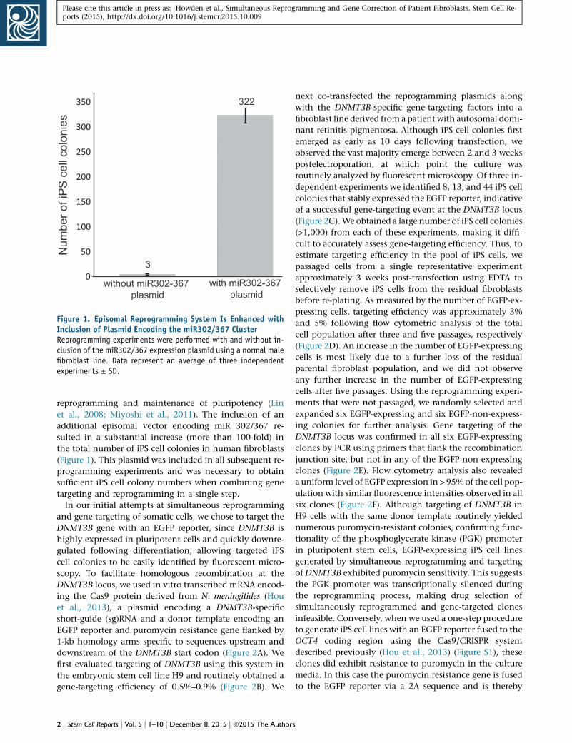
Figure 1. Episomal Reprogramming System Is Enhanced withInclusion of Plasmid Encoding the miR302/367 ClusterReprogramming experiments were performed with and without in-clusion of the miR302/367 expression plasmid using a normal malefibroblast line. Data represent an average of three independentexperiments ± SD.
Please cite this article in press as: Howden et al., Simultaneous Reprogramming and Gene Correction of Patient Fibroblasts, Stem Cell Re-ports (2015), http://dx.doi.org/10.1016/j.stemcr.2015.10.009
reprogramming and maintenance of pluripotency (Lin
et al., 2008; Miyoshi et al., 2011). The inclusion of an
additional episomal vector encoding miR 302/367 re-
sulted in a substantial increase (more than 100-fold) in
the total number of iPS cell colonies in human fibroblasts
(Figure 1). This plasmid was included in all subsequent re-
programming experiments and was necessary to obtain
sufficient iPS cell colony numbers when combining gene
targeting and reprogramming in a single step.
In our initial attempts at simultaneous reprogramming
and gene targeting of somatic cells, we chose to target the
DNMT3B gene with an EGFP reporter, since DNMT3B is
highly expressed in pluripotent cells and quickly downre-
gulated following differentiation, allowing targeted iPS
cell colonies to be easily identified by fluorescent micro-
scopy. To facilitate homologous recombination at the
DNMT3B locus, we used in vitro transcribed mRNA encod-
ing the Cas9 protein derived from N. meningitides (Hou
et al., 2013), a plasmid encoding a DNMT3B-specific
short-guide (sg)RNA and a donor template encoding an
EGFP reporter and puromycin resistance gene flanked by
1-kb homology arms specific to sequences upstream and
downstream of the DNMT3B start codon (Figure 2A). We
first evaluated targeting of DNMT3B using this system in
the embryonic stem cell line H9 and routinely obtained a
gene-targeting efficiency of 0.5%–0.9% (Figure 2B). We
2 Stem Cell Reports j Vol. 5 j 1–10 j December 8, 2015 j ª2015 The Authors
next co-transfected the reprogramming plasmids along
with the DNMT3B-specific gene-targeting factors into a
fibroblast line derived from a patient with autosomal domi-
nant retinitis pigmentosa. Although iPS cell colonies first
emerged as early as 10 days following transfection, we
observed the vast majority emerge between 2 and 3 weeks
postelectroporation, at which point the culture was
routinely analyzed by fluorescent microscopy. Of three in-
dependent experiments we identified 8, 13, and 44 iPS cell
colonies that stably expressed the EGFP reporter, indicative
of a successful gene-targeting event at the DNMT3B locus
(Figure 2C).We obtained a large number of iPS cell colonies
(>1,000) from each of these experiments, making it diffi-
cult to accurately assess gene-targeting efficiency. Thus, to
estimate targeting efficiency in the pool of iPS cells, we
passaged cells from a single representative experiment
approximately 3 weeks post-transfection using EDTA to
selectively remove iPS cells from the residual fibroblasts
before re-plating. As measured by the number of EGFP-ex-
pressing cells, targeting efficiency was approximately 3%
and 5% following flow cytometric analysis of the total
cell population after three and five passages, respectively
(Figure 2D). An increase in the number of EGFP-expressing
cells is most likely due to a further loss of the residual
parental fibroblast population, and we did not observe
any further increase in the number of EGFP-expressing
cells after five passages. Using the reprogramming experi-
ments that were not passaged, we randomly selected and
expanded six EGFP-expressing and six EGFP-non-express-
ing colonies for further analysis. Gene targeting of the
DNMT3B locus was confirmed in all six EGFP-expressing
clones by PCR using primers that flank the recombination
junction site, but not in any of the EGFP-non-expressing
clones (Figure 2E). Flow cytometry analysis also revealed
a uniform level of EGFP expression in > 95%of the cell pop-
ulation with similar fluorescence intensities observed in all
six clones (Figure 2F). Although targeting of DNMT3B in
H9 cells with the same donor template routinely yielded
numerous puromycin-resistant colonies, confirming func-
tionality of the phosphoglycerate kinase (PGK) promoter
in pluripotent stem cells, EGFP-expressing iPS cell lines
generated by simultaneous reprogramming and targeting
of DNMT3B exhibited puromycin sensitivity. This suggests
the PGK promoter was transcriptionally silenced during
the reprogramming process, making drug selection of
simultaneously reprogrammed and gene-targeted clones
infeasible. Conversely, when we used a one-step procedure
to generate iPS cell lines with an EGFP reporter fused to the
OCT4 coding region using the Cas9/CRISPR system
described previously (Hou et al., 2013) (Figure S1), these
clones did exhibit resistance to puromycin in the culture
media. In this case the puromycin resistance gene is fused
to the EGFP reporter via a 2A sequence and is thereby
Please cite this article in press as: Howden et al., Simultaneous Reprogramming and Gene Correction of Patient Fibroblasts, Stem Cell Re-ports (2015), http://dx.doi.org/10.1016/j.stemcr.2015.10.009
driven from the endogenousOCT4 regulatory region rather
than the minimal and non-specific PGK promoter.
We have also successfully used our one-step protocol to
simultaneously reprogram and genetically correct the dis-
ease-causing mutation in the patient fibroblasts, an auto-
somal dominant C > T transition in exon 42 of the PRPF8
gene. This was achieved using a plasmid encoding the
Cas9 protein from S. pyogenes (Mali et al., 2013b), a plasmid
encoding a PRPF8-specific sgRNA that binds 33 bp up-
stream of the disease-causing mutation, and a 184-bp sin-
gle-stranded oligodeoxynucleotide (ssODN) (Figure 3A).
The ssODN was engineered to contain four synonymous
mutations to minimize the possibility of Cas9 protein re-
cutting following homologous recombination and to aid
in the identification of clones that had undergone a gene-
targeting event (Figure 3A). Approximately 3 weeks post-
transfection, we randomly isolated and expanded a total
of 72 iPS cell colonies for further analysis. A PCR product
encoding the region of interest was amplified from the
genomic DNA of all 72 clones using primers flanking the
target site, which was subsequently analyzed by Sanger
sequencing. Cas9-induced modification of one or both
PRPF8 alleles was observed in 22 (31%) of the clones
analyzed, most commonly detected as a nonhomologous
end joining (NHEJ) event within the intended cut site.
Homologous recombination at the target site could be
detected in 6 (8%) clones, as evidenced by the loss of the
disease-causing mutation or the presence of one or more
synonymous mutations carried by the corrective ssODN
(Table 1). Genetic correction of the autosomal dominant
patient-specific mutation was observed in 2 clones, while
targeting of the wild-type allele was observed in 4 clones.
We were unable to determine which allele had undergone
gene targeting in 1 clone (P.57) due to a 151-bp deletion
spanning the site of the mutation. Surprisingly, 1 clone
(P.50) appeared to have undergone bi-allelic homologous
recombination, as evidenced by correction of the patient-
specific mutation and the presence of ssODN-specific syn-
onymous mutations on both alleles (Figures 3B and 3C).
However, this clone also contained a 1-bp deletion approx-
imately 50 bp upstream of the intended site of the Cas9-
induced double-stranded break. We hypothesize that this
is most likely due to homologous recombination with an
incorrectly synthesized ssODN rather than an additional
mutation caused by NHEJ, which normally occurs at the
site of the double-stranded break.
Next we attempted to correct the disease-causing muta-
tion in a fibroblast line isolated from an infant with severe
combined immunodeficiency (SCID), caused by mutations
in the gene encoding adenosine deaminase (ADA). SCID
patients could particularly benefit from a one-step protocol
that facilitates the expedited generation of gene-corrected
iPS cells because without early intervention, such as a
Ste
bone marrow transplant, patients typically die within the
first 1 to 2 years of life. We first attempted to simulta-
neously reprogram and targetDNMT3B in ADA-SCID fibro-
blasts and identified one EGFP-expressing colony (0.9%)
out of a total of 108 iPS cell colonies (Figure S2). PCR anal-
ysis confirmed targeting of the DNMT3B locus (see Fig-
ure 2E). We next attempted to simultaneously reprogram
and correct one of the disease-causing mutations in the
ADA-SCID fibroblasts using our one-step protocol. The fi-
broblasts were derived from a patient who is a compound
heterozygote: one allele has a C > T transition in exon 11
of the ADA gene (1,081C > T), and the second allele has
an A > G transition in the 3-prime splice site of intron 3, re-
sulting in a deletion of exon 4 from mature mRNA. We
chose to correct the C > T transition in exon 11 using an
sgRNA specific to the mutant, but not wild-type, exon 11
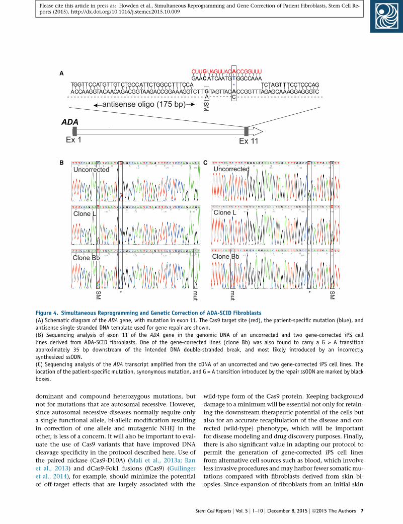
sequence of the ADA gene (Figure 4A). We hypothesized
that this would minimize Cas9 cutting in both alleles, as
seen in the majority of the PRPF8 gene-targeted iPS cell
lines, where only 1 out of the 6 clones did not have a sec-
ond allele modified, either by NHEJ or a second homolo-
gous recombination event. To facilitate gene correction
we used a 175-bp single-stranded corrective ssODN, which
was engineered to contain a single synonymous mutation
within theCas9 target site (Figure 4A). A total of 55 colonies
were expanded and screened, with Cas9-inducedmodifica-
tion of ADA exon 11 observed in 20 (36%) clones, as deter-
mined by Sanger sequencing of a 1.4-kb PCR product
amplified from genomic DNA using primers flanking the
target site. Gene targeting was detected in 3 (5%) clones,
as evidenced by the loss of the disease-causing mutation
and the presence of the synonymous mutation carried by
the corrective ssODN. Genetic correction of the patient-
specific mutation in exon 11 was observed in all three
clones, without modification of the second allele, indi-
cating that Cas9 preferentially favored the mutant exon
11 sequence (over wild-type). This was also evident upon
analysis of the uncorrected clones that had undergone
NHEJ at the target site. Interestingly, in one gene-corrected
iPS cell line, we also detected a G > A transition approxi-
mately 35 bp downstream of the intended site of the
Cas9-induced double-stranded break (Figure 4B). Again,
due to its location relative to the Cas9 target site, we hy-
pothesize that this is most likely the result of homologous
recombination with an incorrectly synthesized ssODN.
Expression of corrected ADA mRNA was also confirmed
in all three gene-corrected iPS cell lines following total
RNA extraction and RT-PCR to amplify the complete ADA
transcript, which was then sequenced (Figure 4C). The
two gene-corrected lines that did not carry the additional
G > A transitionwere characterized further by teratoma for-
mation, G-banding, and PCR analysis to detect residual re-
programming plasmids. Both clones formed teratomas
m Cell Reports j Vol. 5 j 1–10 j December 8, 2015 j ª2015 The Authors 3
DNMT3BEx 23Ex 1 Ex 2
1 kbpA 3’ arm5’ arm EGFP PGKPuroR
105
0
EGFP+0.68%.
104
103
104 1051030
A B
EGFP+2.96%.
105
0
104
103
passage 3
D
105
0
104
103
104 1051030
passage 5
EGFP+5.05%.
104 1051030
C
E
G2
G6
G5
G4
G3
BA
C c
on
AD
AH
9-E
GFP
con
G1
C1
C2
C3
C4
C5
C6
F
con
clone 1
clone 2
clone 3
clone 4
clone 5
clone 6
1 kb
400 μm
1 kb
Cas9 target site
104 1051030
400 μm
FITC-A
SSC-A
SSC-A
SSC-A
FITC-A
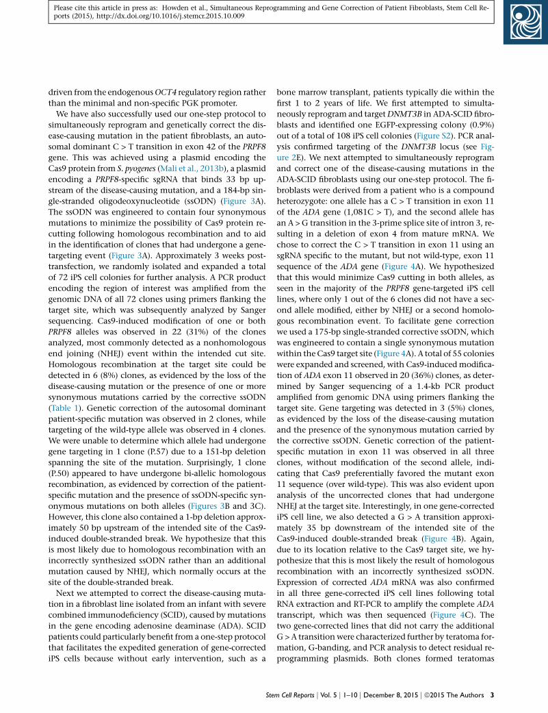
Figure 2. Gene Targeting of the DNMT3B Locus(A) Schematic diagram of the DNMT3B locus and the donor template used for gene targeting. pA, polyA signal; Ex, exon.(B) Flow cytometric analysis of H9 cells 3 days following the transfection of DNMT3B-specific donor template, mRNA encoding Cas9protein, and plasmid carrying DNMT3B-specific sgRNA.(C) Representative phase contrast and fluorescent images of an iPS cell colony generated after simultaneous reprogramming and genetargeting of DNMT3B in fibroblasts derived from a patient with retinitis pigmentosa, acquired 2.5 weeks post-electroporation.(D) Flow cytometric analysis of the total cell population after EDTA passaging, initiated approximately 3 weeks post-electroporation.Analysis was performed after three and five passages.(E) PCR analysis across recombination junction to confirm gene targeting of DNMT3B, using primers specific to sequence upstream of 30
homology arm and EGFP. Analysis was performed on 6 EGFP-expressing iPS cell clones (lanes G1–G6) and 6 EGFP-non-expressing iPS cell
(legend continued on next page)
4 Stem Cell Reports j Vol. 5 j 1–10 j December 8, 2015 j ª2015 The Authors
Please cite this article in press as: Howden et al., Simultaneous Reprogramming and Gene Correction of Patient Fibroblasts, Stem Cell Re-ports (2015), http://dx.doi.org/10.1016/j.stemcr.2015.10.009
A
PRPF8
Ex 43Ex 1
GGG
GCUUCCACAAT TCCCC GGG
AAA G GG
GGCCC U
T AC TGACCCC
CUGG
GGAACTTG
ATGTAC
AGG
AAATATGTTTATAC
AGCTACAGCTGGTCGATGTCGACC
CGAAC T CCGCTTGAGG
AAATTT
GAGCTC
TTCTACCACAAGATGGTG
G A
*
- - - - - - - - - - - - - - - - - - - - - - - - - - - - - - - - - - - - - - - - - - - - - - - - - - - - - - - - - - - - - - - - - - - - - - - - - - - antisense oligo (184 bp)
allele 1 (wt)
allele 2 (mut)
B
1 bp del
SM1
SM2
SM3
SM4
C
GCTTCCACAA AGG CC T CTGG
allele 1
allele 2
SM1
SM2
SM3
SM4
* *
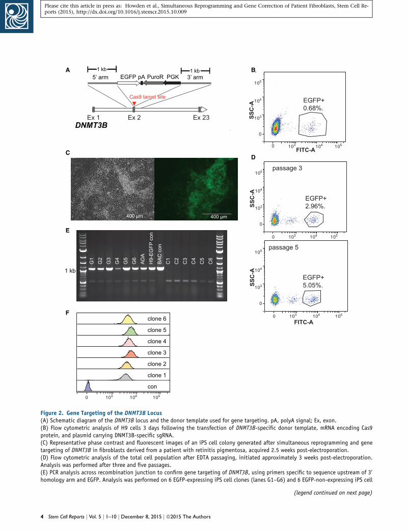
Figure 3. Simultaneous Reprogramming and Genetic Correction of the PRPF8 Gene in Fibroblasts from a Patient with RetinitisPigmentosa(A) Schematic diagram of the PRPF8 gene, with mutation in exon 42. The Cas9 target site (red), the patient-specific mutation (blue), andantisense single-stranded DNA template used for gene repair are shown.(B) Sequencing analysis of exon 42 of the PRPF8 gene in the genomic DNA from uncorrected patient-specific iPS cells. Both wild-type andmutant alleles are shown.(C) Sequencing analysis of genomic DNA from a single iPS cell clone following successful simultaneous reprogramming and geneticcorrection of patient-specific fibroblasts. Both alleles appear to have undergone homologous recombination with the corrective ssODN asevidenced by the presence of the ssODN-specific synonymous mutations (SM 1-4) on both alleles. One allele also has a single base-pairdeletion, which is most likely caused by an ssODN that was incorrectly synthesized. The location of the patient-specific mutation andsynonymous mutations introduced by the repair ssODN are marked by black boxes.
Please cite this article in press as: Howden et al., Simultaneous Reprogramming and Gene Correction of Patient Fibroblasts, Stem Cell Re-ports (2015), http://dx.doi.org/10.1016/j.stemcr.2015.10.009
comprising all three primary germ layers following injec-
tion into immunocompromised mice (Figure S3A), ex-
hibited normal karyotypes (Figure S3B), and were found
to be free of residual plasmid sequences (Figure S3C).
To further investigate the possibility that incorrectly syn-
thesized oligonucleotides may introduce additional muta-
tions following homologous recombination with the host
genome, we performed sequencing analysis of the ADA-
and PRPF8-specific ssODNs used for gene repair in the
experiments described previously. ssODNs were PCR
amplified using primers that bind the 50 and 30 ends and in-
serted into a plasmid vector.We then analyzed the oligonu-
cleotide-specific sequences in individual clones by Sanger
sequencing. In a control experiment, to estimate polymer-
ase error rate, PRPF8-specific ssODN sequence from one
clones (lanes C1–C6). An H9-derived DNMT3B-EGFP knockin and BAC catemplate) were included as positive controls.(F) Flow cytometric analysis of EGFP-expressing iPS cell lines generaDNMT3B locus.
Ste
representative clone was PCR amplified, re-cloned and sub-
sequently analyzed by Sanger sequencing. No mutations
were detected in any of the 30 clones analyzed from the
control experiment. In contrast, mutations were detected
in approximately 28% (9/32) and 29% (10/34) of clones
harboring the PRPF8 and ADA ssODN sequences, respec-
tively (Figure 5). Although single nucleotide deletions
were the most common type of mutation observed, single
base insertions, single base substitutions, and deletions
up to three bases in size were also detected (Table S1).
Furthermore, mutations were observed throughout the
ssODN and did not appear to exhibit any significant posi-
tional bias. Interestingly, a mutation within one ADA-
specific ssODN sequence was also detected at the same
location as that observed in the ADA gene-corrected iPS
rrying EGFP inserted at DNMT3B start codon (used to generate donor
ted after simultaneous reprogramming and gene targeting of the
m Cell Reports j Vol. 5 j 1–10 j December 8, 2015 j ª2015 The Authors 5
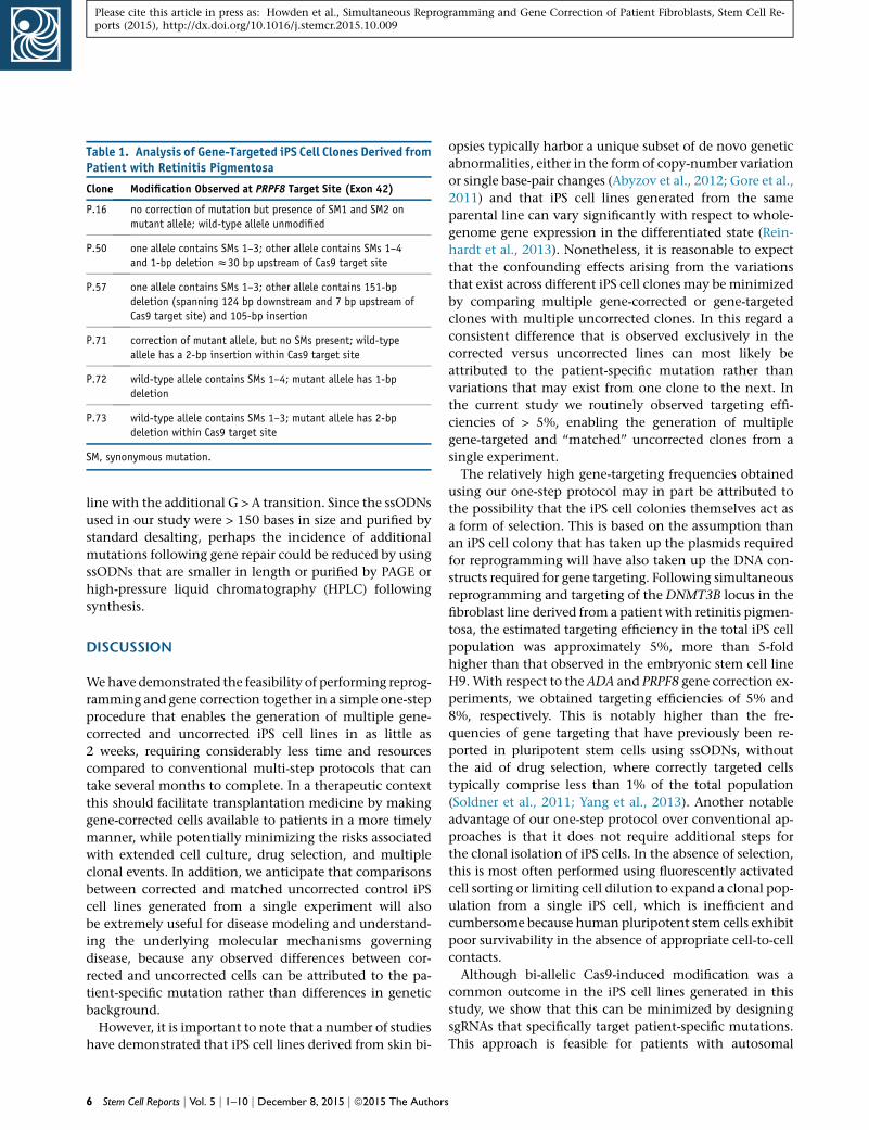
Table 1. Analysis of Gene-Targeted iPS Cell Clones Derived fromPatient with Retinitis Pigmentosa
Clone Modification Observed at PRPF8 Target Site (Exon 42)
P.16 no correction of mutation but presence of SM1 and SM2 on
mutant allele; wild-type allele unmodified
P.50 one allele contains SMs 1–3; other allele contains SMs 1–4
and 1-bp deletion z30 bp upstream of Cas9 target site
P.57 one allele contains SMs 1–3; other allele contains 151-bp
deletion (spanning 124 bp downstream and 7 bp upstream of
Cas9 target site) and 105-bp insertion
P.71 correction of mutant allele, but no SMs present; wild-type
allele has a 2-bp insertion within Cas9 target site
P.72 wild-type allele contains SMs 1–4; mutant allele has 1-bp
deletion
P.73 wild-type allele contains SMs 1–3; mutant allele has 2-bp
deletion within Cas9 target site
SM, synonymous mutation.
Please cite this article in press as: Howden et al., Simultaneous Reprogramming and Gene Correction of Patient Fibroblasts, Stem Cell Re-ports (2015), http://dx.doi.org/10.1016/j.stemcr.2015.10.009
line with the additional G > A transition. Since the ssODNs
used in our study were > 150 bases in size and purified by
standard desalting, perhaps the incidence of additional
mutations following gene repair could be reduced by using
ssODNs that are smaller in length or purified by PAGE or
high-pressure liquid chromatography (HPLC) following
synthesis.
DISCUSSION
Wehave demonstrated the feasibility of performing reprog-
ramming and gene correction together in a simple one-step
procedure that enables the generation of multiple gene-
corrected and uncorrected iPS cell lines in as little as
2 weeks, requiring considerably less time and resources
compared to conventional multi-step protocols that can
take several months to complete. In a therapeutic context
this should facilitate transplantation medicine by making
gene-corrected cells available to patients in a more timely
manner, while potentially minimizing the risks associated
with extended cell culture, drug selection, and multiple
clonal events. In addition, we anticipate that comparisons
between corrected and matched uncorrected control iPS
cell lines generated from a single experiment will also
be extremely useful for disease modeling and understand-
ing the underlying molecular mechanisms governing
disease, because any observed differences between cor-
rected and uncorrected cells can be attributed to the pa-
tient-specific mutation rather than differences in genetic
background.
However, it is important to note that a number of studies
have demonstrated that iPS cell lines derived from skin bi-
6 Stem Cell Reports j Vol. 5 j 1–10 j December 8, 2015 j ª2015 The Authors
opsies typically harbor a unique subset of de novo genetic
abnormalities, either in the form of copy-number variation
or single base-pair changes (Abyzov et al., 2012; Gore et al.,
2011) and that iPS cell lines generated from the same
parental line can vary significantly with respect to whole-
genome gene expression in the differentiated state (Rein-
hardt et al., 2013). Nonetheless, it is reasonable to expect
that the confounding effects arising from the variations
that exist across different iPS cell clones may be minimized
by comparing multiple gene-corrected or gene-targeted
clones with multiple uncorrected clones. In this regard a
consistent difference that is observed exclusively in the
corrected versus uncorrected lines can most likely be
attributed to the patient-specific mutation rather than
variations that may exist from one clone to the next. In
the current study we routinely observed targeting effi-
ciencies of > 5%, enabling the generation of multiple
gene-targeted and ‘‘matched’’ uncorrected clones from a
single experiment.
The relatively high gene-targeting frequencies obtained
using our one-step protocol may in part be attributed to
the possibility that the iPS cell colonies themselves act as
a form of selection. This is based on the assumption than
an iPS cell colony that has taken up the plasmids required
for reprogramming will have also taken up the DNA con-
structs required for gene targeting. Following simultaneous
reprogramming and targeting of the DNMT3B locus in the
fibroblast line derived from a patient with retinitis pigmen-
tosa, the estimated targeting efficiency in the total iPS cell
population was approximately 5%, more than 5-fold
higher than that observed in the embryonic stem cell line
H9.With respect to the ADA and PRPF8 gene correction ex-
periments, we obtained targeting efficiencies of 5% and
8%, respectively. This is notably higher than the fre-
quencies of gene targeting that have previously been re-
ported in pluripotent stem cells using ssODNs, without
the aid of drug selection, where correctly targeted cells
typically comprise less than 1% of the total population
(Soldner et al., 2011; Yang et al., 2013). Another notable
advantage of our one-step protocol over conventional ap-
proaches is that it does not require additional steps for
the clonal isolation of iPS cells. In the absence of selection,
this is most often performed using fluorescently activated
cell sorting or limiting cell dilution to expand a clonal pop-
ulation from a single iPS cell, which is inefficient and
cumbersome because human pluripotent stem cells exhibit
poor survivability in the absence of appropriate cell-to-cell
contacts.
Although bi-allelic Cas9-induced modification was a
common outcome in the iPS cell lines generated in this
study, we show that this can be minimized by designing
sgRNAs that specifically target patient-specific mutations.
This approach is feasible for patients with autosomal
ADA
Ex 11Ex 1
ACCAAGGTACAACG TTGGT TAT CC T C
CAT
G GG CG ATTCTG
TAAGACGCCG
TTTAAA
A
CCAGAAC
GGT
CUUGATCAATG
TGGCC
UAGUUACACCGGAAA
TC AG
UUU
AGAGCTTTAAA
CCTGGA
CC AGGGGTC
*
- - - - - - - - - - - - - - - - - - - - - - - - - - - - - - - - - - - - - - - - - - - - - - - - - - - - - - - - - - - - - - - - - - - antisense oligo (175 bp) S
M
TTA G
C CG
C
- - - - - - - CCTTGTAGTTACACCGGTTT
A
B CUncorrected
Clone L
SM * mut
Clone Bb
SM*mut
Uncorrected
Clone L
Clone Bb
Figure 4. Simultaneous Reprogramming and Genetic Correction of ADA-SCID Fibroblasts(A) Schematic diagram of the ADA gene, with mutation in exon 11. The Cas9 target site (red), the patient-specific mutation (blue), andantisense single-stranded DNA template used for gene repair are shown.(B) Sequencing analysis of exon 11 of the ADA gene in the genomic DNA of an uncorrected and two gene-corrected iPS celllines derived from ADA-SCID fibroblasts. One of the gene-corrected lines (clone Bb) was also found to carry a G > A transitionapproximately 35 bp downstream of the intended DNA double-stranded break, and most likely introduced by an incorrectlysynthesized ssODN.(C) Sequencing analysis of the ADA transcript amplified from the cDNA of an uncorrected and two gene-corrected iPS cell lines. Thelocation of the patient-specific mutation, synonymous mutation, and G > A transition introduced by the repair ssODN are marked by blackboxes.
Please cite this article in press as: Howden et al., Simultaneous Reprogramming and Gene Correction of Patient Fibroblasts, Stem Cell Re-ports (2015), http://dx.doi.org/10.1016/j.stemcr.2015.10.009
dominant and compound heterozygous mutations, but
not for mutations that are autosomal recessive. However,
since autosomal recessive diseases normally require only
a single functional allele, bi-allelic modification resulting
in correction of one allele and mutagenic NHEJ in the
other, is less of a concern. It will also be important to eval-
uate the use of Cas9 variants that have improved DNA
cleavage specificity in the protocol described here. Use of
the paired nickase (Cas9-D10A) (Mali et al., 2013a; Ran
et al., 2013) and dCas9-Fok1 fusions (fCas9) (Guilinger
et al., 2014), for example, should minimize the potential
of off-target effects that are largely associated with the
Ste
wild-type form of the Cas9 protein. Keeping background
damage to a minimumwill be essential not only for retain-
ing the downstream therapeutic potential of the cells but
also for an accurate recapitulation of the disease and cor-
rected (wild-type) phenotype, which will be important
for disease modeling and drug discovery purposes. Finally,
there is also significant value in adapting our protocol to
permit the generation of gene-corrected iPS cell lines
from alternative cell sources such as blood, which involve
less invasive procedures andmay harbor fewer somaticmu-
tations compared with fibroblasts derived from skin bi-
opsies. Since expansion of fibroblasts from an initial skin
m Cell Reports j Vol. 5 j 1–10 j December 8, 2015 j ª2015 The Authors 7

0
25
10
15
20
30
5
35
ADA ultramer PRPF8 ultramer control
% c
lone
s w
ith m
utat
ion
Figure 5. Sequencing Analysis of Oligonucleotides Used inGene Repair ExperimentsThe number of plasmid clones (expressed as percentage of totalclones analyzed) carrying oligonucleotide sequence with at leastone mutational event is shown. As a control, the PRPF8-specificoligonucleotide sequence from a single plasmid clone was PCR-amplified and re-cloned to assess polymerase error rate.
Please cite this article in press as: Howden et al., Simultaneous Reprogramming and Gene Correction of Patient Fibroblasts, Stem Cell Re-ports (2015), http://dx.doi.org/10.1016/j.stemcr.2015.10.009
biopsy can take several weeks, the ability to simultaneously
reprogram and genetically repair cells isolated from blood
draws will also serve to expedite the process of generating
gene-corrected iPS cell lines even further.
EXPERIMENTAL PROCEDURES
Plasmid DNA and mRNAThe episomal vectors (pEP4EO2SEN2L, pEP4EO2SET2K, and
pEP4EO2SEM2K) carrying seven factors for reprogramming have
been described previously (Yu et al., 2009). An additional plasmid
carrying the miR 302/367 cluster was generated by PCR—ampli-
fying an approximately 1.2-kb fragment from genomic DNA (ex-
tracted from human embryonic stem cell line H1) and cloned
into the pSimpleII plasmid (an OriP-containing plasmid) under
the control of the elongation factor-1a promoter. The plasmids
used for targeting OCT4 have been previously described (Hou
et al., 2013). The plasmid hCas9 was a gift from George Church
(Addgene plasmid #41815). All sgRNA targeting the DNMT3B,
PRPF8, or ADA genes were driven from a U6 promoter cloned
into pstBlue-1 (Novagen). The DNMT3B targeting vector was
generated by inserting a DNA cassette encoding an EGFP reporter,
PGK-promoter-driven puromycin gene, and kanamycin gene
flanked by FRT sites into a bacterial artificial chromosome (BAC)
clone containing the complete DNMT3B coding region (CTD-
2608L15) using the Red-ET recombination system (Genebridges).
The kanamycin resistance gene was subsequently removed from
the gene-targeting vector with Flpe-recombinase (Genebridges).
All plasmids were prepared by cesium chloride gradient extraction.
In vitro transcribed mRNA encoding Cas9 and EBNA1 was gener-
ated using the SP6 mMessage mMachine kit (Life Technologies).
8 Stem Cell Reports j Vol. 5 j 1–10 j December 8, 2015 j ª2015 The Authors
ssODNs (Ultramers) were purchased from Integrated DNA
Technologies.
Fibroblast CultureFibroblasts derived from a patient with retinitis pigmentosa were
obtained from the Peirce Lab, Penn State University, and ADA-
SCID fibroblasts were obtained from Coriell Laboratories (ID no.
GM02824). Fibroblasts were cultured in DMEM (Invitrogen) sup-
plemented with 15% fetal bovine serum (FBS) (HyClone) at
37�C, 5% CO2, and 5% O2.
Flow CytometryCells were analyzed for EGFP expression by flow cytometry using a
FACSCanto II flow cytometer (BD Biosciences). Data acquisition
and analysis were performed using FACsDiva software version
6.1.3 (BD Biosciences) and FlowJo (Tree Star) software version
9.5.1. Gating was performed on live cells based on forward and
side scatter analysis.
Reprogramming/Gene CorrectionFibroblasts were harvested 2 days after passaging and resuspended
in OptiMEM (Life Technologies) at a final concentration of 4–6 3
106 cells/ml. 500 ml of the cell suspension was added to a 0.4-cm
cuvette containing the reprogramming and gene-targeting DNA
constructs and electroporated (220 V, 1,000 mF) using the Gene
Pulser II (BioRad). See Table S2 for DNA concentrations used for
each experiment. In vitro transcribed mRNA encoding a truncated
version of the EBNA1proteinwas also included to enhance nuclear
uptake of the oriP-containing reprogramming vectors (Chen et al.,
2011; Howden et al., 2006). Following electroporation, cells were
plated on a single 10-cm Matrigel-coated (Corning) plate and
maintained in fibroblast media until 4 days post-transfection,
and then they were switched to E7 medium (E8 medium without
transforming growth factor b) with 100 mM sodium butyrate and
changed every other day as described previously (Chen et al.,
2011). Sodium butyrate was removed from the media after the
appearance of the first iPS cell colonies at around day 10. After
isolation, iPS cells were maintained and expanded in E8 medium
with daily media changes and passaged every 3–4 days with
EDTA in 13 PBS as previously described (Chen et al., 2011).
PCR, RT-PCR, and Sanger SequencingTotal genomic DNA was extracted by using the DNeasy Blood and
Tissue Kit (QIAGEN). Total RNAwas extracted by using the RNeasy
Plus Mini Kit (QIAGEN). cDNA was synthesized using SuperScript
III (Life Technologies) according to the manufacturer’s protocol.
PCR was performed by using GoTaq Green PCR Mastermix (Prom-
ega). Prior to sequencing, PCR products were treated with rAPid
Alkaline Phosphatase (Roche Life Science) and Endonuclease I
(NEB) for 30 min at 37�C followed by heat inactivation at 80�Cfor 15 min. Sequencing reactions were performed using BigDye
Terminator v3.1 (Life Technologies). Reactions were then purified
and sequenced by the Genome Center, University of Wisconsin.
For sequencing analysis of single alleles, PCR products amplified
from genomic DNA extracted from each of the PRPF8 gene-cor-
rected clones were cloned into the pUC19 vector (Life Technolo-
gies). Plasmid DNA was extracted from 12 randomly selected
Please cite this article in press as: Howden et al., Simultaneous Reprogramming and Gene Correction of Patient Fibroblasts, Stem Cell Re-ports (2015), http://dx.doi.org/10.1016/j.stemcr.2015.10.009
colonies for each of the 6 cloned PCR products and sequenced as
described above.
Oligonucleotide SequencesFor lists of primers used in this study for PCR amplification and
generation of sgRNA plasmids, see Table S3.
SUPPLEMENTAL INFORMATION
Supplemental Information includes three figures and three tables
and can be found with this article online at http://dx.doi.org/10.
1016/j.stemcr.2015.10.009.
AUTHOR CONTRIBUTIONS
S.E.H. and J.A.T. conceived the experiments and wrote the paper;
J.P.M. and B.M.D. performed teratoma injection and harvesting.
A.G.E. and E.G.S. assisted with ssODN analysis experiments.
ACKNOWLEDGMENTS
We thank Eric Peirce for providing the fibroblast line derived from
a patient with retinitis pigmentosa. This work was supported by a
Wynd-Gund Translation Research Award from the Foundation
Fighting Blindness. S.E.H. is supported by a National Health and
Medical Research Council (NHMRC) Overseas Biomedical Fellow-
ship. A.G.E. an E.G.S. are senior research fellows of the NHMRC.
Received: September 22, 2015
Revised: October 14, 2015
Accepted: October 15, 2015
Published: November 12, 2015
REFERENCES
Abyzov, A., Mariani, J., Palejev, D., Zhang, Y., Haney, M.S., Toma-
sini, L., Ferrandino, A.F., Rosenberg Belmaker, L.A., Szekely, A.,Wil-
son, M., et al. (2012). Somatic copy number mosaicism in human
skin revealed by induced pluripotent stem cells. Nature 492,
438–442.
Chen, G., Ye, Z., Yu, X., Zou, J., Mali, P., Brodsky, R.A., and Cheng,
L. (2008). Trophoblast differentiation defect in human embryonic
stem cells lacking PIG-A and GPI-anchored cell-surface proteins.
Cell Stem Cell 2, 345–355.
Chen, G., Gulbranson, D.R., Hou, Z., Bolin, J.M., Ruotti, V., Pro-
basco, M.D., Smuga-Otto, K., Howden, S.E., Diol, N.R., Propson,
N.E., et al. (2011). Chemically defined conditions for human
iPSC derivation and culture. Nat. Methods 8, 424–429.
Gore, A., Li, Z., Fung, H.L., Young, J.E., Agarwal, S., Antosiewicz-
Bourget, J., Canto, I., Giorgetti, A., Israel, M.A., Kiskinis, E., et al.
(2011). Somatic coding mutations in human induced pluripotent
stem cells. Nature 471, 63–67.
Guilinger, J.P., Thompson, D.B., and Liu, D.R. (2014). Fusion of
catalytically inactive Cas9 to FokI nuclease improves the specificity
of genome modification. Nat. Biotechnol. 32, 577–582.
Hockemeyer, D., Soldner, F., Beard, C., Gao, Q., Mitalipova, M.,
DeKelver, R.C., Katibah, G.E., Amora, R., Boydston, E.A., Zeitler,
B., et al. (2009). Efficient targeting of expressed and silent genes
Ste
in human ESCs and iPSCs using zinc-finger nucleases. Nat. Bio-
technol. 27, 851–857.
Hou, Z., Zhang, Y., Propson, N.E., Howden, S.E., Chu, L.F., Son-
theimer, E.J., and Thomson, J.A. (2013). Efficient genome engi-
neering in human pluripotent stem cells using Cas9 fromNeisseria
meningitidis. Proc. Natl. Acad. Sci. USA 110, 15644–15649.
Howden, S.E., Wardan, H., Voullaire, L., McLenachan, S., William-
son, R., Ioannou, P., and Vadolas, J. (2006). Chromatin-binding re-
gions of EBNA1 protein facilitate the enhanced transfection of
Epstein-Barr virus-based vectors. Hum. Gene Ther. 17, 833–844.
Howden, S.E., Gore, A., Li, Z., Fung, H.L., Nisler, B.S., Nie, J., Chen,
G., McIntosh, B.E., Gulbranson, D.R., Diol, N.R., et al. (2011). Ge-
netic correction and analysis of induced pluripotent stem cells
from a patient with gyrate atrophy. Proc. Natl. Acad. Sci. USA
108, 6537–6542.
Laurent, L.C., Ulitsky, I., Slavin, I., Tran, H., Schork, A., Morey, R.,
Lynch, C., Harness, J.V., Lee, S., Barrero, M.J., et al. (2011). Dy-
namic changes in the copy number of pluripotency and cell prolif-
eration genes in human ESCs and iPSCs during reprogramming
and time in culture. Cell Stem Cell 8, 106–118.
Lin, S.L., Chang, D.C., Chang-Lin, S., Lin, C.H., Wu, D.T., Chen,
D.T., and Ying, S.Y. (2008). Mir-302 reprograms human skin cancer
cells into a pluripotent ES-cell-like state. RNA 14, 2115–2124.
Liu, G.H., Suzuki, K., Qu, J., Sancho-Martinez, I., Yi, F., Li, M., Ku-
mar, S., Nivet, E., Kim, J., Soligalla, R.D., et al. (2011). Targeted gene
correction of laminopathy-associated LMNAmutations in patient-
specific iPSCs. Cell Stem Cell 8, 688–694.
Mali, P., Aach, J., Stranges, P.B., Esvelt, K.M., Moosburner, M., Ko-
suri, S., Yang, L., and Church, G.M. (2013a). CAS9 transcriptional
activators for target specificity screening and paired nickases for
cooperative genome engineering. Nat. Biotechnol. 31, 833–838.
Mali, P., Yang, L., Esvelt, K.M., Aach, J., Guell, M., DiCarlo, J.E.,
Norville, J.E., and Church, G.M. (2013b). RNA-guided human
genome engineering via Cas9. Science 339, 823–826.
Miyoshi, N., Ishii, H., Nagano, H., Haraguchi, N., Dewi, D.L., Kano,
Y., Nishikawa, S., Tanemura, M., Mimori, K., Tanaka, F., et al.
(2011). Reprogramming ofmouse andhuman cells to pluripotency
using mature microRNAs. Cell Stem Cell 8, 633–638.
Ran, F.A., Hsu, P.D., Lin, C.Y., Gootenberg, J.S., Konermann, S., Tre-
vino, A.E., Scott, D.A., Inoue, A., Matoba, S., Zhang, Y., and Zhang,
F. (2013). Double nicking by RNA-guided CRISPR Cas9 for
enhanced genome editing specificity. Cell 154, 1380–1389.
Reinhardt, P., Schmid, B., Burbulla, L.F., Schondorf, D.C., Wagner,
L., Glatza, M., Hoing, S., Hargus, G., Heck, S.A., Dhingra, A., et al.
(2013). Genetic correction of a LRRK2 mutation in human iPSCs
links parkinsonian neurodegeneration to ERK-dependent changes
in gene expression. Cell Stem Cell 12, 354–367.
Soldner, F., Laganiere, J., Cheng, A.W., Hockemeyer, D., Gao, Q.,
Alagappan, R., Khurana, V., Golbe, L.I., Myers, R.H., Lindquist,
S., et al. (2011). Generation of isogenic pluripotent stem cells
differing exclusively at two early onset Parkinson pointmutations.
Cell 146, 318–331.
Takahashi, K., Tanabe, K., Ohnuki, M., Narita, M., Ichisaka, T., To-
moda, K., and Yamanaka, S. (2007). Induction of pluripotent stem
m Cell Reports j Vol. 5 j 1–10 j December 8, 2015 j ª2015 The Authors 9

Please cite this article in press as: Howden et al., Simultaneous Reprogramming and Gene Correction of Patient Fibroblasts, Stem Cell Re-ports (2015), http://dx.doi.org/10.1016/j.stemcr.2015.10.009
cells from adult human fibroblasts by defined factors. Cell 131,
861–872.
Yang, L., Guell,M., Byrne, S., Yang, J.L., De Los Angeles, A.,Mali, P.,
Aach, J., Kim-Kiselak, C., Briggs, A.W., Rios, X., et al. (2013). Opti-
mization of scarless human stem cell genome editing. Nucleic
Acids Res. 41, 9049–9061.
Yu, J., Vodyanik, M.A., Smuga-Otto, K., Antosiewicz-Bourget, J.,
Frane, J.L., Tian, S., Nie, J., Jonsdottir, G.A., Ruotti, V., Stewart,
10 Stem Cell Reports j Vol. 5 j 1–10 j December 8, 2015 j ª2015 The Autho
R., et al. (2007). Induced pluripotent stem cell lines derived from
human somatic cells. Science 318, 1917–1920.
Yu, J., Hu, K., Smuga-Otto, K., Tian, S., Stewart, R., Slukvin, I.I., and
Thomson, J.A. (2009). Human induced pluripotent stem cells free
of vector and transgene sequences. Science 324, 797–801.
Zou, J., Mali, P., Huang, X., Dowey, S.N., andCheng, L. (2011). Site-
specific gene correction of a point mutation in human iPS cells
derived from an adult patient with sickle cell disease. Blood 118,
4599–4608.
rs