Embed Size (px)
Citation preview

COMPUTER ANIMATION AND VIRTUAL WORLDSComp. Anim. Virtual Worlds 2015; 26:197–206
Published online in Wiley Online Library (wileyonlinelibrary.com). DOI: 10.1002/cav.1649
SPECIAL ISSUE PAPER
Simultaneous estimation of elasticity for multipledeformable bodiesShan Yang* and Ming Lin
Department of Computer Science, University of North Carolina at Chapel Hill, Brooks Computer Science Building 201 S. ColumbiaSt., Chapel Hill, NC, USA
ABSTRACT
Material property has great importance in deformable body simulation and medical robotics. The elasticity parameters,such as Young’s modulus of the deformable bodies, are important to make realistic animations.
Further, in medical applications, the (recovered) elasticity parameters can assist surgeons to perform better pre-opsurgical planning and enable medical robots to carry out personalized surgical procedures. Previous elasticity parametersestimation methods are limited to recover one elasticity parameter of one deformable body at a time. In this paper, wepropose a novel elasticity parameter estimation algorithm that can recover the elasticity parameters of multiple deformablebodies or multiple regions of one deformable body simultaneously from (at least two sets of) images. We validateour algorithm with both synthetic test cases and real patient computed tomography images. Copyright © 2015 John Wiley& Sons, Ltd.
KEYWORDS
elasticity parameter reconstruction; human tissue simulation; tissue material property
All supporting information may be found in the online version of this article.
*Correspondence
Shan Yang, University of North Carolina at Chapel Hill.E-mail: [email protected]
1. INTRODUCTION
Material properties are important in depicting the charac-teristics of virtual objects for realistic computer animationsof soft bodies.
In addition, virtual surgical simulation has also beenincreasingly used for rapid prototyping of clinical devices,pre-operation planning of medical procedures, virtualtraining exercises for surgeons and medical personnel,and so on. And, tissue elasticity properties are importantparameters for developing accurate and predictive surgicalsimulation. Furthermore, to compute desired and accurateforce feedback for haptic display requires knowledge aboutthe deformation of soft tissues and organs, which are char-acterized by patient-specific elastic parameters for differenttissues and organs.
Elastography[1] was first proposed to determine theelasticity properties by measuring the deformation of thetissue because of the application of the known externalforces. The known external forces are the boundary con-dition needed to recover the exact elasticity parameter.Originally, the deformations were measured using ultra-sound imagery[2], but such techniques produced coarse,
two-dimensional representations of the moving tissue.More sophisticated imaging techniques, such as magneticresonance imaging (MRI)[3,4] and computed tomography(CT), produce three-dimensional images of the deform-ing tissues, allowing a more accurate measurement ofdisplacement.
Toward realizing the concept of three-dimensional phys-iological humans, we propose perhaps one of the firstelasticity parameter estimation algorithm for multiple,heterogeneous deformable bodies simultaneously usingmedical images†. Our approach is based on a multi-dimensional optimization method that iteratively performsdeformable body simulation using a finite element (FE)method on reconstructed organ models with the con-tinuously refined, estimated elasticity parameters. Thegeometric models of organs are reconstructed based onlow-resolution CT images.
Our objective function measures the sum of the dis-tance between the nodes of the organ surface. In contrast to
†In this paper, we use CT images. But, the algorithm is alsoapplicable to MRI images.
Copyright © 2015 John Wiley & Sons, Ltd. 197
Estimation of elasticity for multiple deformable bodies S. Yang and M. Lin
elastography methods [5–7], the only information we needis the displacement of the nodes of the organ surface. Wedo not need every pixel-wise displacement vector, thus, noextra procedures need to be performed on the patient. Twosets of (medical) images are sufficient to recover the elas-ticity parameters using our method. Therefore, our methodcan be widely applicable to different imaging technology.It can be used for animatino of soft bodies (see supple-mentary video) and possibly for cancer staging using onlylow-resolution CT images.
2. RELATED WORK
In computer graphics, extensive research has been donefor deformable body simulation [8–11]. Researchers havealso been studying methods for designing simulation withmaterial properties [12–15] and for realistic bio-structuresimulation [16].
In medical applications, there are mainly two kinds ofsoft tissue elasticity properties estimation methods [17]:invasive and noninvasive techniques. The invasive meth-ods rely on a device to measure the displacement andforce response [18–20]. These methods take organ sampleseither out of the human or animal bodies and perform theexperiment in vitro (out side the body) or do the procedurein situ (inside the body). The collected data are then used tosolve the inverse problem, which is to recover the elasticityproperties, by constructing a polynomial interpolation [21]or by using an FE model [17,22,23].
The noninvasive methods were mostly based on imageanalysis techniques to measure the displacement. In the1980s, several methods were proposed to measure themotion of the soft tissue, such as the one proposed byWilson and Robinson [24] using radio frequency M-modesignals and the one proposed by Birnholz and Farrell [25]using ultrasound B-scans. Researchers have also used med-ical image analysis on 2-D ultrasound and/or MRI imagesto estimate the elastic parameters of soft tissue [26–28].
In the area of elastography, researchers [5–7] proposedalgorithms based on the distance between two medicalimages. By solving the least square problem, the elas-ticity parameters are recovered. Van Houten et al. [4]used elastography methods to estimate the Young’s mod-ulus distribution of a 2D area, then later extendedto solve three-dimensional elastic parameter distributionusing MRI [29]. These methods need high-resolution dis-placement fields to recover the elasticity parameter [27],where the displacement field is typically obtained throughan external device using a vibration actuation mecha-nism. For organs that are located deep-seated inside ahuman body, the vibrator may need to be placed inside theorgan[30], making the procedure much more complex andpossibly uncomfortable for the patient.
Other than distance field based methods, there are alsoother measurement algorithms. The modality-independentelastography method [31] measures the elasticity parame-ters by maximizing the image similarity based on a number
of landmarks. However, this technique does not apply toall the soft tissues, as landmarks cannot always be found insome of the organs such as prostate. Statistical and machinelearning algorithms have also been used to classify soft tis-sues and estimate the parameters using multi-spectral MRimages [32].
Although the existing elastography methods can pro-vide an estimation of the elasticity parameter distribution,they require high-resolution magnetic resonance medi-cal images and a device to measure the external forceexerted on the soft tissues, which is not always possibleor practical. Lee et al. proposed the first model to esti-mate the Young’s modulus based on low-resolution CTimages, and no external force is needed to set the bound-ary condition [33]. By optimizing the distance betweenthe deformed and the reference surface meshes, the elas-tic parameter is estimated. However, this method can onlyrecover the elasticity parameter of one organ. In con-trast, our work can recover the elasticity parameters ofmultiple, heterogeneous soft bodies simultaneously, usinga multi-dimensional optimization method.
3. METHOD
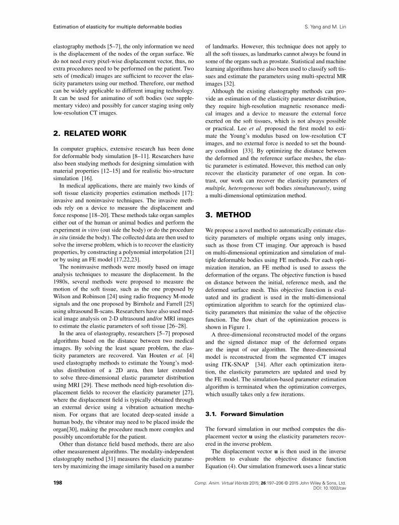
We propose a novel method to automatically estimate elas-ticity parameters of multiple organs using only images,such as those from CT imaging. Our approach is basedon multi-dimensional optimization and simulation of mul-tiple deformable bodies using FE methods. For each opti-mization iteration, an FE method is used to assess thedeformation of the organs. The objective function is basedon distance between the initial, reference mesh, and thedeformed surface mesh. This objective function is eval-uated and its gradient is used in the multi-dimensionaloptimization algorithm to search for the optimized elas-ticity parameters that minimize the value of the objectivefunction. The flow chart of the optimization process isshown in Figure 1.
A three-dimensional reconstructed model of the organsand the signed distance map of the deformed organsare the input of our algorithm. The three-dimensionalmodel is reconstructed from the segmented CT imagesusing ITK-SNAP [34]. After each optimization itera-tion, the elasticity parameters are updated and used bythe FE model. The simulation-based parameter estimationalgorithm is terminated when the optimization converges,which usually takes only a few iterations.
3.1. Forward Simulation
The forward simulation in our method computes the dis-placement vector u using the elasticity parameters recov-ered in the inverse problem.
The displacement vector u is then used in the inverseproblem to evaluate the objective distance functionEquation (4). Our simulation framework uses a linear static
198 Comp. Anim. Virtual Worlds 2015; 26:197–206 © 2015 John Wiley & Sons, Ltd.DOI: 10.1002/cav
S. Yang and M. Lin Estimation of elasticity for multiple deformable bodies
Figure 1. The flow chart of the optimization iteration. The initial guess of the elasticity parameter is provided based on standardtissue values, prior to the start of the optimization. For each optimization iteration, the tissue deformation is recomputed using a finiteelement (FE) method simulation. The value of the distance objective function is also re-evaluated. At the end of each iteration, the
elasticity parameter is updated and used by the FE model to continue the simulated-based optimization process.
FE method [35] (Equation (1)). The weak formulation forelasticity problem is given,Z
�ruT� Rud�C
Z�r�T�d��
Z�ruT bd� �
Z�ruT td� D 0
(1)
where u is the displacement vector and t is the boundarycondition(traction act on the boundary �). For quasi-staticdeformation process, the Ru D 0. We can rewriteEquation (1) as�Z
�r�T�d� �
Z�ruT bd�
��
�Z�ruT td�
�D 0
(2)
with the first part of the equation as the internal force andthe second part of the equation as the external force.
We use linear elastic material model. For isotropic linearelasticity, the stress–strain relation is defined as,
� D D" (3)
where � is the stress tensor, " is the strain tensor, and matrixD is defined by the material elasticity parameters. We useYoung’s modulus E and Poisson’s ratio � for describingmaterial properties.Previous elastography methods use external forces as theboundary condition. Our method, in contrast, does notneed external forces. The initial boundary condition in ouralgorithm is the known displacement vector of the sur-face mesh. This boundary condition is applied only to thenodes of the surface mesh. When the three-dimensionalmodel deformed, the force generated by the deformationwill drive the simulation.
3.2. The Inverse Problem
The inverse problem is the process of elasticity parameterestimation. Our method is based on the multi-dimensionaloptimization method. By solving the least square problemiteratively, we recover the elasticity parameter. We then usethe distance between the initial, reference surface mesh,and the deformed surface mesh to iteratively update theobjective function.
3.2.1. Distance Based Objective Function.
As our simulation framework is based on thelow-resolution CT images, only the displacement of theboundary of the soft tissue is known. Our objective func-tion Equation (4) is constructed using the sum of thedistance between the nodes of initial, reference surface,and that of the deformed surface. By minimizing the valueof the objective function, we find the optimal elasticityparameters �.
ˆ.�m/ D1
2
NXmD1
Xul2Sm,ur2Sr
kd.ul Cul, ur/k2 (4)
where m is the mth organ and N is the number of organsthat is in the simulation scene.
d.ul C ul, ur/ is the bidirectional Hausdorff distancebetween deformed surface mesh Sm and the initial, refer-ence mesh Sr. �m D Em, in which Em is the Young’smodulus of the mth organ. Our method can be extended tooptimize more than one parameter. We could also includethe Poisson’s ratio into �m.The � that minimizes the objective function is the opti-mized set of elasticity parameters.
Comp. Anim. Virtual Worlds 2015; 26:197–206 © 2015 John Wiley & Sons, Ltd. 199DOI: 10.1002/cav
Estimation of elasticity for multiple deformable bodies S. Yang and M. Lin
3.2.2. Multi-Dimensional Numerical
Optimization Method.
We propose to use a multi-dimensional optimiza-tion method to recover the elasticity parameters. Ourthree-dimensional model can have a large number of nodes,so a significant amount of memory would be needed tostore the exact Hessian matrix for the Newton’s optimiza-tion method. Therefore, to solve the least square problem,we choose the Limited Memory Quasi-Newton’s method.
Using this method, the approximation of the Hessianmatrix is maintained instead of the exact Hessian matrix.For each step of this Broyden–Fletcher–Goldfarb–Shannomethod [36],
�kC1 D �k � ˛kHkrˆk (5)
where Hk is the approximated Hessian matrix, xk is thevariable to be optimized,ˆk is the objective function value,k denotes the kth optimization iteration, and rˆ is the gra-dient of the objective function. To compute the gradient,the partial derivative of ˆ is with respect to the elasticityparameter, the Young’s modulus of the mth organ.
4. EXPERIMENT
We have implemented our algorithm and performed threesets of experiments to evaluate its accuracy under differ-ent conditions using both two synthetic sets of models
with known parameters to validate the approach and areconstructed set of organs from CT images to illustrateits robustness.
4.1. Recovering Known Values
The first experiment is designed to test the accuracy ofthe algorithm, if the three organs sharing boundary. As thenumber of the organs increases, the problem becomes evenmore complicated.
4.1.1. Model Construction.
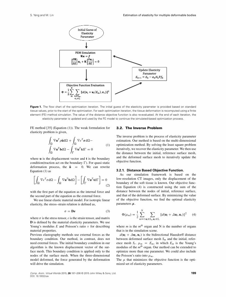
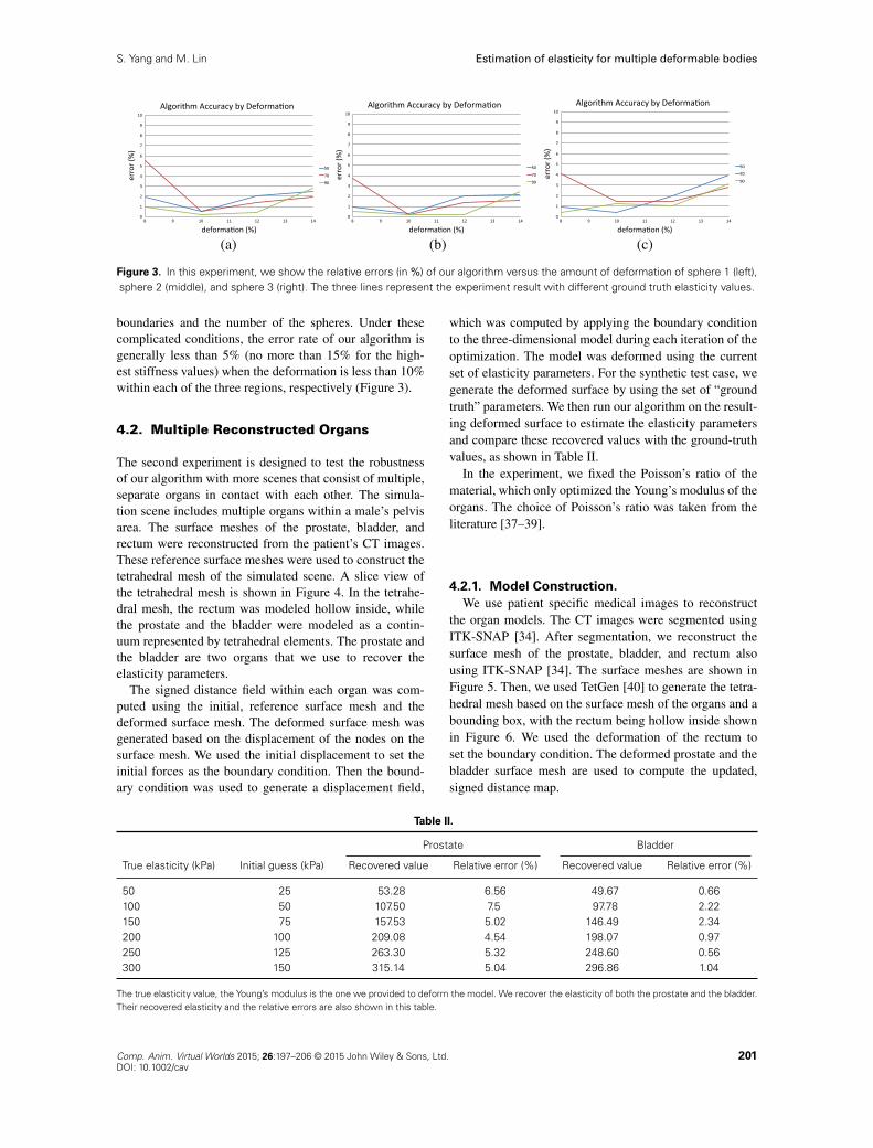
We used 3-concentric spheres to build the test model inexperiment I. In order to measure the elastic parameter ofsphere 1, for the area between sphere 1 and sphere 2 andthe area between sphere 2 and sphere 3, tetrahedralizationis carried out within sphere 1, the area between sphere 1and sphere 2 and the area between sphere 2 and sphere 3.The sliced view of the three-dimensional model is shown inFigure 2. The following table is generated when the threeareas are all deformed by slightly less than 10%.
4.1.2. Result.
The result of this experiment is shown in Table I. In thisexperiment we increase the number of “organs” to test theaccuracy of our algorithm. The result of this experimentis affected by both the fact that the spheres are sharing
Figure 2. A sliced view of the tetrahedral mesh of experiment I. The image on the left shows only the tetrahedral mesh of thespheres, while the image on the right shows the complete tetrahedral mesh.
Table I.
Sphere 1 Sphere 2 Sphere 3
True elasticity Initial guess Recovered Relative Recovered Relative Recovered Relative(kPa) (kPa) value error (%) value error (%) value error (%)
50 25 49.23 1.54 49.54 0.92 49.55 0.970 35 66.14 5.51 67.41 3.7 67.15 4.0790 45 89.18 0.91 89.52 0.53 89.68 0.36110 55 107.93 1.88 108.76 1.13 109.17 0.75130 65 112.89 13.16 118.95 8.5 124.89 3.93150 750 132.87 11.44 138.78 7.48 145.32 3.12
The true elasticity value, the Young’s modulus is the one we provided to deform the model. Sphere 1, the area between sphere 1 and sphere 2, and thearea between sphere 2 and sphere 3 are the three “organs” that we experimented on. The recovered elasticity parameters and the relative errors areshown in this table.
200 Comp. Anim. Virtual Worlds 2015; 26:197–206 © 2015 John Wiley & Sons, Ltd.DOI: 10.1002/cav
S. Yang and M. Lin Estimation of elasticity for multiple deformable bodies
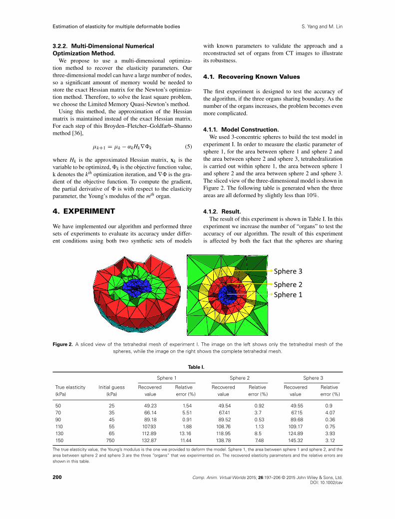
(a) (b) (c)
Figure 3. In this experiment, we show the relative errors (in %) of our algorithm versus the amount of deformation of sphere 1 (left),sphere 2 (middle), and sphere 3 (right). The three lines represent the experiment result with different ground truth elasticity values.
boundaries and the number of the spheres. Under thesecomplicated conditions, the error rate of our algorithm isgenerally less than 5% (no more than 15% for the high-est stiffness values) when the deformation is less than 10%within each of the three regions, respectively (Figure 3).
4.2. Multiple Reconstructed Organs
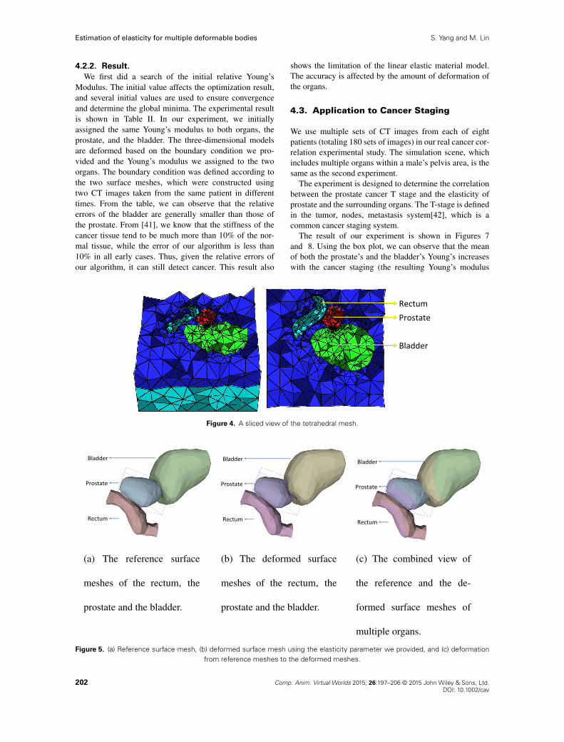
The second experiment is designed to test the robustnessof our algorithm with more scenes that consist of multiple,separate organs in contact with each other. The simula-tion scene includes multiple organs within a male’s pelvisarea. The surface meshes of the prostate, bladder, andrectum were reconstructed from the patient’s CT images.These reference surface meshes were used to construct thetetrahedral mesh of the simulated scene. A slice view ofthe tetrahedral mesh is shown in Figure 4. In the tetrahe-dral mesh, the rectum was modeled hollow inside, whilethe prostate and the bladder were modeled as a contin-uum represented by tetrahedral elements. The prostate andthe bladder are two organs that we use to recover theelasticity parameters.
The signed distance field within each organ was com-puted using the initial, reference surface mesh and thedeformed surface mesh. The deformed surface mesh wasgenerated based on the displacement of the nodes on thesurface mesh. We used the initial displacement to set theinitial forces as the boundary condition. Then the bound-ary condition was used to generate a displacement field,
which was computed by applying the boundary conditionto the three-dimensional model during each iteration of theoptimization. The model was deformed using the currentset of elasticity parameters. For the synthetic test case, wegenerate the deformed surface by using the set of “groundtruth” parameters. We then run our algorithm on the result-ing deformed surface to estimate the elasticity parametersand compare these recovered values with the ground-truthvalues, as shown in Table II.
In the experiment, we fixed the Poisson’s ratio of thematerial, which only optimized the Young’s modulus of theorgans. The choice of Poisson’s ratio was taken from theliterature [37–39].
4.2.1. Model Construction.
We use patient specific medical images to reconstructthe organ models. The CT images were segmented usingITK-SNAP [34]. After segmentation, we reconstruct thesurface mesh of the prostate, bladder, and rectum alsousing ITK-SNAP [34]. The surface meshes are shown inFigure 5. Then, we used TetGen [40] to generate the tetra-hedral mesh based on the surface mesh of the organs and abounding box, with the rectum being hollow inside shownin Figure 6. We used the deformation of the rectum toset the boundary condition. The deformed prostate and thebladder surface mesh are used to compute the updated,signed distance map.
Table II.
Prostate Bladder
True elasticity (kPa) Initial guess (kPa) Recovered value Relative error (%) Recovered value Relative error (%)
50 25 53.28 6.56 49.67 0.66100 50 107.50 7.5 97.78 2.22150 75 157.53 5.02 146.49 2.34200 100 209.08 4.54 198.07 0.97250 125 263.30 5.32 248.60 0.56300 150 315.14 5.04 296.86 1.04
The true elasticity value, the Young’s modulus is the one we provided to deform the model. We recover the elasticity of both the prostate and the bladder.Their recovered elasticity and the relative errors are also shown in this table.
Comp. Anim. Virtual Worlds 2015; 26:197–206 © 2015 John Wiley & Sons, Ltd. 201DOI: 10.1002/cav
Estimation of elasticity for multiple deformable bodies S. Yang and M. Lin
4.2.2. Result.
We first did a search of the initial relative Young’sModulus. The initial value affects the optimization result,and several initial values are used to ensure convergenceand determine the global minima. The experimental resultis shown in Table II. In our experiment, we initiallyassigned the same Young’s modulus to both organs, theprostate, and the bladder. The three-dimensional modelsare deformed based on the boundary condition we pro-vided and the Young’s modulus we assigned to the twoorgans. The boundary condition was defined according tothe two surface meshes, which were constructed usingtwo CT images taken from the same patient in differenttimes. From the table, we can observe that the relativeerrors of the bladder are generally smaller than those ofthe prostate. From [41], we know that the stiffness of thecancer tissue tend to be much more than 10% of the nor-mal tissue, while the error of our algorithm is less than10% in all early cases. Thus, given the relative errors ofour algorithm, it can still detect cancer. This result also
shows the limitation of the linear elastic material model.The accuracy is affected by the amount of deformation ofthe organs.
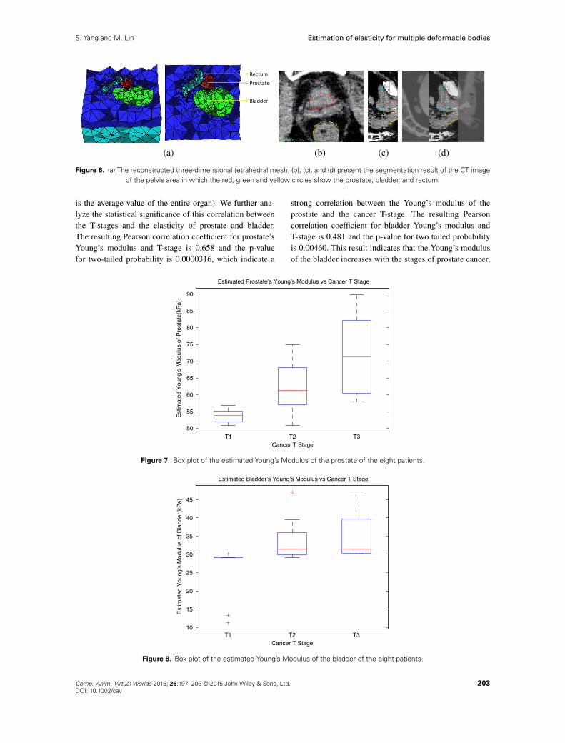
4.3. Application to Cancer Staging
We use multiple sets of CT images from each of eightpatients (totaling 180 sets of images) in our real cancer cor-relation experimental study. The simulation scene, whichincludes multiple organs within a male’s pelvis area, is thesame as the second experiment.
The experiment is designed to determine the correlationbetween the prostate cancer T stage and the elasticity ofprostate and the surrounding organs. The T-stage is definedin the tumor, nodes, metastasis system[42], which is acommon cancer staging system.
The result of our experiment is shown in Figures 7and 8. Using the box plot, we can observe that the meanof both the prostate’s and the bladder’s Young’s increaseswith the cancer staging (the resulting Young’s modulus
Figure 4. A sliced view of the tetrahedral mesh.
Figure 5. (a) Reference surface mesh, (b) deformed surface mesh using the elasticity parameter we provided, and (c) deformationfrom reference meshes to the deformed meshes.
202 Comp. Anim. Virtual Worlds 2015; 26:197–206 © 2015 John Wiley & Sons, Ltd.DOI: 10.1002/cav
S. Yang and M. Lin Estimation of elasticity for multiple deformable bodies
(a) (b) (c) (d)
Figure 6. (a) The reconstructed three-dimensional tetrahedral mesh; (b), (c), and (d) present the segmentation result of the CT imageof the pelvis area in which the red, green and yellow circles show the prostate, bladder, and rectum.
is the average value of the entire organ). We further ana-lyze the statistical significance of this correlation betweenthe T-stages and the elasticity of prostate and bladder.The resulting Pearson correlation coefficient for prostate’sYoung’s modulus and T-stage is 0.658 and the p-valuefor two-tailed probability is 0.0000316, which indicate a
strong correlation between the Young’s modulus of theprostate and the cancer T-stage. The resulting Pearsoncorrelation coefficient for bladder Young’s modulus andT-stage is 0.481 and the p-value for two tailed probabilityis 0.00460. This result indicates that the Young’s modulusof the bladder increases with the stages of prostate cancer,
Figure 7. Box plot of the estimated Young’s Modulus of the prostate of the eight patients.
Figure 8. Box plot of the estimated Young’s Modulus of the bladder of the eight patients.
Comp. Anim. Virtual Worlds 2015; 26:197–206 © 2015 John Wiley & Sons, Ltd. 203DOI: 10.1002/cav
Estimation of elasticity for multiple deformable bodies S. Yang and M. Lin
but they are not strongly correlated. Our findings reconfirmthe studies [43] that prostate cancer increases the proba-bility of bladder cancer. As cancer continues to advance tohigher stages, it spreads to neighboring tissues.
4.4. Discussion
We have used a range of deformation that is larger thanthe normal tissue deformation to stress test our algorithmand to analyze the relationship between the degree of accu-racy versus the amount of deformation. This analysis helpsus understand when nonlinear models should be used.Additional sensitive analysis with respect to segmentation,simulation resolution (i.e. the size of mesh), and use ofnonlinear FEM model can also be performed to provideadditional information to the users. We “jumpstart” theiterative optimization process with some range of defaultvalues and the algorithm usually converges quickly withinless than 10 iterations in practice.
Our implementation currently addressed the possibilityof multiple solutions by using multiple (3–5) initial val-ues sampled over a wide range (50–300) of possible values(say 50, 150, 250) and use multiple sets of the imagedata from few different days to compute the average val-ues, after eliminating possible “outliner value(s)”. Withthis approach, our algorithm is able to find the elasticityparameters that are very close to the “ground truth” valuesin practice.
5. CONCLUSION ANDFUTURE WORK
In this paper, we presented a novel multi-body elastic-ity parameter estimation method using low-resolution CTimages. As our method do not require any external forcesto be measured and only the deformation of the organ sur-face is needed, it can be applied to organs that are locateddeep-seated in the human body. There are limitations, how-ever. The amount of deformation of the soft tissue canaffect the accuracy of the algorithm. The larger the defor-mation, the higher the relative error is from the estimation.This is because of the fact that we have adopted a lin-ear, static FE method and linear elastic material model.Linear models are generally considered accurate and suf-ficient when the deformation is small and within a certainrange where linearity assumption is applicable. Our exper-imental results support this observation. The linear elasticmaterial model is not suitable for the simulation of humanorgans when they undergo a large deformation. In thefuture, we plan to adopt a more complex, nonlinear elas-tic material model for soft tissue simulation, such as theMooney Rivlin model. The accuracy may likely be higherwhen the amount of deformation is significant, althoughwe expect the computation cost to increase as well.
The algorithm we proposed in this paper is based ona multi-dimensional optimization method, which can alsobe used to estimate multiple elasticity parameters of a
single organ of multiple, heterogeneous tissue propertiesfor different regions of a (human) body. Because of theimportance of Young’s modulus in noninvasive cancerdetection, we choose to estimate this parameter for mul-tiple organs simultaneously. However, Poisson’s ratio hasalso been suggested as a significant indicator for breastcancer. Therefore, in the future, we plan to further studythe accuracy of multi-dimensional optimization methodand hope to use it to estimate multiple elasticity parame-ters of a single organ for more accurate cancer screeningand grading.
ACKNOWLEDGEMENTS
This research is supported in part by the JointNSF and NIH Smart and Connected Health Program,NIH#R01EB020426 � 01. We would like to thank Drs.Ronald Chen and Edward Chaney for CT images from theirLab, and Dr. Huai-Ping Lee for data from his thesis inour experiments.
REFERENCES
1. Ophir J, Céspedes I, Ponnekanti H, Yazdi Y, Li X.Elastography: a quantitative method for imaging theelasticity of biological tissues. Ultrasonic Imaging1991; 13(2): 111–134.
2. Krouskop TA, Dougherty DR, Vinson, FS, et al. Apulsed Doppler ultrasonic system for making nonin-vasive measurements of the mechanical properties ofsoft tissue. Journal of Rehabilitation Research andDevelopment 1987; 24(2): 1–8.
3. Chenevert TL, Skovoroda AR, Odonnel M, EmelianovSY. Elasticity reconstructive imaging by meansof stimulated echo MRI. Magnetic Resonance inMedicine 1998; 39(3): 482–490.
4. Van Houten EEW, Paulsen KD, Miga MI, KennedyFE, Weaver JB. An overlapping subzone technique forMR-based elastic property reconstruction. MagneticResonance in Medicine 1999; 42(4): 779–786.
5. Becker M, Teschner M. Robust and efficient estimationof elasticity parameters using the linear finite elementmethod. Proceedings of Simulation and Visualization2007: 15–28.
6. Eskandari H, Salcudean SE, Rohling R, Bell I.Real-time solution of the finite element inverse prob-lem of viscoelasticity. Inverse Problems 2011; 27(8):085002.
7. Zhu Y, Hall TJ, Jiang J. A finite-element approachfor Young’s modulus reconstruction. Medical Imaging,IEEE Transactions on 2003; 22(7): 890 –901.
8. Terzopoulos D, Platt J, Barr A, Fleischer K. Elasticallydeformable models 1987; 21: 205–214.
204 Comp. Anim. Virtual Worlds 2015; 26:197–206 © 2015 John Wiley & Sons, Ltd.DOI: 10.1002/cav

S. Yang and M. Lin Estimation of elasticity for multiple deformable bodies
9. Nealen A, Muller M, Keiser R, Boxerman E,Carlson M. Physically based deformable models incomputer graphics. Computer Graphics Forum 2006;25: 809–836.
10. Müller M, Gross M. Interactive virtual materials.In Proceedings of Graphics Interface 2004, GI ’04,School of Computer Science, University of Water-loo, Waterloo, Ontario, Canada, 2004, CanadianHuman-Computer Communications Society; 239–246.
11. Meehan M, Teschner M, Girod S. Three-dimensionalsimulation and prediction of craniofacial surgery.Orthodontics & Craniofacial Research 2003; 6(s1):102–107.
12. Miguel E, Bradley D, Thomaszewski, B, et al.Data-driven estimation of cloth simulation models.Computer Graphics Forum (Eurographics 2012) 2012;31(2): 519–528.
13. Miguel E, Tamstorf R, Bradley, E, et al. Modeling andestimation of internal friction in cloth. ACM Trans-actions on Graphics (Proc. SIGGRAPH Asia) 2013;32(6).
14. Wang H, Ramamoorthi R, O’Brien J. Data-driven elas-tic models for cloth: modeling and measurement. ACMTransactions on Graphics (SIGGRAPH) 2011; 30(4):71:1–71:12.
15. Bickel B, Bächer M, Otaduy, MA, et al. Designand fabrication of materials with desired deforma-tion behavior. In ACM SIGGRAPH 2010 Papers, LosAngeles, California, 2010; 1–10.
16. Maciel Anderson, Boulic Ronan, Thalmann Daniel.Deformable tissue parameterized by properties of realbiological tissue. In Surgery Simulation and Soft TissueModeling. Springer: Springer Berlin Heidelberg, 2003;74–87.
17. Samur E, Sedef M, Basdogan C, Avtan L, Duzgun O.A robotic indenter for minimally invasive measurementand characterization of soft tissue response. MedicalImage Analysis 2007; 11(4): 361–373.
18. Carter FJ, Frank TG, Davies PJ, McLean D, CuschieriA. Measurements and modelling of the compliance ofhuman and porcine organs. Medical Image Analysis2001; 5(4): 231–236.
19. Kauer M, Vuskovic V, Dual J, Szekely G, Bajka M.Inverse finite element characterization of soft tissues,Vol. 6, 2002.
20. Rosen J, Hannaford B, MacFarlane MP, Sinanan MN.Force controlled and teleoperated endoscopic grasperfor minimally invasive surgery–experimental perfor-mance evaluation, 1999.
21. Bicchi A, Canepa G, De Rossi D, Iacconi P,Scillingo EP. A sensor-based minimally invasivesurgery tool for detecting tissue elastic properties,1996.
22. Gao Z, Kim T, James DL, Desai JP. Semi-automated
soft-tissue acquisition and modeling for surgical simu-
lation, 2009.
23. Misra S, Ramesh KT, Okamura AM. Modelling of
non-linear elastic tissues for surgical simulation. Com-
puter Methods in Biomechanics and Biomedical Engi-
neering 2010; 13(6): 811–818.
24. Wilson LS, Robinson DE. Ultrasonic measurement
of small displacements and deformations of tissue.
Ultrasonic Imaging 1982; 4(1): 71–82.
25. Birnholz JC, Farrell EE. Fetal lung development: com-
pressibility as a measure of maturity. Radiology 1985;
157(2): 495–498.
26. Gao L, Parker KJ, Lerner RM, Levinson SF. Imaging
of the elastic properties of tissue-a review. Ultrasound
in Medicine & Biology 1996; 22(8): 959–977.
27. Manduca A, Oliphant TE, Dresner, MA, et al. Mag-
netic resonance elastography: non-invasive mapping of
tissue elasticity. Medical Image Analysis 2001; 5(4):
237–254.
28. Zheng Y-P, Mak AFT. An ultrasound indentation sys-
tem for biomechanical properties assessment of soft
tissues in-vivo, 1996.
29. Van Houten EEW, Miga MI, Weaver JB, Kennedy FE,
Paulsen KD. Three-dimensional subzone-based recon-
struction algorithm for MR elastography. Magnetic
Resonance in Medicine 2001; 45(5): 827–837.
30. Chopra R, Arani A, Huang, Y, et al. In vivo MR elas-
tography of the prostate gland using a transurethral
actuator. Magnetic Resonance in Medicine 2009;
62(3): 665–671.
31. Miga MI. New approach to elastrograph imaging:
modality-independent elastography 2002: 604–611.
32. Liang Z, MacFall JR, Harrington DP. Parameter esti-
mation and tissue segmentation from multispectral MR
images, 1994.
33. Lee HP, Foskey M, Niethammer M, Krajcevski P,
Lin MC. Simulation-based joint estimation of body
deformation and elasticity parameters for medical
image analysis. Medical Imaging, IEEE Transactions
on 2012; 31(11): 2156 –2168.
34. Yushkevich PA, Piven J, Hazlett, HC, et al.
User-guided 3D active contour segmentation of
anatomical structures: significantly improved effi-
ciency and reliability. NeuroImage 2006; 31(3):
1116–1128.
Comp. Anim. Virtual Worlds 2015; 26:197–206 © 2015 John Wiley & Sons, Ltd. 205DOI: 10.1002/cav

Estimation of elasticity for multiple deformable bodies S. Yang and M. Lin
35. Bro-Nielsen M. Finite element modeling in surgerysimulation. Proceedings of the IEEE 1998; 86(3):490–503.
36. Nocedal J, Wright SJ. Numerical Optimization.Springer Verlag: Springer New York, 1999.
37. Bharatha A, Hirose M, Hata, N, et al. et al. Evaluationof three-dimensional finite element-based deformableregistration of pre- and intraoperative prostate imaging.Medical Physics 2001; 28: 2551–2560.
38. Hensel JM, Ménard C, Chung, PWM, et al. Devel-opment of multiorgan finite element-based prostatedeformation model enabling registration of endorec-tal coil magnetic resonance imaging for radiotherapyplanning. International Journal of Radiation Oncol-ogy, Biology and Physics 2007; 68(5).
39. Tanner C, Schnabel JA, Hill DLG, Hawkes DJ, LeachMO, Hose DR. Factors influencing the accuracy ofbiomechanical breast models. Medical Physics 2006;33: 1758–1769.
40. Si H, TetGen A. A quality tetrahedral mesh generatorand three-dimensional delaunay triangulator. Weier-strass Institute for Applied Analysis and Stochastic,Berlin, Germany 2006.
41. Lyshchik A, Higashi T, Asato, R, et al. et al. Thyroidgland tumor diagnosis at US elastography1. Radiology2005; 237(1): 202–211.
42. Sobin LH, Gospodarowicz MK, Wittekind C. TNMClassification of Malignant Tumours. John Wiley &Sons: New York, 2011.
43. Chun TY. Coincidence of bladder and prostate cancer.The Journal of Urology 1997; 157(1): 65–67.
AUTHORS’ BIOGRAPHIES
Shan Yang is a PhD candidate in theGAMMA Research Lab at the Univer-sity of North Carolina at Chapel Hill.She received her B.E. degree in Com-puter Science from Shanghai Jiao TongUniversity. Her research is focused onphysically based simulation.
Ming Lin received her BS, MS, andPhD degrees in Electrical Engineer-ing and Computer Science from theUniversity of California, Berkeley. Sheis currently the John R. & LouiseS. Parker Distinguished Professor ofComputer Science at the University ofNorth Carolina (UNC), Chapel Hill and
an honorary Visiting Chair Professor (Yangtze Scholar) atTsinghua University in Beijing, China. She has receivedseveral honors and awards, including IEEE VGTC VRTechnical Achievement Award 2010 and nine best paperawards. She is a Fellow of ACM and IEEE. Her researchinterests include physically based modeling, real-timeinteractive 3D graphics, virtual environments, geometricmodeling, and GPU-Computing. She has (co)-authoredmore than 250 refereed publications and coedited/authoredfour books. She is the Editor-in-Chief of IEEE Transac-tions on Visualization and Computer Graphics and a mem-ber of six editorial boards of scientific journals. She hasserved as a program/paper committee member in over 130leading conferences and co-chaired in over 25 internationalconferences and workshops.
206 Comp. Anim. Virtual Worlds 2015; 26:197–206 © 2015 John Wiley & Sons, Ltd.DOI: 10.1002/cav