Embed Size (px)
Citation preview

This is a repository copy of Simulating the sensitivity of cell nutritive environment to composition changes within the intervertebral disc.
White Rose Research Online URL for this paper:http://eprints.whiterose.ac.uk/97949/
Version: Accepted Version
Article:
Wills, C.R., Malandrino, A., Van Rijsbergen, M. et al. (3 more authors) (2016) Simulating the sensitivity of cell nutritive environment to composition changes within the intervertebral disc. Journal of the Mechanics and Physics of Solids, 90. pp. 108-123. ISSN 0022-5096
https://doi.org/10.1016/j.jmps.2016.02.003
Article available under the terms of the CC-BY-NC-ND licence (https://creativecommons.org/licenses/by-nc-nd/4.0/)
[email protected]://eprints.whiterose.ac.uk/
Reuse
This article is distributed under the terms of the Creative Commons Attribution-NonCommercial-NoDerivs (CC BY-NC-ND) licence. This licence only allows you to download this work and share it with others as long as you credit the authors, but you can’t change the article in any way or use it commercially. More information and the full terms of the licence here: https://creativecommons.org/licenses/
Takedown
If you consider content in White Rose Research Online to be in breach of UK law, please notify us by emailing [email protected] including the URL of the record and the reason for the withdrawal request.

Author’s Accepted Manuscript
Simulating the sensitivity of cell nutritive
environment to composition changes within the
intervertebral disc
C. Ruiz Wills, A. Malandrino, MM. van
Rijsbergen, D. Lacroix, K. Ito, J. Noailly
PII: S0022-5096(16)30090-4
DOI: http://dx.doi.org/10.1016/j.jmps.2016.02.003
Reference: MPS2792
To appear in: Journal of the Mechanics and Physics of Solids
Received date: 5 December 2014
Revised date: 27 November 2015
Accepted date: 6 February 2016
Cite this article as: C. Ruiz Wills, A. Malandrino, MM. van Rijsbergen, D.
Lacroix, K. Ito and J. Noailly, Simulating the sensitivity of cell nutritive
environment to composition changes within the intervertebral disc, Journal of theMechanics and Physics of Solids, http://dx.doi.org/10.1016/j.jmps.2016.02.003
This is a PDF file of an unedited manuscript that has been accepted for
publication. As a service to our customers we are providing this early version of
the manuscript. The manuscript will undergo copyediting, typesetting, and
review of the resulting galley proof before it is published in its final citable form.
Please note that during the production process errors may be discovered which
could affect the content, and all legal disclaimers that apply to the journal pertain.
www.elsevier.com/locate/jmps

Simulating the sensitivity of cell nutritive environment to composition changes within the intervertebral disc
C. Ruiz Wills1,2, A. Malandrino2, MM. van Rijsbergen3, D. Lacroix4, K. Ito3, J. Noailly1,2*
1Department of Communication and information Technologies (DTIC), Universitat Pompeu Fabra (UPF), Barcelona, Spain
2Biomechanics and Mechanobiology, Institute for Bioengineering of Catalonia (IBEC),
Barcelona, Spain 3Department of Biomedical Engineering, Eindhoven University of Technology,
Eindhoven, The Netherlands 4INSIGNEO Institute for In Silico Medicine, Department of Mechanical Engineering,
University of Sheffield, Sheffield, UK *Corresponding author. [email protected] (J. Noailly) Postal address: C/ Roc Boronat, 138, 08018 Barcelona, Spain Tel. +34 93 542 15 79
Abstract
Altered nutrition in the intervertebral disc affects cell viability and can generate catabolic cascades contributing to extracellular matrix (ECM) degradation. Such degradation is expected to affect couplings between disc mechanics and nutrition, contributing to accelerate degenerative processes. However, the relation of ECM changes to major biophysical events within the loaded disc remains unclear. A L4-L5 disc finite element model including the nucleus (NP), annulus (AF) and endplates was used and coupled to a transport-cell viability model. Solute concentrations and cell viability were evaluated along the mid-sagittal plane path. A design of experiment (DOE) was performed. DOE parameters corresponded to AF and NP biochemical tissue measurements in discs with different degeneration grades. Cell viability was not affected by any parameter combinations defined. Nonetheless, the initial water content was the parameter that affected the most the solute contents, especially glucose. Calculations showed that altered NP composition could negatively affect AF cell nutrition. Results suggested that AF and NP tissue degeneration are not critical to nutrition-related cell viability at early-stage of disc degeneration. However, small ECM degenerative changes may alter significantly disc nutrition under mechanical loads. Coupling disc mechano-transport simulations and enzyme expression studies could allow identifying spatiotemporal sequences related to tissue catabolism.
Key terms: Intervertebral disc degeneration, Tissue composition, Finite element analysis, Cell nutrition, Multiphysics.
1. Introduction
Low back pain is a common clinical problem, in many cases related to the
degeneration of the intervertebral disc (IVD) (Battié et al. 2007). The disc is a
cartilaginous structure composed by four distinct regions. The outer ring is the annulus
fibrosus (AF), a fibrous cartilage that surrounds a gelatinous core called the nucleus
pulposus (NP). A transition zone (TZ) bridges these two regions, and a thin layer of
hyaline cartilage, i.e. the cartilage endplates (CEP), separates the NP and TZ from the
bone (Guilak et al. 1999; Roberts and Urban 2011). All sub-tissues are structurally and
mechanically different but also highly bounded to each other, contributing to the
functional mechanics of the IVD.
The mechanical and biophysical functionalities of the disc are determined by
both the biochemistry and the ultrastructure of the extracellular matrix (ECM) (Setton
and Chen 2004). The NP has a high concentration of negatively charged proteoglycans.
On one hand, these proteoglycans lead to tissue swelling, which stretches the fibres of
the surrounding AF. Both the mechanical resistance of the latter and the pressurization
of the interstitial fluid of the NP provide the IVD with a unique balance of flexibility
and mechanical strength (Setton and Chen 2004). On the other hand, the concentration
of proteoglycans in the NP affects the rate at which molecules can diffuse through the
tissue (Urban et al. 2004), while it also depends on disc deformations. More generally,
the disc ECM, i.e. a collagen network embedded in a dense proteoglycan gel, acts as a
selective physical barrier to the diffusion of molecules into the disc and controls the
diffusive exchange of molecules with the surrounding tissues.
The IVD has a very low density of cells in comparison to other tissues; only 1%
of the disc volume is occupied by cells (Roberts and Urban 2011). Nevertheless the
continuing activity of cells largely controls the fate of the disc. On one hand, cells
produce the macromolecules that keep the disc tissues functional with the passage of
time (Roberts and Urban 2011). On the other hand, they are able to trigger catabolic
processes that may accelerate the depletion of ECM components. The ECM balance that
results from these processes affects directly the biomechanical function of the
intervertebral disc as well as numerous biochemical processes.

In particular, essential solutes such as oxygen and glucose are supplied to the
IVD from the blood vessels located at the margins of the organ (Urban et al. 2004). The
further transport of these solutes to the cells relies mainly on diffusion within the fluid
phase that saturates the disc ECM (Urban et al. 2004). Disc cells consume oxygen and
glucose and produce lactic acid (glycolysis). While the lack of glucose can be a strong
trigger of catabolic cell responses (Neidlinger-Wilke et al. 2012), the lack of oxygen
was reported to alter the proteoglycan production (Horner and Urban 2001). At the same
time, acid lactic needs to be removed in order to avoid any drop of pH in the
extracellular medium. The local balance of these important chemical entities is
governed by the properties of both the ECM and the solutes (Urban et al. 2004).
In the mechanically loaded IVD, it is intuitive to anticipate that tissue
compaction, i.e. consolidation, and the resulting changes in both diffusion distances and
fluid fractions affect solute diffusion. With disc degeneration, proteoglycan depletion is
the most important biochemical change, and because of the consequent a fall in the
osmotic pressure, the disc becomes less able to maintain hydration when loaded
mechanically (Urban and Roberts 2003). Tissue fibrosis might happen concurrently,
contributing to increase the relative amount of solid phase at the detriment of the fluid
phase. Addressing the difficulty to explore experimentally the effect of these alterations
on disc cell nutrition at the organ level, different finite element (FE) studies have been
proposed.
Malandrino et al. (2011) studied the coupling between disc poromechanics and
metabolic transport. They found that mechanical loads and tissue properties might affect
significantly the distribution of oxygen and lactate when large and prolonged volume
changes are involved. Also, the simulation results obtained by Galbusera et al. (2011)
suggest that water loss inside the disc can induce cell death because of a reduced
diffusion of nutrient and waste products. For a given disc geometry, a predominant
impact of tissue consolidation on nutrition-related cell death was further reported based
on the calculations results obtained by Malandrino et al. (2014a). Interestingly, Zhu et
al. (2012) found that dynamic compression might limit nutrition-related cell death when
degenerated disc properties were simulated, whilst earlier reported simulations
suggested that dynamic loads limit the mechanically-induced water loss from the disc
(Ruiz et al. 2013). However, none of the reported mechano-transport models
incorporated explicit information about ECM composition, e.g. proteoglycan, collagen
and water content, and the precise influence of ECM composition changes on IVD cell
viability remains unaddressed.
In order to clarify the relationships between ECM composition, disc
degeneration, nutrition, and cell viability, a comprehensive analysis is needed with
explicit consideration of degeneration-dependent changes in proteoglycan, collagen and
water. Schroeder et al. (2007) have reported an osmoporoviscoelastic constitutive model
for the AF and NP disc tissues, the parameters of which depended on the biochemical
composition and organization of these tissues. For the first time, such a model allowed
describing the internal mechanical conditions of the disc as a function of assessable
ECM characteristics, through the FE method.
Accordingly, the present study aimed to combine the respective assets of
mechano-transport simulations and composition-based tissue modelling in the IVD, in
order to study how degenerative changes in disc composition may affect cell nutrition
under mechanical loads. The biochemical changes explored were based on previous
measurements of collagen, proteoglycan and water contents in healthy and mildly-
moderately (Pfirrmann grade 3) degenerated discs. Numerical explorations were based
on a systematic parametric analysis of the variation of these composition parameters,
and metabolic transport results were extrapolated to the possible occurrence of
nutrition-induced cell death.
2. Materials and methods
2.1. Disc model
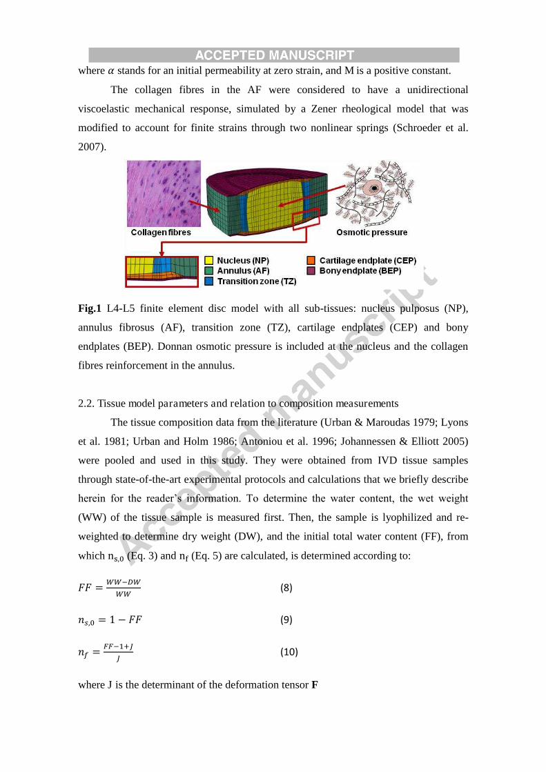
A L4-L5 IVD model including the NP, the AF, the TZ and the CEP was used
(Fig. 1) (Ruiz et al. 2013). For the NP, the TZ, and the AF, tissue constitutive models
described the poromechanical interactions between: a hyperelastic porous matrix,
saturated by intra and extra-fibrillar fluid, a swelling pressure stress simulated the
Donnan osmotic effects, and viscoelastic collagen fibres (AF only) (Roberts and Urban
2011; Schroeder et al. 2007). The total stress tensor, 時, was given by the sum of a pore
pressure component p and the effective stress of the porous solid skeleton, 時蚕讃讃: 時 噺 時蚕讃讃 伐 喧薩 (1)

where I is the identity tensor and where the pore pressure, p, is the sum of the water
chemical potential, 憲栂, and a swelling pressure term ッ講 (Schroeder et al. 2007): 喧 噺 憲栂 髪 つぱ (2)
The macroscopic stress-strain response of the solid matrix was controlled by an
initial shear modulus (罫陳), an initial solid fraction (券鎚┸待) and by the current deformation
of the homogenised poroeleastic continuum. A modified neo-Hookean model was used
to describe the finite strain behaviour of the material (Schroeder et al. 2007): 時蚕讃讃 噺 伐 怠滞 挑津岫徴岻徴 罫陳薩 釆伐な 髪 戴盤徴袋津濡┸轍匪盤貸徴袋津濡┸轍匪 髪 戴徴挑津岫徴岻津濡┸轍盤貸徴袋津濡┸轍匪鉄挽 髪 弔尿徴 岾刷 伐 蛍態 戴斑 薩峇 (3)
where J is the determinant of the deformation gradient tensor F, I is the first invariant
the left Cauchy-Green strain tensor B.
In (2), 憲栂 was related to the velocity of the interstitial fluid through the
permeability, by applying Darcy’s law. As for the osmotic pressure gradient ッ講, it was
given by the expression (Schroeder et al. 2007):
ッ講 噺 剛沈津痛迎劇嵜俵潔庁┸勅掴捗態 髪 ね磐廷賑猫禰罰廷日韮禰罰 卑態 潔勅掴痛態 崟伐 に剛勅掴痛迎劇潔勅掴痛 (4)
where 剛沈津痛 and 剛勅掴痛 are the internal and external osmotic coefficients respectively, 紘沈津痛 and 紘勅掴痛 are the internal and external activity coefficients, 潔勅掴痛 is the external
concentration of salt and 潔庁┸勅掴捗 is the proteoglycan fixed charge density that depends on
both the extra-fibrillar water (券勅掴捗) and the normal fixed charge density in mEq per
millilitre of the total fluid (潔庁), according to the expression: 潔庁┸勅掴捗 噺 津肉頂鈍津賑猫肉 (5)
with, 券勅掴捗 噺 券捗 伐 砿頂沈貢頂┸痛墜痛 (6)
where 券捗 is the total fraction of water, 砿頂沈 is a parameter that defines the intrafibrillar
water per collagen mass, and 貢頂┸痛墜痛 is the collagen content respect to the total wet
weight. The hydraulic permeability of the tissue (腔) was expressed as (Schroeder et al.
2007): 腔 噺 糠盤な 伐 券勅掴捗匪貸暢 (7)
where 糠 stands for an initial permeability at zero strain, and M is a positive constant.
The collagen fibres in the AF were considered to have a unidirectional
viscoelastic mechanical response, simulated by a Zener rheological model that was
modified to account for finite strains through two nonlinear springs (Schroeder et al.
2007).
Fig.1 L4-L5 finite element disc model with all sub-tissues: nucleus pulposus (NP),
annulus fibrosus (AF), transition zone (TZ), cartilage endplates (CEP) and bony
endplates (BEP). Donnan osmotic pressure is included at the nucleus and the collagen
fibres reinforcement in the annulus.
2.2. Tissue model parameters and relation to composition measurements
The tissue composition data from the literature (Urban & Maroudas 1979; Lyons
et al. 1981; Urban and Holm 1986; Antoniou et al. 1996; Johannessen & Elliott 2005)
were pooled and used in this study. They were obtained from IVD tissue samples
through state-of-the-art experimental protocols and calculations that we briefly describe
herein for the reader’s information. To determine the water content, the wet weight
(WW) of the tissue sample is measured first. Then, the sample is lyophilized and re-
weighted to determine dry weight (DW), and the initial total water content (FF), from
which 坦┸待 (Eq. 3) and 脱 (Eq. 5) are calculated, is determined according to:
繋繋 噺 調調貸帖調調調 (8)
券鎚┸待 噺 な 伐 繋繋 (9)
券捗 噺 庁庁貸怠袋徴徴 (10)
where J is the determinant of the deformation tensor F

As for the estimation of the proteoglycan and total collagen content, the dried
samples are digested in papain solution. Digested solutions are then used (i) to
determine the content of sulfated Glycosaminoglycans (sGAG) through Dimethyl
Methylene Blue (DMMB) assay (Farndale et al., 1986), and (ii) to achieve a measure
for collagen content according to Hydroxyproline measurements, through the
Chloramin-T assay (Huszar et al., 1980),
The initial fixed charge density 盤 題┸待匪 per per total hydrated tissue volume, from
which 題 (Eq. 5) is calculated, was calculated by using the expression (Narmoneva et al.
1999): 潔庁┸待 噺 佃迩濡頂迩濡暢調迩濡 (11)
潔庁 噺 潔庁┸待 津鈍┸轍津鈍┸轍貸怠袋徴 (12)
where 達坦, 達坦, and 達坦are the valency (2 mEq/mmol), the molecular weight (513000
g/mmol), and the concentration (in g/mL) of chondroitin sulfate, respectively, and 題┸待 is the intial water content. The sGAG content measured through the DMMB assay
is assumed to be equivalent to the chondroitin sulfate content, i.e. 達坦 is the amount of
sGAG divided by the water content of the sample. To obtain び達┸担誰担 in Eq. (6), the initial
collagen content (たg/mg DW) was estimated from hydroxyproline content by using 7.6
as the mass ratio of collagen to hydroxyproline (Sivan et al., 2006): 貢頂┸痛墜痛 噺 ガ 月検穴堅剣捲検喧堅剣健件券結 茅 ば┻は (13)
The stiffness constants and permeability were obtained in a previous study
through numerical fits of model predictions to experimental measurements (Schroeder
et al. 2008). Confined compression experiments were used to define the shear modulus
Gm (NP: Gm = 1 MPa; AF: Gm = 0.84 MPa), the positive constant M (= 1.2), and the
positive material constant 糠 (= 0.00015 mm4/Ns) of the tissue ground substance.
Uniaxial tensile tests on annulus samples were used for the parameters of the
reinforcing annulus fibres.
2.3. Transport and cell viability model
The composition-based disc model was coupled to a solute transport model
according to a sequential workflow proposed by (Malandrino et al. 2011; 2014a). On

one hand, the solute transport considered the diffusion-reaction of oxygen, lactate and
glucose:
擢擢痛嵜 系潮鉄系鎮銚頂痛系直鎮通頂崟 伐 嵜経潮鉄 ど どど 経鎮銚頂痛 どど ど 経直鎮通頂崟椛態 嵜 系潮鉄系鎮銚頂痛系直鎮通頂崟 噺 嵜 迎潮鉄迎鎮銚頂痛迎直鎮通頂崟 (14)
where 系沈, 経沈 and 迎沈 are the concentrations, tissue diffusion coefficients, and the
reactions of oxygen 岫件 噺 頚態), lactate 岫件 噺 健欠潔建) and glucose 岫件 噺 訣健憲潔) respectively.
On the other hand, tissue deformations and interstitial fluid flow were linked to
the reactive transport of these solutes through integration of the mechanically induced
changes in porosity, as calculated through the poromechanical equations. Moreover, the
effective diffusivity of each solute in the tissue (経鎚墜鎮通痛勅) was related to the solute
diffusivity in water (経栂銚痛勅追), according to the Mackie Meares model (Mackie and
Meares 1955): 拶鎚墜鎮通痛勅 噺 帖葱尼禰賑認薩岫提岻鉄 (15)
where 肯 is the total distance travelled by the solutes relative to a reference distance
between two planes of a cubic lattice defined by the molecules of the solid phase.
Comparing ions/metabolites diffusion in a porous membrane to diffusion in a liquid,
e.g. water, 肯 was reported to scale as (Mackie and Meares 1955): 肯 噺 岫怠袋津濡岻岫怠貸津濡岻 (16)
where 券鎚 is the solid fraction of the matrix, that can be expressed in terms of the water
content (券捗): 券鎚 噺 な 伐 券捗 (17)
Replacing Eq. 17 in Eq. 16, we obtained: 肯 噺 盤態貸津肉匪津肉 (18)
And replacing Eq. 18 in Eq. 15 led to the expression: 拶鎚墜鎮通痛勅 噺 磐 津肉態貸津肉卑態 経栂銚痛勅追薩 (19)

As for the metabolic reactions, the model included the system of reactions
reported by (Bibby et al. 2005):
Oxygen cell consumption 迎潮鉄 噺 伐券捗 ば┻にぱ貢頂勅鎮鎮┸鯨潮鉄 峭 系潮鉄岫喧茎 伐 ね┻ひの岻な┻ねは 髪 系潮鉄 髪 ね┻どぬ岫喧茎 伐 ね┻ひの岻嶌 (20)
Lactate production 迎鎮銚頂痛 噺 貢頂勅鎮鎮結捲喧 峙伐に┻ねば 髪 ど┻ひぬ喧茎 髪 ど┻なは系潮鉄 伐 ど┻どどのぱ盤系潮鉄匪態峩 (21)
Glucose consumption 迎直鎮通頂 噺 伐なに迎鎮銚頂痛 (22)
where 迎潮鉄 is in kPa/h, 迎鎮銚頂痛 is in nmol/(mLh), 迎直鎮通頂 is in nmol/(mLh) , 貢頂勅鎮鎮 is the cell
density of the tissue, 系潮鉄is the oxygen concentration in kPa. The pH was linked to the
lactate concentration, 系鎮銚頂痛 (in nmol/mL), by the expression (Bibby et al. 2005): 喧茎 噺 ば┻ね 髪 畦 ゲ 系鎮銚頂痛 (23)
which is a linearization of experimental results of pH decay with lactate accumulation
(Bibby et al. 2005), and 1/A = - 11.11 nmol/mL is a constant that quantifies change of
pH per unit of lactate concentration.
The cell viability was considered as the ratio 貢頂勅鎮鎮【貢頂勅鎮鎮┸待, being the initial cell
density 貢頂勅鎮鎮┸待 before any cell death occurs with tissue-specific values taken from the
literature (Malandrino et al. 2011) (Table 1). Cell density change over time was based
on the experimental study of Horner and Urban (2001), where 1 millions/mL cells in
wells start to die exponentially without glucose and when the pH is acid. Thereby, the
cell density was given by this expression: 貢頂勅鎮鎮 噺 貢頂勅鎮鎮┸待 岫伐ゎ辿 岻 (24)
where the death rate for pH (糠椎張) is constant and equal to ぬ┻ねぬ捲など貸滞 嫌貸怠, and the rate
for glucose (糠直鎮通頂) is ひ┻にぱ捲など貸滞 嫌貸怠. These constants were directly derived by curve
fitting, from the experiment of cells performed by Horner and Urban (2001) and ensured

an accurate representation of the spatiotemporal cell death patterns for cell density
values relevant to the IVD, i.e. 4x10⁶ cells/mL and 8 x10⁶ cells/mL (Malandrino et al.
2014b). The viability criteria were taken from previous studies (Horner and Urban
2001) where exponential decays start when: a) the glucose concentration decreases
below 0.5 nmol/mL, b) the pH value is below 6.8 and c) both glucose and pH are below
their critical values.
2.4. Boundary conditions
A day cycle of 8 hours of rest under 150 N compressive load and 16 hours of
activity under 800 N in compression was repeated for three days (Wilke et al. 1999).
The load was applied at the upper bony endplate while the lower bony endplate was
fixed. External pressure was nil. Oxygen, lactate and glucose concentrations were
applied at the outer surfaces of the CEP and of the AF (Fig.2a), and literature-based
values (Malandrino et al. 2011) were used (Table 1). Before simulating the effect of any
mechanical load, we initialized the transport model in other to achieve steady state
solute concentrations throughout the IVD model. Transport parameters and cell viability
were evaluated along the mid sagittal plane path: NP centre, anterior and posterior AF
(Fig. 2b).
Table 1 Transport-cell viability parameters applied: oxygen concentration (系潮鉄), lactate
concentration (系鎮銚頂痛), glucose concentration (系直鎮通頂) and cell density (貢頂勅鎮鎮). Tissue
Boundary conditions at the edge Initial condition 系潮鉄 (kPa)
系鎮銚頂痛 (nmol/mL)
系直鎮通頂 (nmol/mL)
貢頂勅鎮鎮 (106 cells mm-3)
CEP 5.1 0.8 4 0.0135
AF 5.8 0.9 5 0.00555

Fig.2 a) Solute boundary conditions applied at the surfaces of the annulus (AF) and
cartilage endplates (CEPs) and b) Points selected for nutrition-cell viability evaluation:
posterior annulus centre (left side), nucleus centre and anterior annulus centre (right
side).
2.5. Design of experiment
A design of experiment (DOE) using a fractional factorial statistical method was
performed, and variations were defined for the following biochemical parameters of the
composition-based AF and NP models (Eqs. 4, 5 and 6): initial water content (券庁待),
initial fixed charge density (潔庁待) and collagen content (貢頂┸痛墜痛) (Table 2). For each
parameter, variations in the DOE consisted in a switch between an upper level and a
lower level parameter value. While upper level values corresponded to the composition
data measured for Thompson grade I IVDs, lower level values corresponded to grade III
IVDs (Thompson et al. 1990).
Table 2 Design of experiment parameters and values: annulus initial water content ( 題待
AF), annulus collagen content (び達┸担誰担 AF), nucleus initial water content (券庁待 NP),
nucleus initial fixed charge density (潔庁待 NP) and nucleus collagen content (貢頂┸痛墜痛 NP).
Level 券庁待 AF
(*)
貢頂┸痛墜痛 AF
(**)
券庁待 NP
(*)
潔庁待 NP
(mEq/mL)
貢頂┸痛墜痛 NP
(**)
Grade I 0.75 0.65 0.80 0.30 0.150
Grade III 0.70 0.78 0.76 0.23 0.285
* Fraction of wet weight ** Fraction of dry weight
A fractional factorial analysis of に泰貸怠 噺 なは runs was used, representing a
resolution V. In fractional factorial designs, such a resolution allows thorough
identification of the main effects, without any confusion with multiple factor
interactions. The significance of each parameter effect was analyzed statistically
(Minitab Inc.) using an analysis of variance (ANOVA) with a significance
threshold 糠 噺 ど┻どの. The standardized effect 劇怠 was calculated using the following
equation: 劇怠 噺 系頚継繋繋怠系頚継繋繋聴帳 (25)
where 系頚継繋繋怠 is the effect coefficient of the parameter and 系頚継繋繋聴帳 is the standard
error of the coefficient.
2.5. Convergence analysis and model validation under creep
The mesh convergence of the model was verified based on a comprehensive
convergence study performed for the mesh template of the IVD model, including
assessment of the stability of the poromechanical predictions (Ruiz et al. 2013).
However, the transport model was sensitive to the time step selected for the
calculations. As such, an additional convergence study was performed for the time
discretisation of the transport model, so as to obtain the best compromise between
accuracy of the results and computational cost at the different time points selected for
the DOE. In order to validate the composition-based model under compression, the disc
height reductions achieved after creep were compared to those reported in the literature
after the experimental boundary conditions were simulated.
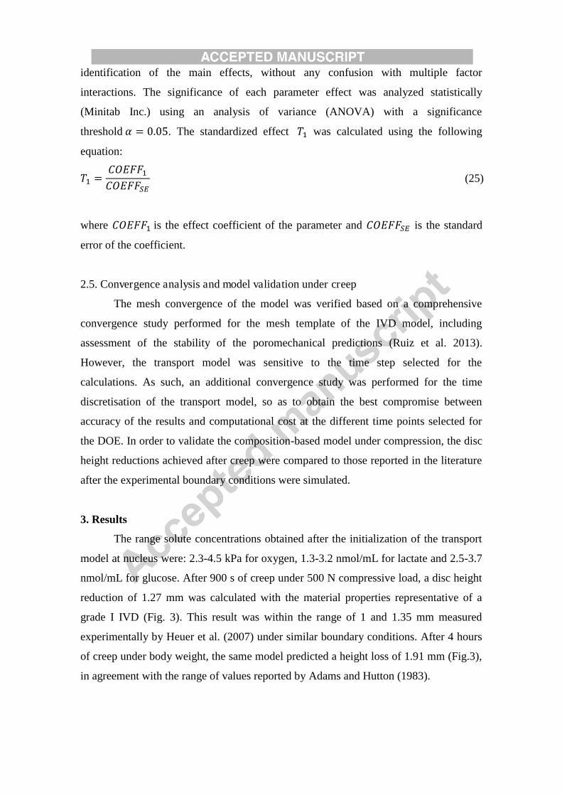
3. Results
The range solute concentrations obtained after the initialization of the transport
model at nucleus were: 2.3-4.5 kPa for oxygen, 1.3-3.2 nmol/mL for lactate and 2.5-3.7
nmol/mL for glucose. After 900 s of creep under 500 N compressive load, a disc height
reduction of 1.27 mm was calculated with the material properties representative of a
grade I IVD (Fig. 3). This result was within the range of 1 and 1.35 mm measured
experimentally by Heuer et al. (2007) under similar boundary conditions. After 4 hours
of creep under body weight, the same model predicted a height loss of 1.91 mm (Fig.3),
in agreement with the range of values reported by Adams and Hutton (1983).
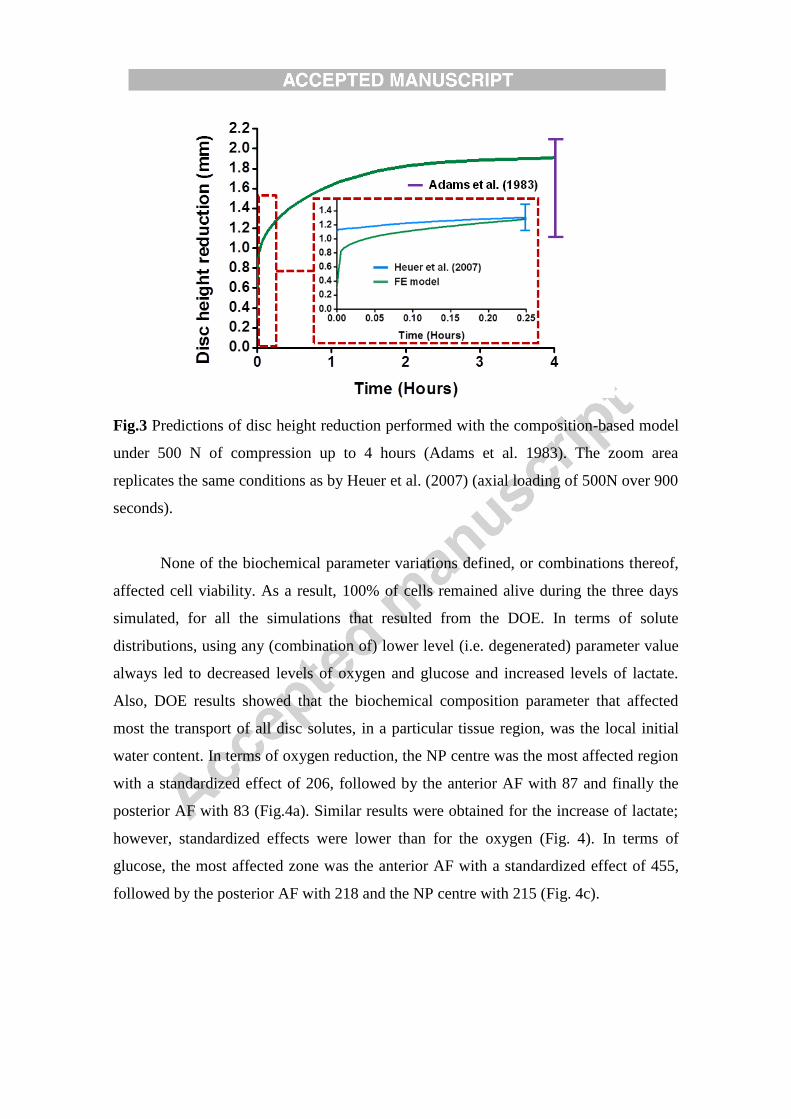
Fig.3 Predictions of disc height reduction performed with the composition-based model
under 500 N of compression up to 4 hours (Adams et al. 1983). The zoom area
replicates the same conditions as by Heuer et al. (2007) (axial loading of 500N over 900
seconds).
None of the biochemical parameter variations defined, or combinations thereof,
affected cell viability. As a result, 100% of cells remained alive during the three days
simulated, for all the simulations that resulted from the DOE. In terms of solute
distributions, using any (combination of) lower level (i.e. degenerated) parameter value
always led to decreased levels of oxygen and glucose and increased levels of lactate.
Also, DOE results showed that the biochemical composition parameter that affected
most the transport of all disc solutes, in a particular tissue region, was the local initial
water content. In terms of oxygen reduction, the NP centre was the most affected region
with a standardized effect of 206, followed by the anterior AF with 87 and finally the
posterior AF with 83 (Fig.4a). Similar results were obtained for the increase of lactate;
however, standardized effects were lower than for the oxygen (Fig. 4). In terms of
glucose, the most affected zone was the anterior AF with a standardized effect of 455,
followed by the posterior AF with 218 and the NP centre with 215 (Fig. 4c).
Fig.4 Standardized effect of disc composition parameters variation from grade I to
grade III, after 3 days of simulation, on oxygen, lactate and glucose concentrations at: a)
NP centre, b) posterior AF and c) anterior AF (Dashed horizontal line is the threshold of
significance).
Analysing the solute distributions along the mid-sagittal plane after 3 days, the
16 models created led to a reduced number of groups of results in terms of regional
solute concentration (Fig. 5). In the AF, four groups were unequivocally defined (Fig.
5a) according to different combinations of initial water contents in the different IVD
subtissues, e.g. per order of decreasing glucose concentrations:
Group 1: 券庁待 AF and 券庁待 NP representative of grade I
Group 2: 券庁待 AF representative of grade I and 券庁待 NP representative of grade III
Group 3: 券庁待 AF representative of grade III and 券庁待 NP representative of grade I
Group 4: 券庁待 AF and 券庁待 NP representative of grade III
In the NP, two groups of results were identified, as illustrated in Fig. 5b in terms
of evolution of the oxygen content over 3 days at the centre of the tissue. The group
with high oxygen concentration corresponded to simulations with a NP initial water
content representative of grade I, i.e. simulations from the Groups 1 and 3 defined

above. In contrast, the group with lower oxygen concentrations gathered the simulation
results with NP initial water content representative of grade III (Groups 2 and 4).
Fig.5 Effect of initial water content 岫券庁待岻 variations on IVD solute concentrations: a)
glucose at the sagittal plane (day 3). The figure shows the groups of results formed
according to the initial water contents of the subtissues; group 1: both 券庁待 AF and 券庁待 NP of grade I, group 2: 券庁待 AF of grade I and 券庁待 NP of grade III, group 3: 券庁待 AF
of grade III and 券庁待 NP of grade I and group 4: both 券庁待 AF and 券庁待 NP of grade III
and b) oxygen at NP centre along three days of simulation. Two groups of results are
identified; one with high oxygen content corresponding to simulations with 券庁待 NP of
grade I and another group with lower oxygen content corresponding to the results with 券庁待 NP of grade III.
In addition to the direct effects of local composition changes, the ANOVA
results also showed that the glucose content in the anterior AF was strongly influenced
by the initial water content in the NP (Fig. 6a). However in the posterior AF, the
glucose distribution was significantly affected by both the initial water content and the
fixed charge density of the NP, and the effect of the local collagen was relatively limited
(Fig. 6b).

Fig.6 Evolution of the effect of composition parameter variation of the nucleus (券庁待 NP, 潔庁待 NP and 貢頂┸痛墜痛 NP) and annulus (貢頂┸痛墜痛 AF) on glucose concentration during a day at:
a) the anterior AF and b) the posterior AF. (Dashed horizontal line is the threshold of
significance)
All the biochemical changes simulated in this study affected the diurnal height
loss after three days. Both at the NP centre and in the posterior AF, the initial fixed
charge density of the NP was the parameter that influenced the most the disc height
(Fig. 7). For the anterior AF, the AF initial water content was the most relevant
parameter followed by the initial fixed charged density of the NP.
Fig.7 Effect of disc composition parameters (annulus initial water content (券庁待 AF),
annulus collagen content (貢頂┸痛墜痛 AF), nucleus initial water content (券庁待 NP), nucleus
initial fixed charge density (潔庁待 NP), and nucleus collagen content (貢頂┸痛墜痛 NP)) variation
on daily disc height: a) after 3 days simulated and b) at the sagittal plane (day 3).
(Dashed horizontal line is the threshold of significance)

4. Discussion
The osmo-poroviscoelastic formdulation used in this study was duly verified
against thermodynamical consistency (Huyghe et al. 2009). At the tissue level, the
model was also validated, with parameter values that represented healthy tissues
(Schroeder et al. 2007). In terms of organ validation, the amount of creep measurements
available in the literature is limited. In the report of their experimental study, Adams
and Hutton (1983) mentioned that the compressive boundary load used corresponded to
the body weight, but they did not give any specific value. As such, we took a generic
value of 500 N of compressive load (Wilke et al. 1999) in order to replicate their creep
study. Assuming that this approximation is acceptable, our results showed that the our
healthy model extrapolated reasonably up to 4 hours the simulated behaviour for 15
minutes creep, validated against the measurements reported by Heuer et al. (2007).
Regarding the validity of the degenerated models, our study did not aim to
simulate a degenerated disc, but aimed to identify those composition changes that might
affect the nutrition of the cells along degeneration, based on biochemically measured
quantities. Hence, the variations of phenomenological tissue model parameters such as
the stiffness moduli and the permeability were not taken into account. This
approximation allowed us focusing our design of experiments on the composition, and
was supported by a previous sensitivity analysis reported by Malandrino et al. (2011):
by using a similar mechano-transport model, the authors showed that the porosity and
the osmotic pressure were the osmo-poromechanical parameters that mostly affected the
nutrient transport, being the effect of the stiffness and the permeability parameters
negligible. In order to prepare the present study, we have confirmed this outcome
through a previous sensitivity analysis that included 糠 and 罫陳, according to Eqs 7, and
3, respectively (results not shown). Hence, our exploration of the effect of composition
changes on the transport of nutrients effectively targeted those tissue model
components, i.e. the porosity and the osmotic pressure, most relevant to the metabolic
transport calculations.
In our simulations, solute contents were always decreased with degenerative
composition changes, but the minimum glucose and pH values were 0.76 nmol/mL and
6.90 respectively, which did not exceed the critical values to see cell death (Horner and
Urban 2001). Accordingly, cell viability remained 100% for all the runs performed

during the three days simulated. Glucose and pH thresholds used for cell viability came
from in vitro experiments (Horner and Urban 2001). It is difficult to know whether
these thresholds are valid in vivo, since no related investigations have been reported so
far. However, these criteria have been already used in many numerical studies that have
explored the possible role of nutrition in disc degeneration. Malandrino et al. (2014a)
used a 3D mechano-transport model to study the mechanical effects on cell viability via
transport variations, depending on simulated CEP calcification, AF and NP tissue
degeneration, and disc height reduction. Though their tissue constitutive modelling was
different from ours, their results supported our findings that moderate AF and NP
degeneration only is unlikely to lead to critical glucose levels under physiological load
magnitudes and for generic disc geometries. Moreover, the calculations of Malandrino
et al. (2014) suggested that simulating CEP calcification (50%) was necessary in order
to predict cell death, the latter occurring because of an increased lack of glucose in the
inner AF. These results were qualitatively similar to those achieved by Zhu et al. (2012)
with a slightly different cell viability function, in which pH was not an explicit trigger
of cell death and where cell death rate (i.e. 糠直鎮通頂 in Eq. 18) was a nonlinear function of
the current glucose concentration. In all these studies, the experiment-based critical
glucose concentration of 0.5 mM was the most important assumption for cell death
predictions, and all results suggested that nutrition-induced cell death might not be
linked to AF and NP degeneration. While our simulations used a similar assumption,
and led to a similar outcome, our model allowed a refined control of the specific
degeneration changes that may alter cell nutrition in the different tissue.
Supporting to our results, several simulation reports pointed out that the glucose
concentration in inner AF is easily affected by any simulated degenerative change
(Galbusera et al. 2011; Zhu et al. 2012; Malandrino et al. 2014a). This phenomenon was
largely attributed to the strong local consolidation of the TZ regions, due to the lateral
pressure exerted on the fibre-reinforced AF by the lateral expansion of the NP under
mechanical loads (Ruiz et al. 2013). Among the different mechanotransport - cell
viability studies published, our full 3D simulations can be compared directly to the
work reported by (Malandrino et al. 2011; 2014a), with the same model geometry.
Whilst using a porohyperelastic constitutive model with a fixed osmotic pressure in the
NP, these authors always identified the inner anterior AF as the disc region where
glucose concentration was mostly reduced. In contrast, we predicted that the cumulated
effect of water and proteoglycan loss within the NP was particularly critical to the
availability of glucose in the posterior AF. Interestingly, this issue seemed to arise from
the particularly high impact of NP proteoglycan loss on disc height reduction and
increased tissue consolidation in the TZ of the posterior AF area. Acknowledging that
limited glucose can trigger inflammatory and catabolic responses by disc cells
(Neidlinger-Wilke et al. 2012), we may infer that early NP degeneration might
contribute to weaken biochemically the posterior AF due to local nutrition issues.
Confirmation of this possible non-mechanical weakening would require, however,
experimental data about disc cell catabolic activity for different glucose contents
between 0.5 and 5 mM.
The composition-based model reproduced the expected disc height reduction
under compression changes due to ECM degeneration, i.e. but we did not consider
permanent disc height loss. Such a consideration could have modified slightly the
computed influence of the diffusion distances (Galbusera et al. 2011; Malandrino et al.
2011). However, this issue is not expected to have a major impact on the current study,
since the influence of tissue consolidation is probably predominant, for our particular
disc model geometry, as suggested by Malandrino et al. (2011) and further discussed in
the next paragraph. Endplate sclerosis was not considered either. Whereas Zhang et al.
(2008) considered that this condition rarely appears in grade III discs, Benneker et al.
(2005) reported that sclerosis in grade III discs is frequently found. As mentioned
earlier, several authors of numerical studies have simulated CEP sclerosis, but the way
to do it is not clear, especially for a specific grade, and exploring such simulation
hypothesis is beyond the scope of this paper.
Overall, the initial water content is the parameter of disc composition that
affected most the transport of solute at the end of the three days simulated. The most
affected solute was glucose, especially in the anterior AF. This trend did not change
when an ANOVA was performed at the end of each simulated day. The predominant
influence of water content is supported by earlier sensitivity studies where a decrease in
porosity was reported to affect the transport of oxygen and lactate, more than any other
poromechanical parameters did (Malandrino et al. 2011). Simulated composition
changes led to disc height reductions in the mechanically loaded disc models, which
reduced the effective diffusion distances. However, our results revealed that without any

initial disc height reduction in an unconsolidated state, the possible benefits of the
mechanically induced height reduction for diffusion are largely countered by increased
tissue consolidation, i.e. decreased current porosity. A similar outcome was suggested
by a parameterization of the porosity and diffusion coefficients proposed by Galbusera
et al. (2011).
We should, however, recall that our diffusion coefficients were porosity-
dependent according to the use of the Mackie-Meares diffusivity law (Eq. 19). This law
has been extensively used for several studies on cartilage and intervertebral disc
(Maroudas 1968; Frank et al. 1990; Lanir et al. 1998), and seems physically reasonable
especially due to likely constrictivity effects (Holzer et al. 2013). Yet, recent
measurements showed that the model of Mackie and Meares could slightly
underestimate experimental diffusivities (Gu et al. 2004). However, these measurements
were performed in agarose gels and not in extracted tissues. Other strain-dependent
diffusivity laws have been proposed (Zhu et al. 2012), and how they influence
predictions in comparison to the Mackie and Meares law would have to be assessed.
Meanwhile, because of the lack of robust experimental studies on disc tissues
diffusivities in vitro or in vivo, exploiting the Mackie-Meares model stands for a
pragmatic and physically sound approach.
Eq. (1) assumes that the solid phase of the matrix is incompressible for all disc
tissues, which implies that the volumetric changes of the poroelastic continuum are due
to the gain or loss of water when the tissue deforms. Should the solid phase be
compressible, our calculations would overestimate the effects of the volumetric
deformations on the diffusion of solutes in the compressed tissue regions. Furthermore,
considering solid phase compressibility would modify the relation between diffusion
distance and porosity changes, and the positive effect of diffusion distance reductions
could become relatively stronger under simulated compression. Yet, it is worth to
mention that the solid phase of the NP and AF matrices are rich in proteoglycans: the
latter can represent up 85% of the solid phase in the NP (Urban and Roberts 2003).
Hence, the steric and electric repulsion forces in and between the glycosaminoglycan
aggregates at the nanoscale (Dean et al. 2006; Han et al. 2007), might favour
incompressibility of proteoglycan-rich phases at the microscale. Also, the swelling
pressure generated by the proteoglycan negative fixed charges, pre-tenses the collagen

fibres and increases the volumetric stiffness of the proteoglycan phase. In our
simulations, this swelling pressure is a spherical stress tensor that pre-stresses the solid
phase. Hence, we implicitly simulate a hydrated and turgid mix of collagen and
proteoglycans at the nanoscale that is likely to appear as a nearly incompressible phase
at the scale of several tens of microns.
Though no quantitative measurements are available to our knowledge to test the
effective compressibility of the modelled solid phase, analogies with articular cartilage
exist in terms of composition and multiphysics interactions among tissue constituents.
Miller and Morgan (2010) analysed the results of micro and macro compression tests of
articular cartilage and found that the micro- and macro-scale poroelastic properties of
the tissue were consistent when derived from a formulation based on Eq. (1). This
outcome suggested that the collagen-proteoglycan hydrated matrix could be reasonably
considered as incompressible at the microscale. At the macroscopic scale, the use of a
poromechanical theory similar to ours could reproduce the response of the articular
cartilage for a variety of experiments, i.e. confined and unconfined compression, stress
relaxation, indentation, swelling (Wilson et al. 2006).
As anticipated earlier, our simulated degenerative changes in the NP may affect
the AF nutrition. In the anterior AF, the effect of 券庁待 in the NP was 39 at the end of one
simulated day. This standardized effect was higher than the separate effect of each NP
parameter on the glucose concentration in the posterior AF, i.e. 21 for 券庁待, 17 for 潔庁待
and 3 for 貢頂┸痛墜痛. However, combining the respective effects of all NP parameters at the
posterior AF gave a cumulative effect of 41, slightly higher than the global effect of NP
alterations on the anterior AF. According to measurements (Iatridis et al. 2007), NP
desiccation and proteoglycan depletion would be concomitant in mildly to moderately
degenerated discs, and our findings suggest that such a situation exposes particularly the
posterior AF. According to the hypothesis of possible biochemical AF weakening made
earlier, such impact of NP alteration on the AF may contribute to explain
mechanistically why radial tears in the posterior AF mostly appear in a moderately to
severely discs (Osti et al. 1992). Interestingly, calculations indicated that NP
dehydration alone affected both the posterior and anterior AF. Here, it becomes worth to
mention the possible effect of any early loss of CEP functionality that might contribute
to reduce the NP water content by altering the balance of water in-flow and out-flow

along daily loading (Ayotte et al. 2001). Hence, assuming that early NP dehydration is
possible, our simulations raise the question whether nutrition issues could play role in
the occurrence of circumferential tears, already present in both the anterior and posterior
AF of normal to moderately degenerated discs (Osti et al. 1992).
Fig.8 Comparative composition parameters for grade I and grade III discs: a) AF water
content (%), b) NP water content (%), c) AF collagen content (% dry weight), d) NP
collagen (% dry weight), e) AF fixed charge density (mEq/mL), and f) NP fixed charge
density (mEq/mL). Literature data referred to grade III IVD on the Thompson scale
(Lyons et al. 1981; Urban and Holm 1986; Antoniou et al. 1996; 2004; Johannessen and
Elliott 2005), while in-house measurements referred to grade III IVD on the Pfirrmann
scale (Dao et al. 2014).
Fig. 8 shows the mean composition parameters used in this study, in comparison
to individual measurements reported in the literature. For the degenerated discs,
literature data referred to grade III IVDs on the Thompson scale, which is different from
the MRI-based Pfirrmann scale usually used in clinics. In-house biochemical
assessments of tissue samples from seven human discs previously graded as grade III on
the Pfirrmann scale (Dao et al. 2014) revealed that the water content and fixed charge
density used in the model represented mildly to moderately degenerated discs,
according to common clinical evaluations (Fig. 8a,b,e,f). As for the collagen content,
the value used in the model seems more similar to an advance grade III/early grade IV
disc on the Pfirrmann scale (Fig. 8c,d). However, our results showed that changes in the
collagen content did not affect the solute concentrations, and only slightly affected the
disc height reduction.
This study has the following limitations: first, the three days chosen for the
simulations might not be enough for reaching a steady-state for a cell viability study;
although we found that a steady-state convergence of two days was enough to observe
cell death with a calcified CEP (data not shown) (Malandrino et al. 2014a). Second, we
did not simulate dynamic load variations around the mean load value chosen for day
activity. Although these loads might limit the loss of water (Schmidt et al. 2010) and
could favour the transport of nutrients (Zhu et al. 2012), simulating dynamic loads
would have increased the computational cost, and earlier studies (Zhu et al. 2012)
suggest that it would not have changed our interpretations. Third, multiaxial loads were
not considered in this study. Nevertheless, under external axial compression, most of
disc regions are subjected to compression. For the regions that present combined
tension-compression, it has been reported that tension-compression nonlinearities affect
the estimation of interstitial velocities (Huang et al. 2001; Huang et al. 2003), and have
little effect on the transport of small solutes, e.g. glucose and oxygen (Huang and Gu
2007). As such, our mechano-transport predictions would not be affected by not
considering such nonlinearities. Fourth, we did not consider any local variations in
proteoglycan concentration and total amount of collagen within each modelled tissue.
Though such a variation could have better informed the suggested sensitivity of the
posterior AF in relation to the anterior AF, we have based our model on specific
degeneration-specific composition measurements, made available for this study. Fifth,
we did not consider the limitation of Eq. 3 to capture the hysteresis presented by
cartilaginous tissues. The Eq. 3 represents the elastic response of the solid phase and it
does not incorporate any term for energy dissipation. Effects of Boltzmann
superposition principle are expected to appear from the viscoelastic collagen fibres.
However, experiments support the idea that energy dissipation in cartilage matrix is
controlled by the fluid flows, captured by poroelastic simulations (Han et al. 2011). As
such, integration of Eq. 3 into Eq. 1 is expected to give the capability to represent the
NP response to cyclic loads. The predominance of poromechanical time effects in disc
tissues was supported by experimental tests on IVD specimens (Costi et al. 2008).
However, no direct validation of our tissue models was reported for cyclic loads.
Finally, the effect of other important factors related to disc nutrition, e.g. inflammatory
factors (Lotz and Ulrich 2006) was not considered, while necessary to assess whether
the local nutrient deprivation calculated can weaken the tissues.
5. Conclusions
A composition-based model coupled to a transport-cell viability model was
presented as tool to explore the influence of measured ECM changes on disc nutrition
and cell viability. This study suggests that small degenerative ECM changes may
produce significant solutes alterations. While these changes between grade I and grade
III degeneration did not seem relevant to nutrition-related cell viability, they allowed
identifying possible mechanisms related to known AF alterations along degeneration.
The computational approach developed provides a powerful tool to achieve improved
understanding of disc degenerative mechanisms. In particular, our results suggest that
the biological and tissue/disc structure changes, able to provoke NP dehydration
independently on the further proteoglycan depletion, should be tackled, in order to
explore the pathogenesis of the disc in a more integrated way. Obviously, the effect of
other important factors related to disc nutrition, e.g. inflammatory factors (Lotz and
Ulrich 2006) would be necessary. Our simulations provide, however, a robust
framework for such kind of development. The presented model could also be highly
relevant to address the challenge of achieving designs of biomimetic IVD implants or
scaffold materials that ensure proper mechanical/multiphysics responses in disc
regenerative therapies (Noailly et al. 2014).
Acknowledgements
Financial funding from the European Commission (My SPINE-269909) and the
Agència de Gestió d'Ajuts Universitaris i de Recerca (AGAUR-2012BE100979) are
acknowledged.
References
Adams, M.A., Hutton, W.C., 1983. The effect of posture on fluid content of lumbar
intervertebral discs. Spine 8(6), 665-671.
Antoniou, J., Steffen, T., Nelson, F., Winterbottom, N., Hollander, A.P., Poole, R.A.,
Aebi, M., Alini, M., 1996. The human lumbar intervertebral disc: evidence for changes
in the biosynthesis and denaturation of the extracellular matrix with growth, maturation,
ageing, and degeneration. J. Clin. Invest. 98(4), 996-1003.
Antoniou, J., Demers, C. N., Beaudoin, G., Goswami, T., Mwale, F., Aebi, M., Alini,
M., 2004. Apparent diffusion coefficient of intervertebral discs related to matrix
composition and integrity. Magn Reson Imaging, 22(7), 963-972.
Ayotte, D.C., Ito, K., Tepic, S., 2001. Direction-dependent resistance to flow in the
endplate of the intervertebral disc: an ex vivo study. J. Orthop. Res. 19, 1073-1077.
Battié, M.C., Videman, T., Levalahti, E., Gill, K., Kaprio, J., 2007. Heritability of low
back pain and the role of disc degeneration. Pain 133, 272-280.
Benneker, L., Heini, P., Anderson, S., Alini, M., Ito, K., 2005. Correlation of
radiographic and MRI parameters to morphological and biochemical assessment of
intervertebral disc degeneration. Eur. Spine J. 14, 27-35.
Bibby, S.R., Jones, D.A., Ripley, R.M., Urban, J.P., 2005. Metabolism of the
intervertebral disc: effects of low levels of oxygen, glucose, and pH on rates of energy
metabolism of bovine nucleus pulposus cells. Spine 30, 487-496.
Costi, J.J., Stokes, I.A., Gardner-Morse, M.G., Iatridis, J.C., 2008. Frequency-
dependent behaviour of the intervertebral disc in response to each of six degree of
freedom dynamic loading: Solid phase and Fluid phase contributions. Spine 33(16),
1731-1738.
Dao, T.T., van Rijsbergen, M., Pouletaut, P., Charleux, F., Ito, K., Ho Ba Tho, M.C.,
2014. In vitro Assessment of Micro-structural Properties of Intervertebral Disc using
1.5T Magnetic Resonance T2 and ADC Mappings. In: 7th World Congress of
Biomechanics, July 6-11, 2014, Boston, Massachusetts, USA.
Dean, D., Han, L., Grodzinsky, A.J., Ortiz, C., 2006. Compressive nanomechanics of
opposing aggrecan macromolecules. J Biomech. 39(14), 2555-2565.
Farndale, R.W., Buttle, D.J., Barrett, A. J., 1986. Improved quantitation and
discimination of sulphated glycosaminoglycans by use of dimethylmethylene blue.
Biochim. Biophy. Acta. 883, 173-177.
Frank, E.H., Grodzinsky, A.J., Phillips, S.L., Grimshaw, P.E., 1990. Physicochemical
and Bioelectrical Determinants of Cartilage Material Properties. Biomech. Diarthrodial.
Joints., 261-282.
Galbusera, F., Mietsch, A., Schmidt, H., Wilke, H.J., Neidlinger-Wilke, C., 2011. Effect
of intervertebral disc degeneration on disc cell viability: a numerical investigation.
Comput. Methods Biomech. Biomed. Engin. 16(3), 328-337.
Gu, W.Y., Yao, H., Vega, A.L., Flagler, D., 2004. Diffusivity of ions in agarose gels
and intervertebral disc: effect of porosity. Ann. Biomed. Eng. 32, 1710-1717.
Guilak, F., Ting-Beall, H.P., Baer, A.E., Trickey, W.R., Erickson, G.R., Setton. L.A.,
1999. Viscoelastic properties of intervertebral disc cells. Spine 24, 2475-2486.
Han, L., Dean, D., Mao, P., Ortiz, C., Grodzinsky, A.J., 2007. Nanoscale shear
deformation mechanisms of opposing cartilage aggrecan macromolecules. Biophys. J.
93(5), L23-L25.
Han, L., Frank, E.H., Greene, J.J., Lee, H.Y., Hung, H.H.K., Grodzinsky, A.J., Ortiz,
C., 2011. Time-dependent nanomechanics of cartilage. Biophys. J. 100(7), 1846-1854.
Heuer, F., Schmitt, H., Schmidt, H., Claes, L., Wilke, H.J., 2007. Creep associated
changes in intervertebral disc bulging obtained with a laser scanning device. Clin.
Biomech. 22, 737-744.

Holzer, L., Wiedenmann, D., Münch, B., Keller, L., Prestat, M., Gasser, Ph., Robertson,
I., Grobéty, B., 2013. The influence of constrictivity on the effective transport
properties of porous layers in electrolysis and fuel cells. J. Mater. Sci. 48(7), 2934-2952.
Horner, H.A., Urban, J.P., 2001. Effect of nutrient supply on the viability of cells from
the nucleus pulposus of the intervertebral disc. Spine 26, 2543-2549.
Huang, C.Y., Mow, V.C., Ateshian, G.A., 2001 The Role of Flow-Independent
Viscoelasticity in the Biphasic Tensile and Compressive Responses of Articular
Cartilage. J. Biomech. Eng. 123(5), 410-417.
Huang, C.Y., Soltz, M.A., Kopacz, M., Mow, V.C., Ateshian, G.A., 2003. Experimental
verification of the roles of intrinsic matrix viscoelasticity and tension-compression
nonlinearity in the biphasic response of cartilage. J. Biomech. Eng. 125(1), 84-93.
Huang, C.Y., Gu, W.Y., 2007. Effects of tension-compression nonlinearity on solute
transport in charged hydrated fibrous tissues under dynamic unconfined compression. J.
Biomech. Eng. 129(3), 423-429.
Huszar, G., Maiocco, J., Naftolin, F., 1980. Monitoring of collagen and collagen
fragments in chromatography of protein mixturs. Anal. Biochem. 105(1), 424-429.
Huyghe, J.M., Wilson, W., Malakpoor, K., 2009. On the thermodynamical admissibility
of the triphasic theory of charged hydrated tissues. J. Biomech. Eng., 131(4), 245-258.
Iatridis, J., MacLean, J., O’Brien, M., Stokes, I., 2007. Measurements of proteoglycans
and water content distribution in human lumbar intervertebral discs. Spine 32(14) 1493-
1497.
Johannessen, W., Elliott, D.M., 2005. Effects of degeneration on the biophasic material
properties of human nucleus pulposus in confined compression. Spine 30(24), 724-729.
Lanir, Y., Schneiderman, R., Huyghe, J.M., 1998. Partition and diffusion of sodium and
chloride ions in soft charged foam: the effect of external salt concentration and
mechanical deformation. Tissue Eng. 4(4), 365-379.
Lyons, G., Eisenstein, S.M., Sweet, M.B., 1981. Biochemical changes in intervertebral
disc. Biochim. Biophys. Acta 673(4), 443-453.

Lotz, J., Ulrich, J.A., 2006. Innervation, inflammation and hypermobility may
characterize pathologic disc degneration: review of animal model data. J. Bone. Joint.
Surg. Am. 88(2), 76-82.
Mackie, J., Meares, P., 1955. The diffusion of electrolytes in a cation-exchange resin
membrane. Proceedings of the Royal Society of London. Series A, Mathematical and
Physical Sciences 232, 498-518.
Malandrino, A., Noailly, J., Lacroix, D., 2011. The Effect of Sustained Compression on
Oxygen Metabolic Transport in the Intervertebral Disc Decreases with Degenerative
Changes. PLoS Comput Biol 7(8).
Malandrino, A., Noailly, J., Lacroix, D., 2014a. Numerical exploration of the combined
effect of nutrient supply, tissue condition and deformation in the intervertebral disc. J.
Biomech. 47, 607-762.
Malandrino, A., Pozo, J.M., Castro-Mateo, I., Frangi, A., van Rijsbergen, M., Ito, K.,
Wilke, H.J., Dao, T.T., Ho Ba Tho, M.C., Noailly, J., 2014b. On the relative relevance
of subject-specific geometries and degeneration-specific mechanical properties for the
study of cell death in human intervertebral disc models. Front. Bioeng. Biotechnol. 3(5),
1-15.
Maroudas, A., 1968. Physicochemical properties of cartilage in the light of ion
exchange theory. Biophys. J. 8(5), 575-595.
Miller, G.J., Morgan, E.F., 2010. Use of microindentation to characterize the
mechanical properties of articular cartilage: comparison of biphasic material properties
across length scales. Osteoarthr. Cartil. 18(8), 1051-1057.
Narmoneva,D.A.,Wang,J.Y.,Setton,L.A.,1999.Nonuniform swelling-induced residual
strains in articular cartilage. J.Biomech.32, 401–408.
Neidlinger-Wilke, C., Mietsch, A., Rinkler, C., Wilke, H.J., Ignatius, A., Urban, J.,
2012. Interactions of environmental conditions and mechanical loads have influence on
matrix turnover by nucleus pulposus cells. J. Orthop. Res. 30, 112-121.
Noailly, J., Malandrino, A., Galbusera, F., 2014. Computational modelling of spinal
implants, in J. Zhongmin (Ed.), Computational Modelling of Biomechanics and

Biotribology in the Musculoskeletal System. Woodhead Publishing Ltd., Cambridge,
pp. 447–484.
Osti, O., Vernon-Roberts, B., Moore, R., Fraser, R.D., 1992. Annular tears and disc
degeneration in the lumbar spine. J. Bone. Joint. Surg. Br. 74(5), 678-682.
Pfirrmann, C.R.W., Metzdorf, A., Zanetti, M., Hodler, J., Boos, N., 2001. Magnetic
resonance classification of lumbar interverterbal disc degeneration. Spine 26, 1873-
1878.
Roberts, S., Urban, J.P.G., 2011. Intervertebral discs, in: H. Riihimäki & E. Viikari-
Juntura (Eds.), Musculoskeletal system, in: J.M. Stellman (Eds.) Encyclopedia of
Occupational Health and Safety. International Labor Organization, Geneva.
Ruiz, C., Noailly, J., Lacroix, D., 2013. Material property discontinuities in
intervertebral disc porohyperelastic finite element models generate numerical
instabilities due to volumetric strain variations. J. Mech. Behav. Biomed. Mater. 26, 1-
10.
Schmidt, H., Shirazi-Adl, A., Galbusera, F., Wilke, H.J., 2010. Response analysis of the
lumbar spine during regular daily activities--a finite element analysis. J. Biomech.
43(10), 1849-1856.
Schroeder, Y., Sivan, S., Wilson, W., Merkher, Y., Huyghe, J., Maroudas, A., Baaijens,
F.P.T., 2007. Are disc pressure, stress, and osmolarity affected by intra and extrafibrillar
fluid exchange?. J. Orthop. Res. 25, 1317-1324.
Setton, L.A., Chen, J., 2004. Cell mechanics and mechanobiology in the intervertebral
disc. Spine 29, 2710-2723.
Sivan, S., Merkher, Y., Wachtel, E., Ehrlich, S., Maroudas, A., 2006. Correlation of
swelling pressure and intrafibrillar water in young and aged human intervertebral discs.
J. Orthopaed. Res. 24(6), 1292-1298.
Thompson, J.P., Pearce, R.H., Schechter, M.T., Adams, M.E., Tsang, I.K.Y., Bishop,
P.B., 1990. Preliminary evaluation of a scheme for grading the gross morphology of the
human intervertebral disc. Spine. 15(5), 411-415.
Urban, J.P., Maroudas, A., 1979. The measurement of fixed charge density in the
intervertebral disc. Biochim. Biophys. Acta 586, 166-178.
Urban, J.P., Holm, S.H., 1986. Intervertebral disc nutrition as related to spinal
movements and fusion, in A Hargens ed., Tissue Nutr. Viability: New York, NY,
Springer-Verlag, 101-119.
Urban, J.P.G., Roberts, S., 2003. Degeneration of the intervertebral disc. Arthritis Res.
Ther. 5(3), 120-130.
Urban, J.P.G., Smith, S., Fairbank, J.C.T., 2004. Nutrition of the intervertebral disc.
Spine 29, 2700-2709.
Wilke, H.J., Neef, P., Caimi, M., Hoogland, T., Claes, L.E., 1999. New in vivo
measurements of pressures in the intervertebral disc in daily life. Spine 24, 755-762.
Wilson, W., Huyghe, J.M., van Donkelaar, C.C., 2006. A composition-based cartilage
model for the assessment of compositional changes during cartilage damage and
adaptation. Osteoarthr. Cartil. 14(6), 554-560.
Zhang, Y., Zhao, C., Jiang, L., Chen, X., Dai, L., 2008. Modic changes: a systematic
review of the literature. Eur. Spine J. 17, 1289-1299.
Zhu, Q., Jackson, A., Gu, W.Y., 2012. Cell viability in intervertebral disc under various
nutritional and dynamic loading conditions: 3d Finite element analysis. J. Biomech. 45,
2769-2777.

Vitae
Carlos Ruiz is a PhD candidate in the group of Biomechanics and Mechanobiology of The Institute for Bioengineering of Catalonia (IBEC), Spain. He got his bachelor degree in Mechanical engineering from The Universidad Nacional Experimental Politécnica “Antonio José de Sucre” (UNEXPO Barquisimeto, Venezuela) and Master degree in Material Science and Engineering from The Technical University of Catalonia (UPC Barcelona, Spain). In January of 2011, Carlos joined to the group of Biomechanics and Mechanobiology of IBEC in order to develop a composition-based IVD finite element model, where nutrient transport, cell metabolism and cellular activity-related tissue maintenance will be simulated.
After obtaining his MSc in Mechanical Engineering at the University of Bologna, Andrea Malandrino worked at the Rizzoli Institute (2006-2007) on the validation of subject-specific finite element bone models. He obtained his PhD in 2012 from the Universitat Politècnica de Catalunya. From 2008 to 2013, Andrea has mainly focused on the intervertebral disc biotransport and mechanobiology through multiscale finite element modelling, as a researcher at the Institute for Bioengineering of Catalonia. He has also explored the microporomechanical characterization of the human vertebral bone. He is currently a Marie Sklodowska-Curie postdoctoral fellow at Massachusetts Institute of Technology.
Marc van Rijsbergen is a PhD candidate in the group of Orthopaedic Biomechanics of the Eindhoven University of Technology, The Netherlands. He got his Master degree in Medical Engineering from Eindhoven University of Technology (TU/e, Eindhoven, the Netherlands). Since September 2011, Marc is part of the Orthopaedic Biomechanics group and has the task to develop a degenerated biochemical based IVD finite element model, based on the individual tissue composition and mechanical behaviour, including cell metabolism and cellular activity-related tissue maintenance both inside the IVD as well as adjacent bone tissue (mechanoregulated tissue adaptation).
Damien Lacroix is Professor of Mechanobiology in the Department of Mechanical Engineering of the University of Sheffield, UK. He has a first degree in Mechanical Engineering from the National Institute of Applied Science (INSA Lyon, France) and a PhD in Biomechanics from Trinity College Dublin. After various post-doc and fellowships in France and Spain, he was Group Leader of Biomechanics and Mechanobiology from 2008 at the Institute of Bioengineering of Catalonia (Spain). Damien joined the University of Sheffield in 2012 when he took a Chair in Biomedical Engineering within the INSIGNEO research institute. His main research activities include virtual physiological human modelling, mechanobiology, tissue engineering, lumbar disc degeneration, implant design, and cell mechanics.
Keita Ito is a professor in Biomedical Engineering at the Eindhoven University of Technology and in Orthopaedics at the University Medical Center Utrecht. He and his group focus on the mechanobiology of degenerative diseases in bone, articular cartilage and intervertebral disc as well as regeneration in these tissues. He holds a PhD in medical engineering and physics from MIT and an MD from Harvard Medical School. He is a board member of ISSLS, the World Council of Biomechanics and the AO Foundation, and serves on the editorial boards of Tissue Eng., J Ortho Res, Eur Spine J, and J Biomech.
Jérôme Noailly began his PhD in 2002 at the Universitat Politècnica de Catalunya (UPC). He explored the mechanical communications within the lumbar spine through finite element modelling, and addressed model approximation and reliability issues. From 2007 to 2011, he was a Marie Sklodowska-Curie postdoctoral fellow, first at the AO Research Institute, and then at the Institute for Bioengineering of Catalonia (IBEC). During this time, he focussed on soft tissue and multiphysics modelling. In 2009, Jérôme received the best PhD thesis award in engineering from the UPC, and in 2012 he became the head of IBEC’s group of Biomechanics and Mechanobiology.
Highlights

Composition-based osmo-poro-hyperelastic lumbar disc models were coupled to a reaction-diffusion model of oxygen, glucose, and lactate, involved in cell nutrition.
We used a design of experiment to find the composition changes in water, proteoglycan, and collagen that mostly altered disc nutrition.
Composition degenerative changes always reduced the glucose contents in the discs.
Tissue dehydration mostly hindered glucose availability throughout the disc. Further proteoglycan depletion in the nucleus affected particularly cell nutrition
in the posterior annulus due to local consolidation effects.

Highlights
Composition-based osmo-poro-hyperelastic lumbar disc models were coupled to a reaction-diffusion model of oxygen, glucose, and lactate, involved in cell nutrition.
We used a design of experiment to find the composition changes in water, proteoglycan, and collagen that mostly altered disc nutrition.
Composition degenerative changes always reduced the glucose contents in the discs.
Tissue dehydration mostly hindered glucose availability throughout the disc.
Further proteoglycan depletion in the nucleus affected particularly cell nutrition in the posterior annulus due to local consolidation effects.