Embed Size (px)
Citation preview
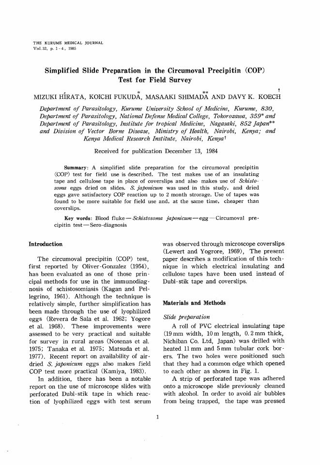
THE KURUME MEDICAL JOURNAL
Vol. 32, p. 1-4, 1985
Simplified Slide Preparation in the Circumoval Precipitin (COP)
Test for Field Survey
MIZUKI HIRATA, KOICHI FUKUDA, MASAAKI SHIMADA AND DAVY K. KOECH
Department of Parasitology, Kurume University School of Medicine, Kurume, 830, Department of Parasitology, National Defense Medical College, Tokorozawa, 359 and Department of Parasitology, Institute for tropical Medicine, Nagasaki, 852 Japan and Division of Vector Borne Disease, Ministry of Health, Nairobi, Kenya; and
Kenya Medical Research Institute, Nairobi, Kenyat
Received for publication December 13, 1984
Summary: A simplified slide preparation for the circumoval precipitin (COP) test for field use is described. The test makes use of an insulating tape and cellulose tape in place of coverslips and also makes use of Schisto-soma eggs dried on slides. S, japonicum was used in this study, and dried eggs gave satisfactory COP reaction up to 2 month strorage. Use of tapes was found to be more suitable for field use and, at the same time, cheaper than coverslips.
Key words : Blood fluke•\Schistosoma japonicum•\egg•\Circumoval pre-
cipitin test•\Sero -diagnosis
Introduction
The Circumoval precipitin (COP) test, first reported by Oliver-Gonzalez (1954), has been evaluated as one of those prin -cipal methods for use in the immunodiag-nosis of schistosomiasis (Kagan and Pel-legrino, 1961). Although the technique is relatively simple, further simplification has been made through the use of lyophilized eggs (Revera de Sala et al. 1962; Yogore et al. 1968). These improvements were assessed to be very practical and suitable for survey in rural areas (Nosenas et al. 1975; Tanaka et al. 1975; Matsuda et al. 1977). Recent report on availability of air-dried S. japonicum eggs also makes field COP test more practical (Kamiya, 1983).
In addition, there has been a notable report on the use of microscope slides with
perforated Dubl-stile tape in which reac-tion of lyophilized eggs with test serum
was observed through microscope coverslips (Lewert and Yogrore, 1969), The present
paper describes a modification of this tech-nique in which electrical insulating and cellulose tapes have been used instead of Dubl- stik tape and coverslips.
Materials and Methods
Slide preparation
A roll of PVC electrical insulating tape
(19 mm width, 10 m length, 0.2 mm thick, Nichiban Co. Ltd, Japan) was drilled with heated 11 mm and 5 mm tubular cork bor-ers. The two holes were positioned such that they had a common edge which opened to each other as shown in Fig. 1.
A strip of perforated tape was adhered
onto a microscope slide previously cleaned with alcohol. In order to avoid air bubbles
from being trapped, the tape was pressed
1
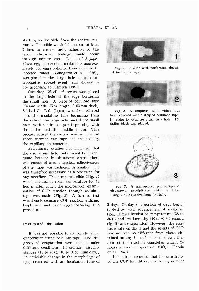
2 HIRATA, ET AL.
starting on the slide from the centre out-
wards. The slide was left in a room at least
2 days to ensure tight adhesion of the
tape, otherwise, leakage would occur
through minute gaps. Ten ƒÊl of S. japo-
nicum egg suspension containing approxi-
mately 100 eggs obtained from an 8-week-
infected rabbit (Yokogawa et al. 1966),
was placed in the large hole using a mi-
cropipette, spread evenly and allowed to
dry according to Kamiya (1983).
One drop (25ƒÊl) of serum was placed
in the large hole at the edge bordering
the small hole. A piece of cellulose tape
(24 mm width, 35 m length, 0.02 mm thick,
Sekisui Co. Ltd, Japan) was then adhered
onto the insulating tape beginning from
the side of the large hole toward the small
hole, with continuous gentle pressing with
the index and the middle finger. This
process caused the serum to enter into the
space between the tape and the slide by
the capillary phenomenon.
Preliminary studies had indicated that the use of one hole only would be inade-
quate because in situations where there was excess of serum applied, adhesiveness of the tape was reduced. A smaller hole was therefore necessary as a reservoir for any overflow. The completed slide (Fig. 2) was incubated at room temperature for 48 hours after which the microscopic exami-nation of COP reaction through cellulose tape was made (Fig.3). A further test was done to compare COP reaction utilizing lyophilized and dried eggs following this
procedure.
Results and Discussion
It was not possible to completely avoid
evaporation using cellulose tape. The de-
grees of evaporation were tested under
different conditions. In ordinary circum-
stances (15 to 28•Ž, 40 to 80% humidity),
no noticiable change in the morphology of
eggs occurred with an incubation time of
Fig. 1. A slide with perforated electri-
cal insulating tape.
Fig. 2. A completed slide which have
been covered with a strip of cellulose tape.
In order to visualize fluid in a hole, 1 %
anilin black was placed.
Fig. 3. A microscopic photograph of
circumoval precipitates which is taken
using •~40 objective lens (•~1280).
2 days. On day 3, a portion of eggs began
to destroy with advancement of evapora-
tion. Higher incubation temperature (28 to
36•Ž) and low humidity (20 to 30%) caused
significant evaporation. However, the eggs
were safe on day 1 and the results of COP
reacton was no different from those ob-
tained on day 2, as has been shown that
alsmost the reaction completes within 24
hours in room temperature (28•Ž)(Garcia
et al. 1981).
It has been reported that the sensitivity
of the COP test differed with egg number
SIMPLIFIED SLIDE PREPARATION FOR COPT 3
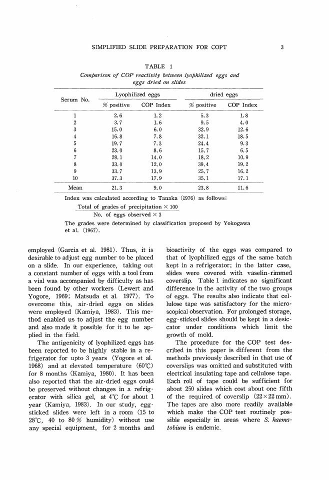
TABLE 1
Comparison of COP reactivity between lyophilized eggs and eggs dried on slides
Index was calculated according to Tanaka (1976) as follows
Total of grades of precipitation •~100
/ No, of eggs observed •~ 3
The grades were determined by classification proposed by Yokogawa et al. (1967).
employed (Garcia et al. 1981). Thus, it is desirable to adjust egg number to be placed on a slide. In our experience, taking out a constant number of eggs with a tool from a vial was accompanied by difficulty as has been found by other workers (Lewert and Yogore, 1969; Matsuda et al. 1977). To overcome this, air-dried eggs on slides were employed (Kamiya, 1983). This me-thod enabled us to adjust the egg number and also made it possible for it to be ap-
plied in the field. The antigenicity of lyophilized eggs has
been reported to be highly stable in a re-
frigerator for upto 3 years (Yogore et al.
1968) and at elevated temperature (60•Ž)
for 8 months (Kamiya, 1980). It has been
also reported that the air-dried eggs could
be preserved without changes in a refrig-
erator with silica gel, at 4•Ž for about 1
year (Kamiya, 1983). In our study, egg-
sticked slides were left in a room (15 to
28•Ž, 40 to 80% humidity) without use
any special equipment, for 2 months and
bioactivity of the eggs was compared to
that of lyophilized eggs of the same batch
kept in a refrigerator; in the latter case,
slides were covered with vaselin-rimmed
coverslip. Table 1 indicates no significant
difference in the activity of the two groups
of eggs. The results also indicate that cel-
lulose tape was satisfactory for the micro-
scopical observation. For prolonged storage,
egg- sticked slides should be kept in a desic-
cator under conditions which limit the
growth of mold.
The procedure for the COP test des-
cribed in this paper is different from the
methods previously described in that use of
coverslips was omitted and substituted with
electrical insulating tape and cellulose tape.
Each roll of tape could be sufficient for
about 250 slides which cost about one fifth
of the required of coverslip (22 •~ 22mm).
The tapes are also more readily available
which make the COP test routinely pos-
sible especially in areas where S. haema-
tobium is endemic.
4 HIRATA, ET AL.
Acknowledgments : This study was done under
the auspices of the Kenya Medical Research
Institute and supported by the Japan Inter-
national Co-operation Agency.
References
GARCIA, E. G., TAPALES, Fe P., VALDEZ, C. A., MIT-CHELL, G. F. and TIU, W.U. (1981). Attempts to standardize the circumoval precipitin test
(COPT) for schistosomiaisis japonica. Sou-theast Asian J. Trop. Med. Pub. Hlth. 12,
384-395.KAGAN, I. G. and PELLEGRINO, J. (1961). A critical
review of immunological methods for diag-nosis of bilharziasis. Bull. W. H. O. 25, 611-674.
KAMIYA, H. (1980). Influence of temperature on the antigenicity of Schistosoma japonicum lyophilized eggs for circumoval precipitin test
(COPT). Jap. J. Vet. Res. 28, 149-154.KAMIYA, H. (1983). Circumoval precipitin (COP)
test using air-dried eggs of Schistosoma ja-
ponicum and S, mansoni. Southeast Asian J. Trop. Med. Pub. Hlth. 14, 451-455.LEWERT, R. M. and YOGORE, M. G. Jr. (1969). A
field circumoval precipitin (COP) test for schistosomiasis japonica. Trans. Roy. Soc. Trop. Med. Hyg. 63, 343-348.MATSUDA, H., NOSENAS, J.S., TANAKA, H., SANTOS, A.J.Jr. and TRINIDAD-PEREZ D. (1977). Com-
parative studies on reading criteria of cir- cumoval precipitin reaction of Schistosoma
japonicum for field survey in highly endemic area. Jap. J. Exp. Med. 47, 369-375.
NOSENAS, J. S., MATSUDA, H., BLAS, B. L., TANAKA, H. and SANTOS, A. J. Jr. (1975). Evaluation of
the circumoval precipitin test using dried blood on filter paper as a diagnostic tool in epidemiological survey for schistosomiasis.
Jap. J. Exp. Med. 45, 367-375.OLIVER-GONZALEZ, J. (1954). Anti-egg precipitins
in the serums from humans infected with Schistosoma mansoni. J. Infect. Dis. 95, 86-
91.RIVERA DE SALA, A., CANCIO, M. and RODRIGUEZ- MOLINA, R. (1962). Preservation of eggs of Schistosoma mansoni for the circumoval pre-
cipitin test. Am. J. Trop. Med. Hyg. 11, 199-200.
TANAKA, H. (1976). Complement fixation and circumoval precipitin reactions for diagnosis and as tests of cure in schistosomiasis. Sou-theast Asian J. Trop. Med. Pub. Hlth. 7, 176-179.
TANAKA, H., MATSUDA, H., BLAS, B. L. and NOSENAS,
J. S. (1975). Evaluation of a technique of circumoval precipitin test using blood taken on filter paper and a microtiter technique of complement fixation test of Schistosoma ja-
ponicum. Jap. J. Exp. Med. 45, 105-111.YOGORE, M. G. Jr., LEWERT, R. M. and SILAN, R. B.
(1968). The circumoval precipitin (COP) test in schistosomiasis japonica. Am. J. Trop.
Med. Hyg. 17, 65-71.YOKOGAWA, M. and SANO, M. (1966). Immunodi-
agnosis of schistosomiasis japonica II. isola - tion tehnique of the Schistosoma eggs from the tissues for circumoval precipitin test.
Jap. J. Parasit. 15, 394-398.YOKOGAWA, M., SANG, M. and ARAKI, K. (1967).
Immunodiagnosis of schistosomiasis japonica III. circumoval precipitin test. Jap. J. Parasit. 16, 77-84.