Embed Size (px)
Citation preview

SILVER IMPREGNATION IN SITU An Aid to Radiographic Interpretation
JACK BOYD, PHD, BVMS,* NORMAN W. RANTANEN, D V M , MST
In evaluating radiographs of the limb joints and head, students encounter difficulty where su- perimposition occurs. By replacing calcium with silver salts in the bone, enhanced radiopacity can be produced. In this study, silver impregnation was used to increase the radiopacity of individual carpal and tarsal bones, selected bones of the skull and the sinuses, and guttural pouch of the horse. This provides an interpretation aid for teaching radiographic anatomy of these regions. Veterinary Radiology, Vol. 25, No. 5, 1984; p p 220-226.
Key words: joints, sinuses, guttural pouch, anatomy, silver nitrate, bone stain.
N A PREVIOUS REPORT,' a method of impregnating I the bones of the skull with silver was described, which could be used as an interpretation aid in teaching radiographic anatomy. It afforded a method of enhancing the radiographic opacity of individual skull bones so that it was possible to compare a stan- dard radiograph with the radiograph of a skull rebuilt with the silver-containing bone, thus clarifying defi- nition in areas of superimposition. The technique was initially demonstrated in cat skulls, which were easily rebuilt, but has since been used for the skulls of larger species to facilitate radiographic interpretation in these species.
Materials and Methods
Previously radiographed immature bovine and equine heads were boiled to give separate skull bones. A single bone was then impregnated with silver nitrate and reassembled into the skull and further radiographs taken. Plasticine was used to mimic the sutures.
Lateromedial and dorsopalmar (plantar) radiographs of pony limbs were obtained before the limbs were boiled. A single bone was selected from each carpal and tarsal specimen and immersed in silver nitrate so- lution according to a previously described meth- od. ' The bones were then reassembled with the use of double-sided adhesive strips placed between apposing surfaces to simulate the correct joint spacing. The reassembled joints were then radiographed in the same positions as the original radiographs. Comparisons were made between the two sets of radiographs. The impregnated bone had become blackened by the silver so that it was easily identified in the reassembled spec- imen. Bovine limbs were treated in a similar fashion,
* Department of Veterinary Anatomy, University of Glasgow Vet-
t Echo Affiliates Inc., Lexington, Kentucky, 40508. erinary School, Bearsden Road, Bearsden, Glasgow, G61 1QH.
and individual epiphyses were treated to highlight their contribution to the radiographic image.
Due to the problem of immobility of the prepared specimens and lack of soft tissue comparison, addi- tional limbs of various domestic species were radio- graphed to provide more normal radiographs of the tarsus and carpus. Joints were then carefully opened, and a single carpal or tarsal bone was removed. Crys- tals of KOH were inserted between the bones and al- lowed to dissolve into the surrounding soft tissue to facilitate removal of the selected bone. The isolated bone was immersed in 95% alcohol for 48 hours and then impregnated with silver by placing in an aqueous 5% solution of silver nitrate for two to three days. After confirming by radiography that sufficient im- pregnation had occurred, the bone was carefully re- placed into the joint that had been kept wrapped in moist cloths in a refrigerator. The skin flap was then sutured with linen. These joints were radiographed in the same positions as the normal fresh joints. Radio- graphs were obtained with air in the joints and with the joint spaces filled with liquid paraffin to simulate synovial fluid. This process was repeated for each bone of the joint producing a collection of carpal and tarsal bones of high radiopacity. The head of a eu- thanized pony was radiographed in a lateral position to demonstrate the maxillary and frontal sinuses. The rostra1 and caudal compartments of the maxillary sinus of one side of the head were filled with 95% alcohol through trephine holes made in the lateral wall of the maxilla. The caudal sinus, having a direct communi- cation with the frontal sinus allowed alcohol to fill the latter sinus. After filling completely, the trephine holes were tightly closed using nylon screws, and the head was placed in lateral recumbency, with the filled side down, for 48 hours. The screws were then removed, and the alcohol allowed to drain. The sinuses were filled with a 1% silver nitrate solution, the screws re- placed, and the head positioned with the filled side
220

VOL. 25. No. 5 SILVER IMPREGNATION 22 1
FIG. 1 . Dorsoventral radiograph of the head of a calf.
down. The sinuses were then evacuated of silver ni- trate by draining for several hours. The head was ra- diographed in the position matching the original radio- graph. The guttural pouch of another pony head was treated in a similar manner using alcohol and silver nitrate. The materials were introduced and withdrawn via an approach through the triangle formed by the ramus of the mandible, the tendon of the sternoce- phalic muscle and the linguofacial vein.
Results
In a routine radiograph of a calf head (Fig. l), there was poor contrast between superimposed skull bones
FIG. 2. Dorsoventral radiograph of an immature bovine skull. Ap- pearance of the fused sphenoid and basioccipital bones (S) is en- hanced by silver nitrate.
when it was compared with Figure 2, in which the silver-impregnated fused sphenoid and basioccipital bones could be readily discerned because of their in- creased radiopacity. A silver impregnated distal me- tacarpal epiphysis was radiographed in a rebuilt spec- imen (Fig. 3) to give greater definition of that center when compared with the appearance in the radiograph of the intact limb (Fig. 4). Radiographs of specimens with silver-impregnated bones can as an aid to inter- pretation, be used in conjunction with the rebuilt bone specimen with its blackened bony center (Figs. 5 , 6). In Figures 7 and 8, radiographs of equine and canine

222 BOYDANDRANTANEN 1984
FIG. 3. Dorsopalmar radiograph of the rebuilt bones of an imma- ture bovine manus. The distal medial metacarpal epiphysis (E) has increased radiopacity due to silver impregnation.
FIG. 4. Dorsopalmar radiograph of an intact immature bovine manus.

VOL. 25, No. 5 SILVER IMPREGNATION 223
FIG. 5. Dorsopalmar radiograph of a reassembled equine carpus. The 3rd carpal bone (3) has increased radiopacity due to silver im- pregnation. FIG. 6. Skeletal models of equine thoracic limbs. The intermediate
carpal bone of the right limb and 3rd carpal bone of the left limb are black due to silver nitrate impregnation.

224 BOYD AND RANTANEN 1984
FIG. 7. Lateromedial radiograph of a flexed equine carpal joint. The intermediate carpal bone (I) has increased radiopacity after silver nitrate impregnation and replacement in the joint.
FIG. 8. Dorsopalmar radiograph of a canine carpal joint. The in- termedioradial carpal bone (R) has increased radiopacity due to silver impregnation and replacement into the joint.
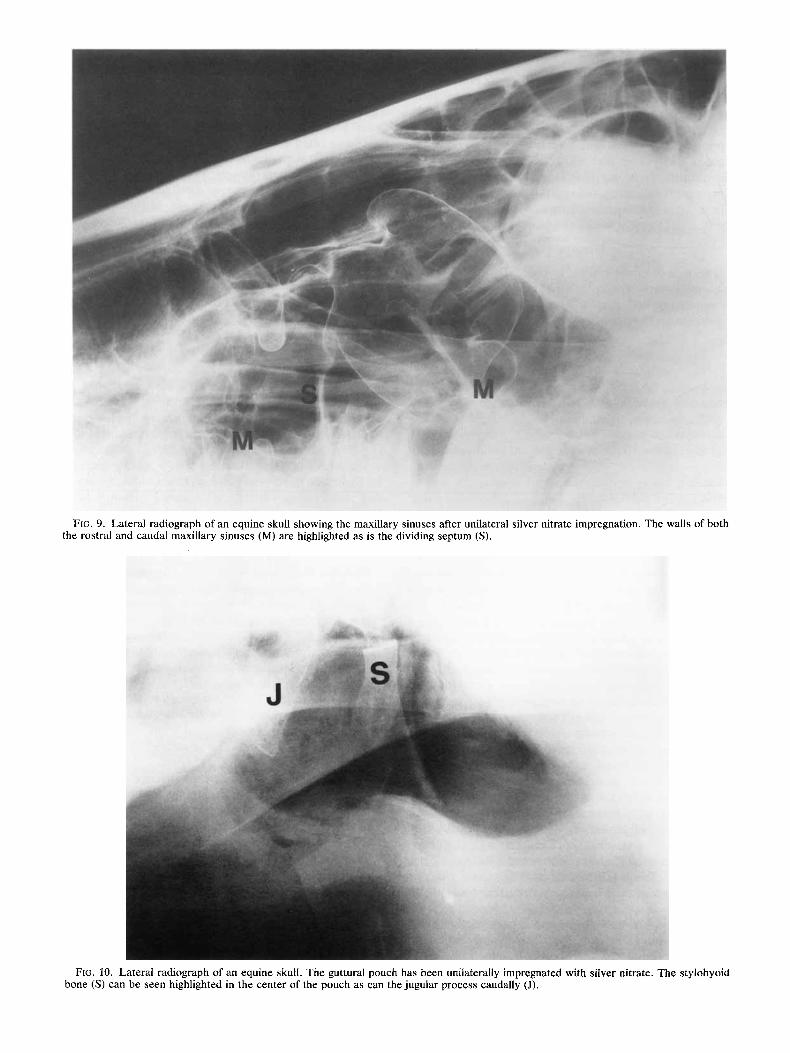
FIG. 9. Lateral radiograph of an equine skull showing the maxillary sinuses after unilateral silver nitrate impregnation. The walls of both the rostra1 and caudal maxillary sinuses (M) are highlighted as is the dividing septum (S).
FIG. 10. Lateral radiograph of an equine skull. The guttural pouch has been unilaterally impregnated with silver nitrate. The stylohyoid bone (S) can be seen highlighted in the center of the pouch as can the jugular process caudally (J).

226 BOYD AND RANTANEN 1984
carpal joints were obtained in which single bones have received silver impregnation before replacement into the joint. The position of these bones within the joint was readily discerned. A radiograph was obtained of a pony head in which unilateral filling of the maxillary sinuses with silver had allowed impregnation of the surrounding bone margins (Fig. 9). In Figure 10, a pony’s guttural pouch had been unilaterally filled with silver nitrate, impregnating bony structures that abutted onto or lay within the pouch.
Discussion
The use of silver nitrate impregnation to enhance the radiopacity of bony structures was of assistance in producing definition of bones within joints when superimposed images caused difficulty in interpreta-
tion. The replacement of the impregnated bone back into the joint allowed the production of radiographs with the joint in a variety of positions, which had pro- duced problems of superimposition. The equine sin- uses have often proved to be difficult for students to interpret, but with absorption of silver, the bony per- ifery of these structures was more readily identified. The guttural pouch, being an air-filled sac, is fairly well-defined in standard radiographs , but the position of the stylohoid bone and the bones dorsal to the pouch are demonstrated more clearly with silver im- pregnation. It would appear that the technique of silver impregnation could be applied to other speci- mens and could facilitate the interpretations of radio- graphs with minimum distortion of the regions being studied.
REFERENCE
1. Boyd JS. Skull radiographs: silver impregnation as an inter- pretation aid. Vet Radio1 1980;21:82-4.
BOOK REVIEW
Veterinary Medicine, 6th edition. D. C. Blood, 0. M. Radostits, and J. A. Henderson. $65.00, 1328 pages, Bailliere Tindall, London.
The sixth edition of Veterinary Medicine succeeds the familiar textbook of large animal diseases by Blood and Henderson. While expanded 150 pages in length, set in a bolder type, and printed by a new publisher, the revision is true to its original objectives, i.e., to provide an instrument of continuing self-education and reference to the veterinary profession. The text is de- signed as a general information resource rather than a tome of great detail. Like its predecessor, the book is divided into two well-organized sections. The first, “General Medicine,” covers diseases of organ sys- tems, e.g., the respiratory, cardiovascular, hepatobil- iary, and integumentary systems. Also included are chapters on general therapeutics, diagnostics, and general systemic states. The second section, “Special Medicine,” classifies diseases by cause; i.e., viral, bacterial, rickettsial, and chemical and physical agents.
The book is easily read and is organized to be used as a teaching textbook. More importantly for the prac- ticing veterinarian, the book is designed to aid in the
systematic process of disease diagnosis. A clinician can enter the index with one or more clinical signs, be led to the section or sections covering the systems implicated in the physical findings, and methodically develop a list of differential diagnoses. Included in each section are common diagnostic tests and proce- dures. Appropriate radiographic studies are described in general terms. Therapeutic plans are included in each subsection. To the American reader, some prod- ucts and procedures to which the authors refer are unfamiliar or unavailable, and some terminology dif- ferences are evident. The strongest attribute of this book is its index and organization.
While few drawings and an absence of pictures or radiographs may be shortcomings in the book, the au- thors do an exceptional job referring the reader to pri- mary sources in the literature and to review articles. Adding more to the text than is already present would compromise its everyday usefulness. This book has overwhelming appeal not only to practicing veterinar- ians but also to students and educators. It is a must for the shelves of anyone interested in large animal medicine.
Anthony L. Kiorpes, DVM, PhD