Embed Size (px)
Citation preview
J. Phys. Chem. 1992,96, 10405-10411 10405
of eqns 2 and 3 and the results described here. A detailed ex- perimental investigation is currently under way, and the results will be reported elsewhere.
References and Notes (1) Israelachvili, J . N.; Adams, G. E. J . Chem. Soc., Furuduy Trans. 1
(2) Parker, J. L.; Christenson, H. K.; Ninham, B. W. Rev. Sci. Instrum.
(3) Israelachvili, J. N. J . Colloid Interface Sci. 1973, 44, 259. (4) Derjaguin, B. V. Kolloid Z . 1934, 69, 155. (5) Attard, P.; Parker, J. L. J . Phys. Chem. 1992, 96, 5086. (6) White, L. R. J . Colloid Interjace Sci. 1983. 95, 286. (7) Attard, P.; B&ard, D. R.; Ursenbach, C. P.; Patey, G. N. Phys. Reu.
(8) Pashley, R. M. J . Colloid Interfuce Sci. 1981, 80, 153. (9) Pashley, R. M. J. Colloid Interfuce Sci. 1981, 83, 531. (10) Pashley, R. M.; Israelachvili, J. N. J. Colloid Interface Sci. 1984,101,
1978, 74, 975.
1989, 60, 3135.
A 1991,44,8224.
511.
(11) Attard, P.; Parker, J. L. Phys. Rev. A, in press. (12) Hughes, B. D.; White, L. R. Quart. J . Mech. Appl. Muth. 1979,32,
(13) Hughes, B. D.; White, L. R. J. Chem. Soc., Faruday Trans. 11980,
(14) Muller, V. M.; Yushchenko, V. S.; Derjaguin, B. V. J . Colloid In-
(15) Hertz, H.; Reine, J. Angew. Murh. 1881. 92, 156. (16) Johnson. K. L.: Kendall, K.; Roberts, A. D. Proc. R. Soc. London A
(17) Derjaguin, 8. V.; Muller, V. M.; Toporov Yu, J. Colloid Interfuce
(18) Parker, J. L.; Stewart, A. M. Prog. Colloid Polym. Sci., in press. (19) Parker, J. L. Lungmuir 1992,8, 551. (20) Israelachvili, J. N.; Tabor, D. Nature (Phys. Sci.) 1972, 236, 106. (21) Horn, R. G.; Israelachvili, J. N.; F'ribac, F. J. Colloid Interface Sei.
(22) Chen, Y. L.; Helm, C. A.; Israelachvili, J. N . J . Phys. Chem. 1991,
(23) Ducker, W. A.; Senden, T. J.; Pashley, R. M. Nature 1991,353,239.
445.
76, 963.
terfuce Sci. 1983, 92, 92.
1971,324. 301
Sci. 1975, 53, 314.
1987, 11 5,480.
95, 10736.
Reduction of the Roughness of Silver Films by the Controlled Application of Surface Forces
John M. Levins and T. Kyle Vanderlick* Department of Chemical Engineering, University of Pennsylvania, Philadelphia, Pennsylvania I91 04-6393 (Received: June 24, 1992; In Final Form: September 17, 1992)
Using the surface forces apparatus, we have brought into contact a thermally evaporated silver film with a smooth mica sheet and have measured the time-dependent changes in the wavelengths of fringes of equal chromatic order generated using multiple beam interferometry. Based on a theoretical analysis of the interference fringes produced for a silver surface with prescribed roughness, the wavelength shifts that we observe can be explained by an irreversible reduction in the roughness of the silver surface. Assuming a sinusoidal profile for the silver surface, our measured wavelength shifts correspond to a decrease in root-mean-squared roughness from between 10 and 30 A. Concurrent with the reduction of silver roughness, we observe an increase in both the area of deformed contact and the pull-off force between mica and silver. We also measured the thicknesses of alkanethiol self-assembled monolayers chemisorbed on the silver surface. Although our thicknesses are lower than those obtained by others using ellipsometry, our monolayer thicknesses increase as expected with the length of the alkanethiol hydrocarbon chain.
Introduction Developed into its present form by Israelachvili,' the surface
forces apparatus (SFA) has become a mainstay experimental technique of colloid and interface science. The apparatus allows the measurement of the force acting between two opposed surfaces as a function of their separation. In nearly all applications of the SFA, mica surfaces have been employed, primarily because mica can be cleaved molecularly smooth. Within the past few years, however, other surfaces-such as silica? poly(ethy1ene tere- phthalate),' sapphire,' and platinum fh5-have been used, with varying degrees of succcss, in the SFA.
In this paper, we demonstrate that silver can be used as one of the two surfaces, mica being the other, without significant loss of resolution in the separation measurement. Moreover, we find that the roughness of thermally evaporated silver films can be r e d u d by mmpnaping the silver between two molecularly smooth mica sheets. Our interest in silver films is based on their use as substrates for alkanethiol self-assembled monolayer formation.61 I Self-assembled monolayers form via the spontaneous chemisorption from solution of functionalized molecules onto a host solid surface. We have formed self-assembled monolayers on silver surfaces in the SFA and demonstrated that angstrom-level resolution in monolayer thickness measurements can be obtained.
The ultimate goal of our research is to use self-assembled monolayers to prepare surfaces with well-defined chemical and
*To whom correspondence should be addressed.
0022-365419212096- 10405S03.0010
structural properties for use in the SFA. We intend to employ these surfaces to study several colloidal interactions that are poorly understood-specifically, hydration f~ rces , ' ~ - '~ steric undulation forces,IsJ6 and hydrophobic forces.17-19 The first step toward achieving this goal is the focus of this paper: demonstrating that a surface suitable for the formation of self-assembled monolayers (silver) can be used in the SFA.
Although alltanethiol self-assembled monolayers are known to form on the other noble metals: silver has the advantage that it is ideally suited for the application of multiple beam interfero- metry, which is used to determine surface separation in the SFA. White light is directed through an interferometer consisting of two highly reflective metallic filar separated by a layer, or layers, of dielectric material with an overall thickness greater than the wavelength of light in the visible spectrum. The light undergoes multiple reflections between the reflective layers and emerges as a series of fringa of equal chromatic order (FECO).M The FECO occur at discrete wavelengths which depend on the distance be- tween the two reflective films. Silver is generally used as the metallic coating because of its high reflectivity.
Israelachvili2' derived analytical expresSons relating the distance between the silver films to the wavelength, A, of any particular FECO. Rather than account explicitly for the optical properties of the reflective films, Israelachvili treated the silver layers as planes with reflectivity approaching unity. Using the multilayer matrix method-which accounts for the optical properties of each layer in the interferometer-Clarknz2 examined the accuracy
Q 1992 American Chemical Societv
10406 The Journal of Physical Chemistry, Vol. 96, No. 25, 1992 Levins and Vanderlick
of Israelachvili’s equations and found them applicable only for the centrosymmetric configuration of layers (Le., silver-mica- medium-mica-silver) that is most typically used in the SFA.
Others before us have attempted to measure forces between a mica surface and a silver surface in the SFA. Soon after the inception of the apparatus, Coakley and Tabor23 measured the van der Waals forces between silver and mica in air. More recently, Parker and Chr i~ tenson~~ measured solvation forces between a silver surface and a mica surface immersed in organic and aqueous solutions. In all of these experiments, the equations of Israelachvili were used to calculate the separation between the mica and silver, and so the reported results are in error. In addition, none of these experimenters addressed explicitly the roughness of the silver and its impact on the accuracy of distance measurements. Only a very brief mention of this was made by Parker and Christenson, who noted that “the contact position is somewhat uncertain because the surfaces appear to be compressible over about 1 nm.”
We have recently used the multilayer matrix method to de- termine the effect of silver roughness on the observable interference fringes.25 The results of our analysis will be used to interpret the experimental findings reported here.
Experimental Section A. Methods and Procedures. Our surface forces apparatus
(SFA) is identical to the one used by Israelachvili and Adams.’ Although they describe the apparatus and the method of force measurement, a more detailed description is given by Vanderlick et a1.26 Our SFA is housed in a class loo00 clean room to reduce the risk of dust contamination. The clean room includes a class 10 laminar flow hood wherein the SFA is assembled and prepared for experiments. Experiments are conducted with the SFA mounted on an electronic antivibration table (EVIS, Newport Corp.) which dampens the vibrations of the force measuring spring well enough to permit accurate distance measurements in a gaseous environment.
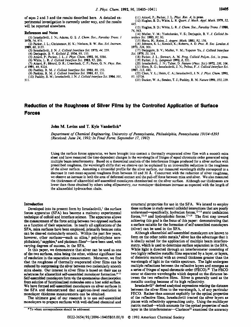
Some of our experiments (those involving the formation of self-assembled monolayers) required immersing one of the surfaces in solution, without disturbing its lateral position relative to the other surface. To do this, we used a small bath assembly, man- ufactured by Anutech, Ltd., Australia, that is designed for the SFA. As shown in Figure 1, the assembly consists of a gooseneck double-cantilever spring that supports the lower surface and a Teflon bath of 10-mL capacity that can be cupped around the spring. Solution is injected through a Teflon port at the bottom of the SFA. The upper micrometer-driven rod is used to lower the spring into the bath. Our gooseneck sprng has a deflection constant of 1.055 X lo2 N/m; this spring was used in all the experiments reported here, including those which did not require the injection of solutions into the SFA.
The surfaces employed in all experiments were prepared as follows. Freshly cleaved sheets of ruby muscovite mica, 2-4 pm thick, were coated on one side only with a thermally evaporated silver f h . Films 500-580 A thick were deposited at rates varying from 2 to 4 A/s, as determined using a quartz crystal monitor. Some depositions were camed out in a diffusion-pumped vacuum system and others in a newly purchased turbo-pumped vacuum chamber; both systems were equipped with a Pyrex bell jar. Resistive evaporation boats made of either tungsten or molyb- denum were used. The pressure during evaporation was always between 1 X low7 and 4 X lov7 Torr. Silver was supplied from Aesar (99.999%) or Ted Pella, Inc. (99.99%). The epoxy resin EPON 1004 was used to glue the silver-coated mica sheets onto silica supporting disks of cylindrical shape (R = 1 cm). The disks were loaded into the SFA with their axes oriented at right angles. The SFA was then sealed and purged with prepurified nitrogen for no less than 15 min.
Monolayers were formed by immersing the silver surface into the small bath assembly containing an ethanolic solution of the thiol of interest. The thiol-based compounds (1-octadecanethiol, l-dodecanethiol, and 1-octanethiol) were purchased from Aldrich and used as received. Absolute ethanol was purchased from
I Medium I
Piezoelectric driven rod crystal
Small teflon - bath
/ Gooseneck leaf spring
Small bath injection port 1
Double cantilever spring
Helical spring
7 driven rod
Figure 1. Schematic of the small bath assembly mounted in the SFA. An expanded view of the surfaces is provided, showing the relative pos- itions of each layer for the silver-mica-mediumsilver-mica interferom- eter. The layer thicknesses are not drawn to scale.
Pharmco and also used as received. The following concentrations and immersion times were used: dodecanethiol, 3.5 mM, 2 h; octanethiol, 3.5 mM, 2 h; octadecanethiol, 0.6 mM, 4 h. The thiol solution was then drained, and the monolayer-coated surface was rinsed by filling the bath with ethanol and draining it after a few minutes; this was repeated three times. The SFA was purged with nitrogen during monolayer formation and for 2 h afterward to
Meam“& TheSFA dry the surface.
can be used to bring together two separated surfaces from ma- croscopic distances into molecular contact. Our key experimental observations are based on the time-dependent behavior of a silver film when it is in contact with an opposed mica surface. The purpose of this section is to describe how the SFA is used to bring two surfaces into and out of contact. In addition, we describe the geometrical deformations which can accompany two surfaces in contact.
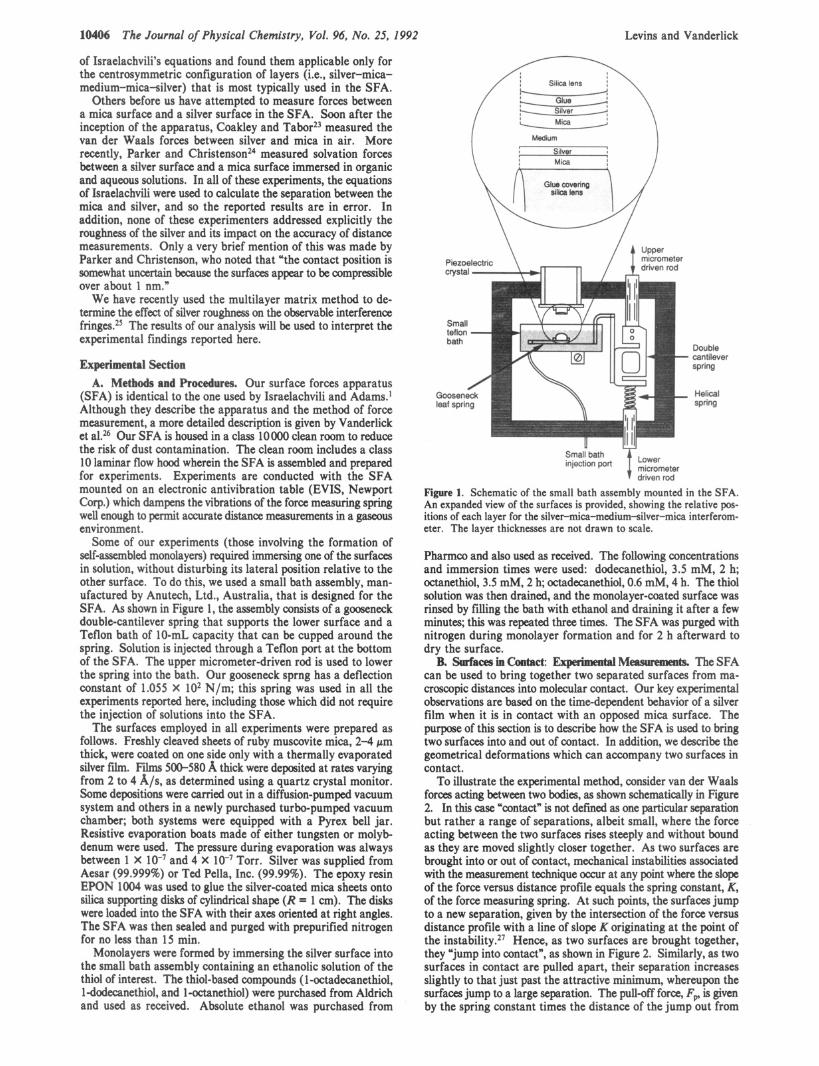
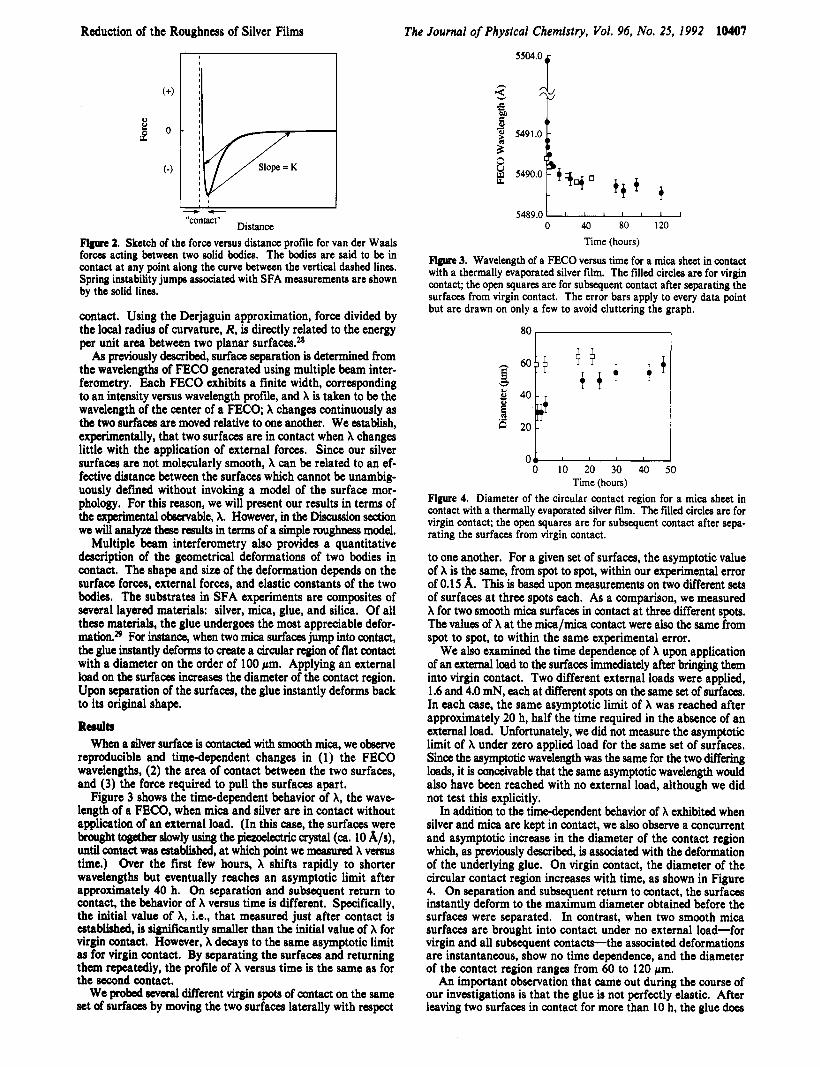
To illustrate the experimental method, consider van der Waals forces acting between two bodies, as shown schematically in Figure 2. In this case “contact” is not defined as one particular separation but rather a range of separations, albeit small, where the force acting between the two surfaces rises steeply and without bound as they are moved slightly closer together. As two surfaces are brought into or out of contact, mechanical instabilities associated with the measurement technique occur at any point where the slope of the force versus distance profile equals the spring constant, K, of the force measuring spring. At such points, the surfaces jump to a new separation, given by the intersection of the force versus distance profile with a line of slope K originating at the point of the in~tab i l i ty .~~ Hence, as two surfaces are brought together, they “jump into contact”, as shown in Figure 2. Similarly, as two surfaces in contact are pulled apart, their separation increases slightly to that just past the attractive minimum, whereupon the surfaces jump to a large separation. The pull-off force, Fp is given by the spring constant times the distance of the jump out from
B. Surfacesincontact:
Reduction of the Roughness of Silver Films The Journal of Physical Chemistry, Vol. 96, No. 25, 1992 10407
I - - Distance “contact”
Figure 2. Sketch of the force versus distance profile for van der Waals forces acting between two solid bodies. The bodies are said to be in contact a t any point along the curve between the vertical dashed lines. Spring instability jumps associated with SFA measurements are shown by the solid lines.
contact. Using the Derjaguin approximation, force divided by the local radius of curvature, R, is directly related to the energy per unit area between two planar surfaces.28 As previously dcacribed, surface separation is determined from
the wavelengths of FECO generated using multiple beam inter- ferometry. Each FECO exhibits a finite width, corresponding to an intensity versus wavelength profile, and X is taken to be the wavelength of the center of a FECO A changes continuously as the two surfaces are moved relative to one another. We establish, experimentally, that two surfaces are in contact when X changes little with the application of external forces. Since our silver surfaces are not molecularly smooth, X can be related to an ef- fective distance between the surfaces which cannot be unambig- uously defined without invoking a model of the surface mor- phology. For this reason, we will present our results in terms of the experimental observable, A. However, in the Discussion section we will analyze these results in terms of a simple roughnm model.
Multiple beam interferometry also provides a quantitative description of the geometrical deformations of two bodies in contact. The shape and size of the deformation depends on the surface forces, external forces, and elastic constants of the two bodies. The substrates in SFA experiments are composites of several layered materials: silver, mica, glue, and silica. Of all these materials, the glue undergoes the most appreciable defor- mation.29 For instance, when two mica surfaces jump into contact, the glue instantly deforms to create a circular region of flat contact with a diameter on the order of 100 pm. Applying an external load on the surfaces increases the diameter of the contact region. Upon separation of the surfaces, the glue instantly deforms back to its original shape.
R d Q When a silver surface is contacted with smooth mica, we observe
reproducible and timedependent changes in (1) the FECO wavelengths, (2) the area of contact between the two surfaces, and (3) the force required to pull the surfaces apart.
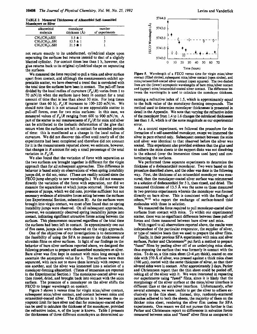
Figure 3 shows the time-dependent behavior of A, the wave- length of a FECO, when mica and silver are in contact without application of an external load. (In this case, the surfaces were brought together s k l y using the piezoelactric crystal (ca. 10 A/@, until contact was established, at which point we measured X versus time.) Over the first few hours, X shifts rapidly to shorter wavelengths but eventually reaches an asymptotic limit after approximately 40 h. On separation and subsequent return to contact, the behavior of X versus time is different. Specifically, the initial value of A, i.e., that measured just after contact is established, is significantly smaller than the initial value of X for virgin contact. However, X decays to the same asymptotic limit as for virgin contact. By separating the surfaces and returning them repeatedly, the profile of X versus time is the same as for the m n d contact.
We probed several different virgin spots of contact on the same set of surfaces by moving the two surfaces laterally with respect
5504.0
h
5.
5489.0 0 40 80 120
Time (hours)
Figure 3. Wavelength of a FECO versus time for a mica sheet in contact with a thermally evaporated silver film. The filled circles are for virgin contact; the open squares are for subsequent contact after separating the surfaces from virgin contact. The error bars apply to every data point but are drawn on only a few to avoid cluttering the graph.
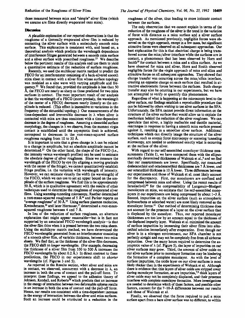
0’ 0 10 20 30 40 50
Time (hours) Figure 4. Diameter of the circular contact region for a mica sheet in contact with a thermally evaporated silver film. The filled circles are for virgin contact; the open squares are for subsequent contact after sepa- rating the surfaces from virgin contact.
to one another. For a given set of surfaces, the asymptotic value of A is the same, from spot to spot, within our experimental error of 0.1 5 A. This is based upon measurements on two different sets of surfaces at three spots each. As a comparison, we measured X for two smooth mica surfaces in contact at three different spots. The values of A at the mica/mica contact were also the same from spot to spot, to within the same experimental error.
We also examined the time dependence of A upon application of an external load to the surfaces immediately after bringing them into virgin contact. Two different external loads were applied, 1.6 and 4.0 mN, each at different spots on the same set of surfaces. In each case, the same asymptotic limit of X was reached after approximately 20 h, half the time required in the absence of an external load. Unfortunately, we did not measure the asymptotic limit of X under zero applied load for the same set of surfaces. Sice the asymptotic wavelength was the same for the two differing loads, it is conceivable that the same asymptotic wavelength would also have been reached with no external load, although we did not test this explicitly.
In addition to the timedependent behavior of X exhibited when silver and mica are kept in contact, we also observe a concurrent and asymptotic increase in the diameter of the contact region which, as previously described, is associated with the deformation of the underlying glue. On virgin contact, the diameter of the circular contact region increases with time, as shown in Figure 4. On separation and subsequent return to contact, the surfaces instantly deform to the maximum diameter obtained before the surfaces were separated. In contrast, when two smooth mica surfaces are brought into contact under no external load-for virgin and all subsequent contacts-the associated deformations are instantaneous, show no time dependence, and the diameter of the contact region ranges from 60 to 120 pm.
An important observation that came out during the course of our investigations is that the glue is not perfectly elastic. After leaving two surfaces in contact for more than 10 h, the glue does
10408 The Journal of Physical Chemistry, Vol. 96, No. 25, 1992 Levins and Vanderlick
TABLE I: Measured Thicknesses of Abaethiol Self-Assembled M d y e r s on Silver
a 1 ka n e t h i o I monolayer number molecule thicknesd (A) of experiments
CHt(CH,LSH 5.5 * 1 1 CH;(CH;j;,SH 13.5 i 1 3 C H A C H J i W 21.5 1 1
not return exactly to its undeformed, cylindrical shape upon separating the surfaces but returns instead to that of a slightly blunted cylinder. For contact times less than 1 h, however, the glue returns back to its original cylindrical shape on separating the surfaces.
We measured the force required to pull a mica and silver surface apart from contact, and although the measurements exhibit a p preciable scatter, we have observed a trend that is correlated with the total time the surfaces have been in contact. The pullsff force divided by the local radius of curvature (Cp/R) varies from 1 to 70 mN/m when the surfaces have been in contact for a total amount of time that is less than about 30 min. For long times (greater than 60 h), Fp/R increases to 100-320 mN/m. We should note that it is not unusual to see appreciable scatter in pull-off forces, even for two mica surfaces: in this case, we measured values of Fp/R ranging from 600 to 900 mN/m. A part of the scatter in our measurements of Fp/R for mica and silver can be attributed to the inelastic deformation of the glue that occurs when the surfaces are left in contact for extended periods of time: this is manifested as a change in the local radius of curvature. We did not discover this effect until nearly all of the experiments had been completed and therefore did not account for it in the measurements reported above; we estimate, however, that changes in R account for only a small percentage of the total variation in Fp/R.
We also found that the variation of force with separation as the two surfaces are brought together is different for the virgin approach than for all subsequent approaches. This difference in behavior is based solely on observations of when spring instability jumps did, or did not, occur. (These are readily noticed since the FECO jump abruptly to new wavelengths.) We did not measure directly the variation of the force with separation, nor did we measure the separations at which jumps occurred. However the presence of jumps, which we did note, provides sufficient but not necessary evidence of attractive forces acting between the surfaces (see Experimental Section, subsection B). As the surfaces were brought into virgin contact, we most often found that no spring instability jumps were observed. On all subsequent approaches, however, we consistently observed spring instability jumps into contact, indicating si@icant attractive forces acting between the surfaces. This phenomenon occurred regardless of the total time the surfaces had been left in contact. In approximately one out of five cases, jumps also were observed on the virgin approach.
One of the objectives of our investigations is to demonstrate the feasibility of using the SFA to measure the thicknesses of ultrathin films on silver surfaces. In light of our findings on the behavior of bare silver surfaces reported above, we designed the following procedure to prepare self-assembled monolayers on silver. Bare silver was first kept in contact with mica long enough to ascertain the asymptotic value for X. The surfaces were then separated, with care not to move them laterally with respect to one another, and the silver was immersed into a solution of a monolayer-forming alkanethiol. (Times of immersion are reported in the Experimental Section.) The monolayer-coated silver was then rinsed, dried, and brought back into contact with the mica surface. The presence of a monolayer on the silver shifts the FECO to longer wavelength on contact.
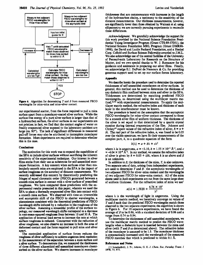
Figure 5 shows X versus time for virgin n;ica/silver contact, subsequent mica/silver contact, and contact between mica and octanethiol-coated silver. The differnce in X between the as- ymptotic limit for bare silver and that for monolayer-coated silver can be used to calculate the tbicknm of the monolayer, provided the refractive index, n, of the layer is known. Table I presents the thicknesses of three different monolayers as determined as-
5744.0
5743.0
0 10 20 30 40 50 60 70
Time (hours) Figure 5. Wavelength of a FECO versus time for virgin mica/silver contact (filed circles), subsequent mica/silver contact (open circles), and mica/octanethiol-coated silver contact (open squares). The two dashed lines are the (lower) asymptotic wavelengths of bare mica/silver contact and (upper) mica/octanethiol-coated silver contact. The difference be tween the wavelengths is used to calculate the monolayer thickness.
suming a refractive index of 1 S, which is approximately equal to the bulk value of the monolayer-forming compounds. The method used to determine monolayer thicknesses is presented in detail in the Appendix. We note that varying the refractive index of the monolayer from 1.4 to 1.6 changes the calculated thicknessea less than 1 A, which is of the same magnitude as our experimental error.
As a control experiment, we followed the procedure for the formation of a self-assembled monolayer, except we immersed the silver in pure ethanol only. Subsequent contact between the mica and silver was identical to that observed before the silver was soaked. This experiment also provided evidence that the glue used to adhere the mica sheets to the support disks was not dissolving in the ethanol (over the immersion times used here) and con- taminating the surfaces.
We performed three separate experiments to determine the thickness of a dodecanethiol monolayer. Two were based on the procedure described above, and the other was done in the following way. First, the thickness of an octanethiol monolayer was mea- sured, then the monolayer-coated silver surface was reimmersed in a solution of dodecanethiol for 5 h, then rinsed, and dried. The measured thickness of 13.5 A was the same as those measured in two previous experiments wherein the monolayer was formed directly on bare silver. This is consistent with the findings of others,3w33 who report the exchange of surface-bound thiol molecules with those in solution.
We measured the force required to pull monolayer-coated silver surfaces from contact with mica. To within our experimental scatter, there was no significant difference between these pull-off forces and those measured between bare silver and mica.
With regard to all observations reported above, the results were independent of the particular evaporator, the supplier of silver, or type of resistive boats that we used to prepare the silver films.
Finally, in their previous SFA experiments with mica and silver surfaces, Parker and ChristensonM put forth a method to prepare “fused” films by peeling silver off of an underlying mica sheet, thus exposing the surface that was formerly in contact with the mica. To do so, a thin mica sheet (24 pm thick), coated on one side with 270 A of silver, was pressed against a thick mica sheet (100 pm), coated with the same thickness of silver, so that their silvered sides were in contact. After approximately 2 days, Parker and Christenson report that the thin sheet could be peeled off, taking all of the silver with it. We were interested in repeating our experiments using “fused” films, since it is likely that the morphology of the silver surface at the mica/silver interface is different than at the air/siIver interface. Unfortunately, after several attempts, we were unable to get the silver to adhere ex- clusively to the thin sheet. Instead, tiny, randomly-scattered patches adhered to both the sheets, the majority of them on the thicker mica sheet, rendering the silver film useless for SFA experiments. One reason we did not pursue this further is that Parker and Christenson report no differences in solvation forces measured between mica and “fused” silver films as compared to
Reduction of the Roughness of Silver Films The Journal of Physical Chemistry, Vol. 96, No. 25, 1992 10409
those measured between mica and ‘simple” silver films (which we assume are films directly evaporated onto mica).
Diseuesion A plausible explanation of our reported observations is that the
roughness of a thermally evaporated silver film is reduced by contacting the silver surface with a molecularly smooth mica surface. This explanation is consistent with, and based on, a theoretical analysis which predicts the wavelength dependence of interference fringes generated between a smooth mica surface and a silver surface with prescribed roughness.25 We describe below the pertinent results of this analysis and use them to yield a quantitative estimate of the reduction of surface roughness.
Recently, we used the multilayer matrix method to predict the FECO for an interferometer consisting of a back-silvered smooth mica sheet in contact with a silver film whose surface topology was modeled as a sine wave with varying amplitude and fre- uency.25 We found that, provided the amplitude is less than 50 %, the FECO are nearly as sharp as those predicted for two mica
surfaces in contact. The most important result of our analysis is that the wavelength of a FECO (more precisely, the wavelength of the center of a FECO) decreases nearly linearly as the am- plitude is reduced. (This effect is insensitive to variations in the frequency of the sinusoidal topology.) Our measurements of the time-dependent and irreversible decrease in X when silver is contacted with mica are thus consistent with a time-dependent decrease in the degree of roughness. Assuming a sinusoidal surface morphology, the changes in X we observe, from the moment virgin contact is established until the asymptotic limit is achieved, correspond to decreases in the root-mean-squared surface roughness ranging from 10 to 30 A.
It is important to note that a given change in X can be related to a change in amplitude, but an absolute amplitude cannot be determined.25 On the other hand, the shape of the FECO (i.e., the intensity versus wavelength profiles) can be used to estimate the absolute degree of silver roughness. Since we measure the wavelength of the FECO by eye (by aligning a moving graticule with the center of the fringe), we cannot quantitatively establish fringe profiles, Le. the variation with wavelength of intensity. However, we can estimate visually the width (in wavelength) of the FECO, which we can use to set an upper bound on the am- plitude of the surface roughness. We fmd this to be approximately 50 A, which is in qualitative agreement with the results of other techniques used to determine the roughness of evaporated silver films. Using scanning-tunneling microscopy, Raether determined a root-mean-squared roughness of 5 A34 and Parker reports an -average roughness” of 30 A.3s Using surface plasmon excitation, Kretschmann,’6 and later Homauer,” determined a root-mean- squared roughness between 4 and 7 A.
In lieu of the reduction of surface roughness, an alternate explanation that might appear reasonable-but is in fact not supported by an associated FECO wavelength analysis-is that the silver film thins when it is pressed between the two mica sheets. Using the multilayer matrix method, we have determined the FECO wavelengths generated from an interferometer consisting of a smooth silver film, of variable thickness, between two mica sheets. We fmd that, as the thickness of the silver film decreases, the FECO shift to longer wavelengths. (For example, decreasing the thickness of a silver film from 550 to 500 A increases the FECO wavelengths by about 0.2 A.) In direct contrast to these predictions, the FECO in our experiments shift to shorter wavelengths (cf. Figures 3 and 5) .
As reported in the Results section, when silver and mica are in contact, we observed, concurrent with a decrease in A, an increase in both the area of contact and the pull-off force. To interpret these findings, we appeal to the adhesion theory of Johnson, Kendall, and Roberts38 which predicts that an increase in the energy of interaction between two deformable sphem results in an increase in both the area of contact and the pull-off force. Hence, our results are consistent with a timedependent increase in the energy of interaction between the silver and mica surfaces. Such an increase could be attributed to a reduction in the
roughness of the silver, thus leading to more intimate contact between the surfaces.
The only observation that we cannot explain in terms of the reduction of the roughness of the silver is the trend in the variation of force with distance as a mica surface and a silver surface approach. As mentioned previously, negligible forces were ob- served on the virgin approach, except in a few -, but sisnifcant attractive forces were observed on all subsequent approaches. Our best explanation for this is that electrical charge is being trans- ferred across the mica/silver interface while the surfaces are in contact, a phenomenon that has been observed by Hom and Smith39 for contact between a mica and a silica surface. As we have observed for mica and silver, Horn and Smith observed negligible forces for the virgin approach of mica and silica and attractive forces on all subsequent approaches. They showed that charge transfer was occurring across the mica/silica interface, creating an opposite charge on each surface that resulted in at- tractive electrostatic forces between the surfaces. Such charge transfer may also be ocurring in our experiments, but we have not attempted to verify or quantify this phenomenon.
Regardless of what is happening at the microscopic level to the silver surface, our fmdings establish a reproducible procedure that can be followed by others wishing to use silver surfaces in the SFA. Unfortunately, the SFA cannot provide direct information on the structure of the silver surface that would allow us to explain the mechanism behind the reduction of the silver roughness. We can speculate that silver, a highly malleable and ductile material, rearranges in response to having a smooth mica surface pressed against it, resulting in a smoother silver surface. Additional techniques which can directly image the structure of the silver surface, such as atomic force microscopy or scanning tunneling microscopy, are needed to understand exactly what is occurring at the surface of the silver.
With regard to our self-assembled monolayer thickness mea- surements (Table I), we have compared them with the ellipso- metrically determined thicknesses of Walczak et a1.t and we find that our measurements are lower. Specifically, our measured dodecanethiol and octadecanethiol thicknews are 7 A lower, and our octanethiol thickness is 10 A lower. Three differences between our experiments and those of Walczak et al. most likely account for the discrepancy. First, our monolayers are confined and compressed between mica and silver. Using the recent results of Israelachvili40 for the compressibility of Langmuir-Blodgett monolayers on mica, we estimate that the self-assembled mono- layers in our experiments are being compressed by about 1-3 A. Second, impurities on the silver surface (such as atmospheric hydrocarbons or adsorbed water) are most likely removed as the monolayer Our method of determining thickness does not account for the presence of a layer on the silver surface that is displaced by the monolyer. Thus, our reported monolayer thicknesses are too low by an amount q u a l to the thickness of the displaced impurity layer. Walczak et al. minimized the amount of surface impurities by immersing their silver films into alka- nethiol solution immediately after evaporation. Even though our silver is in a nitrogen environment, our SFA chamber is not perfectly airtight and may not be completely free from all airborne impurities. Over the many hours required to determine the as- ymptotic value of A (cf. Figure 5) , the layer of impurities on our silver surfaces may grow. Third, the amount of silver oxide on our silver surfaces prior to monolayer formation may be hindering the formation of a complete monolayer. As with the level of surface impurities, the oxide layer on our silver surfaces is most likely thicker than in the experiments of Walczak et al. Although there is evidence that thin layers of silver oxide are stripped away during monolayer formation, as are impurities,7p9 thick layers of silver oxide may not be completely displaced, and their presence interfem with complete monolayer formation. More investigations are needed to determine which of these factors, and possible other factors, account for the 7-10-A differences between our results and those of Walczak et al.
Finally, we observed that the force required to pull a mica surface apart from a bare silver surface was no different, to within
10410 The Journal of Physical Chemistry, Vol. 96, No. 25, 1992 Levins and Vanderlick
I I I 1
Measure two adjacent FECO wavelengths for mica-silver contact at the asymptotic limit
Measure two adjacent FECO wavelengths for
mica-mica contact
Input an initial
Decrease 0 FECO wavelengths match those observed for mica-mica contact
to within 0.1A
Increase B
I I Calculated Calculated
within 0.1 A ?
Figure 6. Algorithm for determining T and @ from measured FECO wavelengths for mica-mica and mica-silver contact.
our experimental scatter, from the force required to pull a mica surface apart from a monolayer-coated silver surface. While the surface free energy of a pure silver surface is larger than that of a hydrocarbon surface, the silver surfaces in our experiments are not pristine; in fact, we find that the contact angles of water on freshly evaporated silver exposed to the laboratory ambient are large (ca. go0), The lack of significant differences in measured pull-off forces may also be attributed to incomplete monolayer formation. More experiments are required to determine whether this is the case.
C O n C l d ~ The motivation for this work was to expand the capabilities of
the SFA to include silver surfaces without sadicing the inherent sensitivity of the experimental technique. Our interest in silver films stems from their use as substrates for self-assembled mon- olayer formation. A key concern when surfaces other than mo- lecularly smooth mica are employed in the SFA is the impact of surface roughness on the accuracy of distance measurements. We recently addressed this concern by theoretically predicting the fringes of equal chromatic order (FECO) generated between a smooth mica surface in contact with a silver surface of prescribed roughness. We have compared those predictions with the ex- perimental results presented in this paper, wherein we used the SFA to place a thermally evaporated silver film into contact with a smooth mica surface. We observed with time an irreversible and asymptotic shift of the FECO to shorter wavelengths, a phenomenon consistent with the theoretical predictions of FECO wavelength shifts induced by a reduction in the roughness of the silver surface. Assuming a sinusoidal profile of the surface to- pology, our measured wavelength shifts compond to a decrease in root-mean-squared roughness from between 10 and 30 A. The application of external load serves to increase the rate at which surface roughness is reduced. Concurrent with the reduction of silver roughness, we also observe an increase in both the area of deformed contact and the force required to pull mica and silver apart.
The controlled application of surface forces reduces the roughness of silver sufkiently to achieve angstrom-level resolution in the measurement of the separation between a mica surface and a silver surface. To demonstrate this, we measured the thickncssea of three different alkanethiol self-assembled monolayers chemi- sorbed on the silver surface. We observe increases in monolayer
thicknesses that are commensurate with increases in the length of the hydrocarbon chains, a testimony to the sensitivity of the distance measurements. Our thickness measurements, however, are significantly lower than those obtained by Walczak et al. using ellipsometry; we are currently pursuing experiments to reconcile these differences.
Acknowledgment. We gratefully acknowledge the support for this work provided by the National Science Foundation Presi- dential Young Investigator Program (Grant CTS-89-57051), the National Science Foundation MRL Program (Grant DMR88- 1998), the David and Lucile Packard Foundation, and a Henkel Corp. Colloid and Surface Science Fellowship awarded to J.M.L. We also acknowledge use of the central facilitica of the University of Pennsylvania Laboratory for Research on the Structure of Matter, and we owe special thanks to W. J. Romanow for his guidance and assistance in preparing our silver films. Finally, we acknowledge E.I. DuPont de Nemours & Co. for providing generous support used to set up our surface forces laboratory.
Appendix Wedescribe herein the procedureused todetermine the reported
thicknesses of self-assembled monolayers on silver surfaces. In general, this method can be used to determine the thickness of any dielectric film codined between mica and silver in the SFA. Thicknesses are determined by matching predicted FECO wavelengths, as determined using the multilayer matrix me- thod,22942 with experimental measurements. To apply the mul- tilayer matrix method, the refractive index and thickness of each layer in the interferometer must be known.
Our procedure is based on the assumption that the asymptotic FECO wavelengths for mica-silver contact correspond to those for a smooth silver film of uniform thickness. The thickness of the silver is set q u a l to that determined by a quartz crystal monitor during thermal evaporation of the film. Johnson and Christf13 report values of the refractive index of silver, if = n + ik. The real part of the refractive index, n, was found to be 0.05 over the visible spectrum; we have fit their measurements of the complex part, k, to a quadratic equation of the form
&(A) = u + bX + cX2
where A is in angstroms, u = -3.10, b = 1.55 X lW3 A-l, and c = -6.04 x 10⁻⁸ A-2. ~n our method, we assume the refractive index of silver is given by if = 0.05 + iBk, where k is as above and @ is an unknown.
In addition to 8, the thickness of the mica, T, is also unknown. Two separate sets of data, arising from independent experiments, are used to determine T and 8: the asymptotic wavelengths of two adjacent FECO for silver-mica contact and the wavelengths of two adjacent FECO for mica-mica contact. All of the mica sheets used in both experiments are cut from the same large sheet of uniform thickness. For the refractive index of mica we usel
4.76 x 10⁵ n(X) = 1.5820 +
A2
where A is the wavelength of light in angstroms. Using the multilayer matrix method, we iteratively converge on values of T and @ such that the predicted FECO wavelengths match those observed in the two separate experiments; the algorithm is shown in Figure 6. For 10 separate experiments, we have found the average of 8 to be 0.86, with a standard deviation of 0.06 and a range from 0.74 to 0.94.
To determine the thicknesses of self-assembled monolayers, we use the multilayer matrix method to predict the FECO wave- lengths when a dielectric layer is inserted between the mica and silver (with T and 8 as determined above). The refractive index of the monolayer is assumed to be 1.5. The monolayer thichess is systematically increased until the wavelength of the measured FECO matches that which is predicted to within 0.1 A. Refenaccs and Notes
( I ) Israelachvili, J. N.; Adam, G. E. J. Chcm. SOC, Furaduy Trans. 1 1978, 74, 915.
J. Phys. Chem. 1992,96, 10411-10418 10411
(2) Horn, R. G.; Smith, D. T.; Haller, W. Chem. Phys. Lett. 1989,162,
(3) Merrill, W. W.; Pocius, A. V.; Thaklrcr, B. V.; Tirrell, M. Lungmuir
(4) Horn, R. G,; Clarke, D. R.; Clarkson, M. T. J. Mater. Res. 1988, 3,
(5) Smith, C. P.; Maecla, M.; Atanssoska, L.; White, H. S.; McClure, D.
(6) Walczak, M. M.; Chung, C.; Stole, S. M.; Widrig, C. A.; Porter, M.
(7) Laibinb,P. E.; Whitesides,G. M.J. Am. Chem.Soc. 1992,114,1990. (8) Bryant, M. A.; Pemberton, J. E. J. Am. Chem. Soc. 1991,113,3629. (9) Laibinis, P. E.; Whitesides, G. M.; Allara, D. L.; Tao, Y.; Parikh, A.
N.; N u w , R. G. J. Am. Chem. Soc. 1991,113,7152. (10) Laibinis, P. E.; Fox, M. A.; Folkers, J. P,.; Whitesides, G. M. Lung-
muir 1991, 7, 3167. (1 1) Fenter, P.; Eisenberger, P.; Li, J.; Camillone 111, N.; Bernasek, S.;
Scala, G.; Ramanarayanan, T. A.; Liang, K. S. Lungmuir 1991, 7, 2013. (12) Parscgian, V. A.; Rand, R P.; Fuller, N. L. J. Phys. Chem. 1991,95,
4777. (13) Israclachvili, J. N. Chem. Scr. 1985, 25, 7. (14) Ninham, B. W. Chem. Scr. 1985,25, 3. (15) Israelachvili, J. N.; Wcnnerstrbm, H. J. Phys. Chem. 1992,96,520. (16) Israelachvili, J. N.; Wennerstr6m, H. Lungmuir 1990, 6, 873. (17) Israelachvili, J. N.; Pashley, R. M. J. Colloid Interface Sci. 1984.98,
(18) Tsao, Y.; Yang, S. X.; Evans, D. F.; Wcnncrstr6m. H. Lungmuir
(19) Clawson, P. M.; Christenson, H. K. J. Phys. Chem. 1988, 92, 1650. (20) Tolansky, S . Multiple-Beam Interferometry of Surfaces and Films;
404.
1991, 7, 1975.
413.
J. J. Phys. Chem. 1988,92, 199.
D. J. Am. Chem. Soc. 1991,113,2370.
500.
1991, 7, 3154,
Oxford University Press: London, 1948.
(21) Israclachvili, J. N. J . Colloid Interfocr Sci. 1973,44, 259. (22) Clarkson, M. T. J. Phys. D 1989,22,475. (23) Coaklev. C. J.: Tabor. D. J. Phvs D 1978. 11. L77. (24j Parker,- J. L.; Christenson, H. K. J. Chem. Phys. 1988.88, 8013. (25) Levins, J. M.; Vandcrlick, T. K. J. Colloid Inrer/cm Sci., submitted
(26) Vanderlick, T. K.; Scrivcn, L. E.; Davis, H. T. Colloids Surf. 1991, for publication.
52. 9. (27) Lodge, K. B.; Mason, R. Proc. R. Soc. London, A 1982,383,279. (28) Derjaguin, B. V. Kolloid Z. 1934,69, 155. (29) Horn, R. G.; Israelachvili, J. N.; Pribac, F. J. Colloid Interfocc Sci.
(30) Chidsey, C. E. D.; Bertozzi, C. R.; Putvinski, T. M.; Mujscc, A. M.
(31) Hiclunan, J. J.; Ofer, D.; Zou, C.; Wrighton, M. S.; Laibinia, P. E.;
(32) Collard, D. M.; Fox, M. A. Lungmuir 1991, 7, 1192. (33) Creager, S. E.; Hockctt, L. A.; Rowe, 0. K. Lungmuir 1992,8,854. (34) Raether, H. Surf. Sci. 1984, 140, 31. (35) Parker, J. L. Langmuir 1992,8,551. (36) Krctschmann, E . Opt. Commun. 1974,10, 353. (37) Hornaucr, D. L. Opt. Commun. 1976,16,76. (38) Johnson, K. L.; Kendall, K.: Roberts, A. D. Proc. R. Soc. London,
(39) Horn, R. G.; Smith, D. T. Science 1992,256,362. (40) Chen, Y. L.; Helm, C. A.; braclachvili, J. N. kuyFtuir 1991,7,2694. (41) Bain, C. D.; Troughton, E. B.; Tao, Y. T.; Evall, J.; Whitesides, G.
(42) Born, M.; Wolf, E. Principles of Optics, 6th cd.; Perpmon Press:
(43) Johnson, P. B.; Christy, R. W. Phys. Rev. B 1972, 6, 4370.
1987, 115, 480.
J. Am. Chem. Soc. 1990,113,4301.
Whitesides, G. M. J. Am. Chem. Soc. 1991,113, 1128.
A 1971,324, 301.
M.; Nuzzo, R. G. J. Am. Chem. Soc. 1989,111,321.
New York, 1980; pp 51-71.
Electron Spln Resonance and Electron Spin Echo Studies of Cu(I1) Ion Location and Coordination Geometry in Na-, K-, and Rb-SAPO-42
Maggie Zamadics and Larry Kevan*
Department of Chemistry, University of Houston, Houston, Texas 77204-5641 (Received: June 24, 1992; In Final Form September 22, 1992)
Cu(I1) ions in Na+, K+, and Rb+ cationic forms of SAPO-42 are investigated. The various Cu(I1) species generated after dehydration and exposure to adsorbates are examined with electron spin resonance and electron spin echo modulation techniques. The results are interpreted in terms of Cu(I1) ion location and coordination geometry. The conclusions are compared to those of Cu(I1) ions in zeolite A, which is the aluminosilicate structural analog of SAPO-42. In rehydrated samples of Na- and K-SAPO-42 the Cu(I1) ions are pentacoordinated to two water molecules and three framework oxygens. In contrast, Cu(I1) ions in rehydrated samples of CuNa-A and CuK-A are octahedrally coordinated to three water ligands and three framework oxygens. In a hydrated sample of CuRb-SAPO-42 the Cu(I1) ion is tetrahedrally coordinated to three zeolitic oxygens and one water molecule. A similar coordination is reported for Cu(I1) ions in CuRb-A zeolite. The Cu(I1) ion in all samples equilibrated with methanol interacts with two molecules of methanol. In CuNa- and CuK-SAPO-42 samples the Cu(1I) ion has rhombohedral symmetry while in CuRb-SAFQ-42 the Cu(I1) ion Coordinam with two molecules of methanol and three framework oxygens in a trigonal bipyramidal geometry. In all the cationic forms of SAPO-42 the Cu(I1) ion is found to interact with one molecule of ethylene and two molecules of ethanol. The differences observed between the various forms of SApo-42 are most probably due to electrostatic effects brought about by the cocations while steric effects are unlikely due to the low cation exchange properties of these materials. The differences in the Cu(I1) ion behavior between SAPO-42 and zeolite A can be attributed to the differences in the cation densities of SAPO-42 versus zeolite A.
Iatrodpctioa Characterization of Cu(I1) ions in the silicoaluminophosphate
(SAPO-n') molecular sieves SAPO-5,2 SAPO-1 1:s' and SAPO- 345 have recently been reported. Both SAPO-S and SAPO-1 1 have structures topologically related to novel structure types of the aluminophosphate class of molecular sieve while SAPO-34 is structurally similar to chabezite, a naturally ocarrhg wlite.6 Comparing results concerning cation location and adsorbate in- teraction of Cu(I1) ions in silicoaluminophosphate materials to their structural analogs from the aluminosilicate class of molecular sieves provides information useful in characterizing these new mol& Sieves. Recent studies Conparing the Cu(I1) ion location
OO22-3654/92/ 2096-1 04 1 lS03 .OO/O
and coordination in SAPO-34 to chabazite illustrates an effect on the coordination geometry of the Cu(I1) ion due to the dif- ferences in the cation densities of these materials.' In this study a similar comparison is made between like cationic forms of SAPO-42 and zeolite A to better characterize SAPO-42.
The basic building units of SAPO-n molecular sieva are AIOi, PO2+, and Si02 tetrahedra. Because the number of AlOl tet- rahedra are greater than the number of PO2+ tetrahedra, the lattice of SAPO-42 is negatively charged. Thus, extraframework cations are required to balance the anionic framework. sAPo-42 material contains both sodium ions from the synthds gel and protons from the dcannpition of the tunplating agent as chargacompensatine
Q 1992 American Chemical Society