Embed Size (px)
Citation preview
2000357 (1 of 10) © 2020 Wiley-VCH GmbH
www.mbs-journal.de
ReseaRch aRticle
Silk Protein Paper with In Situ Synthesized Silver Nanoparticles
Yujia Liang, Bin Tang, Aarushi Sharma, Dinidu Perera, Benjamin James Allardyce, Sourabh Ghosh, Hannes C. Schniepp, and Rangam Rajkhowa*
Dr. Y. Liang, Dr. B. Tang, Dr. B. J. Allardyce, Dr. R. RajkhowaInstitute for Frontier MaterialsDeakin UniversityGeelong, VIC 3216, AustraliaE-mail: [email protected]. A. Sharma, Prof. S. GhoshDepartment of Textile and Fibre EngineeringIndian Institute of Technology DelhiNew Delhi 110016, IndiaDr. D. Perera, Prof. H. C. SchnieppDepartment of Applied ScienceWilliam & MaryWilliamsburg, VA 23187-8795, USA
The ORCID identification number(s) for the author(s) of this article can be found under https://doi.org/10.1002/mabi.202000357.
DOI: 10.1002/mabi.202000357
sponges, etc., to meet various application requirements.[2–8] The silk dissolution pro-cess, however, destroys the hierarchically assembled structures within silk fibers consisting of nanoscale- and mesoscale-width fibrils. In contrast, the exfoliation of silk fibers to isolate fibrils without dis-solving to individual protein molecules can preserve the original secondary struc-ture and β-sheet nanocrystals within these fibrils. Such meso/nanofibrils offer opportunities to create new types of silk materials.[9] Fibrils in the mes-oscales or nanoscales have been success-fully extracted to produce membranes or papers;[10–12] however, these processes still rely on chemical extraction and often take days to weeks to peel the fibrils from silk fibers. We have demonstrated that envi-ronment friendly and scalable approaches can be applied without using any harmful chemicals to produce microfibrillated silk (MFS) for use in silk protein papers.[13,14]
The aim of this work was to use our exfoliated silk nanofibers for in situ synthesis of silver nanopar-ticles (AgNPs) and produce novel protein papers from AgNPs coated MFS (MFS-AgNPs) suspensions. The MFS produced by our shear induced fibrillation was used as templates to reduce silver salt to AgNPs. Noble metal nanoparticles including silver and gold have been used to provide specific functions to fibers/fabrics.[15–19] For example, AgNPs were used in the past with silk-based matrices for biomedical materials and smart textiles/nanotextiles.[18,20] We believe that AgNPs as a type of broad-spectrum antibacterial agents have been extensively used in various fields. The antibacterial activities of AgNPs on different substrates have been widely analyzed.[21–24] AgNPs possess high antimicrobial activity even at a relatively low concentration compared with other metal nanoparticles, while the low release of silver means they are not toxic to human cells.[25] In the lit-erature, no cytotoxicity was found in the substrates with AgNPs for wound healing.[26–28] In addition, by obtaining anisotropic AgNPs, the modified substrates can present various colors due to the localized surface plasmon resonance (LSPR) properties of AgNPs.[17,18] As the LSPR of AgNPs is sensitive to their shape and size,[29,30] the color of AgNPs has been effectively tuned by controlling the morphologies of particles.
One of the challenging problems with metal nanoparticles is their tendency to aggregate on the surface of treated materials.
Silver nanoparticles (AgNPs) are in situ synthesized for the first time on microfibrillated silk (MFS) exfoliated from domesticated Philosamia cynthia ricini (eri) and Bombyx mori (mulberry) silkworm silk fibers. The process is rapid (hours time), does not rely on harmful chemicals, and produces robust and flexible AgNPs coated MFS (MFS-AgNPs) protein papers with excellent handling properties. None of these can be achieved by approaches used in the past to fabricate AgNPs silk systems. MFS bonds the AgNPs strongly, providing good support and stabilization for the NPs, leading to strong wash fastness. The mechanical properties of the MFS-AgNPs papers largely do not change compared to the MFS papers without nanoparticles, except for some higher concentration of AgNPs in the case of mulberry silk. The improved tensile properties of eri silk papers with or without AgNPs compared to mulberry silk papers can be attributed to the higher degree of fibrillation achieved in eri silk and its inherent higher ductility. MFS-AgNPs from eri silk also exhibit strong antibacterial activity. This study provides the basis for the development of smart protein papers based on silk fiber and functional nanomaterials.
1. Introduction
Silk has emerged as an important material for biomedical and other advanced applications including for sensing, electronics, and photonics.[1] There are limited options for using native fiber or silk textiles for such non-textile uses and therefore silk fiber is often dissolved, then reconstituted using liquid–solid-phase transfer to fabricate nanofibers, films, microspheres, hydrogels,
Macromol. Biosci. 2020, 2000357
© 2020 Wiley-VCH GmbH2000357 (2 of 10)
www.advancedsciencenews.com www.mbs-journal.de
It was reported silk nanofibers regenerated from silk solution offered an advantage in this aspect since their large surface area helps to keep AgNPs spatially distributed, acting as tem-ples while preventing aggregation of nanoparticles.[31] However, the process of preparing silk solution is time consuming, costly, and depend on toxic salts. More importantly, the silk nanofibers did not act as a reducing agent for AgNP production. To pro-duce AgNPs, dandelion (Tridax procumbens) leaf extracts have been used to reduce silver salt.[31] Additionally, this prepara-tion of silk nanofibers with AgNPs was also time consuming, requiring up to 2 days to synthesize and stabilize the AgNPs. There is another report of in situ synthesis of AgNPs in a single step using silk solution that is cast into a containing film containing AgNPs.[32] The resulting silk film with 0.5% silver nitrate (AgNO3) maintained antimicrobial performance for more than 1 week. In addition, silk films produced from silk solution are typically brittle when dry, restricting their potential use.
This study synthesized AgNPs using silk nanofibers in a rapid single-step process without relying on harmful chemicals in nanofiber fabrication process. We also aimed to retain the inherent microstructure of native silk to produce nanofiber-based papers that are strong and flexible, and thus make flex-ible materials that have greater potential use than brittle regen-erated materials. Hence, we relied on producing nanofibers following top down approach of milling and homogenization. Both the nanofiber production method and silver nanoparticle (AgNP) production method are achieved without any harmful chemicals, which makes silk nanofibers resistant to fast deg-radation.[13,14,33–35] Here, following our recent work on milling and homogenization, exfoliated MFS were produced, and the fibers were used for the first time for reducing silver salt to syn-thesize AgNPs. Finally, MFS-AgNPs functionalized papers were obtained. The entire process from silk fibers to MFS-AgNPs requires hours compared with the published, more complex methods which typically require days.
Two types of silk were used in this work, mulberry silk from Bombyx mori silkworms and eri silk from Samia cynthia ricini silkworms. Owing to the large-scale cultivation and cost efficiency, mulberry silk has been well explored for a variety of applications.[36–39] Eri silk, which is simple to rear because of stronger disease resistance and widely available host plant castor, is also becoming popular as a commercial silk fiber.[40,41] Although eri silk is relatively less explored, its amino acid com-position and fiber structure are significantly different to mul-berry silk.[42,43] Both silk fibers contain aspartic acid and glu-tamic acid that are known to have the capacity to reduce silver ions to AgNPs[44–47] and are hence used in this work. Two silk-based papers with different loading of AgNPs were compared and the morphological, structural, and antibacterial properties of papers were comprehensively investigated.
There are excellent prospects for use of these materials in healthcare applications. Many studies have shown that keratinocytes, fibroblasts, and other cell types proliferate on silk fibroin scaffolds.[42] Silk membranes have been studied as an epidermal scaffold on the wound bed to stimulate the forma-tion of a new epidermal barrier as well as showing potential as a wound dressing.[48] Paper like material prepared from electro-spun silk nanofibers from eri silk showed promising results for
wound healing in animal model. However, eri silk protein had to be extracted from the silk gland of eri silkworms, making the approach challenging for commercial use. Moreover, elec-trospinning has its challenges for use in regular production of nanofibers. Commercial silk patches have been developed to treat burn and surgical wounds where silk liquid is coated on fabrics and silver is used in many commercial products to prevent infection.[49–52] Direct application of silk film has limita-tions due to its lack of flexibility, brittleness, and low gas per-meability. A flexible paper patch with strong mechanical prop-erties combined with antibacterial properties is likely to be suit-able and a superior alternative to existing products. The ability to develop nanofibers from all types of silk and with potential for large-scale production makes our process unique for devel-oping wound-care paper membranes. We have demonstrated that water penetration properties of our papers can be con-trolled that is critical for wound-care application.[13] Thus, this work of silk paper with AgNPs offers an innovative alternate material for further work in this area. Silk papers with adequate modifications and added functionalities could be useful for many specific applications. One potential application is dental barrier membrane where such paper-like membranes promote penetration of bone-forming cells but prevent infiltration of unwanted epithelial cells.[53] Currently, collagen-based materials are used but face challenges of faster degradation than needed and high costs. Excellent potential of silk in such applications has been cited and use of cocoon membranes has been sug-gested.[54] The nanofiber membrane discussed here is a much advanced material for such applications. This study provides an initial report of fabrication of antibacterial protein paper with AgNPs. It will open opportunities for more studies to explore many potential healthcare applications.
2. Results and Discussion
2.1. Optical Characterization
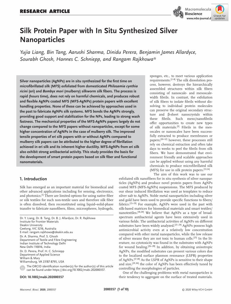
An MFS suspension suitable for paper formation was produced using milling and three passes through the high-pressure homogenizer. Additional passes did improve exfoliation and, considering the energy input in the homogenization process, the process was restricted to three passes. The preparation pro-cess and the method to measure aspect ratio of the MFS were reported earlier[13,55] and are therefore not included here. After treatment with different concentrations of AgNO3 (0, 10−4, 2 × 10⁻⁴, 3 × 10⁻⁴, and 4 × 10⁻⁴ m), the papers were denoted by E-MFS ∼ E-MFS-AgNPs-4 or M-MFS ∼ M-MFS-AgNPs-4, respectively. Figure 1 shows the formation of eri microfibrillated silk (E-MFS) paper and mulberry microfibrillated silk (M-MFS) papers from respective MFS slurries when cast and dried at room temperature. The results shown in Figure 1 confirmed that there was no negative impact on the paper formation ability of MFS due to reaction conditions to produce AgNPs. The color of the paper slightly turned faint beige when AgNPs were syn-thesized with the AgNO3 concentration as low as 1 × 10⁻⁴ m, indicating the existence of the synthesized AgNPs within the E-MFS and M-MFS suspensions. The color of the paper was deepened from light yellow (2 × 10⁻⁴ m), yellow (3 × 10⁻⁴ m) to
Macromol. Biosci. 2020, 2000357
© 2020 Wiley-VCH GmbH2000357 (3 of 10)
www.advancedsciencenews.com www.mbs-journal.de
bright yellow (4 × 10⁻⁴ m), meaning that more AgNPs were syn-thesized with the increase of initial AgNO3 concentration. For the same concentration of AgNO3, the M-MFS papers appeared darker than E-MFS papers, although there was no remarkable difference in whiteness between the two MFS papers without AgNPs. According to the difference in amino acid composition in eri and mulberry silk, this may indicate that more functional groups responsible for reduction of silver ions on M-MFS were presented than E-MFS and assisted the reduction of silver ions and produced more AgNPs (darker colors).
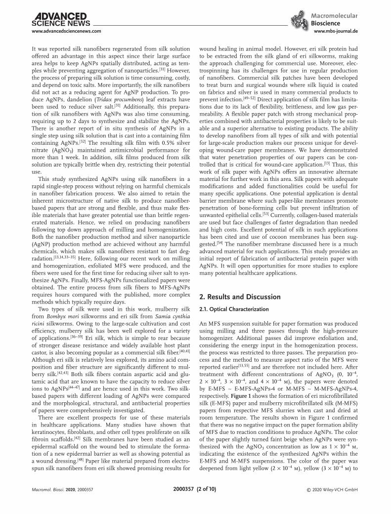
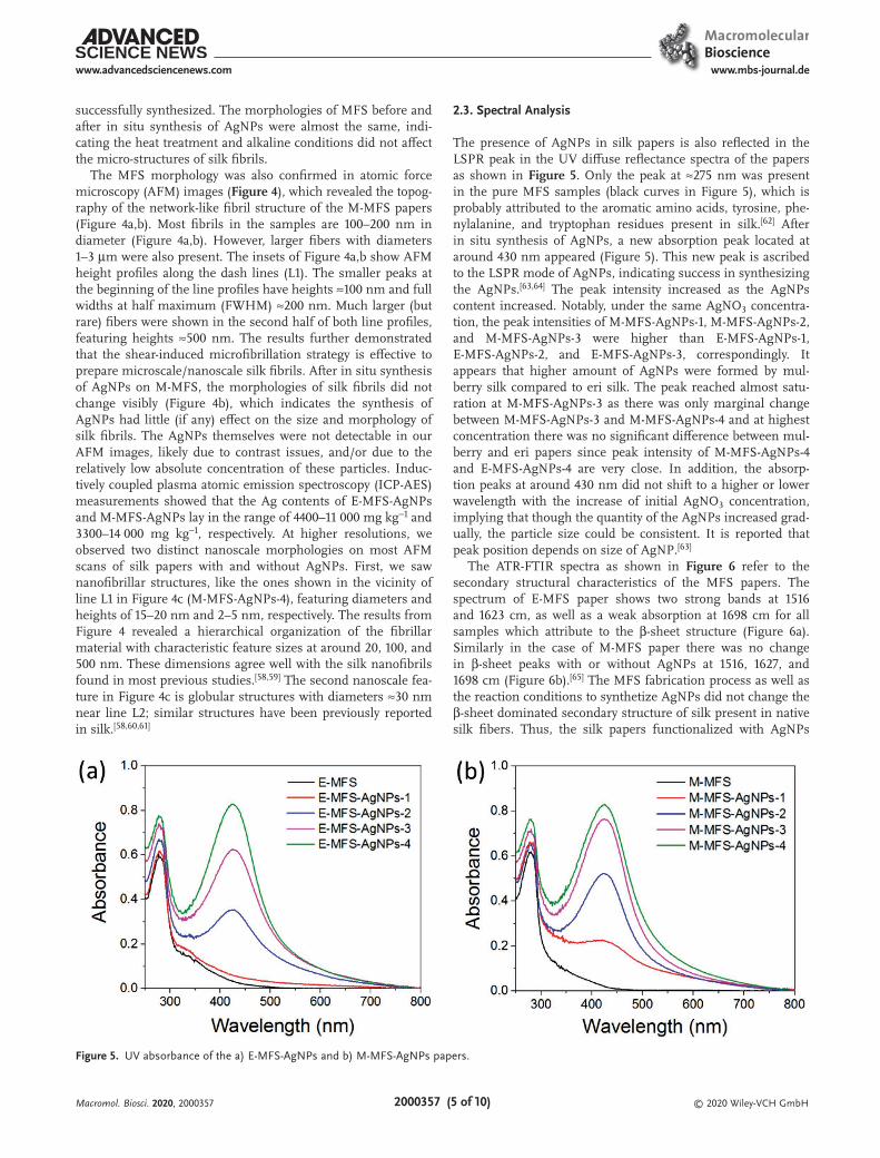
To further investigate the color changes of the papers from the formation of AgNPs, color strength (K/S) plots were
obtained by spectrophotometric measurement. A higher K/S indicates a higher strength of color. All the peaks of K/S curves for the AgNP-modified MFS papers are located around 430 nm (Figure 2a,b). The maximum K/S value of the E-MFS-AgNPs papers increased from 0.05 to 3.58 when the AgNO3 concen-tration increased from 1 × 10⁻⁴ to 4 × 10⁻⁴ m, which is con-sistent with the changes of colors of the E-MFS papers with AgNPs in Figure 1a. The M-MFS-AgNPs papers also showed the similar trend of color changes as E-MFS-AgNPs papers (from 0.55 to 5.89). However, compared with the E-MFS-AgNPs papers, the M-MFS-AgNPs papers displayed higher K/S values for a given AgNO3 concentration, exhibiting dark
Macromol. Biosci. 2020, 2000357
Figure 1. Optical images of the a) E-MFS-AgNPs papers and b) M-MFS-AgNPs papers with different initial AgNO3 concentration.
Figure 2. K/S plots of the a) E-MFS-AgNPs papers, b) M-MFS-AgNPs papers, and c) E-MFS-AgNPs-4 and M-MFS-AgNPs-4 after washing.

© 2020 Wiley-VCH GmbH2000357 (4 of 10)
www.advancedsciencenews.com www.mbs-journal.de
colors in Figure 2b. Durability of AgNPs on MFS papers was inspected by comparing the colors of the papers with AgNPs before and after washing. Color measurement was conducted to find out if the nanoparticles could be removed from the MFS papers by washing. It is evident that there was no notable change in E-MFS-AgNPs-4 and M-MFS-AgNPs-4 after washing (Figure 2c), suggesting that the AgNPs were firmly immobi-lized to MFS. The results reveal that there is a strong interac-tion between the fibrils and the AgNPs.
2.2. Morphological Analysis
The scanning electron microscopy (SEM) images of MFS in Figure 3 show that the exfoliation process assisted by milling
and homogenization produced submicron fibrils which assisted formation of papers due to high degree of entanglement and physical interactions between fibrils. We have already demon-strated that the exfoliation process results in individual fibrils in submicron dimensions as well as branched out from orig-inal fibers allowing to form network structure.[14] The width of these exfoliated fibrils was between 100 and 400 nm (Figure 3a). These MFS can be defined also as nanofiber as electrospun polymeric fibers since most silk fibers of less than 1 µm are termed as nanofiber in the published work.[56,57]
The in situ synthesis of AgNPs on nanofibers was further investigated by SEM. After the reaction of AgNO3 in the MFS suspension (as low as 1 × 10⁻⁴), some particles were observed on the nanofibers (Figure 3b,d) in comparison with pristine silk nanofibers (Figure 3a,c), indicating that the AgNPs were
Macromol. Biosci. 2020, 2000357
Figure 3. SEM images of a) E-MFS, b) E-MFS-AgNPs-1, c) M-MFS, and d) M-MFS-AgNPs-1. The red arrows are pointing to the AgNPs.
Figure 4. 3D-rendered AFM topography images of silk papers. a) M-MFS (no Ag particles). b,c) M-MFS-AgNPs-4. Insets: AFM height profiles along the dashed lines (L1 in (a) and (b) and L1,L2 in (c)).
© 2020 Wiley-VCH GmbH2000357 (5 of 10)
www.advancedsciencenews.com www.mbs-journal.de
successfully synthesized. The morphologies of MFS before and after in situ synthesis of AgNPs were almost the same, indi-cating the heat treatment and alkaline conditions did not affect the micro-structures of silk fibrils.
The MFS morphology was also confirmed in atomic force microscopy (AFM) images (Figure 4), which revealed the topog-raphy of the network-like fibril structure of the M-MFS papers (Figure 4a,b). Most fibrils in the samples are 100–200 nm in diameter (Figure 4a,b). However, larger fibers with diameters 1–3 µm were also present. The insets of Figure 4a,b show AFM height profiles along the dash lines (L1). The smaller peaks at the beginning of the line profiles have heights ≈100 nm and full widths at half maximum (FWHM) ≈200 nm. Much larger (but rare) fibers were shown in the second half of both line profiles, featuring heights ≈500 nm. The results further demonstrated that the shear-induced microfibrillation strategy is effective to prepare microscale/nanoscale silk fibrils. After in situ synthesis of AgNPs on M-MFS, the morphologies of silk fibrils did not change visibly (Figure 4b), which indicates the synthesis of AgNPs had little (if any) effect on the size and morphology of silk fibrils. The AgNPs themselves were not detectable in our AFM images, likely due to contrast issues, and/or due to the relatively low absolute concentration of these particles. Induc-tively coupled plasma atomic emission spectroscopy (ICP-AES) measurements showed that the Ag contents of E-MFS-AgNPs and M-MFS-AgNPs lay in the range of 4400–11 000 mg kg−1 and 3300–14 000 mg kg−1, respectively. At higher resolutions, we observed two distinct nanoscale morphologies on most AFM scans of silk papers with and without AgNPs. First, we saw nanofibrillar structures, like the ones shown in the vicinity of line L1 in Figure 4c (M-MFS-AgNPs-4), featuring diameters and heights of 15–20 nm and 2–5 nm, respectively. The results from Figure 4 revealed a hierarchical organization of the fibrillar material with characteristic feature sizes at around 20, 100, and 500 nm. These dimensions agree well with the silk nanofibrils found in most previous studies.[58,59] The second nanoscale fea-ture in Figure 4c is globular structures with diameters ≈30 nm near line L2; similar structures have been previously reported in silk.[58,60,61]
2.3. Spectral Analysis
The presence of AgNPs in silk papers is also reflected in the LSPR peak in the UV diffuse reflectance spectra of the papers as shown in Figure 5. Only the peak at ≈275 nm was present in the pure MFS samples (black curves in Figure 5), which is probably attributed to the aromatic amino acids, tyrosine, phe-nylalanine, and tryptophan residues present in silk.[62] After in situ synthesis of AgNPs, a new absorption peak located at around 430 nm appeared (Figure 5). This new peak is ascribed to the LSPR mode of AgNPs, indicating success in synthesizing the AgNPs.[63,64] The peak intensity increased as the AgNPs content increased. Notably, under the same AgNO3 concentra-tion, the peak intensities of M-MFS-AgNPs-1, M-MFS-AgNPs-2, and M-MFS-AgNPs-3 were higher than E-MFS-AgNPs-1, E-MFS-AgNPs-2, and E-MFS-AgNPs-3, correspondingly. It appears that higher amount of AgNPs were formed by mul-berry silk compared to eri silk. The peak reached almost satu-ration at M-MFS-AgNPs-3 as there was only marginal change between M-MFS-AgNPs-3 and M-MFS-AgNPs-4 and at highest concentration there was no significant difference between mul-berry and eri papers since peak intensity of M-MFS-AgNPs-4 and E-MFS-AgNPs-4 are very close. In addition, the absorp-tion peaks at around 430 nm did not shift to a higher or lower wavelength with the increase of initial AgNO3 concentration, implying that though the quantity of the AgNPs increased grad-ually, the particle size could be consistent. It is reported that peak position depends on size of AgNP.[63]
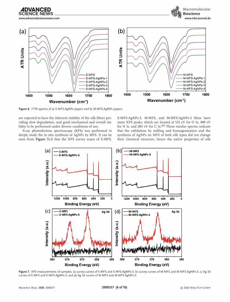
The ATR-FTIR spectra as shown in Figure 6 refer to the secondary structural characteristics of the MFS papers. The spectrum of E-MFS paper shows two strong bands at 1516 and 1623 cm, as well as a weak absorption at 1698 cm for all samples which attribute to the β-sheet structure (Figure 6a). Similarly in the case of M-MFS paper there was no change in β-sheet peaks with or without AgNPs at 1516, 1627, and 1698 cm (Figure 6b).[65] The MFS fabrication process as well as the reaction conditions to synthetize AgNPs did not change the β-sheet dominated secondary structure of silk present in native silk fibers. Thus, the silk papers functionalized with AgNPs
Macromol. Biosci. 2020, 2000357
Figure 5. UV absorbance of the a) E-MFS-AgNPs and b) M-MFS-AgNPs papers.
© 2020 Wiley-VCH GmbH2000357 (6 of 10)
www.advancedsciencenews.com www.mbs-journal.de
are expected to have the inherent stability of the silk fibers pro-viding slow degradation, and good mechanical and overall sta-bility to be performed under diverse conditions of use.
X-ray photoelectron spectroscopy (XPS) was performed to deeply study the in situ synthesis of AgNPs by MFS. It can be seen from Figure 7a,b that the XPS survey scans of E-MFS,
E-MFS-AgNPs-3, M-MFS, and M-MFS-AgNPs-3 films have same XPS peaks, which are located at 531 eV for O 1s, 400 eV for N 1s, and 285 eV for C 1s.[66] These similar spectra indicate that the exfoliation by milling and homogenization and the synthesis of AgNPs on MFS of both silk types did not change their chemical structure, hence the native properties of silk
Macromol. Biosci. 2020, 2000357
Figure 6. FTIR spectra of a) E-MFS-AgNPs papers and b) M-MFS-AgNPs papers.
Figure 7. XPS measurements of samples: a) survey curves of E-MFS and E-MFS-AgNPs-3, b) survey curves of M-MFS and M-MFS-AgNPs-3, c) Ag 3d curves of E-MFS and E-MFS-AgNPs-3, and d) Ag 3d curves of M-MFS and M-MFS-AgNPs-3.
© 2020 Wiley-VCH GmbH2000357 (7 of 10)
www.advancedsciencenews.com www.mbs-journal.de
were maintained, offering the potential for new functionality to be added to the modified materials. In the XPS Ag 3d curves (Figure 7c,d), there are no peaks in E-MFS and M-MFS. After the treatment with AgNPs, two new peaks merged in the red spectra of E-MFS-AgNPs-3 (Figure 7c) and M-MFS-AgNPs-3 (Figure 7d). The characteristic XPS peaks of metallic silver at 374 eV (Ag3d3/2) and 368 eV (Ag 3d5/2) were obtained, indi-cating that the silver ions were reduced to form AgNPs.[67] The tyrosine in silk fiber could contribute to reduction of silver ions in the in situ synthesis process.[68,69]
2.4. Mechanical Properties
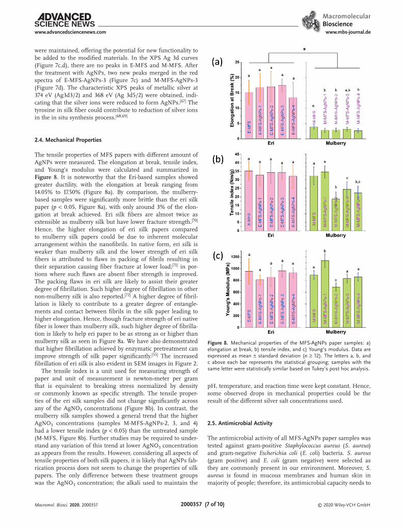
The tensile properties of MFS papers with different amount of AgNPs were measured. The elongation at break, tensile index, and Young’s modulus were calculated and summarized in Figure 8. It is noteworthy that the Eri-based samples showed greater ductility, with the elongation at break ranging from 14.05% to 17.50% (Figure 8a). By comparison, the mulberry-based samples were significantly more brittle than the eri silk paper (p < 0.05, Figure 8a), with only around 3% of the elon-gation at break achieved. Eri silk fibers are almost twice as extensible as mulberry silk but have lower fracture strength.[70] Hence, the higher elongation of eri silk papers compared to mulberry silk papers could be due to inherent molecular arrangement within the nanofibrils. In native form, eri silk is weaker than mulberry silk and the lower strength of eri silk fibers is attributed to flaws in packing of fibrils resulting in their separation causing fiber fracture at lower load;[71] in por-tions where such flaws are absent fiber strength is improved. The packing flaws in eri silk are likely to assist their greater degree of fibrillation. Such higher degree of fibrillation in other non-mulberry silk is also reported.[72] A higher degree of fibril-lation is likely to contribute to a greater degree of entangle-ments and contact between fibrils in the silk paper leading to higher elongation. Hence, though fracture strength of eri native fiber is lower than mulberry silk, such higher degree of fibrilla-tion is likely to help eri paper to be as strong as or higher than mulberry silk as seen in Figure 8a. We have also demonstrated that higher fibrillation achieved by enzymatic pretreatment can improve strength of silk paper significantly.[55] The increased fibrillation of eri silk is also evident in SEM images in Figure 2.
The tensile index is a unit used for measuring strength of paper and unit of measurement is newton-meter per gram that is equivalent to breaking stress normalized by density or commonly known as specific strength. The tensile proper-ties of the eri silk samples did not change significantly across any of the AgNO3 concentrations (Figure 8b). In contrast, the mulberry silk samples showed a general trend that the higher AgNO3 concentrations (samples M-MFS-AgNPs-2, 3, and 4) had a lower tensile index (p < 0.05) than the untreated sample (M-MFS, Figure 8b). Further studies may be required to under-stand any variation of this trend at lower AgNO3 concentration as appears from the results. However, considering all aspects of tensile properties of both silk papers, it is likely that AgNPs fab-rication process does not seem to change the properties of silk papers. The only difference between these treatment groups was the AgNO3 concentration; the alkali used to maintain the
pH, temperature, and reaction time were kept constant. Hence, some observed drops in mechanical properties could be the result of the different silver salt concentrations used.
2.5. Antimicrobial Activity
The antimicrobial activity of all MFS-AgNPs paper samples was tested against gram-positive Staphylococcus aureus (S. aureus) and gram-negative Escherichia coli (E. coli) bacteria. S. aureus (gram positive) and E. coli (gram negative) were selected as they are commonly present in our environment. Moreover, S. aureus is found in mucous membranes and human skin in majority of people; therefore, its antimicrobial capacity needs to
Macromol. Biosci. 2020, 2000357
Figure 8. Mechanical properties of the MFS-AgNPs paper samples: a) elongation at break, b) tensile index, and c) Young’s modulus. Data are expressed as mean ± standard deviation (n ≥ 12). The letters a, b, and c above each bar represents the statistical grouping; samples with the same letter were statistically similar based on Tukey’s post hoc analysis.
© 2020 Wiley-VCH GmbH2000357 (8 of 10)
www.advancedsciencenews.com www.mbs-journal.de
Macromol. Biosci. 2020, 2000357
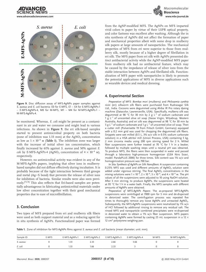
be monitored. Whereas, E. coli might be present as a contami-nant in air and water we consume and might lead to various infections. As shown in Figure 9, the eri silk-based samples started to present antimicrobial property on both bacteria (zone of inhibition was 1.33 mm) at the AgNO3 concentration as low as 1 × 10⁻⁴ m (Table 1). The inhibition zone was larger with the increase of initial silver ion concentration, which finally increased by 61% against S. aureus and 50% against E. coli by E-MFS-AgNPs-4 (AgNO3 concentration of 4 × 10⁻⁴ m), respectively.
However, no antimicrobial activity was evident in any of the M-MFS-AgNPs papers, implying that silver ions in mulberry-based samples did not diffuse effectively during incubation. It is probably because of the tight interaction between thiol groups and metal (AgS bond) that prevents the release of silver ions for inhibition of bacteria. Similar results were also seen previ-ously.[73,74] This also reflects that Eri-based samples are poten-tially advantageous in fabricating antimicrobial materials under low silver concentration together with their good mechanical properties due to ease of microfibrillation.
3. Conclusion
Two types of MFS prepared from eri and mulberry silk fibers were used as both support material and as a reducing agent for in situ synthesis of AgNPs. Functionalized paper was formed
from the AgNP-modified MFS. The AgNPs on MFS imparted vivid colors to paper by virtue of their LSPR optical property, and color fastness was excellent after washing. Although the in situ synthesis of AgNPs did not affect the formation of paper and mechanical properties albeit with some drop in mulberry silk papers at large amounts of nanoparticles. The mechanical properties of MFS from eri were superior to those from mul-berry silk, mostly because of a higher degree of fibrillation in eri silk. The MFS paper from eri silk with AgNPs presented dis-tinct antibacterial activity while the AgNP-modified MFS paper from mulberry silk had no antibacterial feature, which may be caused by the impedance of release of silver ions from the stable interaction between AgNPs and fibrillated silk. Function-alization of MFS paper with nanoparticles is likely to promote the potential applications of MFS in diverse applications such as wearable devices and medical dressing.
4. Experimental SectionPreparation of MFS: Bombyx mori (mulberry) and Philosamia cynthia
ricini (eri) silkworm silk fibers were purchased from Rudrasagar Silk Ltd., India. Cocoons were degummed using Ahiba IR Pro rotary dyeing machine (Datacolor, Lawrenceville, USA). Specifically, mulberry silk was degummed at 98 °C for 30 min by 2 g L−1 of sodium carbonate and 1 g L−1 of unscented olive oil soap (Vasse Virgin, Wilyabrup, Western Australia, Australia) and eri silk was degummed at 98 °C for 30 min by 5 g L−1 of sodium carbonate and 1 g L−1 of unscented olive oil soap. Then a cutter mill (Pulverisette 19 from Fritsch GmbH, Germany) equipped with a 0.2 mm grid was used for chopping the degummed silk fibers. Snippets were wet milled (0.5 L, 3% w/v silk in 0.5% sodium carbonate solution) in a HSA attritor mill (Union Process, USA) containing 5 kg, 2 mm zirconia media using 200 rpm stirring speed for 2 h. Milled fiber suspensions were further treated at 70 °C for 1 h in a beaker, followed by multiple washing steps until a neutral pH was obtained. To produce MFS, the fibers were then suspended in water and passed through a laboratory high-pressure homogenizer (GEA Niro Soavi, model: PandaPLUS 2000) for three times. Silk content was 1% w/v and homogenization pressure was 700 bar.
In Situ Synthesis of AgNPs on Silk Nanofibers: A suspension containing 0.25% MFS was used and different amounts of AgNO3 solution were added under vigorous stirring. The final AgNO3 concentrations in the mixing solutions were 1 × 10⁻⁴, 2 × 10⁻⁴, 3 × 10⁻⁴, and 4 × 10⁻⁴ m. The pH values of all the suspensions were adjusted to 10 using NaOH solution. After 5 min stirring, to produce AgNPs, the suspensions were heated at 95 °C for 1 h in a water bath. Finally, the MFS samples with different amounts of AgNPs were obtained.
Preparation of MFS-AgNPs Papers: The as-prepared MFS-AgNPs suspensions were centrifuged at 7000 rpm for 5 min and de-dispersed in deionized water. The centrifugation process was repeated five times to thoroughly remove any loose AgNPs and unreacted AgNO3. Subsequently, the MFS-AgNPs suspensions were neutralized by 1% w/v of HCl followed by additional rinsing to remove any residual salt. The rinsed MFS and nanoparticle combined precipitates were re-dispersed in deionized water to obtain a 1% w/v fiber suspension. MFS papers containing AgNPs were formed by casting 25 mL suspension in a 12 × 12 cm2 polystyrene weighing pan.
Figure 9. Disc diffusion assay of MFS-AgNPs paper samples against S. aureus and E. coli bacteria: E0 for E-MFS, E1 ∼ E4 for E-MFS-AgNPs-1 ∼ E-MFS-AgNPs-4, M0 for M-MFS, M1 ∼ M4 for M-MFS-AgNPs-1 ∼ M-MFS-AgNPs-4.
Table 1. Zone of inhibition for MFS-AgNPs films against S. aureus and E. coli bacteria (mean diameter; unit: mm).
Sample ID E-MFS E-MFS-AgNPs-1 E-MFS-AgNPs-2 E-MFS-AgNPs-3 E-MFS-AgNPs-4 M-MFS M-MFS-AgNPs
S. aureus 0 1.33 2.33 2.66 3.66 0 0
E. coli 0 1.33 1.66 2.33 3 0 0
© 2020 Wiley-VCH GmbH2000357 (9 of 10)
www.advancedsciencenews.com www.mbs-journal.de
Macromol. Biosci. 2020, 2000357
Characterization: The morphology of the papers made under different conditions was examined in a Supra 55VP field emission scanning electron microscope (Zeiss, Oberkochen, Germany). SEM images were acquired using the secondary electron detector at an accelerating voltage of 3 kV and a working distance of 5–7 mm. Prior to imaging, samples were coated with a 5 nm layer of carbon using an EM ACE600 high-vacuum sputter coater (Leica Microsystems, Wetzlar, Germany). Dynamic-mode AFM images were obtained by an NTEGRA Prima Scanning Probe Laboratory (NT-MDT, Zelenograd, Russia) at ambient conditions using the Universal Head (SF005NTF, NT-MDT) and a 100 µm × 100 µm × 10 µm closed-loop piezo scanner (SC100NTF, NT-MDT). The probes used for imaging were HQ:NSC15/Al BS silicon AFM probes (µmasch, Sofia, Bulgaria) with a typical tip radius of 8 nm, a force constant of ≈40 Nm, and a typical resonant frequency of 325 kHz. Three dimensionally rendered AFM height images were obtained by Blender 3D creation suite version 2.8 (Blender Foundation, Amsterdam, The Netherlands, https://www.blender.org).
The color changes of the papers colored with AgNPs were analyzed based on K/S curves. K/S values of papers with AgNPs were calculated using the Kubelka–Munk equation, K/S = (1-R)2/2R, where, K is the absorption coefficient of the substrate, S is the scattering coefficient of the substrate, and R is the reflectance of the paper at maximum absorption, as measured by a Datacolor spectrophotometer (Spectraflash, SP 600 Plus-CT, USA). The papers were immersed into 40 °C water bath for 2 h to investigate their color fastness. The papers were dried at room temperature and tested by Datacolor again to obtain the plots of K/S. The chemical structures of silk during the modification was analyzed by ATR-FTIR (LUMOS Bruker Biosciences Pty, Australia) with 128 scans at a resolution of 2 cm from 4000 to 600 cm. The UV diffuse reflectance spectra were acquired with a Cary 5000 UV–vis spectrophotometer equipped with an integrating sphere. The instrument was first calibrated by a standard whiteboard, and the papers were clamped and scanned from 200 to 800 nm on the port of the integrating sphere successively. An IRIS Intrepid IIXSP ICP-AES instrument was employed to determine the silver content of different samples. XPS measurements were carried out on a Kratos XSAM800 XPS system with Kα source and a charge neutralizer.
The mechanical properties of the MFS papers with AgNPs were tested on an Instron 5967 tester equipped with 50 N load cell (Instron, Norwood, MA, USA). The protein papers were cut into 10 × 50 mm strips (N = 12) and conditioned to 20 ± 2 °C and 65% ± 2% relative humidity for at 24 h. Each sample was weighed with a four decimal place balance first, and then tested at a gauge length of 20 mm with a crosshead speed of 2 mm min–1.
Statistical Analysis: The tensile mechanical properties (tensile index, Young’s modulus, and elongation at break) were compared both between eri and mulberry silk samples and between the samples made with the different silver concentrations. The data were confirmed to be normally distributed using Shapiro–Wilk normality tests. Then, the sample groups were compared using one-way analysis of variance (ANOVA) followed by Tukey’s post hoc analysis. Differences between the means were considered statistically significant at p < 0.05.
Evaluation of Antibacterial Activity: The antimicrobial activity of papers against E. coli and S. aureus was carried out using disk inhibition zone method in accordance to ASTM-AATCC 147. The microorganisms were cultured in plates with Luria–Bertani medium and agar gel prior to use. Each specimen was cut into circular discs and sterilized by autoclaving, followed by placing in agar plates with different microorganisms, respectively. After incubated at 37 °C for 48 h, the length of the inhibition zone around each sample was measured, and the antimicrobial performance was evaluated. The test was carried out in triplicate for each sample.
AcknowledgementsY.L. and B.T. contributed equally to this work. The authors acknowledge funding from the Australian Academy of Science through the Australia–India Early and Mid-Career Fellowships and ARC Research Hub for
Future Fibers (IH140100018). H.C.S. acknowledges funding from the U.S. National Science Foundation under grant numbers DMR-1352542 and DMR-1905902.
Conflict of InterestThe authors declare no conflict of interest.
Keywordsantibacterial, coloration, papers, silk nanofibers, silver nanoparticles
Received: October 14, 2020Revised: November 18, 2020
Published online:
[1] H. Tao, D. L. Kaplan, F. G. Omenetto, Adv. Mater. 2012, 24, 2824.[2] A. Leal-Egaña, T. Scheibel, Biotechnol. Appl. Biochem. 2010, 55, 155.[3] Z. Zhao, Y. Li, M.-B. Xie, Int. J. Mol. Sci. 2015, 16, 4880.[4] F. Zhang, Z. Zhang, X. Zhu, E.-T. Kang, K.-G. Neoh, Biomaterials
2008, 29, 4751.[5] Y. Wang, D. D. Rudym, A. Walsh, L. Abrahamsen, H.-J. Kim,
H. S. Kim, C. Kirker-Head, D. L. Kaplan, Biomaterials 2008, 29, 3415.[6] R. Fedic, M. Zurovec, F. Sehnal, J. Insect Biotechnol. Sericol. 2002, 71,
1.[7] D. N. Rockwood, R. C. Preda, T. Yücel, X. Wang, M. L. Lovett,
D. L. Kaplan, Nat. Protoc. 2011, 6, 1612.[8] M. Kazemimostaghim, R. Rajkhowa, T. Tsuzuki, X. Wang, Powder
Technol. 2013, 241, 230.[9] S. Ling, K. Jin, D. L. Kaplan, M. J. Buehler, Nano Lett. 2016, 16, 3795.
[10] K. Zheng, J. Zhong, Z. Qi, S. Ling, D. L. Kaplan, Adv. Funct. Mater. 2018, 28, 1806380.
[11] B. Subia, S. Chandra, S. Talukdar, S. C. Kundu, Integr. Biol. 2013, 6, 203.
[12] S. Ling, W. Chen, Y. Fan, K. Zheng, K. Jin, H. Yu, M. J. Buehler, D. L. Kaplan, Prog. Polym. Sci. 2018, 85, 1.
[13] Y. Liang, B. J. Allardyce, S. Kalita, M. G. Uddin, S. Shafei, D. Perera, R. C. N. Remadevi, S. L. Redmond, W. J. Batchelor, C. J. Barrow, R. J. Dilley, H. C. Schniepp, X. Wang, R. Rajkhowa, Biomacromol-ecules 2020, 21, 1303.
[14] M. G. Uddin, W. Batchelor, B. J. Allardyce, N. Byrne, C. J. Barrow, X. Wang, R. Rajkhowa, ACS Sustainable Chem. Eng. 2020, 8, 1155.
[15] B. Tang, J. Kaur, L. Sun, X. Wang, Cellulose 2013, 20, 3053.[16] B. Tang, L. Sun, J. Kaur, Y. Yu, X. Wang, Dyes Pigm. 2014, 103, 183.[17] B. Tang, M. Zhang, X. Hou, J. Li, L. Sun, X. Wang, Ind. Eng. Chem.
Res. 2012, 51, 12807.[18] B. Tang, J. Li, X. Hou, T. Afrin, L. Sun, X. Wang, Ind. Eng. Chem. Res.
2013, 52, 4556.[19] B. Tang, L. Sun, J. Li, J. Kaur, H. Zhu, S. Qin, Y. Yao, W. Chen,
X. Wang, Dyes Pigm. 2015, 113, 289.[20] S. Calamak, E. A. Aksoy, C. Erdogdu, M. Sagıroglu, K. Ulubayram, J.
Nanopart. Res. 2015, 17, 87.[21] P. V. AshaRani, G. L. K. Mun, M. P. Hande, S. Valiyaveettil, ACS
Nano 2009, 3, 279.[22] J. S. Kim, E. Kuk, K. N. Yu, J.-H. Kim, S. J. Park, H. J. Lee, S. H. Kim,
Y. K. Park, Y. H. Park, C.-Y. Hwang, Y.-K. Kim, Y.-S. Lee, D. H. Jeong, M.-H. Cho, Nanomedicine 2007, 3, 95.
[23] Q. Li, S. Mahendra, D. Y. Lyon, L. Brunet, M. V. Liga, D. Li, P. J. J. Alvarez, Water Res. 2008, 42, 4591.
[24] C. Marambio-Jones, E. M. V. Hoek, J. Nanopart. Res. 2010, 12, 1531.[25] S. Pal, R. Nisi, M. Stoppa, A. Licciulli, ACS Omega 2017, 2, 3632.
© 2020 Wiley-VCH GmbH2000357 (10 of 10)
www.advancedsciencenews.com www.mbs-journal.de
Macromol. Biosci. 2020, 2000357
[26] J. Wu, Y. D. Zheng, W. H. Song, J. B. Luan, X. X. Wen, Z. G. Wu, X. H. Chen, Q. Wang, S. L. Guo, Carbohydr. Polym. 2014, 102, 762.
[27] H. L. Ye, J. W. Cheng, K. Yu, Int. J. Biol. Macromol. 2019, 121, 633.[28] M. M. Zangeneh, Appl. Organomet. Chem. 2019, 33, 16.[29] J. N. Anker, W. P. Hall, O. Lyandres, N. C. Shah, J. Zhao, R. P. Van
Duyne, Nat. Mater. 2008, 7, 442.[30] K. A. Willets, R. P. Van Duyne, Annu. Rev. Phys. Chem. 2007, 58, 267.[31] C. M. Srivastava, R. Purwar, A. P. Gupta, Int. J. Biol. Macromol. 2019,
130, 437.[32] S. Patil, N. Singh, Colloids Surf., B 2019, 176, 150.[33] R. Rajkhowa, L. Wang, X. Wang, Powder Technol. 2008, 185, 87.[34] R. Rajkhowa, L. Wang, J. Kanwar, X. Wang, Powder Technol. 2009,
191, 155.[35] M. Kazemimostaghim, R. Rajkhowa, T. Tsuzuki, X. Wang, Powder
Technol. 2013, 249, 253.[36] A. Ude, R. Eshkoor, R. Zulkifili, A. Ariffin, A. Dzuraidah, C. Azhari,
Mater. Des. 2014, 57, 298.[37] R. Rajkhowa, X. Hu, T. Tsuzuki, D. L. Kaplan, X. Wang, Biomacro-
molecules 2012, 13, 2503.[38] K. Singh, R. Jayasomu, Pharm. Biol. 2002, 40, 28.[39] S. Chawla, S. Midha, A. Sharma, S. Ghosh, Adv. Healthcare Mater.
2018, 7, 1701204.[40] R. S. Peigler, S. Naumann, A Revision of the Silkmoth Genus Samia,
University of the Incarnate Word, San Antonio, TX 2003.[41] S. Pal, J. Kundu, S. Talukdar, T. Thomas, S. C. Kundu, Macromol.
Biosci. 2013, 13, 1020.[42] B. Kundu, R. Rajkhowa, S. C. Kundu, X. Wang, Adv. Drug Delivery
Rev. 2013, 65, 457.[43] R. Rajkhowa, V. Gupta, V. Kothari, J. Appl. Polym. Sci. 2000, 77, 2418.[44] T. Asakura, Y. Nakazawa, Macromol. Biosci. 2004, 4, 175.[45] K. Sen, M. Babu K, J. Appl. Polym. Sci. 2004, 92, 1098.[46] A. Rafey, K. B. L. Shrivastavaa, S. A. Iqbal, Z. Khan, J. Colloid Inter-
face Sci. 2011, 354, 190.[47] Z. Zaheer, Rafiuddin, Int. J. Chem. Kinet. 2012, 44, 680.[48] Z. Karahaliloğlu, B. Ercan, E. B. Denkbaş, T. J. Webster, J. Biomed.
Mater. Res. 2015, 103, 135.[49] C. Holland, K. Numata, J. Rnjak-Kovacina, F. P. Seib, Adv. Healthcare
Mater. 2019, 8, 1800465.
[50] P. J. Babu, M. Doble, A. M. Raichur, J. Colloid Interface Sci. 2018, 513, 62.
[51] P. Kamalathevan, P. S. Ooi, Y. L. Loo, Adv. Skin Wound Care 2018, 31, 565.
[52] M. Pollini, F. Paladini, Materials 2020, 13, 3361.[53] S.-W. Lee, S.-G. Kim, Maxillofac. Plast. Reconstr. Surg. 2014, 36, 239.[54] K.-J. Kwon, H. Seok, Appl. Sci. 2018, 8, 1214.[55] M. G. Uddin, B. J. Allardyce, D. R. D. C. Leal, N. Byrne, X. Wang,
W. Batchelor, R. Rajkhowa, Int. J. Biol. Macromol. 2020, 164, 2389.[56] S. H. Kim, Y. S. Nam, T. S. Lee, W. H. Park, Polym. J. 2003, 35, 185.[57] D. H. Reneker, A. L. Yarin, Polymer 2008, 49, 2387.[58] N. Du, Z. Yang, X. Y. Liu, Y. Li, H. Y. Xu, Adv. Funct. Mater. 2011, 21,
772.[59] Q. J. Wang, H. C. Schniepp, JOM 2019, 71, 1248.[60] I. Greving, M. Z. Cai, F. Vollrath, H. C. Schniepp, Biomacromolecules
2012, 13, 676.[61] S. R. Koebley, D. Thorpe, P. Pang, P. Chrisochoides, I. Greving,
F. Vollrath, H. C. Schniepp, Biomacromolecules 2015, 16, 2796.[62] A. Sionkowska, A. Planecka, Polym. Degrad. Stab. 2011, 96, 523.[63] B. Tang, S. Xu, X. Jian, J. Tao, W. Xu, Appl. Spectrosc. 2010, 64, 1407.[64] Y. Sun, Y. Xia, Analyst 2003, 128, 686.[65] Q. Lu, B. Zhang, M. Li, B. Zuo, D. L. Kaplan, Y. Huang, H. Zhu, Bio-
macromolecules 2011, 12, 1080.[66] D. Briggs, Handbook of X-Ray and Ultraviolet Photoelectron Spectros-
copy, Heyden, London 1977.[67] H. Kong, J. Jang, Chem. Commun. 2006, 3010.[68] Q. Dong, H. Su, D. Zhang, J. Phys. Chem. B 2005, 109, 17429.[69] E. Kharlampieva, D. Zimnitsky, M. Gupta, K. N. Bergman,
D. L. Kaplan, R. R. Naik, V. V. Tsukruk, Chem. Mater. 2009, 21, 2696.[70] R. Rajkhowa, V. B. Gupta, V. K. Kothari, 2000, 77, 2418.[71] R. Rajkhowa, J. Kaur, X. G. Wang, W. Batchelor, J. R. Soc., Interface
2015, 12, 20150177[72] Y. Kawahara, T. Hananouchi, M. Tsuji, T. Kimura, S. Kohjiya, J. Seric.
Sci. Jpn. 2001, 70, 109.[73] S. Calamak, E. A. Aksoy, N. Ertas, C. Erdogdu, M. Sagıroglu,
K. Ulubayram, Eur. Polym. J. 2015, 67, 99.[74] H. A. Currie, O. Deschaume, R. R. Naik, C. C. Perry, D. L. Kaplan,
Adv. Funct. Mater. 2011, 21, 2889.


![In Situ TEM Observation of Electrochemical Lithiation of ...Template-synthesized multiwalled carbon nanotube (CNT) [ 12,13 ] was used as a reaction vessel in order to prevent S evaporation](https://img.pdfslide.us/doc/110x75/5fa8e0088cb1191a6a373909/in-situ-tem-observation-of-electrochemical-lithiation-of-template-synthesized.jpg)