Embed Size (px)
Citation preview
|| 315 || | European Journal of General Dentistry | Vol 2 | Issue 3 | September-December 2013 |
Silicone prosthesis for a patient with unilateral ear defect: A clinical case report
ABSTRACTCongenital or acquired loss of body parts is of common occurrence and replacement of such parts for restoring their lost function and esthetics is done by using various bio‑compatible materials. Proper assessment of the disfigured body parts and a feasible approach to rehabilitating them has for long, been the target of clinical maxillofacial prosthodontics. The aim of this article is to present a case report of such a silicone prosthesis for a patient with a congenital unilateral auricular defect.
Key wordsAuricular defect, maxillofacial prosthesis, room temperature vulcanizing silicone
Ajay Singh, Shounak Ghosh1, Sumana Kar1, Imran Ahmed2
Professor and Head, 1Post Graduate Student, Department of Prosthodontics, Sardar Patel Post Graduate Institute of Dental and Medical Sciences, Lucknow, 2MDS Prosthodontics, India
Address for correspondence: Dr. Ajay Singh,
Department of Prosthodontics, Sardar Patel Post Graduate Institute of
Dental and Medical Sciences, Lucknow, Uttar Pradesh, India.
E-mail: [email protected]
INTRODUCTION
Loss of facial tissue regardless of its cause that is congenital or acquired causes serious functional and psychological problems. Maxillofacial prosthodontics deals with prosthetic reconstruction of missing/disfigured head and neck tissue. Prosthetic replacement of the exterior part (Epithesis) is probably as old as civilization. References to it are available in Indian, Greek, Roman, Egyptian Civilizations.[1]
Ambroise Pare is credited with making various contributions to the materials and techniques in facial prosthetics. Fabrication of an extra‑oral prosthesis is probably more of an art than science. The form, color, and texture of the prosthesis must closely match with the surrounding natural tissue. The various treatment options available today include traditional mechanically retained prosthesis, bio‑adhesive retained prosthesis, implant retained, and the recently developed rapid prototyping,[2] and Computer Aided Designing – Computer Aided Machining (CAD‑CAM) developed prosthesis.
Prosthetic reconstruction of missing facial tissue scores over surgical or plastic reconstruction as being simpler, less time taking, cost‑effective, with lesser morbidity, allows periodic evaluation, and cleaning of the site. The prosthodontist has a complete control of color, shape, and position of the prosthesis.
Disadvantages are – need for remaking the prosthesis, dependence on adhesives or other devices for retention and possible irritation of the tissues. The materials, which have been used in the 400 year history of anaplastology include metals, ivory, porcelain, natural rubber, gelatin, and latex.
Modern day materials such as Polymethyl Methacrylate (PMMA) and Silicone enjoy popularity and consistent results.[3]
Long‑term success of a facial prosthesis depends mainly on retention. These prostheses are retained with different methods of retention such as medical adhesives, anatomical undercuts, and mechanical devices such as spectacles, hair bands, magnets, and implants.
However, patients who refuse to undergo any surgical procedures required for the implant placement or cases where surgical intervention is contraindicated, alternate means of retention need to be considered.
Biological adhesives provide adequate retention of the prosthesis. Other aids such as auditory canal hearing
CASE rEPorT
Access this article onlineQuick Response Code:
Website: www.ejgd.org
DOI: 10.4103/2278-9626.115997
Published online: 2021-11-01
Singh, et al.: Silicone ear prosthesis
| European Journal of General Dentistry | Vol 2 | Issue 3 | September-December 2013 | || 316 ||
aids can also come in handy to provide the added retention to the prosthesis.
CASE rEPorT
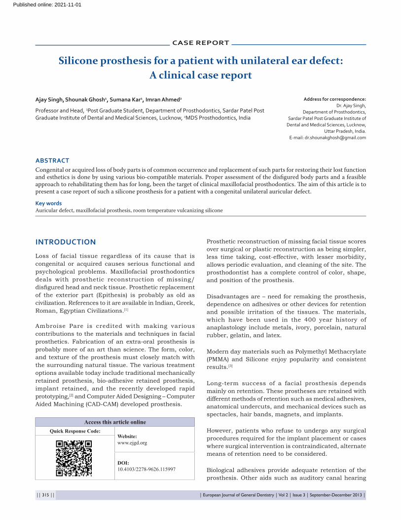
A 35‑year‑old male patient reported to the Department of Prosthodontics, with unilateral auricular deformity on the left side. He gave a history of a congenitally present rudimentary left ear. Never had there been any surgical attempt to correct the deformity, nor had he ever made any prosthesis for the defect [Figure 1].
Clinical examination revealed deformed helix, antihelix, concha, anti‑helical fold, and lobules. However, rudimentary lobular portions were present. The cartilaginous portions were completely missing. Only the dermis was present.
His hearing was slightly affected. There was a 36% hearing loss, which was assessed by audiometry as evaluated by speech recognition tests and signals to noise ratio by an otorhinolaryngologist. Auricular prosthesis was planned for him.
Patient education and counseling regarding the nature function and limitation of the prosthesis was carefully carried out prior to any procedures. A written informed consent was duly obtained.
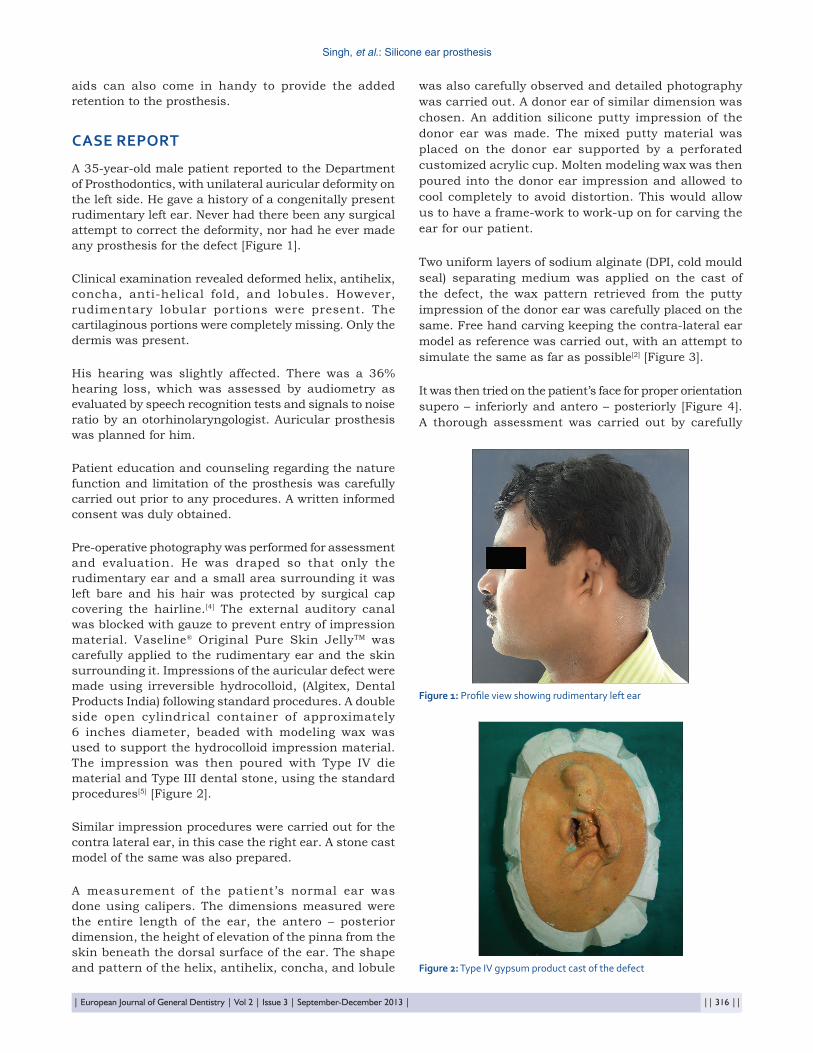
Pre‑operative photography was performed for assessment and evaluation. He was draped so that only the rudimentary ear and a small area surrounding it was left bare and his hair was protected by surgical cap covering the hairline.[4] The external auditory canal was blocked with gauze to prevent entry of impression material. Vaseline® Original Pure Skin JellyTM was carefully applied to the rudimentary ear and the skin surrounding it. Impressions of the auricular defect were made using irreversible hydrocolloid, (Algitex, Dental Products India) following standard procedures. A double side open cylindrical container of approximately 6 inches diameter, beaded with modeling wax was used to support the hydrocolloid impression material. The impression was then poured with Type IV die material and Type III dental stone, using the standard procedures[5] [Figure 2].
Similar impression procedures were carried out for the contra lateral ear, in this case the right ear. A stone cast model of the same was also prepared.
A measurement of the patient’s normal ear was done using calipers. The dimensions measured were the entire length of the ear, the antero – posterior dimension, the height of elevation of the pinna from the skin beneath the dorsal surface of the ear. The shape and pattern of the helix, antihelix, concha, and lobule
was also carefully observed and detailed photography was carried out. A donor ear of similar dimension was chosen. An addition silicone putty impression of the donor ear was made. The mixed putty material was placed on the donor ear supported by a perforated customized acrylic cup. Molten modeling wax was then poured into the donor ear impression and allowed to cool completely to avoid distortion. This would allow us to have a frame‑work to work‑up on for carving the ear for our patient.
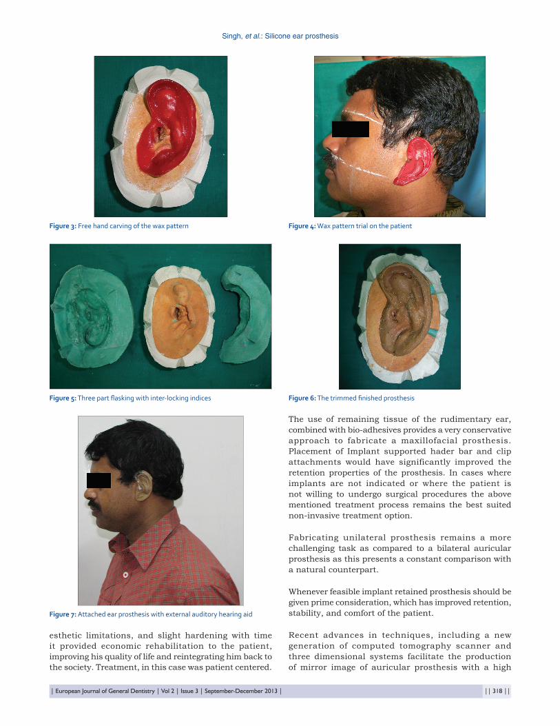
Two uniform layers of sodium alginate (DPI, cold mould seal) separating medium was applied on the cast of the defect, the wax pattern retrieved from the putty impression of the donor ear was carefully placed on the same. Free hand carving keeping the contra‑lateral ear model as reference was carried out, with an attempt to simulate the same as far as possible[2] [Figure 3].
It was then tried on the patient’s face for proper orientation supero – inferiorly and antero – posteriorly [Figure 4]. A thorough assessment was carried out by carefully
figure 1: Profile view showing rudimentary left ear
figure 2: Type IV gypsum product cast of the defect
Singh, et al.: Silicone ear prosthesis
|| 317 || | European Journal of General Dentistry | Vol 2 | Issue 3 | September-December 2013 |
checking the prosthesis both from frontal and profile views. Due importance was given to patients feedback regarding any modification in pattern. The wax pattern was optimally retentive and stable on his face.[6]
Final finishing of the wax pattern wax performed on the cast of the rudimentary ear. The finished pattern was then sealed on the cast model. A three part flasking with indices marking the interlocking of the pieces was planned [Figure 5]. The first part was the die of the rudimentary ear with its Type II gypsum base. Indices to aid repositioning of the counter parts were made on all sides of the base. Modelling wax was used to provide support to the second pour of type II gypsum for the flasking. Sodium alginate separating media was added and the middle part of the customized flask was poured. It was made sure that no gypsum flowed into any undercut of the wax pattern as this would cause a deformation of the pattern when removal of the middle part of the flask would be attempted. Following this another modeling wax supported pour of type II gypsum was made covering the ventral surface of the auricular wax pattern. Dewaxing procedures in a hot water bath, using the standard directives then were carried out.
Shade selection using intrinsic coloration procedures were decided upon. Intrinsic stains (MP Sai, Enterprise) provided with the room temperature vulcanizing (RTV) silicone (MP Sai, Enterprise) was used for shade matching. Basic colors used were yellow, white, brown, purple, and red. Small amounts of the base and catalyst pastes were mixed and incremental adding of the stains gradually was carried out with constant comparison with the skin of the approximating area and the contralateral ear. Separate shades were decided upon to accurately replicate the different parts of the patient’s natural ear. Different shades were selected for the lobule, concha, helix and anti‑helix.[7] Nearest possible simulation was attempted to be achieved by performing shade selection under different light sources, which included incandescent, fluorescent and natural sun light sources.
Packing of the tinted silicon material was carried out and the three parts of the flask were again reattached and seated carefully to ensure that all the margins were flushed together.
At 48 h were allowed to elapse as per the manufacturer’s instructions prior to opening the flask. The silicone prosthesis was then examined for defects and porosities prior to being trimmed and finished using a sharp pair of Parrot Beak scissors [Figure 6].
The final prosthesis was then tried on the patient, and his external auditory hearing aid was also placed, which considerably improved retention. Bio‑adhesive (Cosmosil) was then used to attach the auricular prosthesis to the rudimentary ear[8] [Figure 7].
The patient was advised to use the prosthesis regularly and avoid exposure to direct sunlight due to the limitations of the silicone used for fabrication of the same. He was also instructed to regularly clean the prosthesis using a mild sodium lauryl sulphate solution. Use of strong detergent solutions and hard brushes for cleansing the prosthesis was discouraged. He was instructed not to wear the prosthesis while sleeping as accidental pressure would result in distortion or tearing of the prosthesis.
A regular follow‑up and evaluation of the patient and the prosthesis was undertaken to ensure that there were no eruptions and proper maintenance of the prosthesis was being carried out.
DISCUSSION
Patients with auricular deformity or absence of auricle endures psychological affliction. The aim of maxillofacial rehabilitation is to provide a suitable prosthesis for patients with facial defects so that they can be confident enough to face the society and accept the challenges of life.
Auricular defects can be repaired or reconstructed with autogenous tissue, but this may not be feasible for personal/medical reasons. A good alternative is to develop an auricular prosthesis with a suitable material Silicone is the material for choice for facial prosthesis because of its flexibility and life like appearance. In this case, RTV silicone (MP Sai Enterprise) was used. Intrinsic stains were used for the prosthesis coloration as these are more color stable[9] and provided better esthetic results. Accelerated ageing studies and color evaluation studies using the reflection spectrophotometer analysis have also showed that intrinsic stains undergo considerably less amount of color alteration as compared to extrinsic coloration methods. Furthermore, inorganic stains proved to be more color stable as compared to organic stains derived from plants and other natural sources.
Hardening of the RTV silicones is also a drawback of the material. Hot, humid conditions and contact of the material with sweat, dust, pollen, and other offenders only hastens the hardening and discoloration process. Gradual hardening and discoloration takes place over a period of time, but the material still remains in considerably acceptable condition for about 9‑12 months of time.
For retention of the prosthesis Bio‑adhesive Cosmosil™ was used. Sufficient retention was provided by the material. The adhesive is not soluble in water so provided better retention for longer duration of time as it did not get dissolved when in contact with sweat.
Although this material has shortcomings of having
Singh, et al.: Silicone ear prosthesis
| European Journal of General Dentistry | Vol 2 | Issue 3 | September-December 2013 | || 318 ||
esthetic limitations, and slight hardening with time it provided economic rehabilitation to the patient, improving his quality of life and reintegrating him back to the society. Treatment, in this case was patient centered.
The use of remaining tissue of the rudimentary ear, combined with bio‑adhesives provides a very conservative approach to fabricate a maxillofacial prosthesis. Placement of Implant supported hader bar and clip attachments would have significantly improved the retention properties of the prosthesis. In cases where implants are not indicated or where the patient is not willing to undergo surgical procedures the above mentioned treatment process remains the best suited non‑invasive treatment option.
Fabricating unilateral prosthesis remains a more challenging task as compared to a bilateral auricular prosthesis as this presents a constant comparison with a natural counterpart.
Whenever feasible implant retained prosthesis should be given prime consideration, which has improved retention, stability, and comfort of the patient.
Recent advances in techniques, including a new generation of computed tomography scanner and three dimensional systems facilitate the production of mirror image of auricular prosthesis with a high
figure 7: Attached ear prosthesis with external auditory hearing aid
figure 3: Free hand carving of the wax pattern figure 4: Wax pattern trial on the patient
figure 5: Three part flasking with inter‑locking indices figure 6: The trimmed finished prosthesis
Singh, et al.: Silicone ear prosthesis
|| 319 || | European Journal of General Dentistry | Vol 2 | Issue 3 | September-December 2013 |
level of accuracy, alleviating most of the limitations of conventional prosthesis. Limitation to its use is high‑cost. Development in the field of tissue engineering has resulted in the formation of new tissue equivalents of bone and cartilage that will augment the outcome of prosthodontic rehabilitation in the future.
CONCLUSION
Maxillofacial defects can be emotionally traumatizing and many a times cause a social stigma due to a distorted physical appearance. An attempt to provide a cost‑effective and cosmetically acceptable auricular prosthesis for a male patient was made, which was esthetically and functionally acceptable to him.
Communication and education is the key for accepting the prosthesis. Successful use of prosthesis may depend on the patient’s psychological acceptance of it. Patient’s participation in the decision making process with realistic expectations is also of vital significance.
The patient in this case was highly satisfied with the prosthesis and did report during the follow‑up phase stating significant improvement in confidence in day to day dwellings. He appreciated the limitations of the prosthesis and seemed reasonably happy with what was delivered to him.
rEfErEnCES
1. Mantri SS, Thombre RU, Pallavi D. Prosthodontic rehabilitation of a patient with bilateral auricular deformity. J Adv Prosthodont 2011;3:101‑5.
2. Al Mardini M, Ercoli C, Graser GN. A technique to produce a mirror‑image wax pattern of an ear using rapid prototyping technology. J Prosthet Dent 2005;94:195‑8.
3. Andres C. Survey of materials used in extra‑oral maxillofacial prosthetics. In: Proceedings of Materials Research in Maxillofacial Prosthetic Academy of Dental Materials; Chicago, IL. 1992. p. 25‑40.
4. Kubon TM, Kurtz KS, Piro JD. Impression procedure for creating a partial auricular prosthesis. J Prosthet Dent 2000;83:648‑51.
5. Wang R. Preoperative auricular wax pattern duplication for surgical template fabrication. J Prosthet Dent 1999;81:634‑7.
6. El Charkawi HG, El Sharkawy AG. A simplified technique for orientation of a bone anchored auricular prostheses: A clinical report. J Oral Maxillofac Res 2012;3:e6.
7. Goiato MC, Haddad MF, Sinhoreti MA, dos Santos DM, Pesqueira AA, Moreno A. Influence of opacifiers on dimensional stability and detail reproduction of maxillofacial silicone elastomer. Biomed Eng Online 2010;9:85. Available from: http://www.biomedical‑engineering‑online.com/content/9/1/85.
8. Kiat‑amnuay S, Gettleman L, Khan Z, Goldsmith LJ. Effect of adhesive retention on maxillofacial prostheses. Part I: Skin dressings and solvent removers. J Prosthet Dent 2000;84:335‑40.
9. Andres, C.J. sss and Haugh, S.P. (2000) Facial prosthesis fabrication: Technical aspects. In Clinical Maxillofacial prosthetics, Taylor, T.D., Ed, ISBN 086715‑391‑1, Illions.
How to cite this article: Singh A, Ghosh S, Kar S, Ahmed I. Silicone prosthesis for a patient with unilateral ear defect: A clinical case report. Eur J Gen Dent 2013;2:315-9.
Source of Support: Nil, Conflict of Interest: None declared.
![Intelligent Prosthesis - tams. · PDF fileI Electrooculography (EOG) I Electrocorticogram (EcoG) [ ] Irina Intelligent Prosthesis 4/21. ... Irina Intelligent Prosthesis 21/21](https://img.pdfslide.us/doc/110x75/5aab10c57f8b9aa9488b839d/intelligent-prosthesis-tams-electrooculography-eog-i-electrocorticogram-ecog.jpg)