Embed Size (px)
Citation preview

IntroductionStrongyloidiasis is endemic in tropical and
semitropical regions. Strongyloides exists as afree-living organism that does not require a hostto replicate. There is reported data of skin dis-ease that contacts the soil. Fillariform larvaecan penetrate any area of the skin, after whichthey migrate through the dermis to enter thevasculature. The larvae circulate in the venousblood until they reach the lungs, where theybreak into the alveoli and ascend the bronchialtree. The worms are then swallowed with salivaand pass into the small bowel, where they em-bed in the duodenojejunal mucosa and mature.Most patients with strongyloidiasis have nofrank symptoms. Patients may have an urticari-
al rash (larvae currens) caused by dermal mi-gration of fillariform larvae and non-productivecough with or without hemoptysis [1] due tolung migration stage. Occasionally, patientshave abdominal pain, anorexia, nausea, vomit-ing and diarrhea due to gastrointestinal involve-ment. S. stercoralis can survive in the body for along time due to autoinfection.
In immunocompromised hosts, disseminatedorgan involvement, (lungs, CNS, GI-tract, liv-er,…) may occur. Bacteremia and septic shockmay cause a high mortality [2].
Infection can be diagnosed by finding rhab-ditiform larvae in direct smears of the stool,positive serologic tests, study of duodenal aspi-rate and intestinal biopsy. Strongyloidiasis istreated with ivermectin, albendazole and thi-abendazole [3]. As mentioned above, abdomi-
Case ReportMedical Journal of the Islamic Republic of Iran.Vol. 22, No. 3, November, 2008. pp. 149-151
Significant weight loss, nausea, and vomiting due to strongyloidiasis:a case report
Hossein Froutan, MD,1, Ayatollah Bayatian, MD.2, Seied Mohsen Razavizadeh, MD.3,Mohsen Nasiri-Toosi, MD.4, Afshin Shafaghi, MD.5
Department of Internal Medicine, Imam Khomeini Hospital, Tehran, Iran.
AbstractStrongyloidiasis is caused by infestation with Strongyloides stercoralis, a free
living tropical and semitropical soil helminth that has a larval form that penetratesintact skin. Clinical manifestations may be varied from an asymptomatic infectionin immunocompetent hosts to a diffuse and fatal form in immunocompromisedhosts.
We report a 56-year-old man from Dezful (south-west of Iran) with a 6-monthhistory of nausea, vomiting and significant weight loss (greater than 10%). Abdom-inal ultrasonography had no significant findings. Upper gastrointestinal series andabdominal CT were performed. Dilated bowel loops especially in the jejunum, withdecreased mucosal folds were seen. A biopsy specimen from the third part of duo-denum showed strongyloides larvae, thus albendazole 400 mg twice a day for 3days was initiated. He responded well to this treatment regimen.
Keywords: strongyloidiasis, nausea, vomiting, albendazole.
1. Professor of Gastroenterology, Tehran University of Medical Sciences, Chairman of Gastroenterology Dept.2. Corresponding author, Assistant Professor of Internal Medicine, Internal Medicine Ward, Imam Khomeini Hospital, Tehran, Iran.Tel:+989122497819, email: [email protected]. Assistant Professor of Gastroenterology, Imam Khomeini Hospital.4. Associate Professor of Gastroenterology, Tehran University of Medical Sciences, Tehran, Iran. 5.Assistant Professor of Gastroenterology, Imam Khomeini Hospital.

nal symptoms are the usual presentation of thisinfection. Our patient presented not only withabdominal symptoms, but a significant loss ofweight that may herald malignancies.
Case reportA56 year old man from Dezful (south west of
Iran) referred to our hospital with a 6 month his-tory of bloating, epigastric and left lower quad-rant pain, nausea and vomiting after eating. Hehad a significant loss of weight (18kg) (greaterthan 10% loss of his baseline body weight). Hehad a normal appetite. He had no history of di-arrhea and melena. His abdominal pain did notcorrelate with body position. He had a historyof pelvic fracture due to trauma and minor tha-lassemia. The patient was cachectic with a paleappearance. Vital signs were normal. Lung andheart exams were normal. The abdomen wassoft with normal bowel sounds. There was noorganomegaly or ascites. There was (+1) pittingedema in both lower limbs. The laboratory val-ues were as follow:
WBC = 11000/mm3 PMN= 60%lymph= 35% Eos= 4% Mono= 1% Hemoglobin= 9.2 g/dlMCV = 57 f.L MCH=19.6 PlT = 201000/ mm3 AST = 34 u/LALT = 33 u/L AlP = 251u/LBil (Total)= 0.6 mg/dl Bil direct = 0.1 mg/dL PT = 15 sec PTT = 38 secINR = 1.2 HIV (Ab) = negativeAntigliadin Ab = negative Antiendomysial Ab = negativeFBS = 85 mg/dL BUN = 15 mg/dLCreat = 0.6 mg/dL Na+ = 144 mmol/LK+ = 4.9 mmol/L LDH = 507 u/LSerum iron = 91 micg/mL TIBC = 190 micg/mLFerritin = 103 micg/mL U/A = normalESR = 3 mm/h CRP = 2.9 mg/LAlbumin = 2.6 gr/dL Protein = 5.5 gr/dL
Stool exam was negative for ova and para-sites and occult blood on three occasions.
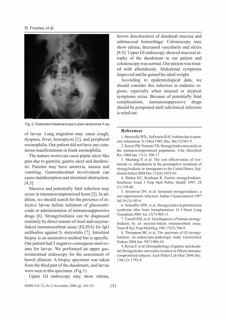
Abdominal ultrasonography was normal.Plain film of the abdomen (Fig.1a) showed di-lated bowel loops. Upper gastrointestinal series(Fig.1b) and abdominal CT were performed.Dilated bowel loops especially in the jejunum
with decreased mucosal folds were seen. Abiopsy specimen from the third part of the duo-denum showed moderate villous atrophy andmarked crypt hyperplasia with mild to moder-ate infiltration of lymphocytes and plasma cellsin the lamina propria. Adult worm, larvae andeggs of strongyloides stercoralis were seen inlamina properia and lumen of the crypts. Thusalbendazole 400 mg twice a day for 3 days wasinitiated.
DiscussionMost immunocompetent hosts of S. sterco-
ralis are asymptomatic. Symptomatic patientsmay have an urticarial rash caused by migration
Significant weight loss...
MJIRI.Vol. 22, No.3, November, 2008. pp. 149-151150
Fig. 1. Strongyloides larva in duodenal biopsy.
a
b
of larvae. Lung migration may cause cough,dyspnea, fever, hemoptysis [1], and peripheralesoinophilia. Our patient did not have any cuta-neous manifestations or frank esoinophilia.
The mature worm can cause peptic ulcer-likepain due to gastritis, gastric ulcer and duodeni-tis. Patients may have anorexia, nausea andvomiting. Gastrointestinal involvement cancause malabsorption and intestinal obstruction.[4,5]
Massive and potentially fatal infection mayoccur in immunocompromised hosts [2]. In ad-dition, we should search for the presence of in-fective larvae before initiation of glucocorti-coids or administration of immunosuppressivedrugs [6]. Strongyloidiasis can be diagnosedroutinely by direct smears of stool and enzyme-linked immunosorbent assay (ELISA) for IgGantibodies against S. stercoralis [7]. Intestinalbiopsy is an insensitive method but is specific.Our patient had 3 negative consequent stool ex-ams for larvae. We performed an upper gas-trointestinal endoscopy for the assessment ofbowel dilation. A biopsy specimen was takenfrom the third part of the duodenum, and larvaewere seen in this specimen. (Fig.1)
Upper GI endoscopy may show edema,
brown discoloration of duodenal mucosa andsubmucosal hemorrhage. Colonoscopy mayshow edema, decreased vascularity and ulcers[8.9]. Upper GI endoscopy showed mucosal at-rophy of the duodenum in our patient andcolonoscopy was normal. Our patient was treat-ed with albendazole. Abdominal symptomsimproved and he gained his ideal weight.
According to epidemiological data, weshould consider this infection in endemic re-gions, especially when unusual or atypicalsymptoms occur. Because of potentially fatalcomplications, immunosuppressive drugsshould be postponed until subclinical infectionis ruled out.
References1. Strazzella WD., Safirstein B.H. Asthma due to para-
sitic infestation. N J Med 1989, Dec; 86(12):947-9.2. Keiser PB, Nutman TB, Strongyloides stercoralis in
the immunocompromised population. Clin MicrobiolRev 2004 Jan. 17(1): 208-17.
3. Muennig P, et al. The cost effectiveness of iver-mectin vs. albendazole in the presumptive treatment ofstrongyloidiasis in immigrants to the United States. Epi-demiol Infect 2004 Dec 132(6):1055-63.
4. Shekar KC, Krishnan R. Gastric strongyloidiasis.Southeast Asian J Trop Med Public Health 1997. 28(1):158-60.
5. Sreenivas DV, et al: Intestinal strongyloidiasis; arare opportunistic infection. Indian J Gastroenterol 1997Jul;16 (3):105-6.
6. Scheaffer MW, et al: Strongyloides hyperinfectionsyndrome after heart transplantation. G J Heart LungTransplant 2004 Jul. 23(7):905-11.
7. Carroll SM, et al. Serodiagnosis of human strongy-loidiasis by an enzyme-linked immunosrbent assay.Trans R Sco Trop Med Hyg 1981;75(5):706-9.
8. Thompson BF, et al. The spectrum of GI strongy-loidiasis: an endoscopic-pathologic study. GastrointestEndosc 2004 Jun. 59(7) 906-10.
9. Rivasi F, et al: Histopathology of gastric and duode-nal Strongyloides stercoralis location in fifteen immuno-compromised subjects. Arch Pathol Lab Med 2006 Dec.130(12): 1792-8.
H. Froutan, et al.
151MJIRI.Vol. 22, No.3, November, 2008. pp. 149-153
Fig. 2. Distended intestinal loops in plain abdominal X ray.