Embed Size (px)
Citation preview
Significance of White Matter Hyperintensities in MCI
Charles DeCarliUniversity of California at Davis
Alzheimer’s Disease CenterImaging of Dementia and Aging
(IDeA) Laboratory
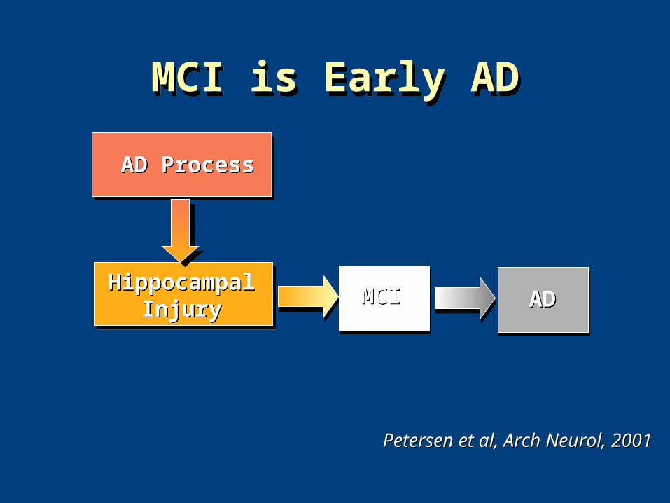
MCI is Early ADMCI is Early AD
Petersen et al, Arch Neurol, 2001Petersen et al, Arch Neurol, 2001
AD ProcessAD Process
HippocampalInjury
HippocampalInjury MCIMCI ADAD
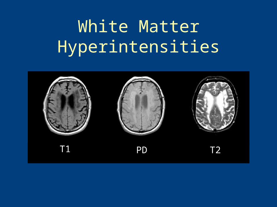
White Matter Hyperintensities
T1 PD T2
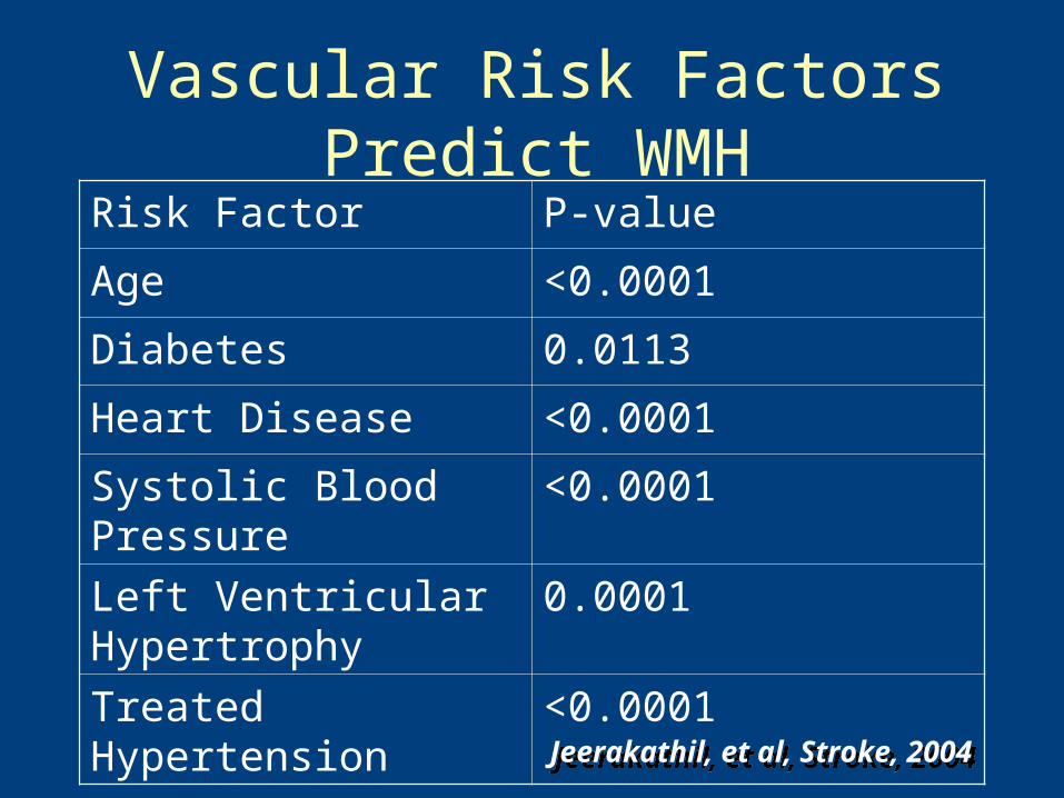
Vascular Risk Factors Predict WMH
Risk Factor P-value
Age <0.0001
Diabetes 0.0113
Heart Disease <0.0001
Systolic Blood Pressure <0.0001
Left Ventricular Hypertrophy
0.0001
Treated Hypertension <0.0001
Jeerakathil, et al, Stroke, 2004Jeerakathil, et al, Stroke, 2004
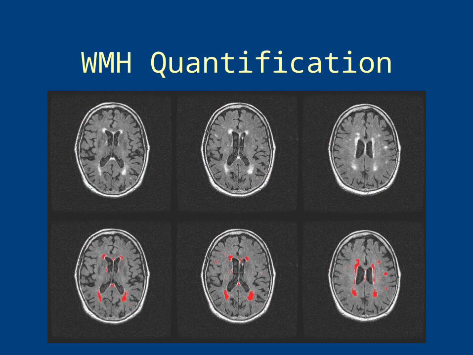
WMH Quantification
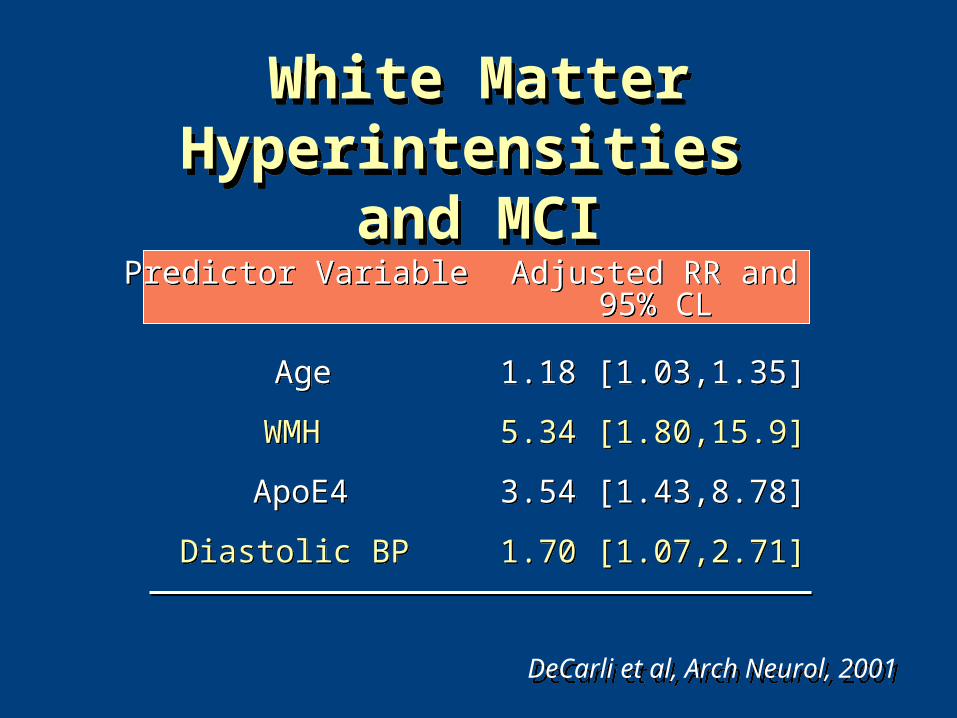
White Matter Hyperintensities and MCI
White Matter Hyperintensities and MCI
Predictor VariablePredictor Variable Adjusted RR andAdjusted RR and95% CL95% CL
AgeAge 1.18 [1.03,1.35]1.18 [1.03,1.35]
WMHWMH 5.34 [1.80,15.9]5.34 [1.80,15.9]
ApoE4ApoE4 3.54 [1.43,8.78]3.54 [1.43,8.78]
Diastolic BPDiastolic BP 1.70 [1.07,2.71]1.70 [1.07,2.71]
DeCarli et al, Arch Neurol, 2001DeCarli et al, Arch Neurol, 2001
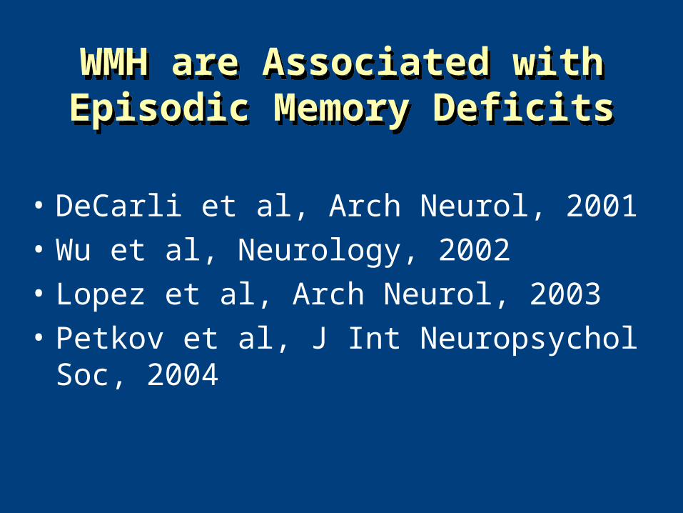
• DeCarli et al, Arch Neurol, 2001
• Wu et al, Neurology, 2002
• Lopez et al, Arch Neurol, 2003
• Petkov et al, J Int Neuropsychol Soc, 2004
WMH are Associated with Episodic Memory DeficitsWMH are Associated with Episodic Memory Deficits
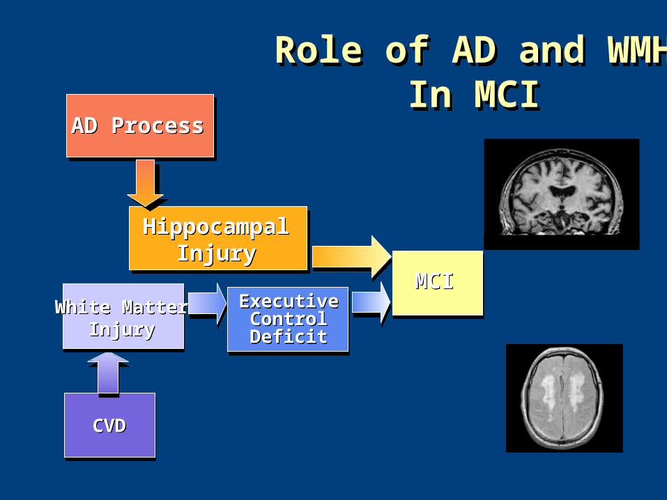
AD ProcessAD Process
HippocampalInjury
HippocampalInjury
MCIMCI
CVDCVD
White MatterInjury
White MatterInjury
ExecutiveControlDeficit
ExecutiveControlDeficit
Role of AD and WMHIn MCI
Role of AD and WMHIn MCI
Different Mechanisms of Episodic Memory Failure in MCI
IDeA Lab
Christine Wu Nordahl, Charan Ranganath, Andrew Yonelinas, Charles DeCarli,Bruce Reed,William J. Jagust
Neuropsychologia 43(11) 1688-1697; 2005
0
5
10
15
20
25
30
35
40
Cou
nt
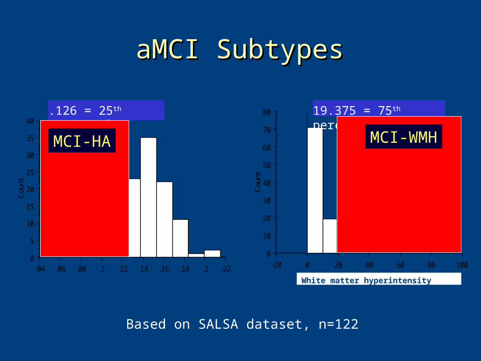
.04 .06 .08 .1 .12 .14 .16 .18 .2 .22
Normalized hippocampal volume
.126 = 25th percentile
0
10
20
30
40
50
60
70
80
Cou
nt-20 0 20 40 60 80 100
WMH volume
19.375 = 75th percentile
White matter hyperintensity volume
aMCI SubtypesaMCI Subtypes
Based on SALSA dataset, n=122
MCI-HA MCI-WMH
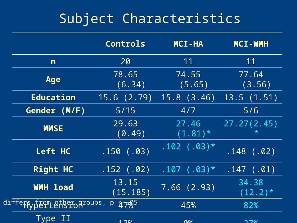
Controls MCI-HA MCI-WMH
n 20 11 11
Age 78.65 (6.34) 74.55 (5.65) 77.64 (3.56)
Education 15.6 (2.79) 15.8 (3.46) 13.5 (1.51)
Gender (M/F) 5/15 4/7 5/6
MMSE 29.63 (0.49) 27.46 (1.81)* 27.27(2.45)*
Left HC .150 (.03) .102 (.03)* .148 (.02)
Right HC .152 (.02) .107 (.03)* .147 (.01)
WMH load 13.15 (15.185) 7.66 (2.93) 34.38 (12.2)*
Hypertension 47% 45% 82%
Type II Diabetes 12% 0% 27%
Subject Characteristics
* differs from other groups, p < .05
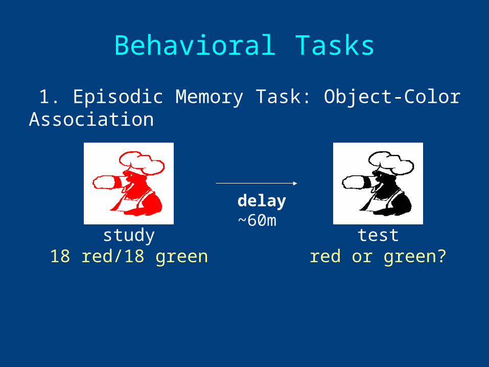
Behavioral Tasks
1. Episodic Memory Task: Object-Color Association
study18 red/18 green
delay~60m
testred or green?
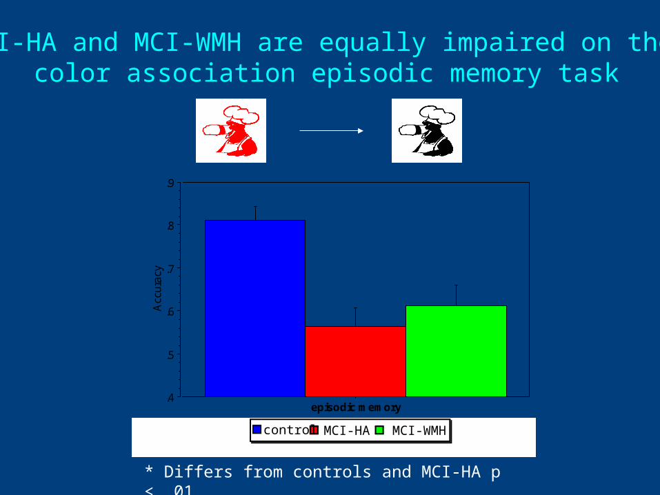
MCI-HA and MCI-WMH are equally impaired on the color association episodic memory task
.4
.5
.6
.7
.8
.9
Acc
ura
cy
episodic memory
* *
* Differs from controls and MCI-HA p < .01
MCI-WMHMCI-HAcontrol
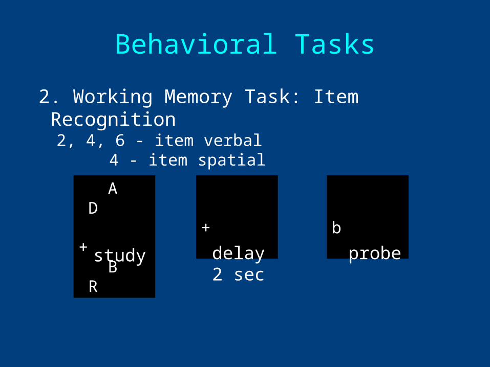
A D + B R
+ b
study delay2 sec
probe
Behavioral Tasks
2. Working Memory Task: Item Recognition2, 4, 6 - item verbal
4 - item spatial
.4
.5
.6
.7
.8
.9
1A
ccu
racy
2-item verbal 4-item verbal 6-item verbal 4-item spatial
MCI-WMHMCI-HAcontrol
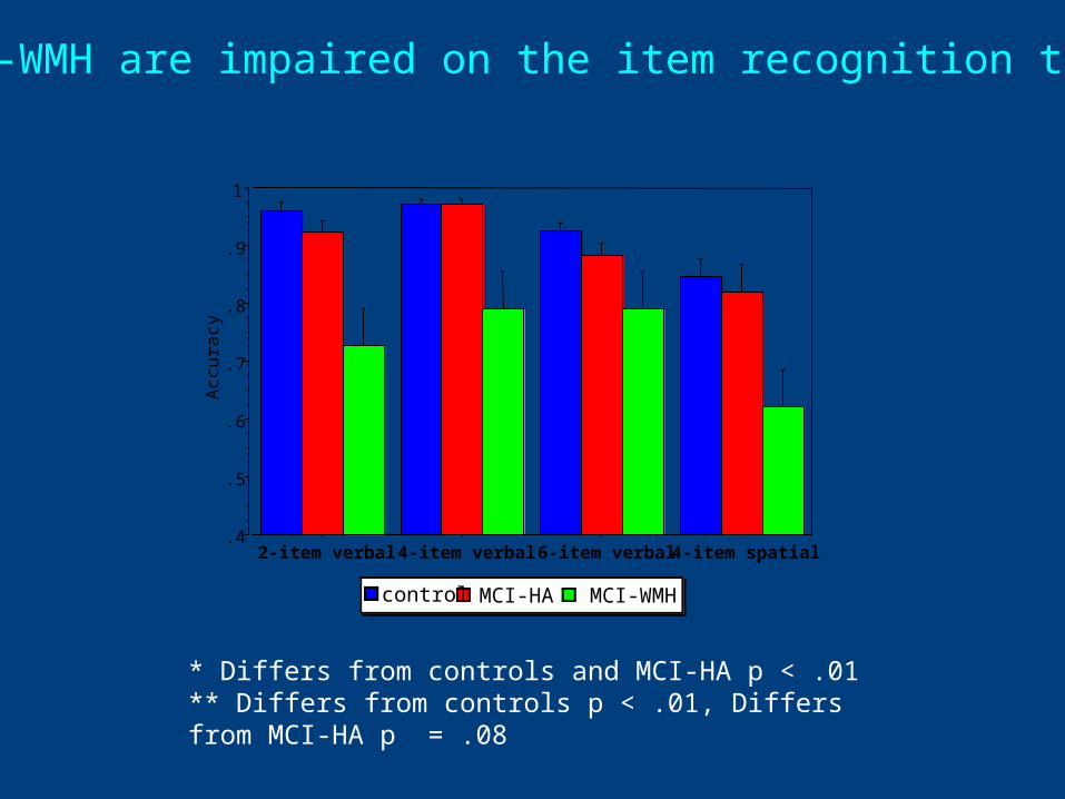
MCI-WMH are impaired on the item recognition task
* * ***
* Differs from controls and MCI-HA p < .01** Differs from controls p < .01, Differs from MCI-HA p = .08
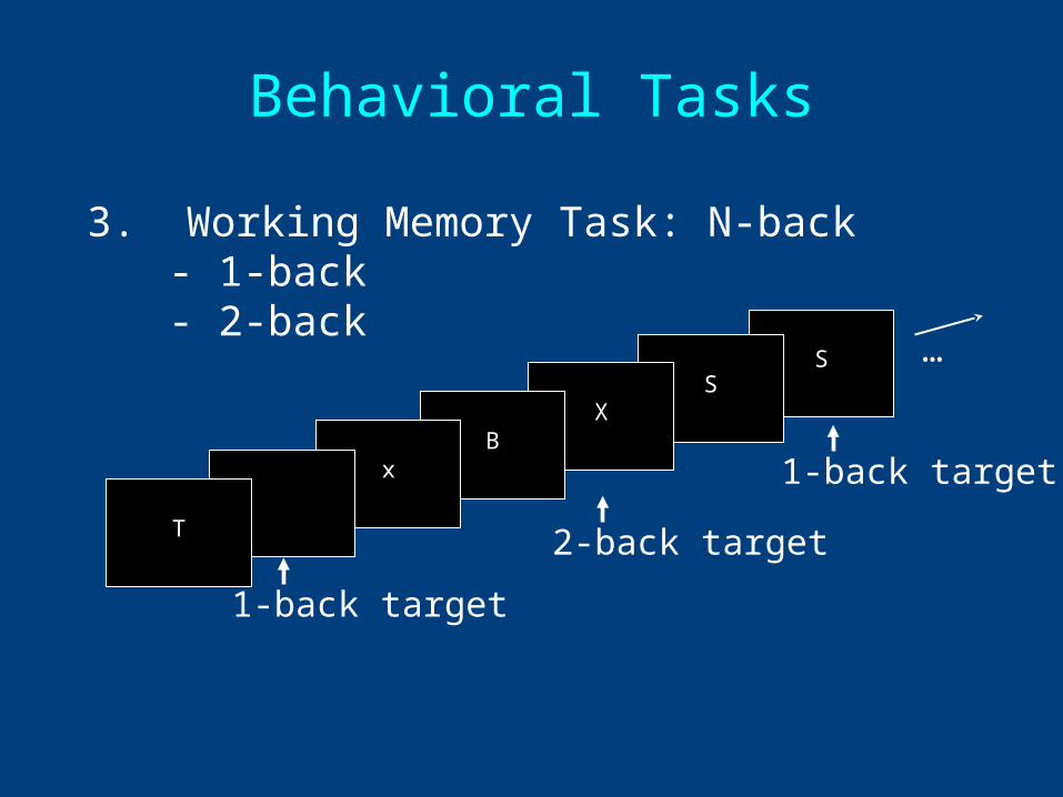
Behavioral Tasks
3. Working Memory Task: N-back- 1-back- 2-back
SS
XB
x t
T
…
1-back target
1-back target
2-back target
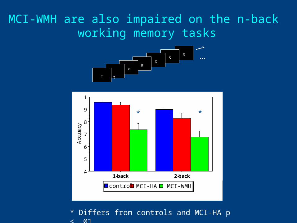
MCI-WMH are also impaired on the n-back working memory tasks
SS
XB
x t
T
…
* Differs from controls and MCI-HA p < .01
.4
.5
.6
.7
.8
.9
1
Acc
ura
cy
1-back 2-back
MCI-WMMCI-HCcontrol
* *
MCI-WMHMCI-HAcontrol
Summary of Behavioral Data
• MCI-HC and MCI-WMH are equally impaired on the episodic memory task
• MCI-WMH are impaired on working memory, both in simple maintenance as well as a more complicated task involving manipulation and maintenance
White Matter Changes Compromise Prefrontal Cortex Function in Healthy Elderly
IDeA Lab
Christine Wu Nordahl, Charan Ranganath, Andrew Yonelinas, Charles DeCarli,Evan Fletcher,William J. Jagust
In Press: Journal Cognitive Neuroscience
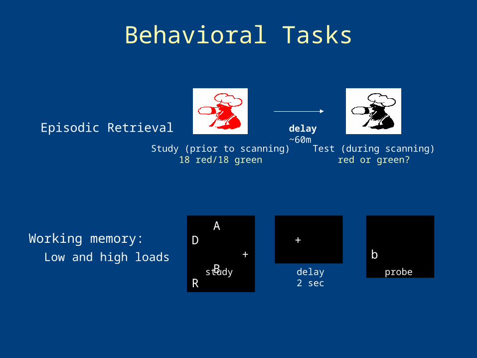
Behavioral Tasks
Episodic Retrieval Study (prior to scanning)
18 red/18 green
delay~60m
Test (during scanning)red or green?
A D + B R
b
study delay2 sec
probe
+ Working memory:
Low and high loads
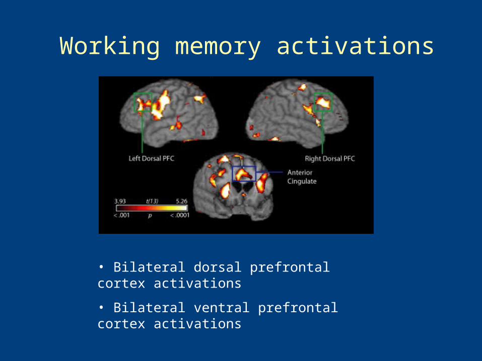
Working memory activations
• Bilateral dorsal prefrontal cortex activations
• Bilateral ventral prefrontal cortex activations
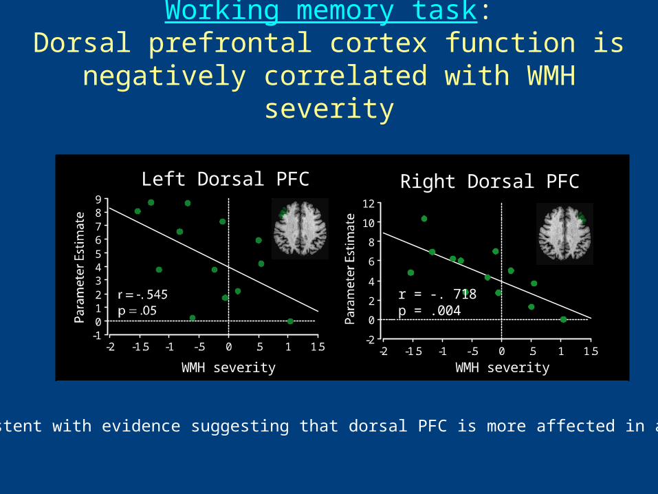
Left Dorsal PFC Right Dorsal PFC
-2
0
2
4
6
8
10
12
-2 -1.5 -1 -.5 0 .5 1 1.5
WMH severity
r = -. 718p = .004
-10123456789
10
-2 -1.5 -1 -.5 0 .5 1 1.5
WMH severity
WMH severity
-10123456789
-2 -1.5 -1 -.5 0 .5 1 1.5
Anterior Cingulate
Working memory task:Dorsal prefrontal cortex function is negatively
correlated with WMH severity
Consistent with evidence suggesting that dorsal PFC is more affected in aging
Left Dorsal PFC Right Dorsal PFC
-2
0
2
4
6
8
10
12
-2 -1.5 -1 -.5 0 .5 1 1.5
WMH severity
r = -. 718p = .004
-10123456789
10
-2 -1.5 -1 -.5 0 .5 1 1.5
WMH severity
WMH severity
-10123456789
-2 -1.5 -1 -.5 0 .5 1 1.5
Anterior Cingulate
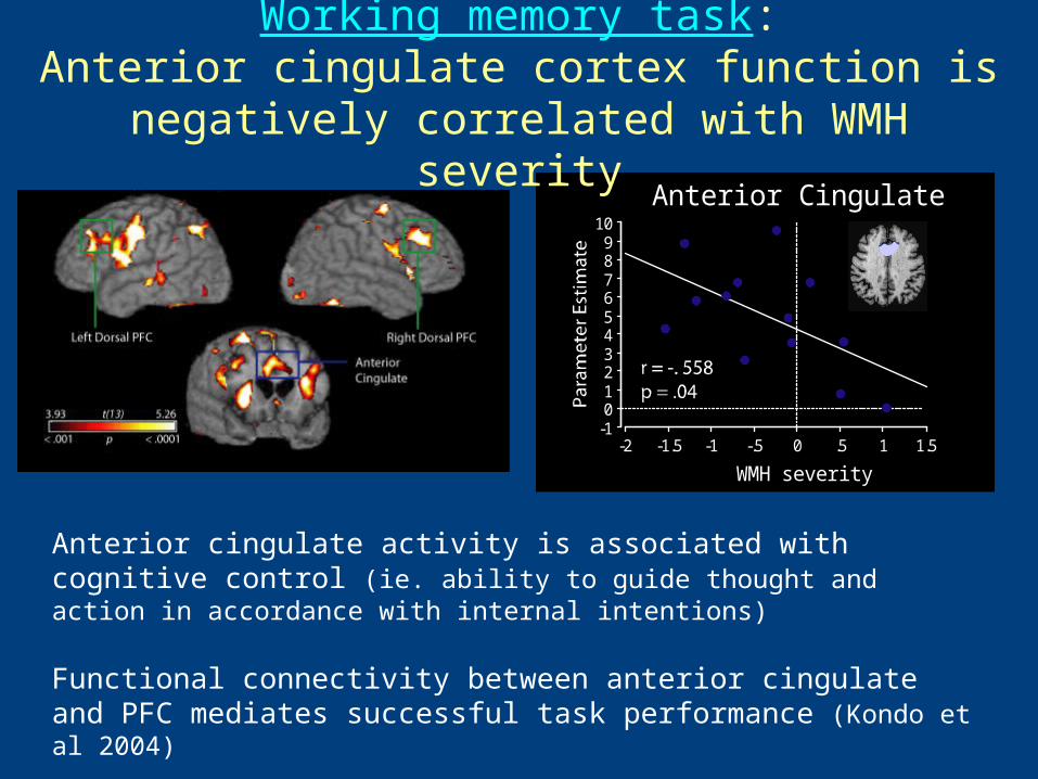
Anterior cingulate activity is associated with cognitive control (ie. ability to guide thought and action in accordance with internal intentions)
Functional connectivity between anterior cingulate and PFC mediates successful task performance (Kondo et al 2004)
Working memory task:Anterior cingulate cortex function is negatively
correlated with WMH severity
Summary of fMRI Data
• WMH a presumed indicator of cerebrovascular brain injury is associated with working memory impairment
• Executive control processes are likely involved and related to amount of WMH
Disconnection of Working Memory Processes by White Matter Hyperintensities
Adriane Mayda
Graduate Student, IDeA Lab
University of California at Davis
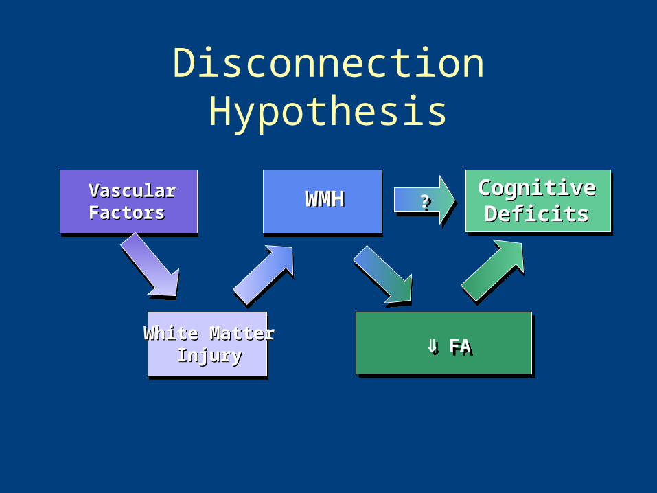
Disconnection Hypothesis
VascularFactorsVascularFactors
White MatterInjury
White MatterInjury
WMHWMH Cognitive Deficits
Cognitive Deficits??
FA FA
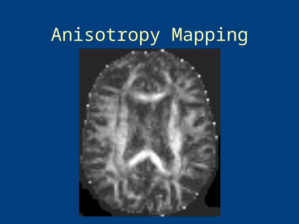
Anisotropy Mapping
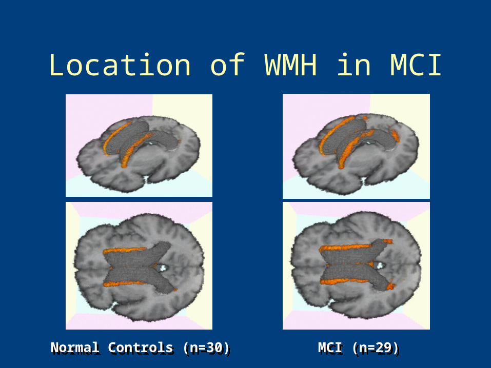
Location of WMH in MCI
Normal Controls (n=30)Normal Controls (n=30) MCI (n=29)MCI (n=29)
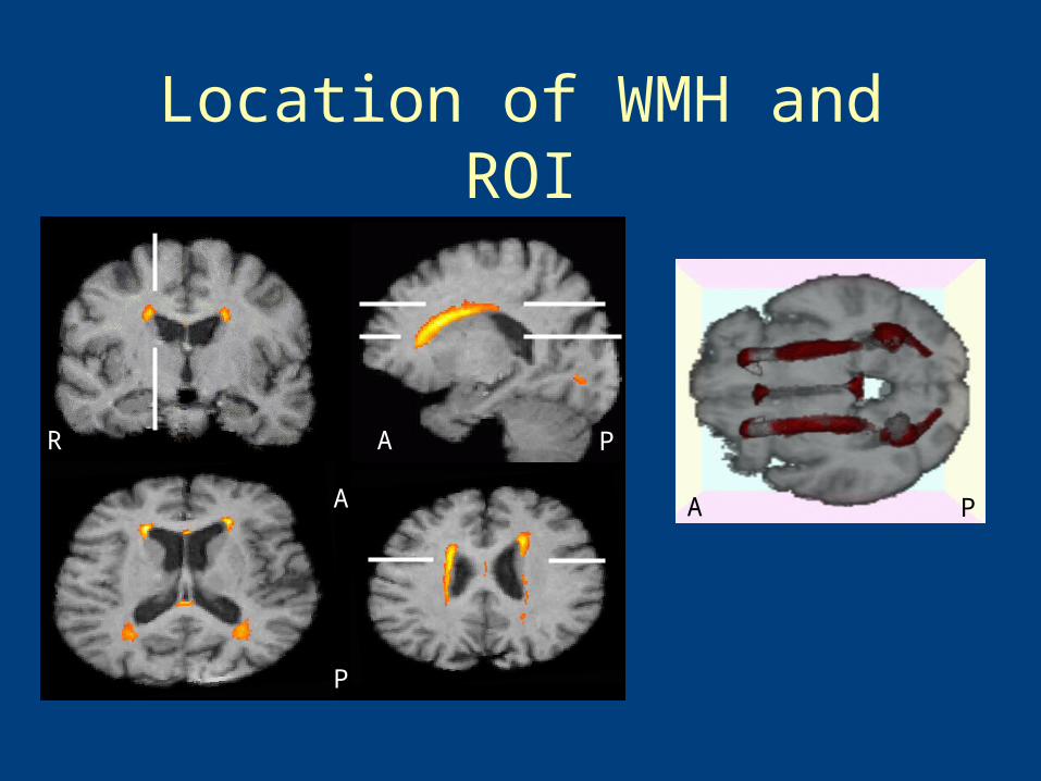
Location of WMH and ROI
A
A
P
PR
A P
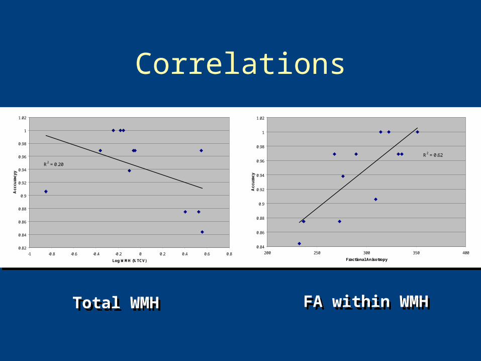
Correlations
R2 = 0.20
0.82
0.84
0.86
0.88
0.9
0.92
0.94
0.96
0.98
1
1.02
-1 -0.8 -0.6 -0.4 -0.2 0 0.2 0.4 0.6 0.8
Log WMH (%TCV)
Ac
cc
ura
cy
y
R2 = 0.62
0.84
0.86
0.88
0.9
0.92
0.94
0.96
0.98
1
1.02
200 250 300 350 400
Fractional Anisotropy
Ac
cu
rac
y
Total WMHTotal WMH FA within WMHFA within WMH
Summary
• aMCI has at least 2 subtypes– Hi WMH– Small Hippocampi
• Amnesia in MCI-WMH results from– Impairments in executive control– Reduced prefrontal activation– Disconnection from posterior targets
Conclusions
• WMH are common to the elderly
• WMH alone can be associated with episodic memory impairments
• WMH likely contribute to susceptibility to late life cognitive impairment and dementia
• WMH are potentially modifiable
• Adriane Mayda• Christine Nordahl• Evan Fletcher• Owen Carmichael• IDeA Lab
Supported by NIH: P30 AG10129, R01 AG09029,R01 AG021028, R01 NS 29993,P01 AG12435, P01 AG0027232,R01 AG111101, R01 AG08122,R01 AG16495, U01 AG024904
http://neuroscience.ucdavis.edu/idealab/