Embed Size (px)
Citation preview
Urška ŽagerGregor AndeBorut Božič
SIGNIFICA(PATHO)P
Urška Žager,
CorrespondinUrška Žager Department
E‐mail: urska
ABSTRACTβ2‐glycoprot
syndrome. T
its physiolog
K(L/V)WX(I/L
Based on thi
some already
(patho)physi
INTRODUCβ2GPI is the
associated w
characterized
β2GPI is a 45
polypeptide
conformatio
the β2GPI’s f
binding β2GP
The physiolo
cascade, the
was suggeste
innate immu
(macromolec
in the selecti
We suggeste
In the presen
by ELISA and
r, Mojca Lundeerluh, Saša Ču
ANCE OF KPHYSIOLOG
Mojca Lunde
ng author
of Rheumato
azager@yahoo
T tein I (β2GPI)
The exact mec
gical role. We
L/V)P motif, p
s motif we co
y known and
ological role.
CTION most commo
with clinical fea
d by vascular
5kDa plasma g
is comprised
n and a “fish‐
fifth domain e
PI opens up an
ogical role of β
removal of a
ed to be invol
unity (8, 9). Nu
cules). Determ
ion of a comm
ed Lipid A of L
nt study we te
d surface plasm
er, Vesna Hodučnik, Tanja Kv
K(L/V)WXGIC FUNCT
er, Vesna Hodn
logy, Universi
o.com
) is a major
chanism unde
e used rando
primarily desi
onfirmed the p
some new pu
n and best‐ch
atures of diffe
thrombosis a
glycoprotein w
of five domai
hook” like op
enables it’s bin
nd thus conve
β2GPI is not en
poptotic bodi
ved in the cle
umerous puta
mination of am
mon motif that
PS as a possib
ested and con
mon resonanc
dnik, veder,
S(p
X(I/L/V)PTION
nik, Gregor An
ity Medical Ce
autoantigen
rlying the β2G
om phage pep
gnated as ta
previously sug
utative β2GPI
haracterized a
erent autoimm
nd pregnancy
with plasma co
ns existing in
en conformat
nding to nega
erts from circu
ntirely elucida
es, triglycerid
arance of lipo
tive functions
mino acid sequ
t has been de
ble selection d
firmed the dis
ce. Based on t
Significance (patho)physiol
P EPITOPE
nderluh, Saša
entre Ljubljan
of autoimmu
GPI’s involvem
ptide library
rget unrelate
ggested role o
binding prot
ntigenic targe
mune disorde
y morbidity, an
oncentration
at least two d
tion. The posit
tively charged
ular to “fish‐h
ated; in vitro s
de metabolism
opolysaccharid
s imply that β
uences recogn
etermined also
decoy and thu
scriminating a
the selected m
of K(L/V)WXlogic function
OF THE Β
Čučnik, Tanja
a, SI‐1000 Lju
une thrombo
ment in the di
y to identify s
ed, was confir
of polar residu
eins. The latt
et for antipho
rs especially t
nd also system
of 50–300 mg
different confo
tively charged
d surfaces suc
ook” like conf
studies have s
m and depletio
de (LPS) and it
2GPI interacts
nized by β2GP
o by several o
s the true bin
and moderate
motif we prese
X(I/L/V)P ep
2GPI IN IT
a Kveder, Boru
bljana, Sloven
philia, known
isease is not f
sequences bi
rmed as the s
ues in β2GPI i
er can help to
spholipid auto
he antiphosph
mic lupus eryt
g/L (2). This 32
ormations: a c
d patch (C281K
ch as anionic p
formation (2,
shown its invo
on of free radi
t was identifie
s with many d
PI, using phage
ther groups st
ding partner o
e affinity of se
ent putative b
pitope of t
TS
ut Božič
nia
n as the ant
fully elucidate
nding to β2G
selective bind
interactions, a
o further eluc
oantibodies (a
holipid syndro
thematosus (S
26 amino acid
circulating pla
KNKEKKC288)
phospholipids
3).
olvment in the
icals (4‐7). Re
ed as a compo
different prote
e display libra
tudying differ
of the selecte
elected peptid
binding partne
he β2GPI i
iphospholipid
ed, as it is not
GPI. Obtained
der of β2GPI.
and identified
cidate β2GPI’s
aPL). aPL are
ome, which is
SLE) (1).
ds long
asma
) located on
s. Upon
e coagulation
cently, β2GPI
onent of
eins
ary, resulted
rent targets.
ed motif (10).
es to β2GPI
ers of β2GPI.
in its
d
t
d
.
d
s
Page 118eJIFCC2011Vol22No4pp118-124
Urška Žager, Mojca Lunder, Vesna Hodnik, Gregor Anderluh, Saša Čučnik, Tanja Kveder, Borut Božič
Significance of K(L/V)WX(I/L/V)P epitope of the β2GPI in its(patho)physiologic function
METHODS
BIOPANNING PROCEDURE
To select β2GPI binding phage peptides a random linear heptamer peptide library (Ph.D.‐7, New England
Biolabs,Beverly, MA, USA) was used. The selection was carried out according to the manufacturer’s instructions (11).
Briefly, phage library (2×10¹¹ pfu in 0.1% Tween 20 in phosphate buffered saline (PBST)) was added to β2GPI‐coated
(30 μg/ml) and blocked (1% bovine serum albumin (BSA) in phosphate buffered saline (PBS)) microtitre plate (High
binding, Costar, Cambridge, MA, USA). Unbound phage clones were removed by intensive washing with 0.1% PBST
and 0.5% PBST. The bound phage clones were eluted in a specific manner by monoclonal anti‐β2GPI (HCAL, Inova
Diagnostics Inc., San Diego, CA, USA), purified high or low avidity anti‐β2GPI (12), or oxidized native IgGs (13). After
four rounds of selection the affinity of recovered phage clones was determined, and a single‐stranded DNA from
selected phage clones was isolated and sequenced (MWG Biotech, Munich, Germany). Two peptides, one presenting
most commonly observed motif and other exhibiting the highest affinity towards β2GPI, were synthesized (GenScript
USA Inc., NJ, USA).
IMMUNOASSAYS
Evaluation of phages’ affinity towards β2GPI
The binding affinity of selected phage clones was confirmed by standard phage ELISA. Briefly, microtitre plate (High
binding, Costar) was coated with β2GPI (100μl/well; 10mg/l) and blocked with 1% BSA in PBS pH 7.4. A separate set of
wells was coated with 1% BSA in PBS pH 7.4 without previous β2GPI immobilization, as negative controls. Each
recovered phage clone (5×10⁹ pfu/well in 0.1% PBST) was added to the β2GPI coated and to the BSA coated wells and
incubated for 1h at room temperature (RT). Wells were rinsed (0.05% PBST) and 200 μl/well of horseradish
peroxidase‐labelled mouse anti‐M13 monoclonal antibody (Amersham Biosciences, Little Chalfont, UK) in 1% BSA in
0.1% PBST (dilution 1:5000), was added and incubated for 1 h at RT. Finally, 100 μl/well of substrate solution (0.22
mg/mL diammonium 2,2’‐azino‐di‐(3‐ethylbenzthiazoline sulfonate in 50 mM citric acid and 1.7 μL of 30% H2O2 /mL,
pH 4.0) was added and the absorbance at 405 nm was measured.
Evaluation of phages’ affinity towards β2GPI and lipid A mixtures
To evaluate putative affinity of selected phage clones towards lipid A, the standard phage ELISA (described above) was
used. Two mixtures of β2GPI and lipid A, containing 20 μg/ml of β2GPI and 10 or 60 μg/ml of lipid A, were prepared.
β2GPI at concentrations 10 and 20 mg/l, and the two mixtures were coated on separate sets of wells and incubated
for 2h at RT. After rinsing (0.1% PBST) 3×10⁹ pfu/well of two phage clones corresponding to the common motif K‐
(L/V)‐W‐X‐(I/L)‐P‐X were added and incubated for 1h at RT. For detection a horseradish peroxidase‐labelled mouse
anti‐M13 monoclonal antibody (Amersham Biosciences) and substrate 3,3’,5,5’‐Tetramethylbenzidine (Sigma‐Aldrich,
MO, USA) were used. After colour development, stop solution (50 μl/well of 2M H2SO4) was added and the
absorbance was measured at 450nm.
Competition ELISA
A competitive ELISA was used to evaluate phages’ or peptides’ ability to inhibit binding of anti‐β2GPI to the β2GPI.
6.25‐25 ng/ml of HCAL or 50‐100 ng/ml of anti‐β2GPI were incubated with various amounts of each phage clone (0,
1.25×10⁹‐ 6×10¹¹) or synthetic peptide (250‐200 nM) for 2h at RT. The mixtures were subsequently applied to β2GPI
coated and BSA blocked microtitre plates (High Binding, Costar). The detection of anti‐β2GPI binding proceeded as in
anti‐β2GPI ELISA (14).
SURFACE PLASMON RESONANCE ANALYSIS OF SELECTED PEPTIDES
The binding affinity of synthetic peptides towards β2GPI was evaluated by surface plasmon resonance (SPR), using
Biacore T100 instrument (GE Healthcare, Uppsala, Sweden). Two peptides selected over unrelated target were used as
negative controls. β2GPI was amine coupled to the CM5 chip (13) reaching the immobilization level of ~2500 RU.
Page 119eJIFCC2011Vol22No4pp118-124
Urška ŽagerGregor AndeBorut Božič
Reference ce
buffer was 0
concentratio
sensorgrams
running buffe
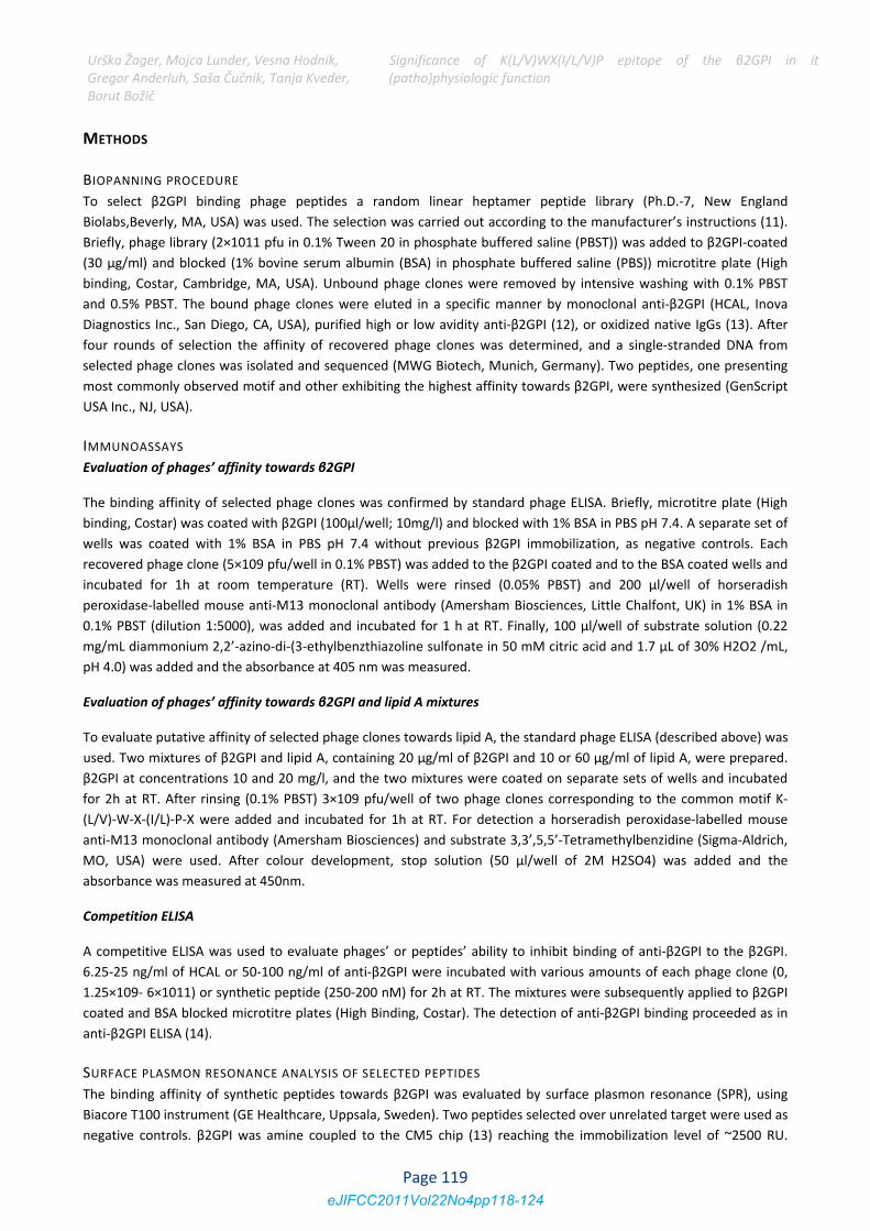
RESULTS ANIn an attemp
involved spe
polyclonal lo
Table 1, the
consensus m
possess prop
Table 1: Amino
known target un
r, Mojca Lundeerluh, Saša Ču
ell was prepa
0.005% PBST,
ons of each pe
s were correc
er.
ND DISCUSSIO
pt to charact
ecific elution o
ow and high a
e selection yi
motif K‐(L/V)‐W
pagation adva
acid sequences o
nrelated sequenc
er, Vesna Hodučnik, Tanja Kv
red by the sa
pH 7.4, and
eptide (250‐20
ted by double
N erize sequenc
of bound pha
vidity anti‐β2
elded series
W‐X‐(I/V/L)‐(P
ntage were se
of selected phage
ces are marked ye
dnik, veder,
S(p
ame immobiliz
the analysis w
000 nM) were
e subtracting
ces binding t
ages with vari
2GPI and oxidi
of similar se
P)‐X. In additio
elected (mark
e displayed pepti
ellow (10); seque
Significance (patho)physiol
zation proced
was performe
e injected ove
the signal ob
o β2GPI by P
ious subgrou
ized native Ig
equences. 53%
on some sequ
ked blue and y
ides. Residues ca
ences correspond
of K(L/V)WXlogic function
dure without
ed at 25°C at
er the sensor
btained on a
Ph.D.‐7 library
ps of anti‐β2G
G with acquir
% of the sel
uences startin
yellow respect
pable of forming
ding to Q‐T‐(L/Q)‐
X(I/L/V)P ep
the addition
a flow rate o
surface for a
reference sur
y, we used a
GPI antibodie
red anti‐β2GP
ected sequen
ng with Q‐T‐(L
tively in Table
g electrostatic or
‐ motif are marke
pitope of t
of the ligand
of 15 μl/ml. F
period of 1 m
rface and the
screening pr
es (monoclona
PI activity). As
nces correspo
L/Q)‐ and mo
e 1) (10).
hydrogen bonds
ed blue.
he β2GPI i
. The running
Four different
min. Obtained
signal of the
rocedure that
al anti‐β2GPI,
presented in
onded to the
tifs known to
are marked red;
in its
g
t
d
e
t
,
n
e
o
;
Page 120eJIFCC2011Vol22No4pp118-124
Urška ŽagerGregor AndeBorut Božič
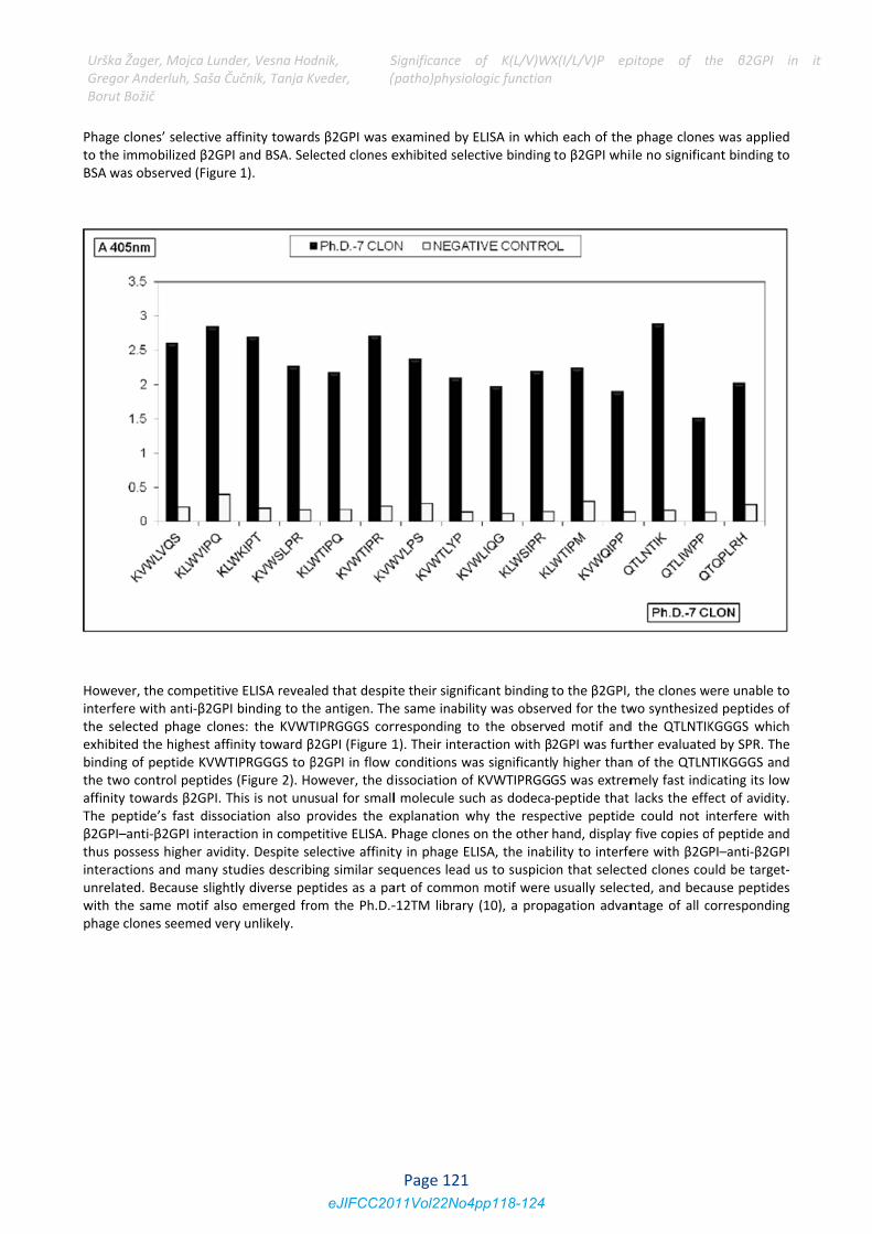
Phage clonesto the immoBSA was obs
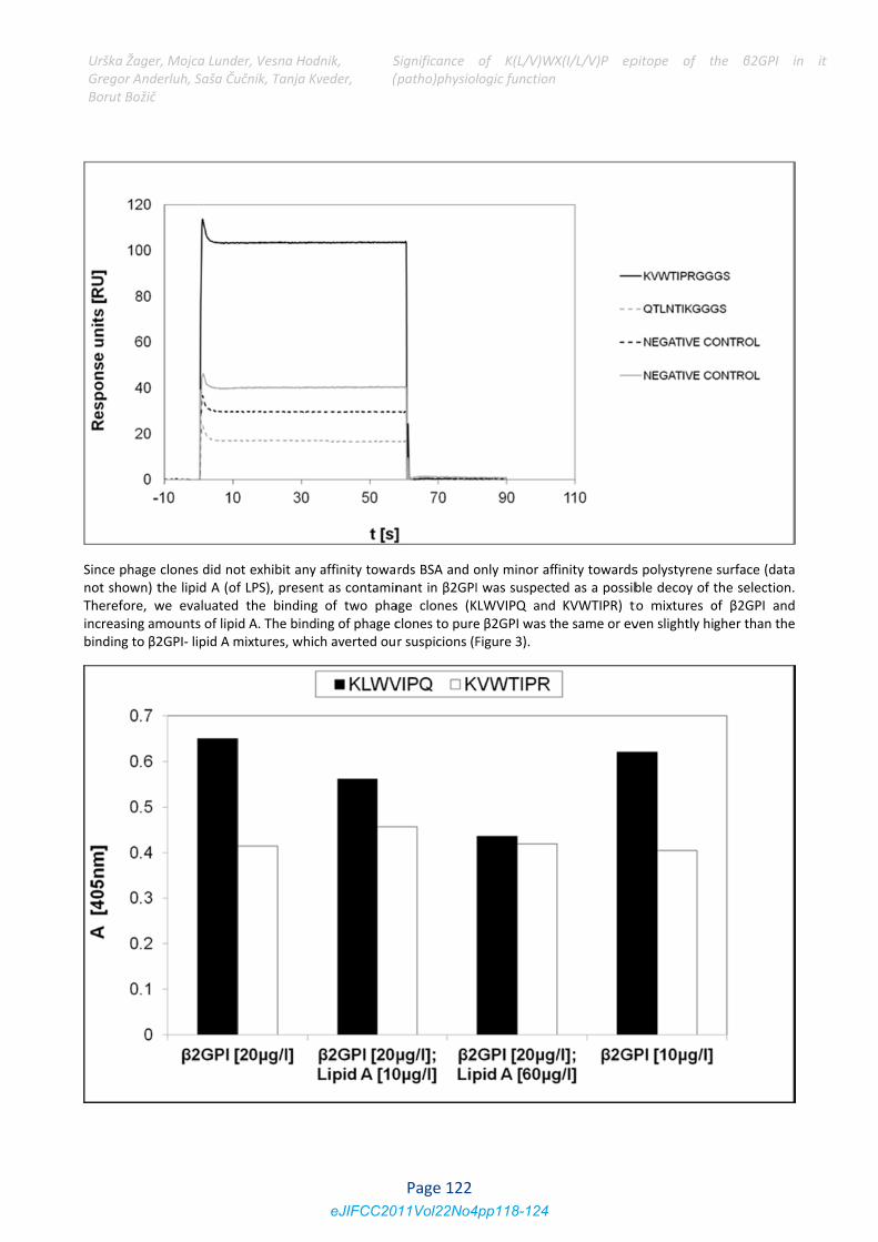
However, theinterfere witthe selectedexhibited thebinding of pethe two contaffinity towaThe peptide’β2GPI–anti‐βthus possessinteractions unrelated. Bwith the samphage clones
r, Mojca Lundeerluh, Saša Ču
s’ selective afbilized β2GPI erved (Figure
e competitiveth anti‐β2GPI phage clonee highest affineptide KVWTItrol peptides ards β2GPI. Th’s fast dissocβ2GPI interacts higher aviditand many stuecause slightlme motif alsos seemed very
er, Vesna Hodučnik, Tanja Kv
ffinity towardand BSA. Sele 1).
e ELISA revealebinding to thees: the KVWTnity toward β2IPRGGGS to β(Figure 2). Hohis is not unuiation also prtion in compety. Despite seudies describily diverse pepo emerged froy unlikely.
dnik, veder,
S(p
s β2GPI was eected clones e
ed that despite antigen. TheTIPRGGGS cor2GPI (Figure 1β2GPI in flow owever, the dsual for smallrovides the eetitive ELISA. Pelective affinitng similar seqptides as a paom the Ph.D.‐
Significance (patho)physiol
examined by exhibited sele
te their signifie same inabilrresponding to1). Their interconditions waissociation ofl molecule sucexplanation wPhage clones ty in phage ELquences lead art of common‐12TM library
of K(L/V)WXlogic function
ELISA in whicective binding
icant binding ity was observo the observeraction with βas significantlf KVWTIPRGGGch as dodeca‐why the respeon the other LISA, the inabus to suspicion motif were y (10), a prop
X(I/L/V)P ep
h each of theto β2GPI whi
to the β2GPI, ved for the twed motif and2GPI was furty higher thanGS was extrem‐peptide that ective peptidehand, display ility to interfeon that selectusually selectagation advan
pitope of t
e phage cloneile no significa
the clones wwo synthesized the QTLNTIKther evaluaten of the QTLNmely fast indilacks the effee could not iy five copies oere with β2Gted clones couted, and becantage of all c
he β2GPI i
s was appliedant binding to
were unable toed peptides ofKGGGS whichd by SPR. TheTIKGGGS andicating its lowect of avidity.nterfere withf peptide andPI–anti‐β2GPIuld be target‐ause peptidescorresponding
in its
d o
o f h e d w . h d I ‐s g
Page 121eJIFCC2011Vol22No4pp118-124
Urška ŽagerGregor AndeBorut Božič
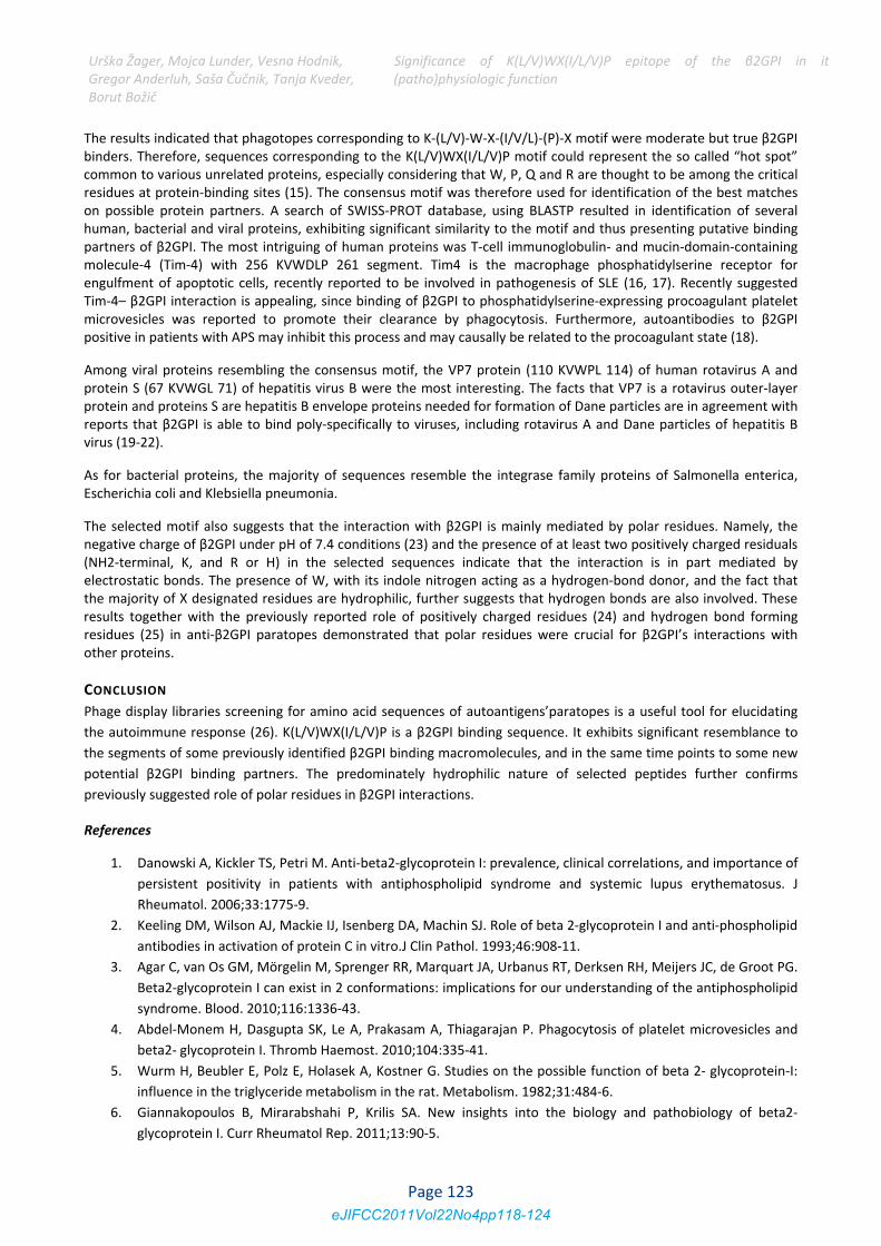
Since phage not shown) tTherefore, wincreasing ambinding to β2
r, Mojca Lundeerluh, Saša Ču
clones did nothe lipid A (ofwe evaluatedmounts of lipi2GPI‐ lipid A m
er, Vesna Hodučnik, Tanja Kv
ot exhibit any f LPS), presen the bindingd A. The bindmixtures, whic
dnik, veder,
S(p
affinity towant as contamin of two phaing of phage cch averted ou
Significance (patho)physiol
rds BSA and onant in β2GPIage clones (Kclones to purer suspicions (F
of K(L/V)WXlogic function
only minor affI was suspectKLWVIPQ ande β2GPI was thFigure 3).
X(I/L/V)P ep
finity towardsed as a possibKVWTIPR) t
he same or ev
pitope of t
s polystyrene ble decoy of tto mixtures oven slightly hi
he β2GPI i
surface (datathe selection.of β2GPI andgher than the
in its
a . d e
Page 122eJIFCC2011Vol22No4pp118-124
Urška Žager, Mojca Lunder, Vesna Hodnik, Gregor Anderluh, Saša Čučnik, Tanja Kveder, Borut Božič
Significance of K(L/V)WX(I/L/V)P epitope of the β2GPI in its(patho)physiologic function
The results indicated that phagotopes corresponding to K‐(L/V)‐W‐X‐(I/V/L)‐(P)‐X motif were moderate but true β2GPI binders. Therefore, sequences corresponding to the K(L/V)WX(I/L/V)P motif could represent the so called “hot spot” common to various unrelated proteins, especially considering that W, P, Q and R are thought to be among the critical residues at protein‐binding sites (15). The consensus motif was therefore used for identification of the best matches on possible protein partners. A search of SWISS‐PROT database, using BLASTP resulted in identification of several human, bacterial and viral proteins, exhibiting significant similarity to the motif and thus presenting putative binding partners of β2GPI. The most intriguing of human proteins was T‐cell immunoglobulin‐ and mucin‐domain‐containing molecule‐4 (Tim‐4) with 256 KVWDLP 261 segment. Tim4 is the macrophage phosphatidylserine receptor for engulfment of apoptotic cells, recently reported to be involved in pathogenesis of SLE (16, 17). Recently suggested Tim‐4– β2GPI interaction is appealing, since binding of β2GPI to phosphatidylserine‐expressing procoagulant platelet microvesicles was reported to promote their clearance by phagocytosis. Furthermore, autoantibodies to β2GPI positive in patients with APS may inhibit this process and may causally be related to the procoagulant state (18).
Among viral proteins resembling the consensus motif, the VP7 protein (110 KVWPL 114) of human rotavirus A and protein S (67 KVWGL 71) of hepatitis virus B were the most interesting. The facts that VP7 is a rotavirus outer‐layer protein and proteins S are hepatitis B envelope proteins needed for formation of Dane particles are in agreement with reports that β2GPI is able to bind poly‐specifically to viruses, including rotavirus A and Dane particles of hepatitis B virus (19‐22).
As for bacterial proteins, the majority of sequences resemble the integrase family proteins of Salmonella enterica, Escherichia coli and Klebsiella pneumonia.
The selected motif also suggests that the interaction with β2GPI is mainly mediated by polar residues. Namely, the negative charge of β2GPI under pH of 7.4 conditions (23) and the presence of at least two positively charged residuals (NH2‐terminal, K, and R or H) in the selected sequences indicate that the interaction is in part mediated by electrostatic bonds. The presence of W, with its indole nitrogen acting as a hydrogen‐bond donor, and the fact that the majority of X designated residues are hydrophilic, further suggests that hydrogen bonds are also involved. These results together with the previously reported role of positively charged residues (24) and hydrogen bond forming residues (25) in anti‐β2GPI paratopes demonstrated that polar residues were crucial for β2GPI’s interactions with other proteins.
CONCLUSION
Phage display libraries screening for amino acid sequences of autoantigens’paratopes is a useful tool for elucidating
the autoimmune response (26). K(L/V)WX(I/L/V)P is a β2GPI binding sequence. It exhibits significant resemblance to
the segments of some previously identified β2GPI binding macromolecules, and in the same time points to some new
potential β2GPI binding partners. The predominately hydrophilic nature of selected peptides further confirms
previously suggested role of polar residues in β2GPI interactions.
References
1. Danowski A, Kickler TS, Petri M. Anti‐beta2‐glycoprotein I: prevalence, clinical correlations, and importance of
persistent positivity in patients with antiphospholipid syndrome and systemic lupus erythematosus. J
Rheumatol. 2006;33:1775‐9.
2. Keeling DM, Wilson AJ, Mackie IJ, Isenberg DA, Machin SJ. Role of beta 2‐glycoprotein I and anti‐phospholipid
antibodies in activation of protein C in vitro.J Clin Pathol. 1993;46:908‐11.
3. Agar C, van Os GM, Mörgelin M, Sprenger RR, Marquart JA, Urbanus RT, Derksen RH, Meijers JC, de Groot PG.
Beta2‐glycoprotein I can exist in 2 conformations: implications for our understanding of the antiphospholipid
syndrome. Blood. 2010;116:1336‐43.
4. Abdel‐Monem H, Dasgupta SK, Le A, Prakasam A, Thiagarajan P. Phagocytosis of platelet microvesicles and
beta2‐ glycoprotein I. Thromb Haemost. 2010;104:335‐41.
5. Wurm H, Beubler E, Polz E, Holasek A, Kostner G. Studies on the possible function of beta 2‐ glycoprotein‐I:
influence in the triglyceride metabolism in the rat. Metabolism. 1982;31:484‐6.
6. Giannakopoulos B, Mirarabshahi P, Krilis SA. New insights into the biology and pathobiology of beta2‐
glycoprotein I. Curr Rheumatol Rep. 2011;13:90‐5.
Page 123eJIFCC2011Vol22No4pp118-124
Urška Žager, Mojca Lunder, Vesna Hodnik, Gregor Anderluh, Saša Čučnik, Tanja Kveder, Borut Božič
Significance of K(L/V)WX(I/L/V)P epitope of the β2GPI in its(patho)physiologic function
7. Blank M, Asherson RA, Cervera R, Shoenfeld Y. Antiphospholipid syndrome infectious origin. J Clin Immunol.
2004;24:12‐23.
8. Ağar C, de Groot PG, Marquart JA, Meijers JC. Evolutionary conservation of the lipopolysaccharide binding
site of β2‐glycoprotein I. Thromb Haemost. 2011;30;106:1069‐75.
9. Agar C, de Groot PG, Mörgelin M, Monk SD, van Os G, Levels JH, de Laat B, Urbanus RT, Herwald H, van der
Poll T, Meijers JC. β₂‐glycoprotein I: a novel component of innate immunity. Blood. 2011;117:6939‐47.
10. Vodnik M, Zager U, Strukelj B, Lunder M. Phage display: selecting straws instead of a needle from a haystack.
Molecules. 2011;16:790‐817.
11. Protein Tools, Ph.D.TM Phage Display Libraries, Instruction Manual
(http://www.neb.com/nebecomm/manualfiles/manuale8101.pdf)
12. Zager U, Irman S, Lunder M, Skarabot M, Musevic I, Hodnik V, Anderluh G, Cucnik S, Kveder T, Rozman B,
Bozic B. Immunochemical properties and pathological relevance of anti‐{beta}2‐glycoprotein I antibodies of
different avidity. Int Immunol. 2011;23:511‐8.
13. Omersel J, Jurgec I, Cucnik S, Kveder T, Rozman B, Sodin‐Semrl S, Bozic B. Autoimmune and proinflammatory
activity of oxidized immunoglobulins. Autoimmun Rev. 2008;7:523‐9.
14. Cucnik S, Ambrozic A, Bozic B, Skitek M, Kveder T. Anti‐beta2‐glycoprotein I ELISA: methodology,
determination of cut‐off values in 434 healthy Caucasians and evaluation of monoclonal antibodies as
possible international standards. Clin Chem Lab Med. 2000;38:777‐83.
15. Hu, Z.; Ma, B.; Wolfson, H.; Nussinov, R. Conservation of polar residues as hot spots at protein interfaces.
Proteins 2000, 39, 331‐342.
16. Zhao P, Xu L, Wang P, Liang X, Qi J, Liu P, Guo C, Zhang L, Ma C, Gao L. Increased expression of human T‐cell
immunoglobulin‐ and mucin‐domain‐containing molecule‐4 in peripheral blood mononuclear cells from
patients with system lupus erythematosus. Cell Mol Immunol. 2010;7:152‐6.
17. Miyanishi M, Tada K, Koike M, Uchiyama Y, Kitamura T, Nagata S. Identification of Tim4 as a
phosphatidylserine receptor. Nature. 2007;450:435‐9.
18. Abdel‐Monem H, Dasgupta SK, Le A, Prakasam A, Thiagarajan P. Phagocytosis of platelet microvesicles and
beta2‐ glycoprotein I. Thromb Haemost. 2010;104:335‐41.
19. Adlhoch C, Kaiser M, Hoehne M, Mas Marques A, Stefas I, Veas F, Ellerbrok H. Highly sensitive detection of
the group A Rotavirus using Apolipoprotein H‐coated ELISA plates compared to quantitative real‐time PCR.
Virol J. 2011;8:63.
20. Aoki ST, Settembre EC, Trask SD, Greenberg HB, Harrison SC, Dormitzer PR. Structure of rotavirus outer‐layer
protein VP7 bound with a neutralizing Fab. Science. 2009;324:1444‐7.
21. Stefas I, Rucheton M, D'Angeac AD, Morel‐Baccard C, Seigneurin JM, Zarski JP, Martin M, Cerutti M, Bossy JP,
Misse D, Graafland H, Veas F. Hepatitis B virus Dane particles bind to human plasma apolipoprotein H.
Hepatology. 2001;33:207–217.
22. Ueda K, Tsurimotob T, Matsubara K. Three envelope proteins of hepatitis B virus: large S, middle S, and major
S proteins needed for the formation of Dane particles. Virol. 1991;65:3521‐9.
23. Sodin‐Semrl S, Frank M, Ambrozic A, Pavlic J, Sustar V, Cucnik S, Bozic B, Kveder T, Rozman B. Interactions of
Phospholipid Binding Proteins with Negatively Charged Membranes: β2‐Glycoprotein I as a Model
Mechanism in Advances in Planar Lipid Bilayers and Liposomes 2008;8:243‐273.
24. Giles I, Lambrianides A, Rahman A. Examining the non‐linear relationship between monoclonal
antiphospholipid antibody sequence, structure and function. Lupus. 2008;17:895‐903.
25. Jurgec I, Lunder M, Bratkovic T, Cucnik S, Kveder T, Rozman B, Bozic B. Hydrogen bonds are crucial for the
anti‐beta 2‐glycoprotein I antibody bindings to the antigen. Ann Rheum Dis. 2009, 68(Suppl I), A4.
26. Zager U, Lunder M, Bozic B. Using Phage Display in Autoimmunity Research. ACSi. 2011, in press.
Page 124eJIFCC2011Vol22No4pp118-124