Embed Size (px)
Citation preview
JOURNAL OF PLANT PROTECTION RESEARCH Vol. 55, No. 2 (2015)
*Corresponding address: [email protected]
Signaling molecules from Lactuca sativa L. induced quorum sensing phenotypes in bacteria
Esmaeil Mahmoudi*
Department of Plant Protection, Islamic Azad University, Isfahan (Khorasgan) Branch, P.O. Box 81595-158 Isfahan, Iran
Received: August 30, 2014 Accepted: April 13, 2015
Abstract: The outcome of the present investigation revealed that lettuce (Lactuca sativa L.) extract strongly interferes with acyl ho-moserine lactone (AHL) regulated physiological functions in the bioreporter strain, Chromobacterium violaceum CV026. Extracts of L. sativa also promoted production of virulence factors in plant pathogen, Pectobacterium carotovorum, and significantly increased tis-sue maceration on potato tubers when there was a low concentration (103 cfu · ml–1) of the bacterium was used. The thin layer chro-matogram which visualised with AHL bioreporter strain, CV026, showed that L. sativa extract produced a circular spot with a diffuse edge tail and migrated with the same mobility as standard N-hexanoyl-L-homoserine lactone (C6-HSL). Gas Chromatography Mass Spectroscopy (GC-MS) analysis of the lettuce extract resulted in the identification of 19 compounds of which homoserine was identi-fied for the first time in plants. Homoserine accounted for 2.37% of the total constituents. It is a new finding that lettuce contains AHL like substances (homoserine) which excite AHL related quorum sensing (QS) in bacteria.
Key words: homoserine, lettuce, Pectobacterium carotovorum, quorum sensing
IntroductionBacterial quorum sensing (QS) is a density-dependent regulatory system that enables bacteria to make commu-nal decisions. This form of gene expression which relies on the secretion and perception of small signal molecules (called “Quoromons”), by bacterial cells, utilise to ensure that some functions are only expressed when a particular population density has been reached (Mahmoudi et al. 2013a; Lade et al. 2014). The two most widely studied QS signals are the peptide based QS system in Gram-positive bacteria, which operate through membrane bound recep-tor histidine kinases, and acyl homoserine lactones (AHL) produced by more than 70 species of Gram-negative bac-teria, which diffuse across the cell membrane and bind to regulatory proteins within the cell (Waters and Bassler 2005; Amara et al. 2011). In many pathogenic bacteria, the production of virulence determinants are regulated by QS. This allows the bacteria to remain invisible to the host defense systems until the population is sufficiently large enough to successfully establish the infection and overwhelm the host (Essarts et al. 2013).
It could be that the AHL signaling system is a prom-ising target for developing novel approaches to control bacterial infections. Recently, some chemicals were found to act as AHL antagonists and inhibit bacterial QS (Cotar 2013; Mohamed 2014). The known small chemicals with QS inhibition activity were grouped into two categories according to their structures and functions. One group is the structural mimics of QS signals, such as the halogenat-ed furanones. These inhibitors act by interfering with the
corresponding signal binding to the receptor (Lyon et al. 2000) or decreasing the receptor concentration (Manefield et al. 2002; Lade et al. 2014). The other group is the enzyme inhibitors, such as triclosan and closantel that inhibit eno-yl-ACP reductase, whose products are the essential inter-mediate in AHL biosynthesis (Czajkowski and Jafra 2009).
Both bacterial pathogens and symbionts require QS to colonise and invade their hosts (Loh et al. 2002). In view of the bacterial dependence on QS for the infection of hosts, it makes good evolutionary sense that eukaryotes have acquired the ability to recognise and respond to bacterial QS signals (Smith and Iglewski 2003) and the ability to ac-tively interfere with bacterial QS through the production of compounds that mimic or inhibit the bacterial signals (Bauer and Teplitski 2001). Plants have been shown to pro-duce secondary metabolites (Zare et al. 2012; Mohamed 2014) and plants also produce AHL mimic compounds, possibly to manipulate the microbial rhizosphere popula-tion (Tolmacheva et al. 2014). Seedlings of various plant species and exudates from pea (Pisum sativum L.) seed-ling induce swarming in Serratia liquefaciens (Grimes and Hennerty 1931) and activate several AHL reporter sys-tems, in addition to inhibiting QS-regulated phenotypes in Chromobacterium violaceum (Schröter 1872) (Teplitski et al. 2000). Various higher plants including pea seedlings, garlic (Allium sativum L.), alfalfa (Medicago sativa L.), vanil-la (Vanilla sp. Mill.), sausage tree (Kigelia africana Benth.), chamomile (Matricaria sp.), water lily (Nymphaea sp.), various peppers (Capsicum spp.), black salsify (Scorzonera sandrasica L.) and jelly fungus (Tremella fuciformis L.) were
Signaling molecules from Lactuca sativa 167
also shown to secrete AHL signal mimicing substances and were also found to have anti-QS activity (Teplitski et al. 2000; Gao et al. 2003; Chenia 2013).
Because of the lack of effective and safe plant protec-tion products, the control of bacterial diseases is much more difficult than the control of fungal diseases (Man-soori et al. 2013; Samavat et al. 2014). Using QS as a target to control and handle detrimental infections caused by human, animal, and plant pathogens, is potentially an at-tractive strategy. Although QS systems are used by many bacterial pathogens to regulate virulence, they are not essential for survival. Thus, disruption of QS (so-called “quorum quenching”) should attenuate pathogenicity without imposing the level of selective pressure associ-ated with antibacterial treatments (Cirou et al. 2009; Mah-moudi et al. 2013a; Yap et al. 2014). This study aimed to test the QS interfering activity of lettuce against bacterial QS, in order to detect effective compounds which assayed to induce QS regulation behaviors in bioreporter strain and plant pathogenic bacterium.
Materials and Methods
Plant material and extraction
Seeds of lettuce (Lactuca sativa L.) were surface disinfect-ed using sodium hypochlorite solution (5%) for 1 min. The seeds were then washed twice with sterilised wa-ter and put in a sterilised peat substrate, to grow under greenhouse conditions (28°C, with 50–70% humidity and 30,000 lux light) with a 16 : 8 h light-dark photoperiod. Plants were taken for extraction after 5 weeks, the roots were cut and the aerial parts of the plants were washed several times using sterile water. Five grams of fresh aer-ial plant material were frozen with liquid nitrogen and ground using a mortar. The fine powder obtained was suspended in ethyl acetate after acidifying with 0.1% gla-cial acetic acid, and mixed for 5 min. The plant residues were removed using paper filter (Whatman paper no. 1) and the extracts stored at –20°C for 1 h. Samples of ex-tracts to be analysed were dried under a stream of sterile air and the residue redissolved in proper solvents.
Violacein induction in C. violaceum CV026 by plant extracts
In a disc-diffusion assay, 50 μl of C. violaceum CV026 with 1.8 × 10⁶ cfu · ml–1 was streaked on Luria Bertani Agar (LBA) medium. Four sterilised discs (6 mm diameter) con-taining 20 μl of concentrated lettuce extract were placed on each plate. The plates were then incubated overnight at 30°C and QS induction was detected as a purple colou-ration of the colony of the bioreporter strain. To ensure the sterility of the extracts and to minimise any introduc-tion of exogenous QS induction compounds, extracts were sterilised using 0.45 μm membrane and extracts were tested for microbial contamination before the viola-cein induction assay by streaking onto LBA plates. Incu-bation was done at 27°C overnight. In a second method, surface disinfected aerial parts of plants were crushed with a sterilised mortar. One gram of plant residue was
placed on LBA that had been previously inoculated with 50 μl of a 1.8 × 10⁶ cfu · ml–1 suspension of CV026 strain. The plates were then incubated at 30°C for 48 h. In the both methods, 5 mg · l–1 of standard C6-HSL (purchased from Sigma-Aldrich, Inc., St. Louis, Mo., USA) was used as the positive control for exogenous QS signal.
Determination of chemical compounds in L. sativa
Thin Layer Chromatography (TLC): Crude extract in the ethyl acetate of L. sativa (see above) was separated by thin layer chromatography on a C18-reversed phase plate (Sigma Aldrich, Inc., St. Louis, Mo., USA). Concen-trated extract (20 μl) was spotted on the plate and 5 μl of C6-HSL (5 mg · l–1) was used as the positive control. The plate was developed with a solvent system of methanol-water (60 : 40, v/v) according to Shaw et al. (1997). After development, the solvent was evaporated, and the dried plates were overlaid with a culture of CV026, as described by McClean et al. (1997).
Gas chromatography/mass spectrometry (GC-MS) analysis was performed using an Agilent 5975 gas chro-matograph equipped with a HP-5MS column (30 m × × 0.25 mm, film thickness 0.25 μm). Oven temperature was maintained at 50°C for 5 min and then programmed to 265°C at a rate of 2.5°C · min–1. The injector and detec-tor (FID) temperature was 250°C and helium was used as the carrier gas. Percentages were calculated by electronic integration of FID peak areas without the use of response factor correction. The MS was run in the electron ionisa-tion mode, using ionisation energy of 70 eV.
Soft rot induction in tuber maceration assay
Potato tubers of Solanum tuberosum cv. Agria were surface sterilised with 10% sodium hypochlorite for 2 min, rinsed with tap water, and air-dried. In the middle of each tuber, one puncture wound (6 mm in diameter and 3 mm deep) was made. An overnight culture (27°C with 200 rpm shak-ing) of the Pectobacterium carotovorum subsp. carotovorum strain EMPCC (Pcc) in LBA medium was collected by cen-trifugation (2,000 g for 10 min at room temperature) and washed twice using 0.8% NaCl. The bacterial pellet was resuspended in 0.8% NaCl and each tuber (n = 4 per condi-tions) was inoculated with 20 μl of Pcc suspensions at two concentrations (2.5 × 10⁶ and 2.5 × 10³ cfu · ml–1) in the pres-ence of 20 μl of the lettuce extract. The inoculated tubers were incubated at 27°C in a moist chamber (80% humid-ity). Four days after inoculation, the tubers were cut trans-versely, photographed, and the disease severity was esti-mated according to the percentage of tissue maceration.
ResultsIn this research, aerial parts of L. sativa were tested for QS interfering activity as described above. Adding 20 μl of lettuce extract to paper discs on the media, led to the production of a violet pigment in colonies of C. violaceum CV026. The amount of pigment production was generally very low, but these few purplish colonies showed that the extract could stimulate the QS system in CV026 bacteri-

168 Journal of Plant Protection Research 55 (2), 2015
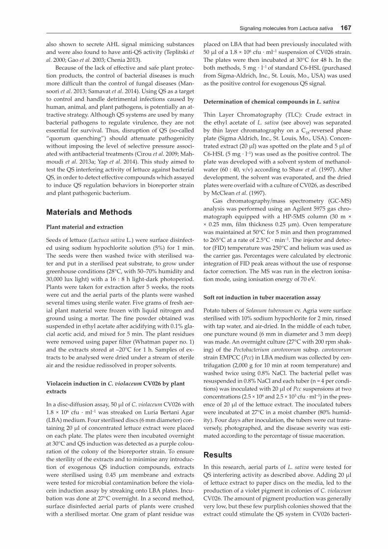
um. These results were further confirmed when crushed plant material was directly put on the media and a high pigment production was observed in CV026 colonies (Fig. 1A). In this method, all plant tissues and their exu-dates were directly presented to the indicator bacterium. Apparently, there was a high concentration of AHL simi-lar molecules in the bacterium environment which caused the increase in pigment production. Inhibition of bacte-rial growth around the crushed plants, was not observed.
The induction of violacein synthesis in areas adjacent to the plant material was similar to that caused by the ad-dition of C6-HSL to a lawn on CV026 medium.
The thin layer chromatogram showed that bioreporter strain, CV026, was only detected on one spot on lettuce extract. The lettuce extract produced a pale, tailing, dif-fuse edge as the control treatment were used 5 μl of C6- -HSL solution (5 mg · l–1). The five μl of C6-HSL solution caused a large spot on the TLC plate (Fig. 1B).
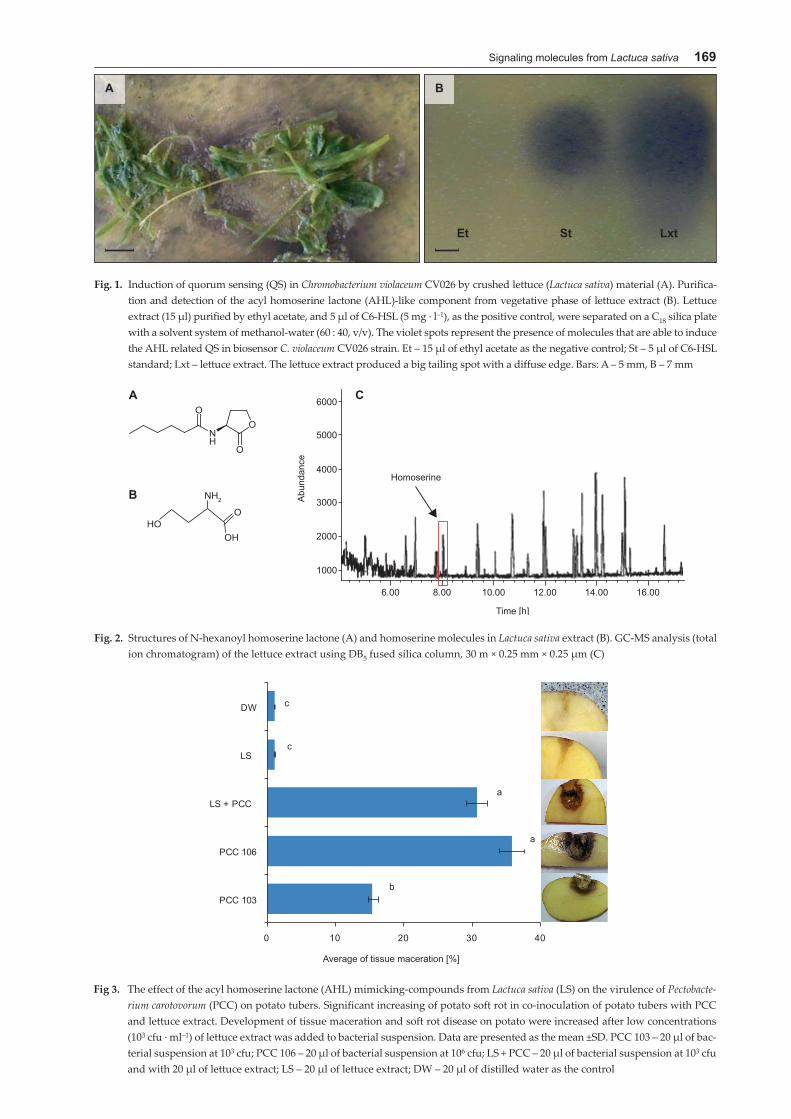
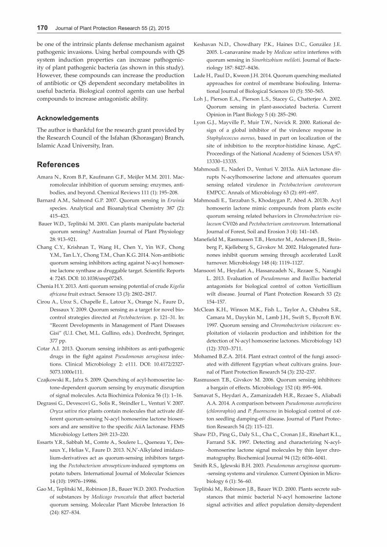
The lettuce extract’s GC-MS and GC-FID analysis re-vealed that the lettuce extract consisted of 19 compounds, of which homoserine accounted for 2.37% of the total constituents (Fig. 2). The ability of L. sativa extract (con-taining herbal homoserine) to suppress the QS system was assessed. As was expected, no QS inhibition activity was observed in the L. sativa extract in the presence of C6-HSL. Finally, the ability of the QS stimulating extract on pathogenicity of P. carotovorum was studied at differ-ent concentrations of bacterium. Adding 20 μl of lettuce extract to the bacterial suspension (with concentrations of 103 cfu · ml-1) caused more severe soft rot symptoms in po-tato tubers than tubers inoculated with only 103 cfu · ml–1 of pathogenic bacterium (Fig. 3). On the other hand, in-jecting a low concentration (103 times less) of Pcc simul-taneously with the plant extract showed the same disease level as tubers inoculated with a concentration of 106 cfu · · ml–1 of Pcc alone. As shown in figure 3, adding extract containing AHL mimic compounds to the bacterial sus-pension brought about an increase in disease caused by Pcc. In other words, P. carotovorum might be able to also identify AHL mimic molecules of plants, and use them as signal molecules. An increase in the concentration of bacterial AHL molecules (because of bacterial multiplica-tion), and AHL similar molecules (from adding the plant extract) in the bacterial environment caused an over-expression of cell wall degradation enzymes and these caused an increase in soft rot symptoms in potato tuber. This was true despite the fact that the population of bac-teria (cell density) was 103 times less than that used in the control (106 cfu · ml–1).
DiscussionIn plant pathology, there is much interest in finding natu-ral or synthetic compounds active in small quantities, that are capable of interfering with QS in pathogenic bacteria in order to disrupt the bacteria’s pathogenicity/virulence production factor. In attempting to learn whether L. sa-tiva secretes substances that mimic AHL signal molecules in regulating population density-dependent behaviors, we relied on the bacterium reporter strain, C. violaceum strain CV026. This strain was carefully mutated by oth-
ers to detect exogenous AHLs (McClean et al. 1997). The addition of one gram of lettuce residue to the LBA me-dium produced pigment in C. violaceum CV026 colonies. There was no QS inhibition activity of L. sativa observed in the presence of C6-HSL in biosensor colonies. Only stimulation of the signal behaviour in the QS system of bioreporter strain took place. The detection of the pres-ence of homoserine in lettuce extract is an important fact because homoserine is a part of the bacterial signal mol-ecules, known as AHL. Acyl homoserine lactones are the main QS signal molecules of Gram-negative bacteria. All AHL are composed of the homoserine lactone ring and the acyl side chain, their signaling activities are related to the lactone ring. These molecules are very sensitive to elevated temperatures and also to alkaline pH. With an increase in the pH or the temperature, AHL rapidly degrade. The mechanism involved in these degradations is lactonolysis, which leads to the generation of acyl-ho-moserine that does not function as a QS signal molecule (Yates et al. 2000). The bioassay results of this research re-vealed that lettuce extract induced QS in the bioreporter strain, CV026. Ours is the first report showing that lettuce contains AHL-like substances (homoserine) which excite AHL related QS in bacteria. This homoserine might be a part or a residue of plant AHL lactone that degraded during in the extraction process. Up till now, the chemical structure of the AHL mimic-compounds have not been well identified, and most of these compounds have QS inhibitory activity that interferes with QS related behav-ior in studied bacteria (Chang et al. 2014; Yap et al. 2014).
It is known that plant extracts have, indeed, a QS in-terfering activity in C. violaceum. The possibility that these plant extracts may also be able to affect QS-mediated phenotypes in other Gram-negative bacteria, was investi-gated. The Pectobacterium carotovorum subsp. carotovorum was chosen for this purpose because of its known QS sys-tems that control a number of genes involved in biofilm formation and production of virulence factors such as pectinolytic enzymes (Barnard and Salmond 2007; Es-sarts et al. 2013; Mahmoudi et al. 2013a). Quorum sens-ing mutants of P. carotovorum have been shown to have a lower production of pectinolytic enzymes than wild-type strains (Mahmoudi et al. 2013a). We found that the presence of the lettuce extract in bacterial suspension sig-nificantly increased the symptoms of soft rot disease as compared to the inoculation of Pcc alone. These results indicate that plant extracts may act as signals for the regulation of bacterial virulence. Our findings support previous work which suggested that fenugreek (Trigo-nella foenum-graecum L.), white clover (Trifolium repens L.) (Mahmoudi et al. 2013b) and rice plants (Oryza sativa L.) (Degrassi et al. 2007) contain molecules which stimulated QS related functions in bioreporter strains. The existence of imitator compounds of bacteria signal molecules, ei-ther in an exciting and induction form or in a destruction and suppression form, can be an appropriate platform for improving chemical control methods based on environ-ment protection and using effective compounds of plants (Keshavan et al. 2005; Rasmussen and Givskov 2006). The chemical nature of the active AHL mimics compounds from plants and active AHL effect on bacterial QS could
Signaling molecules from Lactuca sativa 169
Fig. 1. Induction of quorum sensing (QS) in Chromobacterium violaceum CV026 by crushed lettuce (Lactuca sativa) material (A). Purifica-tion and detection of the acyl homoserine lactone (AHL)-like component from vegetative phase of lettuce extract (B). Lettuce extract (15 μl) purified by ethyl acetate, and 5 μl of C6-HSL (5 mg · l–1), as the positive control, were separated on a C18 silica plate with a solvent system of methanol-water (60 : 40, v/v). The violet spots represent the presence of molecules that are able to induce the AHL related QS in biosensor C. violaceum CV026 strain. Et – 15 μl of ethyl acetate as the negative control; St – 5 μl of C6-HSL standard; Lxt – lettuce extract. The lettuce extract produced a big tailing spot with a diffuse edge. Bars: A – 5 mm, B – 7 mm
Fig. 2. Structures of N-hexanoyl homoserine lactone (A) and homoserine molecules in Lactuca sativa extract (B). GC-MS analysis (total ion chromatogram) of the lettuce extract using DB5 fused silica column, 30 m × 0.25 mm × 0.25 μm (C)
Fig 3. The effect of the acyl homoserine lactone (AHL) mimicking-compounds from Lactuca sativa (LS) on the virulence of Pectobacte-rium carotovorum (PCC) on potato tubers. Significant increasing of potato soft rot in co-inoculation of potato tubers with PCC and lettuce extract. Development of tissue maceration and soft rot disease on potato were increased after low concentrations (103 cfu · ml–1) of lettuce extract was added to bacterial suspension. Data are presented as the mean ±SD. PCC 103 – 20 μl of bac-terial suspension at 103 cfu; PCC 106 – 20 μl of bacterial suspension at 106 cfu; LS + PCC – 20 μl of bacterial suspension at 103 cfu and with 20 μl of lettuce extract; LS – 20 μl of lettuce extract; DW – 20 μl of distilled water as the control
170 Journal of Plant Protection Research 55 (2), 2015
be one of the intrinsic plants defense mechanism against pathogenic invasions. Using herbal compounds with QS system induction properties can increase pathogenic-ity of plant pathogenic bacteria (as shown in this study). However, these compounds can increase the production of antibiotic or QS dependent secondary metabolites in useful bacteria. Biological control agents can use herbal compounds to increase antagonistic ability.
Acknowledgements
The author is thankful for the research grant provided by the Research Council of the Isfahan (Khorasgan) Branch, Islamic Azad University, Iran.
ReferencesAmara N., Krom B.P., Kaufmann G.F., Meijler M.M. 2011. Mac-
romolecular inhibition of quorum sensing: enzymes, anti-bodies, and beyond. Chemical Reviews 111 (1): 195–208.
Barnard A.M., Salmond G.P. 2007. Quorum sensing in Erwinia species. Analytical and Bioanalytical Chemistry 387 (2): 415–423.
Bauer W.D., Teplitski M. 2001. Can plants manipulate bacterial quorum sensing? Australian Journal of Plant Physiology 28: 913–921.
Chang C.Y., Krishnan T., Wang H., Chen Y., Yin W.F., Chong Y.M., Tan L.Y., Chong T.M., Chan K.G. 2014. Non-antibiotic quorum sensing inhibitors acting against N-acyl homoser-ine lactone synthase as druggable target. Scientific Reports 4: 7245. DOI: 10.1038/srep07245.
Chenia H.Y. 2013. Anti quorum sensing potential of crude Kigelia africana fruit extract. Sensore 13 (3): 2802–2817.
Cirou A., Uroz S., Chapelle E., Latour X., Orange N., Faure D., Dessaux Y. 2009. Quorum sensing as a target for novel bio-control strategies directed at Pectobacterium. p. 121–31. In: “Recent Developments in Management of Plant Diseases Gisi” (U.I. Chet, M.L. Gullino, eds.). Dordrecht, Springer, 377 pp.
Cotar A.I. 2013. Quorum sensing inhibitors as anti-pathogenic drugs in the fight against Pseudomonas aeruginosa infec-tions. Clinical Microbiology 2: e111. DOI: 10.4172/2327-5073.1000e111.
Czajkowski R., Jafra S. 2009. Quenching of acyl-homoserine lac-tone-dependent quorum sensing by enzymatic disruption of signal molecules. Acta Biochimica Polonica 56 (1): 1–16.
Degrassi G., Devescovi G., Solis R., Steindler L., Venturi V. 2007. Oryza sativa rice plants contain molecules that activate dif-ferent quorum-sensing N-acyl homoserine lactone biosen-sors and are sensitive to the specific AiiA lactonase. FEMS Microbiology Letters 269: 213–220.
Essarts Y.R., Sabbah M., Comte A., Soulere L., Queneau Y., Des-saux Y., Helias V., Faure D. 2013. N,N’-Alkylated imidazo-lium-derivatives act as quorum-sensing inhibitors target-ing the Pectobacterium atrosepticum-induced symptoms on potato tubers. International Journal of Molecular Sciences 14 (10): 19976–19986.
Gao M., Teplitski M., Robinson J.B., Bauer W.D. 2003. Production of substances by Medicago truncatula that affect bacterial quorum sensing. Molecular Plant Microbe Interaction 16 (24): 827–834.
Keshavan N.D., Chowdhary P.K., Haines D.C., González J.E. 2005. L-canavanine made by Medicao sativa interferes with quorum sensing in Sinorhizobium meliloti. Journal of Bacte-riology 187: 8427–8436.
Lade H., Paul D., Kweon J.H. 2014. Quorum quenching mediated approaches for control of membrane biofouling. Interna-tional Journal of Biological Sciences 10 (5): 550–565.
Loh J., Pierson E.A., Pierson L.S., Stacey G., Chatterjee A. 2002. Quorum sensing in plant-associated bacteria. Current Opinion in Plant Biology 5 (4): 285–290.
Lyon G.J., Mayville P., Muir T.W., Novick R. 2000. Rational de-sign of a global inhibitor of the virulence response in Staphylococcus aureus, based in part on localization of the site of inhibition to the receptor-histidine kinase, AgrC. Proceedings of the National Academy of Sciences USA 97: 13 330–13 335.
Mahmoudi E., Naderi D., Venturi V. 2013a. AiiA lactonase dis-rupts N-acylhomoserine lactone and attenuates quorum sensing related virulence in Pectobacterium carotovorum EMPCC. Annals of Microbiology 63 (2): 691–697.
Mahmoudi E., Tarzaban S., Khodaygan P., Abed A. 2013b. Acyl homoserin lactone mimic compounds from plants excite quorum sensing related behaviors in Chromobacterium vio-laceum CV026 and Pectobacterium carotovorum. International Journal of Forest, Soil and Erosion 3 (4): 141–145.
Manefield M., Rasmussen T.B., Henzter M., Andersen J.B., Stein-berg P., Kjelleberg S., Givskov M. 2002. Halogenated fura-nones inhibit quorum sensing through accelerated LuxR turnover. Microbiology 148 (4): 1119–1127.
Mansoori M., Heydari A., Hassanzadeh N., Rezaee S., Naraghi L. 2013. Evaluation of Pseudomonas and Bacillus bacterial antagonists for biological control of cotton Verticillium wilt disease. Journal of Plant Protection Research 53 (2): 154–157.
McClean K.H., Winson M.K., Fish L., Taylor A., Chhabra S.R., Camara M., Dayykin M., Lamb J.H., Swift S., Bycroft B.W. 1997. Quorum sensing and Chromobacterium violaceum: ex-ploitation of violacein production and inhibition for the detection of N-acyl homoserine lactones. Microbiology 143 (12): 3703–3711.
Mohamed B.Z.A. 2014. Plant extract control of the fungi associ-ated with different Egyptian wheat cultivars grains. Jour-nal of Plant Protection Research 54 (3): 232–237.
Rasmussen T.B., Givskov M. 2006. Quorum sensing inhibitors: a bargain of effects. Microbiology 152 (4): 895–904.
Samavat S., Heydari A., Zamanizadeh H.R., Rezaee S., Aliabadi A.A. 2014. A comparison between Pseudomonas aureofaciens (chlororaphis) and P. fluorescens in biological control of cot-ton seedling damping-off disease. Journal of Plant Protec-tion Research 54 (2): 115–121.
Shaw P.D., Ping G., Daly S.L., Cha C., Cronan J.E., Rinehart K.L., Farrand S.K. 1997. Detecting and characterizing N-acyl- -homoserine lactone signal molecules by thin layer chro-matography. Biochemical Journal 94 (12): 6036–6041.
Smith R.S., Iglewski B.H. 2003. Pseudomonas aeruginosa quorum- -sensing systems and virulence. Current Opinion in Micro-biology 6 (1): 56–60.
Teplitski M., Robinson J.B., Bauer W.D. 2000. Plants secrete sub-stances that mimic bacterial N-acyl homoserine lactone signal activities and affect population density-dependent

Signaling molecules from Lactuca sativa 171
behaviors in associated bacteria. Molecular Plant-Microbe Interaction 13 (6): 637–648.
Tolmacheva A.A., Rogozhin E.A., Deryabini D.G. 2014. Antibac-terial and quorum sensing regulatory activities of some traditional Eastern-European medicinal plants. Acta Phar-maceutica 64 (2): 173–186.
Yap P.S.X., Krishnan T., Yiap B.C., Hu C.P., Chan K.G., Lim S.H.E. 2014. Membrane disruption and anti-quorum sensing ef-fects of synergistic interaction between Lavandula angusti-folia (lavender oil) in combination with antibiotic against plasmid-conferred multi-drug-resistant Escherichia coli. Journal of Applied Microbiology 116 (5): 1119–1128.
Yates E.A., Philipp B., Buckley C., Atkinson S., Chhabra S.R., Sockett R.E., Goldner M., Dessaux Y., Camara M.M., Smith H. 2002. N-Acylhomoserine lactones undergo lactonolysis in a pH-, temperature-, and acyl chain length-dependent manner during growth of Yersinia pseudotuberculosis and Pseudomonas aeruginosa. Infection and Immunity 70 (10): 5635–5646.
Zare Z., Majd A., Sattari T.N., Iranbakhsh A., Mehrabian S. 2012. Antimicrobial activity of leaf and flower extracts of Lippia nodiflora L. (Verbenacea). Journal of Plant Protection Re-search 52 (4): 401–403.