Embed Size (px)
Citation preview
Signal Transduction
G-ProteinsPhosphotidyl InositolTyrosine Kinase
Signal FlowThe transmission of a message across a cell membrane is initiated by binding of a signal molecule to the outer surface of the cell. Receptor, transducer and effector proteins are involved as well as an agonist.
General signal flow pattern: Agonist is an extracellular signal molecule. Receptor protein is a serpentine, integral (transmembrane, 7-TMS) protein. Transducer (intracellular) transmits the signal from the receptor protein to an effector protein. Effector protein elicits a response within the cell. Usually activation or inhibition of a system.
Receptor Protein
• A G-protein coupled receptor (GPCR) is a receptor protein that acts through a G-protein.• When agonist (A) binds to that part of the receptor protein exposed outside of the cell a conformational change is induced in the receptor protein. This change is transmitted to that part of the protein protruding inside the cell.• The resulting conformational change activates another signal system within the cell (second messenger system) doing so through a transducer.• An antagonist when bound to that part of the protein exposed the outside cell can prevent binding by the agonist.
Transducers
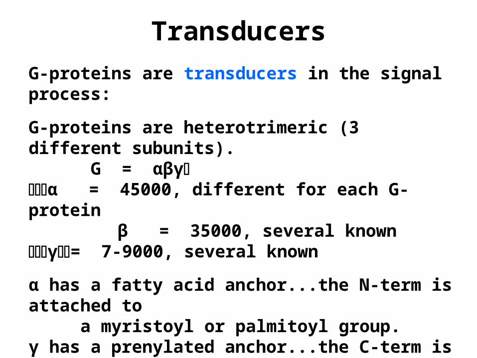
G-proteins are transducers in the signal process:
G-proteins are heterotrimeric (3 different subunits). G = αβγα = 45000, different for each G-protein
β = 35000, several knownγ= 7-9000, several known
α has a fatty acid anchor...the N-term is attached to a myristoyl or palmitoyl group.γ has a prenylated anchor...the C-term is attached to a farnesyl or geranyl group.βis associated strongly with γ.
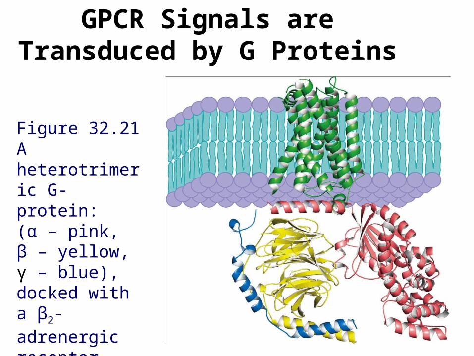
GPCR Signals are Transduced by G Proteins
Figure 32.21 A heterotrimeric G-protein: (α – pink, β – yellow, γ – blue), docked with a β2-adrenergic receptor (green).
Proposed G-Protein Function
A G-protein forms a stable, inactive complex with GDP: GDP•αβγ (inactive)
• A specific GDP•αβγ binds to the part of a specific receptor protein exposed to the cytosol.• The agonist-receptor protein complex enhances dissociation of GDP from GDP•αβγ.• This dissociation is normally slow. Imputed to be regulated by βγ and M++.• The agonist-receptor protein complex can also be considered to be a signal amplifier, activating 10-1000 G-protein molecules depending upon the specific GDP•αβγ.
G-Protein Events
Dissociation of GDP:GDP•αβγ ----> GDP + αβγ
GTP replaces GDP in a fast step:GTP + αβγ ----> GTP•αβγ
GTP binding decreases βγ affinity to α:GTP•αβγ ----> GTP•α + βγ
GTP•α is now active and free to move along the membrane (held by its anchor) to an effector protein.
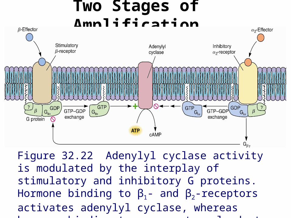
Two Stages of Amplification
Figure 32.22 Adenylyl cyclase activity is modulated by the interplay of stimulatory and inhibitory G proteins. Hormone binding to β1- and β2-receptors activates adenylyl cyclase, whereas hormone binding to α2-receptors leads to inhibition of adenylyl cyclase.
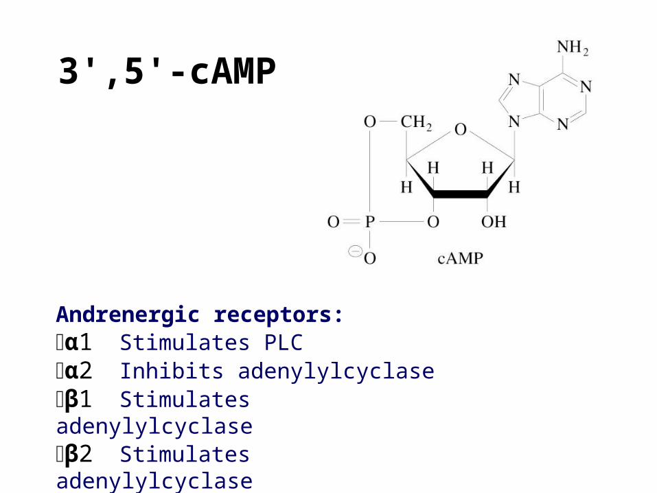
3',5'-cAMP
Andrenergic receptors: α1 Stimulates PLCα2 Inhibits adenylylcyclaseβ1 Stimulates adenylylcyclaseβ2 Stimulates adenylylcyclase
An Effector Protein System
Adenylyl cyclase (effector) is activated by GTP•α and catalyzes formation of c-AMP.
ATP ----> 3',5'-c-AMP + PPi
The enzyme pyrophosphatase hydrolyzes PPi and pulls the reaction to completion: PPi ----> 2 Pi
c-AMP is a second messenger that activates c-AMP dependent protein kinase (PKA or cAMPdPK).
As long as GTP•α is bound to the effector protein the response continues.
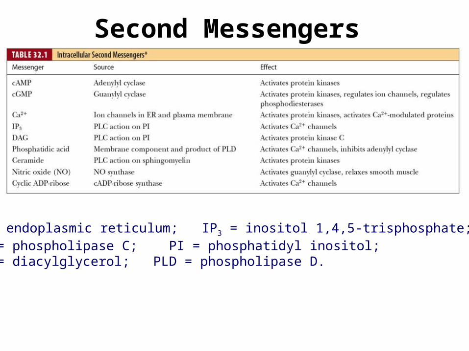
Second Messengers
*ER = endoplasmic reticulum; IP3 = inositol 1,4,5-trisphosphate; PLC = phospholipase C; PI = phosphatidyl inositol; DAG = diacylglycerol; PLD = phospholipase D.
More on G-Proteins
The enzyme phosphodiesterase hydrolyzes c-AMP to 5'-AMP which has no 2nd messenger capabilities.
Note: Several types of hormone receptors may activate the same G-protein. For instance, in liver,there is a receptor site for glucagon (a polypeptide hormone) and another for epinephrine (a β -andrenergic or catecholamine hormone) these both activate Gs which in turn stimulates adenylyl cyclase to make c-AMP. Also, βγ is a transducer for several systems, e.g. activates PL2A, MAPK, etc.

More on G-Proteins
GTP•α is also a GTPase (it slowly hydrolyzes GTP to GDP and Pi): GTP•α ----> GDP•α + Pi
GDP•α is inactive and dissociates from effector protein. GDP•α then returns to receptor protein and binds to βγ and the receptor: GDP•αβγ•GPCR
If agonist is still bound to the receptor protein, GDP dissociates, reactivation occurs, and another cycle of G-protein transduction starts.
Cholera and pertussis toxins modify the α subunit of G-protein resulting in abnormal functioning.
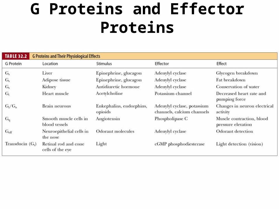
G Proteins and Effector Proteins
Phosphotidyl Inositol (PI) System
Using different receptor, G-protein and effector combinations, a wide assortment of signal systems are available.
• Example agonists for the phosphatidyl inositol system are angiotensin and histamine. These communicate with the G-protein, Gq.
• Phosphatidyl inositol in presence of PI kinase forms PI-4,5-bisP (PIP2).
PI + 2 ATP ----> PI-4,5-bisP + 2 ADP
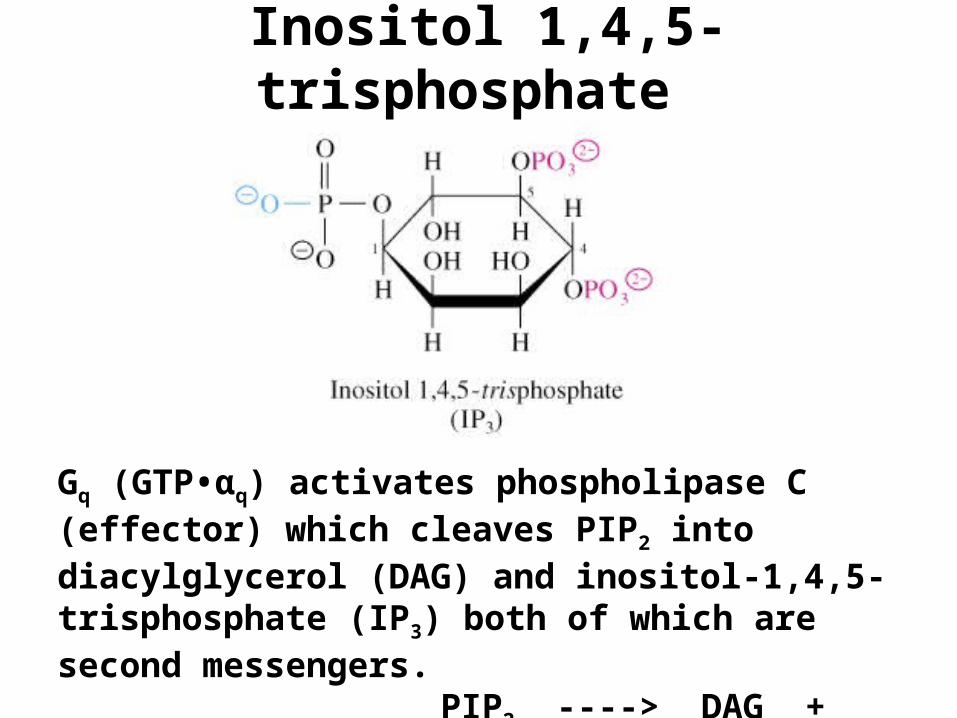
Inositol 1,4,5-trisphosphate
Gq (GTP•αq) activates phospholipase C (effector) which cleaves PIP2 into diacylglycerol (DAG) and inositol-1,4,5-trisphosphate (IP3) both of which are second messengers.
PIP2 ----> DAG + IP3
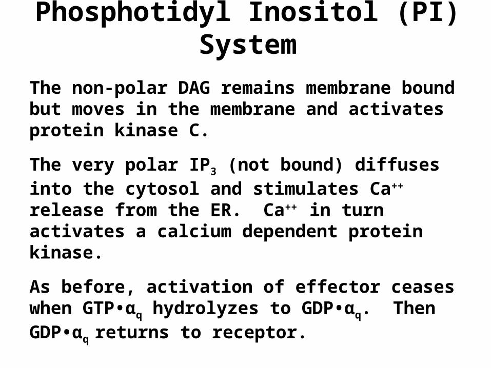
Phosphotidyl Inositol (PI) System
The non-polar DAG remains membrane bound but moves in the membrane and activates protein kinase C.
The very polar IP3 (not bound) diffuses into the cytosol and stimulates Ca++ release from the ER. Ca++ in turn activates a calcium dependent protein kinase.
As before, activation of effector ceases when GTP•αq hydrolyzes to GDP•αq. Then GDP•αq returns to receptor.
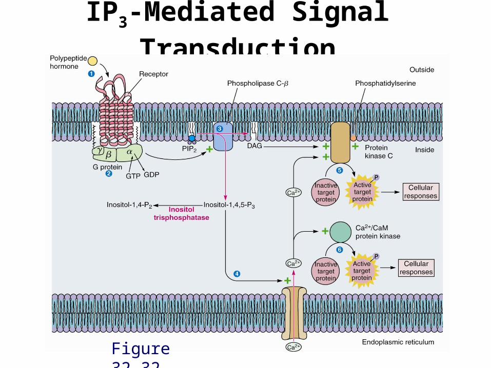
IP3-Mediated Signal Transduction
Figure 32.32
Tyrosine Kinase System
• Tyrosine kinase exists as transmembrane β protein with α protein associated to β on the outside of the membrane. Each αβ complex is referred to as a monomeric subunit.
• Binding of insulin to the RP promotes dimerization of subunits and activation of TK. Active TK phosphorylates itself at Tyr in both β subunits of the αβαβ dimer (autophosphorylation) and also phosphorylates certain cytosolic proteins at Tyr.
Tyrosine Kinase System
• Some TK can also respond to insulin and act through PIP2. PI 3-kinase uses PIP2 as substrate and phosphorylates the 3 position of inositol.
PIP2 + ATP ----> PI-3,4,5-trisP (PIP3) + ADP
• PIP3 is a second messenger that activates certain other protein kinases.
End of Signal Transduction