Embed Size (px)
Citation preview
DEVELO
PMENT
889RESEARCH ARTICLE
INTRODUCTIONFor decades the morphogen hypothesis has helped to explain a widerange of tissue patterning processes (Crick, 1970; Turing, 1952). Thehypothesis states that chemical signals, termed ‘morphogens’, aresecreted from signaling centers, and that the resulting staticextracellular morphogen concentration gradient emanating from thecenter spatially organizes and patterns tissue architecture. That is,rapid morphogen transport creates a concentration gradient invariantover the time window of tissue patterning (i.e. at steady state). Sonichedgehog (Shh), which forms a concentration gradient to pattern thelimb bud (Riddle et al., 1993), midbrain (Britto et al., 2002),forebrain (Ericson et al., 1995) and spinal cord (Roelink et al., 1995)during vertebrate development, is ostensibly a canonical example ofthe morphogen hypothesis. However, very recent studies suggestthat Shh concentration gradient dynamics play a crucial role in tissuepatterning. Both the time of exposure of a cell to a given Shhconcentration (Ahn and Joyner, 2004; Harfe et al., 2004; Kohtz etal., 1998; Park et al., 2004; Wolff et al., 2003; Yang et al., 1997) andthe timing of Shh source secretion (Ericson et al., 1996) are crucialdeterminants of Shh tissue patterning. The classical morphogenhypothesis does not account for such dynamics in gradient formationand cellular response.
Beyond passive diffusion, morphogen systems can have a numberof ‘accessory’ mechanisms that modulate ligand transport.Specifically, each transport mechanism potentially modifies not onlythe steady state concentration gradient, but also the rate ofmorphogen transport at various times in the patterning process. Forexample, studies in Drosophila have identified numerous genesessential for actively transporting morphogens upon their releasefrom secreting cells, and mutating such transporters disrupts tissue
patterning (Chen et al., 2004; Han et al., 2004; Takei et al., 2004).Also, high affinity interactions of morphogens with cell surface(Chuang and McMahon, 1999) and extracellular matrix (ECM)components (The et al., 1999) serve the putative roles of depletingor immobilizing extracellular-diffusing morphogens to limit long-range signaling. Any of these mechanisms can affect the temporalevolution of concentration gradients in developing tissue, which isoverlooked by the focus of the morphogen hypothesis on the steadystate concentration gradient.
To investigate temporal effects of transport and signaling, wemodel Shh regulation of dorsoventral spinal cord patterning in chickembryonic development stages 10-26 [~33-116 hours after egglaying (Ricklefs and Starck, 1998)]. Shh, secreted from thefloorplate, diffuses into the neural tube (Roelink et al., 1995), and,as its concentration decreases within the tissue from approximately15 nM at the floorplate to 0.5 nM at dorsal edge of the neural tube,target cells switch at threshold values from mature ventral to dorsalphenotypes [e.g. from V3rMNrV2rV1 in Fig. 1A, as has beenpreviously reviewed (Persson et al., 2002)]. Our model begins afterthe neural fold appears and essentially as the neural tube closes atstage 10, when Shh is first secreted from the floorplate. Wesubsequently track the cell fate switch between V3 interneurons(V3) and motoneurons (MN) that occurs through stage 26.
The structure of Shh, as well as its various interacting proteins,has complicated a simple understanding of how its transportestablishes a gradient during this process. Shh is covalently modifiedby hydrophobic moieties, including a C-terminal cholesterol and aN-terminal palmitic acid (Pepinsky et al., 1998; Porter et al., 1996),which may anchor the ligand to cell membranes and therebysignificantly reduce its diffusivity. However, Shh is still capable ofsignaling at a large distance, up to 20 cell diameters, away from itssource. In addition, the Shh receptor Patched (Ptc) is upregulated byShh signaling, and its subsequent binding and receptor-mediatedinternalization of Shh depletes the ligand from the extracellularspace (Chen and Struhl, 1996; Marigo and Tabin, 1996).Furthermore, Shh and its Drosophila homolog Hedgehog can formmultimers, and the transmembrane protein Dispatched (Dis) is likely
Signal dynamics in Sonic hedgehog tissue patterningKrishanu Saha and David V. Schaffer*
During development, secreted signaling factors, called morphogens, instruct cells to adopt specific mature phenotypes. However,the mechanisms that morphogen systems employ to establish a precise concentration gradient for patterning tissue architecture arehighly complex and are typically analyzed only at long times after secretion (i.e. steady state). We have developed a theoreticalmodel that analyzes dynamically how the intricate transport and signal transduction mechanisms of a model morphogen, Sonichedgehog (Shh), cooperate in modular fashion to regulate tissue patterning in the neural tube. Consistent with numerous recentstudies, the model elucidates how the dynamics of gradient formation can be a key determinant of cell response. In addition, thiswork yields several novel insights into how different transport mechanisms or ‘modules’ control pattern formation. The modelpredicts that slowing the transport of a morphogen, such as by lipid modification of the ligand Shh, by ligand binding toproteoglycans, or by the moderate upregulation of dedicated transport molecules like Dispatched, can actually increase thesignaling range of the morphogen by concentrating it near the secretion source. Furthermore, several transcriptional targets ofShh, such as Patched and Hedgehog-interacting protein, significantly limit its signaling range by slowing transport and promotingligand degradation. This modeling approach elucidates how individual modular elements that operate dynamically at various timesduring patterning can shape a tissue pattern.
KEY WORDS: Morphogen, Sonic hedgehog, Diffusion, Transport, Modeling
Development 133, 889-900 doi:10.1242/dev.02254
Department of Chemical Engineering and the Helen Wills Neuroscience Institute,University of California, Berkeley, CA 94720-1462, USA.
*Author for correspondence (e-mail: [email protected])
Accepted 5 December 2005
DEVELO
PMENT
890 RESEARCH ARTICLE Development 133 (5)
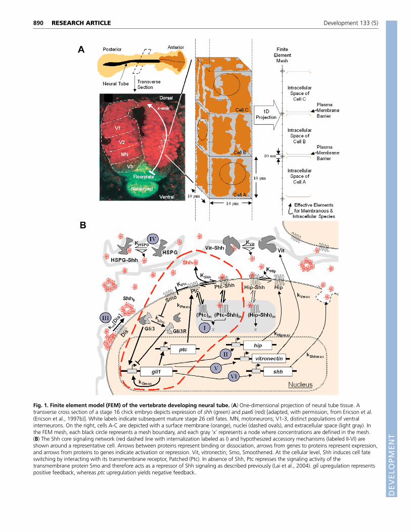
Fig. 1. Finite element model (FEM) of the vertebrate developing neural tube. (A) One-dimensional projection of neural tube tissue. Atransverse cross section of a stage 16 chick embryo depicts expression of shh (green) and pax6 (red) [adapted, with permission, from Ericson et al.(Ericson et al., 1997b)]. White labels indicate subsequent mature stage 26 cell fates. MN, motoneurons; V1-3, distinct populations of ventralinterneurons. On the right, cells A-C are depicted with a surface membrane (orange), nuclei (dashed ovals), and extracellular space (light gray). Inthe FEM mesh, each black circle represents a mesh boundary, and each gray ‘x’ represents a node where concentrations are defined in the mesh.(B) The Shh core signaling network (red dashed line with internalization labeled as I) and hypothesized accessory mechanisms (labeled II-VI) areshown around a representative cell. Arrows between proteins represent binding or dissociation, arrows from genes to proteins represent expression,and arrows from proteins to genes indicate activation or repression. Vit, vitronectin; Smo, Smoothened. At the cellular level, Shh induces cell fateswitching by interacting with its transmembrane receptor, Patched (Ptc). In absence of Shh, Ptc represses the signaling activity of thetransmembrane protein Smo and therefore acts as a repressor of Shh signaling as described previously (Lai et al., 2004). gli upregulation representspositive feedback, whereas ptc upregulation yields negative feedback.
DEVELO
PMENT
to be involved in regulating their assembly and intercellular transport(Kawakami et al., 2002). Moreover, a membrane glycoprotein,Hedgehog-interacting protein (Hip), binds Shh with high affinity tomodulate its signaling activity (Chuang and McMahon, 1999). ECMproteins also regulate Shh transport, as high-affinity binding of Shhto vitronectin in the neural tube has been suggested to aid in theproper presentation of Shh to differentiating motoneurons (Ponsand Marti, 2000). Finally, the effective transport of DrosophilaHedgehog depends upon the activity of heparan sulfateproteoglycans (HSPG) (Bornemann et al., 2004; Takei et al., 2004;The et al., 1999), and Shh has also been shown to bind HSPG (Rubinet al., 2002). The individual and synergistic contributions of each ofthese highly complex elements to the ability of Shh to pattern tissueare unclear. Shh transport via diffusion was previously modeledin the vertebrate limb bud, using a simple signal transductionmechanism without consideration of these accessory transportmechanisms (Dillon et al., 2003). Therefore, to complement,synthesize, and guide experimental work, we have applied a systemsbiology analysis to explore the effects of diffusion, receptor-liganddynamics and gene regulation dynamics on Shh gradient formationand tissue patterning.
We build upon a previous single cell model (Lai et al., 2004) toanalyze Shh patterning of the developing spinal cord and find thatthe concentration gradient initially established by diffusion can bemodified by several long timescale mechanisms, includingmorphogen binding to ECM and gene expression. Modeling resultsare able to reproduce the experimental profiles and clarify thepotential roles of six modular mechanisms involved in Shh gradientformation and cellular signaling (Fig. 1B). This investigationsuggests that different components can be assembled in a modularfashion to dynamically pattern a morphogen gradient according tothe needs of specific tissues.
MATERIALS AND METHODSGeometry of the neural tubeWe consider dorsoventral patterning of the chick neural tube arising fromShh transport in one dimension. The spatial axis of our model tracks thediffusion of Shh away from its floorplate source and through the neural tube,and partitions the neural tube into a mesh of discrete 10 �m cubic elements,each containing one cell (Fig. 1A). Inside each 10 �m cube, the extracellularspace consists of interconnected channels of unspecified geometry, but thatin sum occupy 20% of the volume based on empirically measured voidfractions of neural tissue (Incardona et al., 2002). The diffusion coefficientwas modified to account for tortuosity (Lander et al., 2002). A Shh moleculemoving through the continuous extracellular space of the mesh (Fig. 1A) cansmoothly diffuse through the extracellular regions in cubes A, B and then C.By contrast, all cell surface and intracellular species (e.g. receptors andreceptor-ligand complexes) are completely restricted within their cellularvolume compartment, which is surrounded by a small 10 nm plasmamembrane element/barrier.
Developmental time windowAt the ventral-most region of the chick neural tube, high level Shhexpression is initiated exclusively in the floorplate (Fig. 1A) during stages10-12 (~34 hours after egg laying). At this time (t=0), all cells in the tubehave the same initial gene expression profile. As time progresses, Shhdiffuses dorsally from its floorplate source through the mesh and binds toreceptors or other components, and high Shh signal levels induce a cellphenotype switch (Lai et al., 2004). The position of the mature phenotypesseen in wild-type embryos after stages 26 (>80 hours after laying) is shown(Fig. 1A). Recent work in mouse embryos indicates that early Shh secretionfrom the notochord may diffuse far into the neural tube to affect MNcommitment (Jeong and McMahon, 2005), and such scenarios in chickembryos could readily be incorporated by adding extra elements for thenotochord cells (at x<0), with appropriate Shh secretion dynamics.
Mathematical formulation of Shh transport by diffusion andreceptor kineticsThe Shh signaling network is represented as a set of differential equationsthat track the rates of change in the concentrations of network constituents,and whose individual terms represent rates of diffusion, protein synthesisand degradation (Fig. 1B, Fig. 2A). At the single cell level, we build uponthe Shh signaling network derived by previously (Lai et al., 2004) to includecellular internalization effects (see Figs S3, S4 in the supplementarymaterial). As Shh is increased above a threshold concentration, it stimulatesGli production to the point where Gli positively feeds back upon its ownexpression and rapidly switches the state of the network to ‘on’ (Fig. 2B).The activities of Gli2, which overlap with those of both Gli1 and Gli3, arehighly context dependent, and its molecular interactions in the neural tubeprogenitor cells require further characterization (Bai et al., 2004; Ruiz iAltaba, 1999). As a result, we have effectively parsed the effects of Gli2 intotwo types: either a pure transcriptional activator, the ‘Gli1’ type, or atranscription factor of both repressor and activator functions, the ‘Gli3’ type.Thus, in the model, the effects of Gli2 are effectively lumped into a Gli1 termand a Gli3 term. We report the Gli1 concentration as the important systemoutput, as the on/off gli1 expression interface demarcates the V3/MNboundary.
Parameters and computational techniquesKinetic, diffusive and binding parameter values were either directly takenfrom the literature or estimated based upon analogous biological systems(Fig. 2 legend, see also Table S1 in the supplementary material). Parameterestimates were chosen to meet the three following experimentalobservations: switching ‘on’ of homeodomain nkx2.2 (which serves as thegli1 domain marker in the case of the model) expression at a ~3 nM Shhthreshold at steady state (Ericson et al., 1995; Ericson et al., 1997b); ~50-hour kinetic timescale of Shh secretion from the floorplate [based upontimescales for MN specification from figure 3D in Ericson et al. (Ericson etal., 1997b)]; and a Nkx2.2 protein fluorescence intensity spatial profile in awild-type chick embryo [see figure 3B in Ericson et al. (Ericson et al.,1997b)]. To satisfy the last criterion, because the Shh secretion rate from thefloorplate has not been quantitatively determined, we chose it such that ourpattern matched the Nkx2.2 switching interface seen at 70 �m from thefloorplate (Ericson et al., 1997b). For the ventral-most cells (close to x=0),floorplate induction, marked by an increase in hnf3�, occurs above a 10 nMShh concentration (Briscoe et al., 2000). Such floorplate induction, probablydue to additional downstream targets of gli1 or other signals not included inthis model, accounts for the decrease in nkx2.2 expression seenexperimentally in and near the floorplate (Fig. 3B). For each parameter inthe core-signaling pathway, we conducted sensitivity analysis for parametervalues over four orders of magnitude to observe whether the single cellresponse to Shh varied (Fig. 2C). Model behavior was investigated forparameter values that changed the 3 nM Shh switching threshold at steadystate from 1 to 10 nM and pattern evolution time from 30 to 150 hours Shhsecretion, and all conclusions and trends discussed below remainedqualitatively the same.
The set of equations in Fig. 2A are presented in dimensional form.However, relationships between groups of variables can be intuitively easierto interpret than individual parameters. In addition, grouping variablesreduces the number of independent parameters that are necessary to describethe model. As a result, the following types of variables were non-dimensionalized by the corresponding parameters: concentrations by KGli3,space by 1 cm, and time by 1/kdeg. The corresponding non-dimensionalequations are shown in Figs S3, S4 (in the supplementary material) and werecoded into the FEMLAB software.
RESULTSDynamic expression of transcription factors (e.g. Gli family)governs the V3/MN/V2/V1 pattern of the neural tube (Fig. 1A), aprocess experimentally analyzed by staining for numeroushomeodomain proteins (Briscoe et al., 2000; Jessell, 2000),among the earliest of which are the markers Nkx2.2 in the V3region and Pax6 in the remainder of the tube. In this work, we
891RESEARCH ARTICLESignal dynamics in Shh tissue patterning
DEVELO
PMENT
892 RESEARCH ARTICLE Development 133 (5)
D
G
E F
H
A
5.7
att = 43 hr
t = 43 hr
att = 43 hr
att = 43 hr
att = 43 hr
>2-fold [Gli1] change
V3/MN interfaceat 70 μm
C 100
10
1
0.1
0.001
kperturb
klit
Perturbed Parameter
kPout
Bistable Switch
Irreversible SwitchMonostable Behavior
kGmaxkg3rkPin
B
[Shh] [Shh][Shh]
[Gli1
]
[Gli1
]
[Gli1
] OnOnOn
Off Off Off
MonostableBehavior
BistableBistable
IrreversibleCell Fate Switch
BistableCell Fate Switch
Fig. 2. See next page for legend.
DEVELO
PMENT
focus on the ventral-most binary cell fate switch delineating theV3/MN boundary, which results in the Nkx2.2/Pax6 histologicaldemarcation. Homeodomain protein expression patterns are alsoregulated by BMP, FGF and retinoid signals (reviewed by Jessell,2000), events that can be incorporated into the model in futurework to evolve the model behavior from a binary switch into aladder of cell fates. Given that the V3 domain and Nkx2.2expression are entirely lost in mutants with compromised Shhsignaling [e.g. Shh–/– (Litingtung and Chiang, 2000), Smo–/–
(Wijgerde et al., 2002), Shh–/–/Gli3–/– (Bai et al., 2004; Litingtungand Chiang, 2000), Gli3–/–/Smo–/– (Wijgerde et al., 2002), andGli1–/–/Gli2–/– (Park et al., 2000) mice], we use Nkx2.2 purely asa robust marker of cells where Shh signaling is active, and whereGli is presumably expressed. Also, because Nkx2.2 is rapidlyupregulated following Shh signaling, we assume that it can beused as a marker of Shh signaling activity, without requiringknowledge of the mechanistic and molecular interactions thatconnect Gli to Nkx2.2.
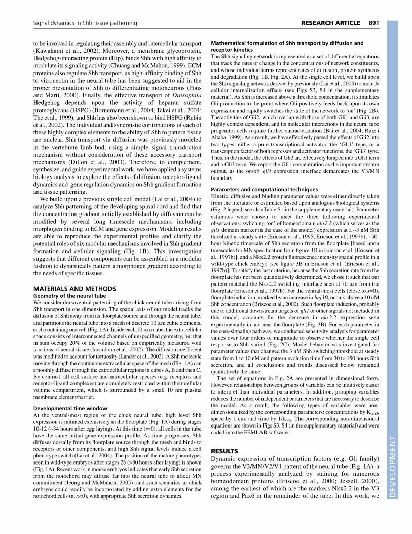
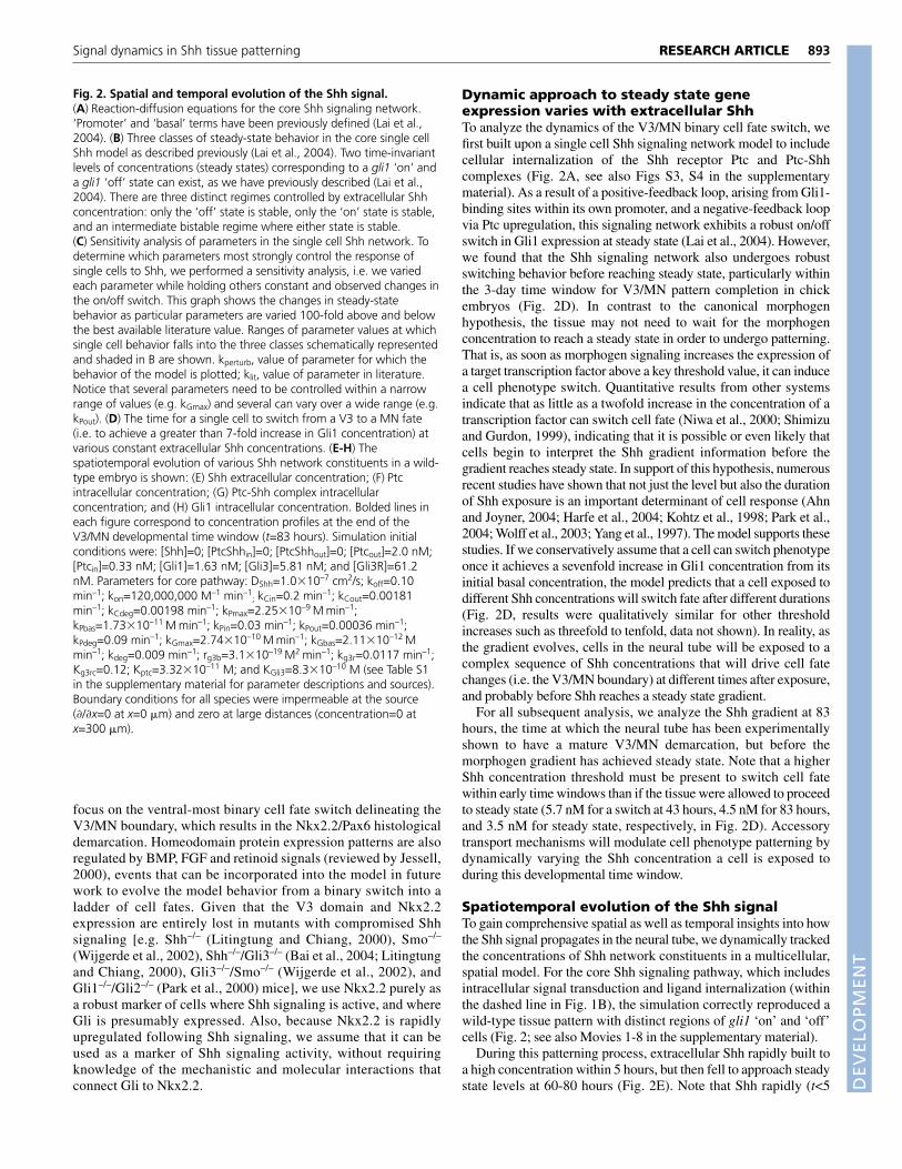
Dynamic approach to steady state geneexpression varies with extracellular ShhTo analyze the dynamics of the V3/MN binary cell fate switch, wefirst built upon a single cell Shh signaling network model to includecellular internalization of the Shh receptor Ptc and Ptc-Shhcomplexes (Fig. 2A, see also Figs S3, S4 in the supplementarymaterial). As a result of a positive-feedback loop, arising from Gli1-binding sites within its own promoter, and a negative-feedback loopvia Ptc upregulation, this signaling network exhibits a robust on/offswitch in Gli1 expression at steady state (Lai et al., 2004). However,we found that the Shh signaling network also undergoes robustswitching behavior before reaching steady state, particularly withinthe 3-day time window for V3/MN pattern completion in chickembryos (Fig. 2D). In contrast to the canonical morphogenhypothesis, the tissue may not need to wait for the morphogenconcentration to reach a steady state in order to undergo patterning.That is, as soon as morphogen signaling increases the expression ofa target transcription factor above a key threshold value, it can inducea cell phenotype switch. Quantitative results from other systemsindicate that as little as a twofold increase in the concentration of atranscription factor can switch cell fate (Niwa et al., 2000; Shimizuand Gurdon, 1999), indicating that it is possible or even likely thatcells begin to interpret the Shh gradient information before thegradient reaches steady state. In support of this hypothesis, numerousrecent studies have shown that not just the level but also the durationof Shh exposure is an important determinant of cell response (Ahnand Joyner, 2004; Harfe et al., 2004; Kohtz et al., 1998; Park et al.,2004; Wolff et al., 2003; Yang et al., 1997). The model supports thesestudies. If we conservatively assume that a cell can switch phenotypeonce it achieves a sevenfold increase in Gli1 concentration from itsinitial basal concentration, the model predicts that a cell exposed todifferent Shh concentrations will switch fate after different durations(Fig. 2D, results were qualitatively similar for other thresholdincreases such as threefold to tenfold, data not shown). In reality, asthe gradient evolves, cells in the neural tube will be exposed to acomplex sequence of Shh concentrations that will drive cell fatechanges (i.e. the V3/MN boundary) at different times after exposure,and probably before Shh reaches a steady state gradient.
For all subsequent analysis, we analyze the Shh gradient at 83hours, the time at which the neural tube has been experimentallyshown to have a mature V3/MN demarcation, but before themorphogen gradient has achieved steady state. Note that a higherShh concentration threshold must be present to switch cell fatewithin early time windows than if the tissue were allowed to proceedto steady state (5.7 nM for a switch at 43 hours, 4.5 nM for 83 hours,and 3.5 nM for steady state, respectively, in Fig. 2D). Accessorytransport mechanisms will modulate cell phenotype patterning bydynamically varying the Shh concentration a cell is exposed toduring this developmental time window.
Spatiotemporal evolution of the Shh signalTo gain comprehensive spatial as well as temporal insights into howthe Shh signal propagates in the neural tube, we dynamically trackedthe concentrations of Shh network constituents in a multicellular,spatial model. For the core Shh signaling pathway, which includesintracellular signal transduction and ligand internalization (withinthe dashed line in Fig. 1B), the simulation correctly reproduced awild-type tissue pattern with distinct regions of gli1 ‘on’ and ‘off’cells (Fig. 2; see also Movies 1-8 in the supplementary material).
During this patterning process, extracellular Shh rapidly built toa high concentration within 5 hours, but then fell to approach steadystate levels at 60-80 hours (Fig. 2E). Note that Shh rapidly (t<5
893RESEARCH ARTICLESignal dynamics in Shh tissue patterning
Fig. 2. Spatial and temporal evolution of the Shh signal.(A) Reaction-diffusion equations for the core Shh signaling network.‘Promoter’ and ‘basal’ terms have been previously defined (Lai et al.,2004). (B) Three classes of steady-state behavior in the core single cellShh model as described previously (Lai et al., 2004). Two time-invariantlevels of concentrations (steady states) corresponding to a gli1 ‘on’ anda gli1 ‘off’ state can exist, as we have previously described (Lai et al.,2004). There are three distinct regimes controlled by extracellular Shhconcentration: only the ‘off’ state is stable, only the ‘on’ state is stable,and an intermediate bistable regime where either state is stable.(C) Sensitivity analysis of parameters in the single cell Shh network. Todetermine which parameters most strongly control the response ofsingle cells to Shh, we performed a sensitivity analysis, i.e. we variedeach parameter while holding others constant and observed changes inthe on/off switch. This graph shows the changes in steady-statebehavior as particular parameters are varied 100-fold above and belowthe best available literature value. Ranges of parameter values at whichsingle cell behavior falls into the three classes schematically representedand shaded in B are shown. kperturb, value of parameter for which thebehavior of the model is plotted; klit, value of parameter in literature.Notice that several parameters need to be controlled within a narrowrange of values (e.g. kGmax) and several can vary over a wide range (e.g.kPout). (D) The time for a single cell to switch from a V3 to a MN fate(i.e. to achieve a greater than 7-fold increase in Gli1 concentration) atvarious constant extracellular Shh concentrations. (E-H) Thespatiotemporal evolution of various Shh network constituents in a wild-type embryo is shown: (E) Shh extracellular concentration; (F) Ptcintracellular concentration; (G) Ptc-Shh complex intracellularconcentration; and (H) Gli1 intracellular concentration. Bolded lines ineach figure correspond to concentration profiles at the end of theV3/MN developmental time window (t=83 hours). Simulation initialconditions were: [Shh]=0; [PtcShhin]=0; [PtcShhout]=0; [Ptcout]=2.0 nM;[Ptcin]=0.33 nM; [Gli1]=1.63 nM; [Gli3]=5.81 nM; and [Gli3R]=61.2nM. Parameters for core pathway: DShh=1.0�10–7 cm2/s; koff=0.10min–1; kon=120,000,000 M–1 min–1
; kCin=0.2 min–1; kCout=0.00181min–1; kCdeg=0.00198 min–1; kPmax=2.25�10–9 M min–1;kPbas=1.73�10–11 M min–1; kPin=0.03 min–1; kPout=0.00036 min–1;kPdeg=0.09 min–1; kGmax=2.74�10–10 M min–1; kGbas=2.11�10–12 Mmin–1; kdeg=0.009 min–1; rg3b=3.1�10–19 M2 min–1; kg3r=0.0117 min–1;Kg3rc=0.12; Kptc=3.32�10–11 M; and KGli3=8.3�10–10 M (see Table S1in the supplementary material for parameter descriptions and sources).Boundary conditions for all species were impermeable at the source(�/�x=0 at x=0 �m) and zero at large distances (concentration=0 atx=300 �m).
DEVELO
PMENT
894
hours) rose well above the static levels found to induce a MN to V3phenotype switch [~3 nM (Ericson et al., 1997b)]. Because proteinexpression from Shh target genes does not build appreciably withinthis relatively short timescale, receptor-ligand internalization andpassive diffusion alone governed the early evolution of the Shhconcentration profile. Shh levels then more rapidly declined as Ptc,a direct Shh transcriptional target, increased in the ventral-mostportions of the embryo and began to mediate Shh degradation viareceptor-mediated endocytosis (Fig. 2F). In contrast to the morecontinuous Shh profile that is smoothened by transport, Ptc profilesexhibited discrete on/off regions due to induced ptc expressionabove the Shh switching threshold (Fig. 2D,E). Near the floorplate,free Ptc initially decreased as extracellular Shh levels rapidlyelevated and bound free Ptc. The resulting Ptc-Shh complexes wererapidly internalized but degraded more slowly, so that thepredominant form of Ptc was an internal, complexed form (Fig. 2G),consistent with previous observations (Incardona et al., 2002;Incardona et al., 2000). After 20 hours, ptc was highly upregulateddue to high Shh signaling near the floorplate.
Shh also drove a dynamic Gli expression pattern. Cells at theinterface exhibited a transient increase followed by a decrease inGli1 concentration, and after 83 hours of Shh secretion, an 8- to 10-fold higher Gli1 protein level was seen for the ventral ‘on’ cellsversus the dorsal ‘off’ cells (Fig. 2H). All concentrations in theneural tube approach but do not reach steady-state levels in thedevelopmental time window of 3 days for wild-type chick V3/MNpatterning (Fig. 2E-H). The resulting intricate concentration profilesof all the core pathway components at various times during
patterning would be difficult to predict intuitively due to the multiplemechanisms occurring at many different timescales: ligand diffusionover a cell diameter over tenths of seconds [(10�10–4 cm)2/1�10–7
cm2/s], ligand binding and internalization on the order of minutes(1/Kshhkon and 1/kCin; see Table S1 in the supplementary material),and gene expression and protein degradation over a number of hours(1/kdeg, 1/kPout and 1/kCdeg).
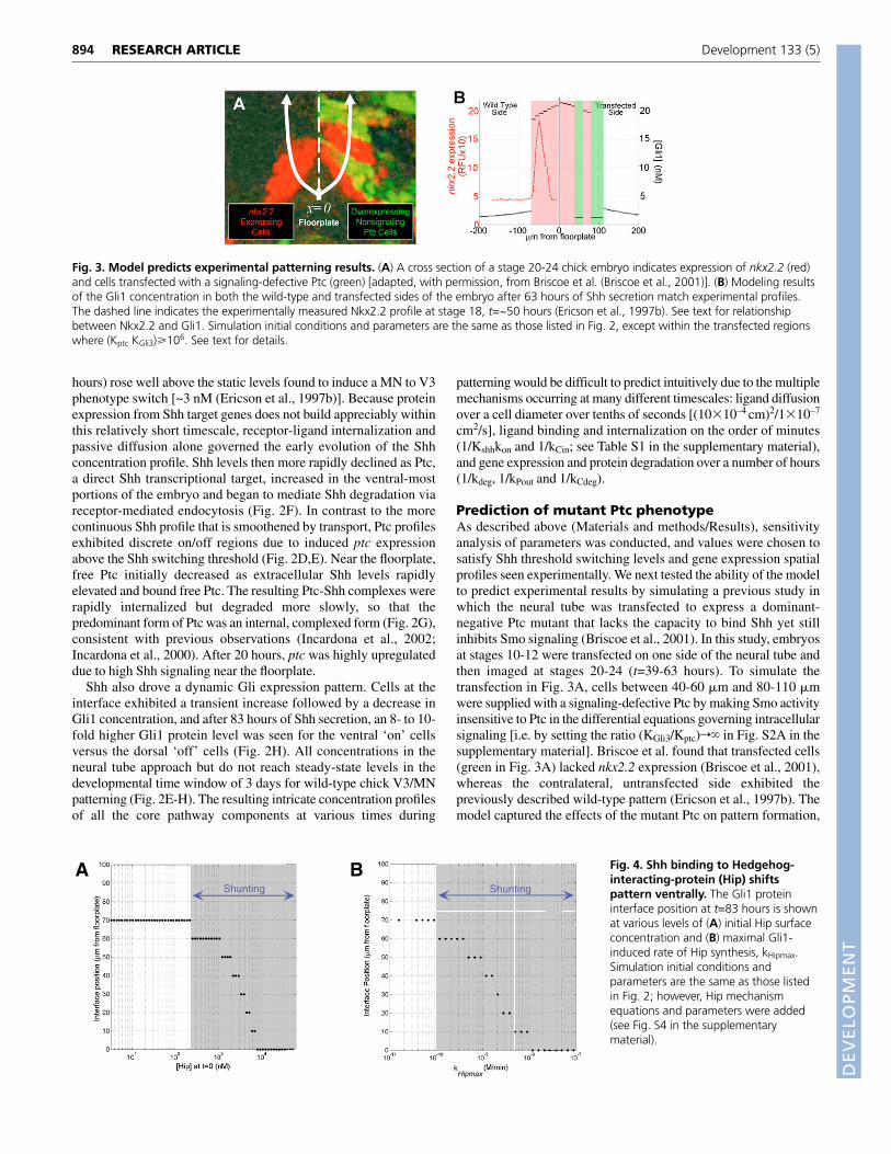
Prediction of mutant Ptc phenotypeAs described above (Materials and methods/Results), sensitivityanalysis of parameters was conducted, and values were chosen tosatisfy Shh threshold switching levels and gene expression spatialprofiles seen experimentally. We next tested the ability of the modelto predict experimental results by simulating a previous study inwhich the neural tube was transfected to express a dominant-negative Ptc mutant that lacks the capacity to bind Shh yet stillinhibits Smo signaling (Briscoe et al., 2001). In this study, embryosat stages 10-12 were transfected on one side of the neural tube andthen imaged at stages 20-24 (t=39-63 hours). To simulate thetransfection in Fig. 3A, cells between 40-60 �m and 80-110 �mwere supplied with a signaling-defective Ptc by making Smo activityinsensitive to Ptc in the differential equations governing intracellularsignaling [i.e. by setting the ratio (KGli3/Kptc)r� in Fig. S2A in thesupplementary material]. Briscoe et al. found that transfected cells(green in Fig. 3A) lacked nkx2.2 expression (Briscoe et al., 2001),whereas the contralateral, untransfected side exhibited thepreviously described wild-type pattern (Ericson et al., 1997b). Themodel captured the effects of the mutant Ptc on pattern formation,
RESEARCH ARTICLE Development 133 (5)
Fig. 3. Model predicts experimental patterning results. (A) A cross section of a stage 20-24 chick embryo indicates expression of nkx2.2 (red)and cells transfected with a signaling-defective Ptc (green) [adapted, with permission, from Briscoe et al. (Briscoe et al., 2001)]. (B) Modeling resultsof the Gli1 concentration in both the wild-type and transfected sides of the embryo after 63 hours of Shh secretion match experimental profiles.The dashed line indicates the experimentally measured Nkx2.2 profile at stage 18, t=~50 hours (Ericson et al., 1997b). See text for relationshipbetween Nkx2.2 and Gli1. Simulation initial conditions and parameters are the same as those listed in Fig. 2, except within the transfected regionswhere (Kptc KGli3)�106. See text for details.
AShunting
B
kHipmax
Shunting
Fig. 4. Shh binding to Hedgehog-interacting-protein (Hip) shiftspattern ventrally. The Gli1 proteininterface position at t=83 hours is shownat various levels of (A) initial Hip surfaceconcentration and (B) maximal Gli1-induced rate of Hip synthesis, kHipmax.Simulation initial conditions andparameters are the same as those listedin Fig. 2; however, Hip mechanismequations and parameters were added(see Fig. S4 in the supplementarymaterial).
DEVELO
PMENT
as the Shh signaling range increased in both the experimental andmodeling results for the transfected side (transfected range ~90�m>~70 �m for wild type in Fig. 3B). The transfected cells cannotsense Shh signal, and therefore do not upregulate ptc, which on theuntransfected side serves as a barrier to Shh transport.
After the model had successfully reproduced patterning perturbedby dominant-negative Ptc, it was used to analyze the effects of severalaccessory mechanisms likely to have a strong effect on Shh transportand tissue patterning (I-VII in Fig. 1A). These mechanisms operateover a wide range of time scales, ranging from transport-hinderingmechanisms that exert rapid effects on the developing Shh gradientto negative-feedback loops operating at long timescales. First, the Hipnegative-feedback loop is shown to restrict the range of Shhpatterning by acting on both short and long timescales. Then, weanalyze various mechanisms that retard or promote Shh diffusion,which leads us to classify patterning into three distinct regimes.
Internalization via Hedgehog interacting proteincauses ventral patterning shiftsHip is a transmembrane glycoprotein that functions as an inducibleantagonist of Shh signaling, because it is a non-signalingtranscriptional target of Shh signaling that binds and sequesters Shh(Chuang and McMahon, 1999; Jeong and McMahon, 2005). Wesimulated Gli upregulation of Hip and allowed it to bind Shh
reversibly, as well as to undergo internalization to accelerate Shhdegradation (Fig. 1B, II). Ventral shifts in the wild-type pattern occurwhen Hip is added to the model (Fig. 4). By sequesteringextracellular Shh, Hip acts as a ‘shunt’ to remove free Shh from thetissue. Therefore, as the initial (t=0) Hip concentration (Fig. 4A) orthe maximal Hip synthesis rate (Fig. 4B) was increased, theextracellular Shh concentration progressively decreased and shiftedthe interface ventrally, consistent with recent experimental results(Stamataki et al., 2005).
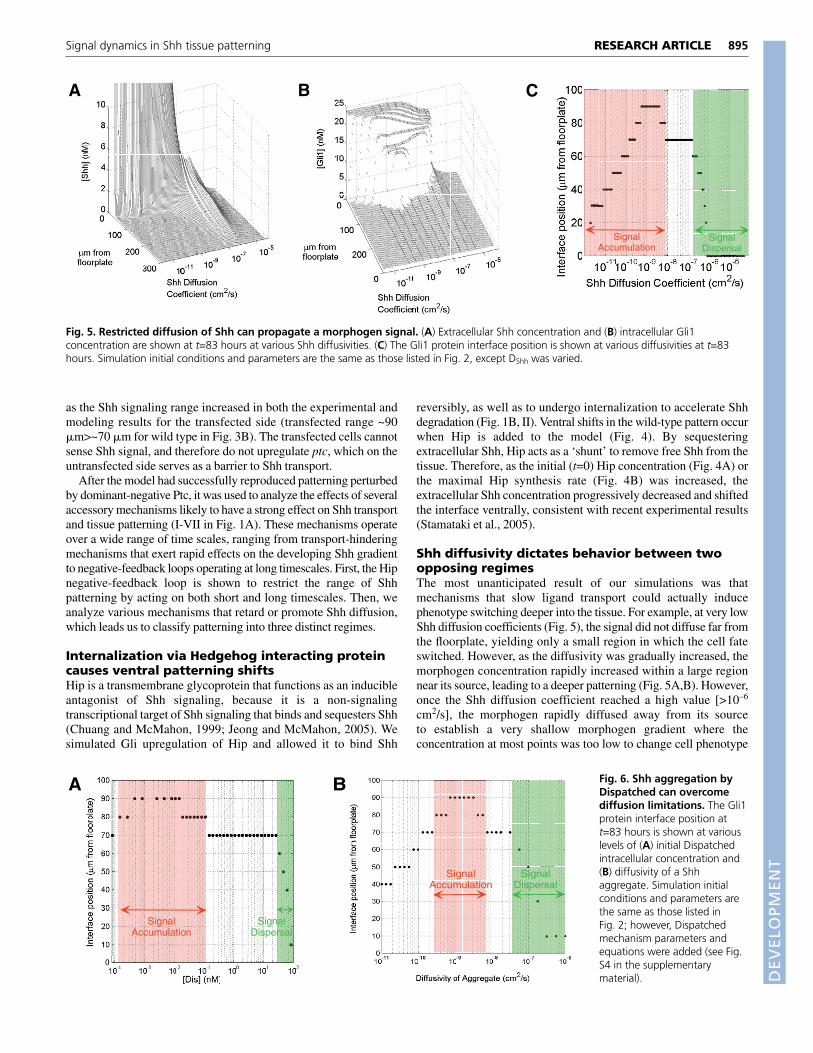
Shh diffusivity dictates behavior between twoopposing regimesThe most unanticipated result of our simulations was thatmechanisms that slow ligand transport could actually inducephenotype switching deeper into the tissue. For example, at very lowShh diffusion coefficients (Fig. 5), the signal did not diffuse far fromthe floorplate, yielding only a small region in which the cell fateswitched. However, as the diffusivity was gradually increased, themorphogen concentration rapidly increased within a large regionnear its source, leading to a deeper patterning (Fig. 5A,B). However,once the Shh diffusion coefficient reached a high value [>10–6
cm2/s], the morphogen rapidly diffused away from its sourceto establish a very shallow morphogen gradient where theconcentration at most points was too low to change cell phenotype
895RESEARCH ARTICLESignal dynamics in Shh tissue patterning
BA C
SignalAccumulation
SignalDispersal
Fig. 5. Restricted diffusion of Shh can propagate a morphogen signal. (A) Extracellular Shh concentration and (B) intracellular Gli1concentration are shown at t=83 hours at various Shh diffusivities. (C) The Gli1 protein interface position is shown at various diffusivities at t=83hours. Simulation initial conditions and parameters are the same as those listed in Fig. 2, except DShh was varied.
A
SignalAccumulation
SignalDispersal
B
SignalAccumulation
SignalDispersal
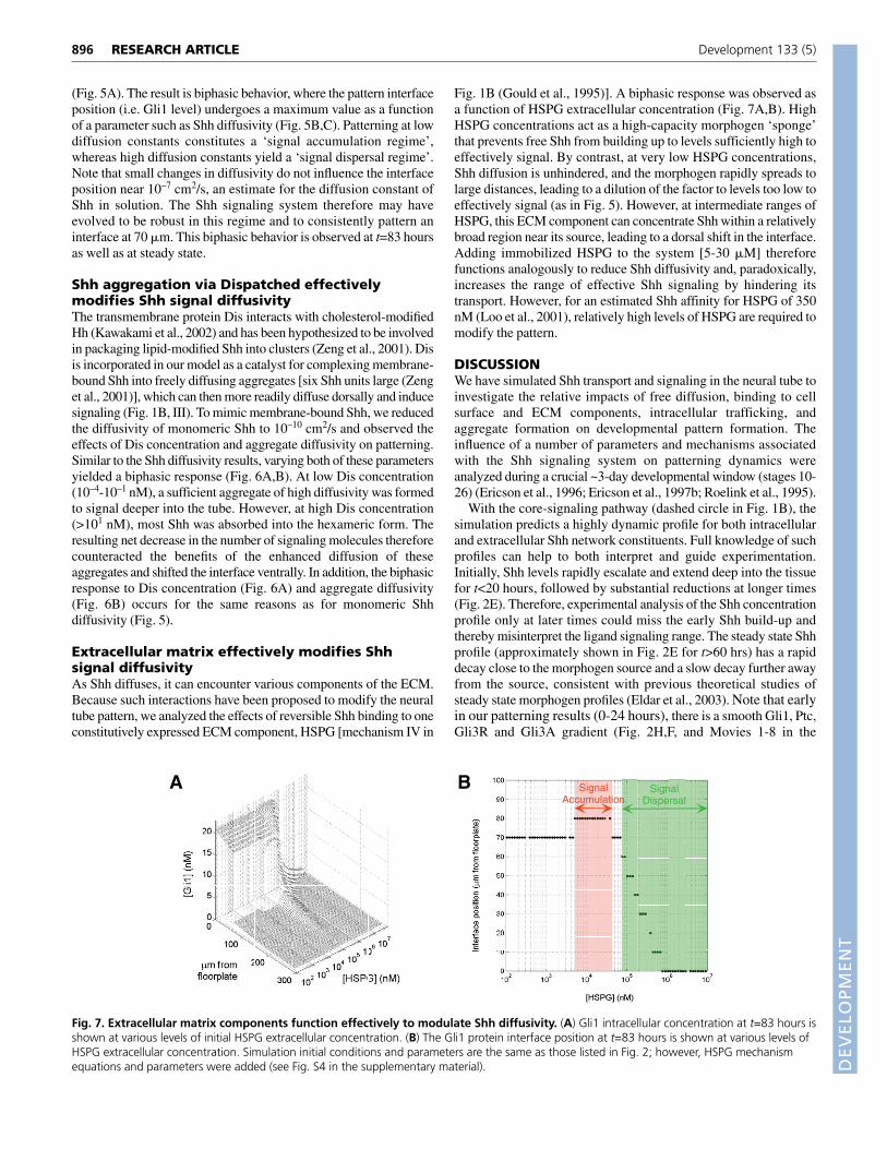
Fig. 6. Shh aggregation byDispatched can overcomediffusion limitations. The Gli1protein interface position att=83 hours is shown at variouslevels of (A) initial Dispatchedintracellular concentration and(B) diffusivity of a Shhaggregate. Simulation initialconditions and parameters arethe same as those listed inFig. 2; however, Dispatchedmechanism parameters andequations were added (see Fig.S4 in the supplementarymaterial).
DEVELO
PMENT
896
(Fig. 5A). The result is biphasic behavior, where the pattern interfaceposition (i.e. Gli1 level) undergoes a maximum value as a functionof a parameter such as Shh diffusivity (Fig. 5B,C). Patterning at lowdiffusion constants constitutes a ‘signal accumulation regime’,whereas high diffusion constants yield a ‘signal dispersal regime’.Note that small changes in diffusivity do not influence the interfaceposition near 10–7 cm2/s, an estimate for the diffusion constant ofShh in solution. The Shh signaling system therefore may haveevolved to be robust in this regime and to consistently pattern aninterface at 70 �m. This biphasic behavior is observed at t=83 hoursas well as at steady state.
Shh aggregation via Dispatched effectivelymodifies Shh signal diffusivityThe transmembrane protein Dis interacts with cholesterol-modifiedHh (Kawakami et al., 2002) and has been hypothesized to be involvedin packaging lipid-modified Shh into clusters (Zeng et al., 2001). Disis incorporated in our model as a catalyst for complexing membrane-bound Shh into freely diffusing aggregates [six Shh units large (Zenget al., 2001)], which can then more readily diffuse dorsally and inducesignaling (Fig. 1B, III). To mimic membrane-bound Shh, we reducedthe diffusivity of monomeric Shh to 10–10 cm2/s and observed theeffects of Dis concentration and aggregate diffusivity on patterning.Similar to the Shh diffusivity results, varying both of these parametersyielded a biphasic response (Fig. 6A,B). At low Dis concentration(10–4-10–1 nM), a sufficient aggregate of high diffusivity was formedto signal deeper into the tube. However, at high Dis concentration(>101 nM), most Shh was absorbed into the hexameric form. Theresulting net decrease in the number of signaling molecules thereforecounteracted the benefits of the enhanced diffusion of theseaggregates and shifted the interface ventrally. In addition, the biphasicresponse to Dis concentration (Fig. 6A) and aggregate diffusivity(Fig. 6B) occurs for the same reasons as for monomeric Shhdiffusivity (Fig. 5).
Extracellular matrix effectively modifies Shhsignal diffusivityAs Shh diffuses, it can encounter various components of the ECM.Because such interactions have been proposed to modify the neuraltube pattern, we analyzed the effects of reversible Shh binding to oneconstitutively expressed ECM component, HSPG [mechanism IV in
Fig. 1B (Gould et al., 1995)]. A biphasic response was observed asa function of HSPG extracellular concentration (Fig. 7A,B). HighHSPG concentrations act as a high-capacity morphogen ‘sponge’that prevents free Shh from building up to levels sufficiently high toeffectively signal. By contrast, at very low HSPG concentrations,Shh diffusion is unhindered, and the morphogen rapidly spreads tolarge distances, leading to a dilution of the factor to levels too low toeffectively signal (as in Fig. 5). However, at intermediate ranges ofHSPG, this ECM component can concentrate Shh within a relativelybroad region near its source, leading to a dorsal shift in the interface.Adding immobilized HSPG to the system [5-30 �M] thereforefunctions analogously to reduce Shh diffusivity and, paradoxically,increases the range of effective Shh signaling by hindering itstransport. However, for an estimated Shh affinity for HSPG of 350nM (Loo et al., 2001), relatively high levels of HSPG are required tomodify the pattern.
DISCUSSIONWe have simulated Shh transport and signaling in the neural tube toinvestigate the relative impacts of free diffusion, binding to cellsurface and ECM components, intracellular trafficking, andaggregate formation on developmental pattern formation. Theinfluence of a number of parameters and mechanisms associatedwith the Shh signaling system on patterning dynamics wereanalyzed during a crucial ~3-day developmental window (stages 10-26) (Ericson et al., 1996; Ericson et al., 1997b; Roelink et al., 1995).
With the core-signaling pathway (dashed circle in Fig. 1B), thesimulation predicts a highly dynamic profile for both intracellularand extracellular Shh network constituents. Full knowledge of suchprofiles can help to both interpret and guide experimentation.Initially, Shh levels rapidly escalate and extend deep into the tissuefor t<20 hours, followed by substantial reductions at longer times(Fig. 2E). Therefore, experimental analysis of the Shh concentrationprofile only at later times could miss the early Shh build-up andthereby misinterpret the ligand signaling range. The steady state Shhprofile (approximately shown in Fig. 2E for t>60 hrs) has a rapiddecay close to the morphogen source and a slow decay further awayfrom the source, consistent with previous theoretical studies ofsteady state morphogen profiles (Eldar et al., 2003). Note that earlyin our patterning results (0-24 hours), there is a smooth Gli1, Ptc,Gli3R and Gli3A gradient (Fig. 2H,F, and Movies 1-8 in the
RESEARCH ARTICLE Development 133 (5)
A B SignalAccumulation
SignalDispersal
Fig. 7. Extracellular matrix components function effectively to modulate Shh diffusivity. (A) Gli1 intracellular concentration at t=83 hours isshown at various levels of initial HSPG extracellular concentration. (B) The Gli1 protein interface position at t=83 hours is shown at various levels ofHSPG extracellular concentration. Simulation initial conditions and parameters are the same as those listed in Fig. 2; however, HSPG mechanismequations and parameters were added (see Fig. S4 in the supplementary material).
DEVELO
PMENT
supplementary material), but this profile begins to sharpen into adiscrete interface at 72 hours (stage 16-26). Quantitative assays ofthe expression of Gli1, Gli3 or Ptc at a broad range of times fromstage 12 through 26 can further test directly whether our frameworkaccurately predicts their expression patterns, and many experimentalstains for Gli or Ptc expression at times before steady-state areconsistent with smooth concentration gradients in the MN domain(Lei et al., 2004; Stamataki et al., 2005). In particular, snapshots ofthe Ptc profile in the neural tube indicate a highly dynamic, initiallygraded profile consistent with our results (see Fig. S5 in thesupplementary material), and at later times processes not includedin the model (e.g. Shh-independent signals influencing the relativelyuncharacterized Ptc promoter) are likely to modulate ptc expression,especially in the more dorsal sections. Recent work supports a rolefor a graded Gli3 profile in early patterning (Stamataki et al., 2005),although several experimental details preclude a direct, quantitativecomparison between these results and our simulations. In particular,endogenous Gli1, Gli2 or Gli3 expression is not directly measured,and exogenously introduced Gli3 is expressed at levels that vary overtime. However, this important work provides strong evidence for therole of gene expression dynamics in tissue patterning.
The mechanism by which a single cell interprets a morphogengradient can occur at the transcription factor level (Niwa et al., 2000;Shimizu and Gurdon, 1999). Quantitative differences in Gli have notbeen experimentally tested for V3/MN patterning in the vertebrateneural tube; however, given the Shh threshold and kinetic data fromchick V3/MN patterning used for our parameter estimates (seeMaterials and methods), the model predicts that V3 cell fatespecification could be established at a sevenfold increase in Gli1from its initial basal concentration [i.e. a Gli1 concentration at thetime of V3 specification of >11 nM (or 7�Gli1 concentration att=0)]. We tested model behavior for V3 cell fate specificationoccurring at a range of thresholds from three- to tenfold Gli (4.9-16.3nM) increases, and all of our conclusions remain qualitatively thesame (data not shown). The sevenfold increase above basal/initialGli expression levels corresponds to a two- to threefold Gli1difference across the V3/MN interface position at approximatelyt=50 hours (Fig. 2H). Therefore, the model behavior is consistentwith the twofold increase in Oct3/Oct4 expression in embryonicstem cells (Niwa et al., 2000) and the threefold increase in SMADcomplexes in a Xenopus blastula cell (Shimizu and Gurdon, 1999)that have been found to trigger cell fate switching. It is notable thatcertain borderline cells (Fig. 2H) experience transient two- orthreefold increases in Gli1, and a deeper investigation of theinduction kinetics for the next generations of transcription factorsdownstream of Gli may reveal whether these cells transientlyexpress MN markers (Ericson et al., 1996) or permanently committo an MN fate. As we have previously discussed in a single cellmodel, stochastic effects, which can in the future be incorporatedinto this spatial model, may account for the transient co-expressionof markers of different cell fates (Lai et al., 2004). Finally, futureincorporation of more detailed mechanisms of interaction betweenthe transcription factors Gli1-Gli3, Nkx2.2, Pax6 and others, as theseinteractions are further elucidated, would help update the model tomatch or predict future patterning results.
Intracellular degradation can shunt the Shh signalVertebrate Shh patterning can be further complicated by the fact thatPtc is not the only receptor that mediates ligand degradation. LikePtc, Hip also binds Shh with high affinity and is a Shh transcriptionaltarget, yet Hip mediates Shh endocytosis and degradation withouttransducing a signal. A shunt in an electrical circuit is an alternate
pathway that diverts current away from the remainder of the circuit,analogous to the receptor-mediated endocytosis and ensuingdegradation that divert a morphogen from signaling. Intracellularshunting by Ptc and Hip, as seen in the modeling results (Fig. 4A,B),attenuates Shh signaling, consistent with several studies in variousorganisms (Chuang and McMahon, 1999; Jeong and McMahon,2005). Negative-feedback loops, which establish shunts viamolecules like Hip, limit Shh penetration and can ‘stabilize’patterning, a mechanism previously proposed for morphogengradients (Eldar et al., 2003).
Although Hip expression has been detected near all Shh signalingcenters, its basal concentration and extent of upregulation upon Shhsignaling are both parameters that vertebrates may use to regulateHedgehog signaling with great spatial precision (Chiang et al., 1999;Chuang et al., 2003; Tojo et al., 2002). For example, Hip preventsthe spread of excess Shh ligand beyond odontogenic mesenchymein tooth development, thus restricting the Shh signaling to specificregions of the oral axis (Coulombe et al., 2004). Other than basalconcentration and extent of Hip upregulation, other rates in the Hippathway may be modulated in different organisms, as in the mouseneural tube where Hip-Shh complex internalization appears to beslow (Chuang and McMahon, 1999; Jeong and McMahon, 2005).The ventralization of the tube observed when Hip is overexpressed(Fig. 4), and the non-cell-autonomous nature of this expansion, isvery consistent with recent experimental work (Stamataki et al.,2005). Interestingly, soluble, diffusible forms of Hip have recentlybeen found in the mature brain (Coulombe et al., 2004). Ourmodeling results suggest that this new mechanism may potentiallyextend the Shh signaling range by protecting Shh from binding tothe cell surface Hip variant, Ptc, or even HSPG (data not shown).
Restricting diffusion can propagate a morphogensignalAlthough receptor binding can restrict the morphogen signalingrange, other mechanisms may unexpectedly enhance it. Twoostensibly opposing experimental observations have been difficultto reconcile: the long-range signaling ability of Shh and membraneanchorage of the ligand by hydrophobic modification. Shhassociates with the membrane through the addition of two lipidtethers during its synthesis, a N-terminal palmitic acid (Pepinsky etal., 1998) and a C-terminal cholesterol (Porter et al., 1996). Despitethe reduced diffusivity accompanying membrane association, manystudies have demonstrated the long range signaling ability of Shh inthe neural tube over 20 cell diameters (>200 �m) (Briscoe et al.,2001; Ericson et al., 1997a; Gritli-Linde et al., 2001).
The simulation counterintuitively predicts that lower diffusionconstants can actually concentrate the signal in the ventral neuraltube and thereby extend its signaling range (Fig. 5A). Specifically,a typical 20 kDa protein in solution has a diffusivity of order~1�10–7 cm2/s, and hydrophobic modification is likely to decreasethe diffusivity to a range between 10–8 and 10–10 cm2/s (Creighton,1992). This reduction may actually help Shh to extend its signalingrange two additional cell layers further from the floorplate (Fig. 5C).In one study in the vertebrate limb, Shh was detected ~200 �m fromthe source, whereas knock-in of a non-lipid modified Shh led toligand detection only at lower levels and much closer to the source(see Lewis et al., 2001). The interpretation was that lipidmodification was necessary for long-range transport. By contrast,our results indicate that the rapidly diffusing, non-lipid-modifiedform may be rapidly diluted within the tissue to fall below theexperimental threshold of detection, whereas the lipid modificationconcentrates the ligand.
897RESEARCH ARTICLESignal dynamics in Shh tissue patterning
DEVELO
PMENT
898
Mechanisms modulating ligand diffusionVia independent mechanisms, both Dis and ECM can also extendthe signaling range of Shh. First, although experiments have yet toquantify a spatial profile for Shh aggregates in the neural tube, ourmodeling results show that Dis-catalyzed aggregation of highlydiffusible Shh aggregates can propagate a Shh signal (Fig. 6A,B).Analogous to the monomeric Shh results (Fig. 5), there is a biphasicresponse in the V3/MN pattern to both the diffusivity of theaggregate and the Dis-catalyzed rate of aggregate generation. In theneural tube of dis mutant mice, ventral fates are not properlyspecified, and Shh immunoreactivity is detected only in Shh-producing cells in the ventral neural tube, somite and limb(Kawakami et al., 2002), indicating that this mechanism exertssignificant control over the range of signaling. Although Dis mayhave additional functions in the Hedgehog pathway, Dis-catalyzedShh aggregation may be a general transport modulating mechanismoperating in Drosophila (Burke et al., 1999) and zebrafish (Nakanoet al., 2004).
Distinguishing among the potential roles of ECM in ligandpresentation, stabilization and accumulation has been difficult(Bornemann et al., 2004; Giráldez et al., 2002; Pons and Marti,2000; Rubin et al., 2002; The et al., 1999). Our results indicate thata very simple mechanism, the reversible binding of Shh to HSPG,can either lengthen or restrict the signaling range depending on theHSPG concentration (Fig. 7A). Both HSPGs and the EXT genesinvolved in their synthesis are abundantly expressed in adevelopmentally regulated manner in the mammalian centralnervous system, suggesting their functional roles in neural tubepatterning (Gould et al., 1995; Inatani et al., 2003; Yamaguchi,2001). As HSPG is actively remodeled by proteases in many tissues,
and the ligand affinity for HSPG can be correspondingly modulated,HSPG concentration and affinity can serve as robust, tunableparameters to modulate ligand signaling in particular tissues. Ourresults also indicate that vitronectin, a direct Shh transcriptionaltarget, can also modulate ligand transport (see Fig. S2 in thesupplementary material).
Simulations of both the Dis-catalyzed ligand aggregationmechanism (Fig. 6) and ECM function (Fig. 7) indicate a similar‘biphasic’ behavior (as seen in Fig. 5 with Shh diffusivity), wherethe effective signaling range undergoes a maximum as a function ofa key parameter. This behavior occurs because aggregating Shh intofreely diffusible compounds allows it to overcome diffusion barriers,whereas ECM components function as additional diffusion barriersto concentrate the signal.
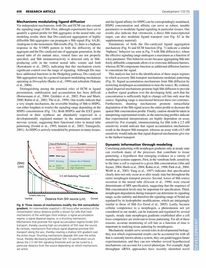
This analysis has led to the identification of three major regimesin which accessory Shh transport mechanisms modulate patterning(Fig. 8). Signal accumulation mechanisms limit Shh diffusion byrestricting morphogen accumulation to near the source. By contrast,signal dispersal mechanisms promote high Shh diffusion to providea shallow signal gradient over the developing field, such that theconcentration is sufficiently high to change cell fate only near thesource. Signaling range is maximized between these two extremes.Furthermore, shunting mechanisms promote intracellulardegradation of the Shh signal across the entire profile to decrease thespatial Shh concentration profile. Finally, caution should be taken ininterpreting experimental results, as the intersecting profiles indicatethat experimental interpretations are highly dependent on assaysensitivity. For example, immunostaining for Shh with a 2.5 nMsensitivity would indicate that signal accumulation mechanismsresult in the deepest Shh transport, whereas an assay with a 0.5 nMsensitivity would indicate that signal dispersal mechanisms give riseto the farthest transport.
Dynamic information through modelingCorrelating patterning with morphogen gradients only at steady statecan overlook many of the processes that contribute to tissuepatterning, a hypothesis that emerging evidence in a variety ofmorphogen systems supports. First, in the vertebrate limb, sensitivityto the time a cell is exposed to a given Shh concentration (Ahn andJoyner, 2004; Harfe et al., 2004; Kohtz et al., 1998; Park et al., 2004;Wolff et al., 2003; Yang et al., 1997) indicates that specificationclearly does not only occur at or after steady state but throughout theentire morphogen transport process. Second, waves of Shh sourcesecretion in the neural tube (Ericson et al., 1996) were crucialdeterminants of MN specification, suggesting that the sequence ofShh concentration levels may be important for specification. Third,morphogen degradation during transport can determine its signalingrange, as the stability and therefore the signaling range of Nodal areregulated by its hydrophobic modifications, which are intriguinglysimilar to those of Shh (Le Good et al., 2005). Lastly, becausecellular competence to a morphogen, a phenomenon not yetconsidered in our model, can be transient and regulated by severalsignals, steady state morphogen gradients established after a cellloses competence are irrelevant to tissue patterning. For all of thesereasons, accurate monitoring of cell fate as a function of time isimportant in studying tissue pattering by morphogens.
Mechanistic models serve several roles in developmental biology:they test which experimental results can be accounted for with allthat is currently known about a system, they can guide/suggest futureexperimentation, and they can test whether several hypothesizedmechanisms can account for a novel phenotype. For example, highthroughput siRNA approaches have recently identified novel
RESEARCH ARTICLE Development 133 (5)
Fig. 8. Three classes of mechanisms modify the Shh extracellulargradient. An intermediate snapshot (~30 hours after secretion) of theconcentration versus distance profile is shown for cells that havemechanisms of the wild-type chick embryo: a signal accumulationregime, a signal dispersal regime, or a shunting mechanism.Mechanisms that promote the signal accumulation regime hinder Shhtransport, thereby causing high accumulation of Shh near the source.By contrast, mechanisms that induce signal dispersal promote Shhtransport along the axis, thereby creating a shallow Shh gradient overthe entire tissue. Shunting mechanisms degrade Shh over the entiretissue, thereby decreasing extracellular Shh. V3 specification occursabove the 2.5 nM Shh signaling threshold and can be tuned to aparticular distance from the source depending on which mechanismsare active.
DEVELO
PMENT
components of the Hh pathway (Lum et al., 2003), and modeling canbe used to test their potential mechanisms of action. Other moleculescan also be included, such as Ptc2, Megalin, and the You class ofproteins, whose precise mechanism in the Hedgehog pathway is notyet known (Carpenter et al., 1998; Ding et al., 1998). For example,our preliminary modeling work does not support a function ofMegalin as an active Shh transporter through the cytoplasm(McCarthy et al., 2002), as long-range signaling is not observed,even with extremely fast intracellular transport rates of Shh-Megalincomplexes (data not shown). Furthermore, a unique advantage ofthis finite element numerical approach is that it can incorporate theeffects of cell division and death during patterning. Future work canexpand the current static model geometry to a ‘living mesh’, so thatelements are added (or subtracted) as they arise (or die) in irregulargeometries and at various times. Finally, the simulation indicates thatthe rate of Shh secretion from the floorplate, which feeds thepatterning process through the activity of a highly complex promoternot yet modeled (Epstein et al., 1999), is a highly importantparameter that should be experimentally measured.
The model guides and predicts numerous additional experimentsto analyze Shh patterning in the neural tube, and potentially othertissues where Shh transport is involved. First, knock-in of variousforms of Shh with varying extents of lipid modification (Feng et al.,2004) is predicted to pattern to differing depths, as a function of Shhdiffusivity (Fig. 5). Likewise, Shh diffusivity can be tuned byinjecting soluble HSPG. Similar to experiments in the brain wherethe spread of molecules with an HSPG interaction domain have beenincreased (Nguyen et al., 2001), high levels of soluble HSPG wouldinduce Shh signal dispersal (Fig. 8). Second, the secretion rate ofShh can be varied in the floorplate through the use of a regulablepromoter, and the model can be used to predict the precise positionof the V3/MN interface as more MN are produced at the expense ofV3 neurons with increasing secretion. Finally, neural tube explantscan be incubated in blocking antibodies against Hip, Dis, HSPG orvitronectin. The concentration of such antibodies would tune thelevels of active Hip, Dis, HSPG and vitronectin to the levels shownin Figs 4, 6 and 7, and Fig. S1 (in the supplementary material),respectively, to test the predicted biphasic responses.
In conclusion, we have analyzed how complex mechanismsacting at various times regulate morphogen transport and modulatetissue patterning. The results guide and suggest further experimentson how these mechanisms work in concert to provide robust Shhneural tube patterning. Future modeling work can explore the effectsof numerous modular mechanisms in the Hedgehog pathway, andmay intriguingly suggest further experiments on how these moduleshave been evolutionary ‘plugged’ into particular tissues to restrict orpropagate the Hedgehog signal when required.
This work was supported by a National Science Foundation GraduateFellowship to K.S. and a University of California Cancer Research CoordinatingCommittee Award and NIH NS048248 to D.V.S.
Supplementary materialSupplementary material for this article is available athttp://dev.biologists.org/cgi/content/full/133/5/889/DC1
ReferencesAhn, S. and Joyner, A. L. (2004). Dynamic changes in the response of cells to
positive hedgehog signaling during mouse limb patterning. Cell 118, 505-516.Bai, C. B., Stephen, D. and Joyner, A. L. (2004). All mouse ventral spinal cord
patterning by hedgehog is gli dependent and involves an activator function ofGli3. Dev. Cell 6, 103-115.
Bornemann, D. J., Duncan, J. E., Staatz, W., Selleck, S. and Warrior, R. (2004).Abrogation of heparan sulfate synthesis in Drosophila disrupts the Wingless,Hedgehog and Decapentaplegic signaling pathways. Development 131, 1927-1938.
Briscoe, J., Pierani, A., Jessell, T. M. and Ericson, J. (2000). A homeodomainprotein code specifies progenitor cell identity and neuronal fate in the ventralneural tube. Cell 101, 435-445.
Briscoe, J., Chen, Y., Jessell, T. M. and Struhl, G. (2001). A hedgehog-insensitiveform of patched provides evidence for direct long-range morphogen activity ofsonic hedgehog in the neural tube. Mol. Cell 7, 1279-1291.
Britto, J., Tannahill, D. and Keynes, R. (2002). A critical role for sonic hedgehogsignaling in the early expansion of the developing brain. Nat. Neurosci. 5, 103-110.
Burke, R., Nellen, D., Bellotto, M., Hafen, E., Senti, K.-A., Dickson, B. J. andBasler, K. (1999). Dispatched, a novel sterol-sensing domain protein dedicatedto the release of cholesterol-modified hedgehog from signaling cells. Cell 99,803-815.
Carpenter, D., Stone, D. M., Brush, J., Ryan, A., Armanini, M., Frantz, G.,Rosenthal, A. and Sauvage, F. J. d. (1998). Characterization of two patchedreceptors for the vertebrate hedgehog protein family. Proc. Natl. Acad. Sci. USA95, 13630-13634.
Chen, M.-H., Li, Y.-J., Kawakami, T., Xu, S.-M. and Chuang, P.-T. (2004).Palmitoylation is required for the production of a soluble multimeric Hedgehogprotein complex and long-range signaling in vertebrates. Genes Dev. 18, 641-659.
Chen, Y. and Struhl, G. (1996). Dual roles for patched in sequestering andtransducing hedgehog. Cell 87, 553-563.
Chiang, C., Swan, R. Z., Grachtchouk, M., Bolinger, M., Litingtung, Y.,Robertson, E. K., Cooper, M. K., Gaffield, W., Westphal, H., Beachy, P. A.et al. (1999). Essential role for Sonic hedgehog during hair folliclemorphogenesis. Dev. Biol. 205, 1-9.
Chuang, P.-T. and McMahon, A. P. (1999). Vertebrate Hedgehog signalingmodulated by induction of a Hedgehog-binding protein. Nature 397, 617-621.
Chuang, P.-T., Kawcak, T. N. and McMahon, A. P. (2003). Feedback control ofmammalian Hedgehog signaling by the Hedgehog-binding protein, Hip1,modulates Fgf signaling during branching morphogenesis of the lung. GenesDev.17, 342-347.
Coulombe, J., Traiffort, E., Loulier, K., Faure, H. and Ruat, M. (2004).Hedgehog interacting protein in the mature brain: membrane-associated andsoluble forms. Mol. Cell. Neurosci. 25, 323-333.
Creighton, T. E. (1992). Proteins: Structures and Molecular Properties. New York:W. H. Freeman.
Crick, F. (1970). Diffusion in embryogenesis. Nature 255, 40-42.Dillon, R., Gadgil, C. and Othmer, H. G. (2003). Short- and long-range effects of
Sonic hedgehog in limb development. Proc. Natl. Acad. Sci. USA 100, 10152-10157.
Ding, Q., Motoyama, J., Gasca, S., Mo, R., Sasaki, H., Rossant, J. and Hui, C.(1998). Diminished Sonic hedgehog signaling and lack of floor platedifferentiation in Gli2 mutant mice. Development 125, 2533-2543.
Eldar, A., Rosin, D., Shilo, B. Z. and Barkai, N. (2003). Self-enhanced liganddegradation underlies robustness of morphogen gradients. Dev. Cell 5, 635-646.
Epstein, D. J., McMahon, A. P. and Joyner, A. L. (1999). Regionalization ofSonic hedgehog transcription along the anteroposterior axis of the mousecentral nervous system is regulated by Hnf3-dependent and -independentmechanisms. Development 126, 281-292.
Ericson, J., Muhr, J., Placzek, M., Lints, T., Jessell, T. M. and Edlund, T. (1995).Sonic hedgehog induces the differentiation of ventral forebrain neurons: acommon signal for ventral patterning within the neural tube. Cell 81, 747-756.
Ericson, J., Morton, S., Kawakami, A., Roelink, H. and Jessell, T. M. (1996).Two critical periods of Sonic Hedgehog signaling required for the specification ofmotor neuron identity. Cell 87, 661-673.
Ericson, J., Briscoe, J., Rashbass, P., van Heyningen, V. and Jessell, T. M.(1997a). Graded sonic hedgehog signaling and the specification of cell fate inthe ventral neural tube. Cold Spring Harb. Symp. Quant. Biol. 62, 451-466.
Ericson, J., Rashbass, P., Schedl, A., Brenner-Morton, S., Kawakami, A., vanHeyningen, V., Jessell, T. M. and Briscoe, J. (1997b). Pax6 controls progenitorcell identity and neuronal fate in response to graded Shh signaling. Cell 90, 169-180.
Feng, J., White, B., Tyurina, O. V., Guner, B., Larson, T., Lee, H. Y., Karlstrom,R. O. and Kohtz, J. D. (2004). Synergistic and antagonistic roles of the Sonichedgehog N- and C-terminal lipids. Development 131, 4357-4370.
Giráldez, A. J., Copley, R. R. and Cohen, S. M. (2002). HSPG modification bythe secreted enzyme notum shapes the wingless morphogen gradient. Dev. Cell2, 667-676.
Gould, S. E., Upholt, W. B. and Kosher, R. A. (1995). Characterization ofchicken syndecan-3 as a heparan sulfate proteoglycan and its expression duringembryogenesis. Dev. Biol. 168, 438-451.
Gritli-Linde, A., Lewis, P., McMahon, A. P. and Linde, A. (2001). Thewhereabouts of a morphogen: direct evidence for short- and graded long-rangeactivity of hedgehog signaling peptides. Dev. Biol. 236, 364-386.
Han, C., Belenkaya, T. Y., Wang, B. and Lin, X. (2004). Drosophila glypicanscontrol the cell-to-cell movement of Hedgehog by a dynamin-independentprocess. Development 131, 73-82.
899RESEARCH ARTICLESignal dynamics in Shh tissue patterning
DEVELO
PMENT
900
Harfe, B. D., Scherz, P. J., Nissim, S., Tian, H., McMahon, A. P. and Tabin, C. J.(2004). Evidence for an expansion-based temporal Shh gradient in specifyingvertebrate digit identities. Cell 118, 517-528.
Inatani, M., Irie, F., Plump, A. S., Tessier-Lavigne, M. and Yamaguchi, Y.(2003). Mammalian brain morphogenesis and midline axon guidance requireheparan sulfate. Science 5647, 1044-1046.
Incardona, J. P., Lee, J. H., Robertson, C. P., Enga, K., Kapur, R. P. andRoelink, H. (2000). Receptor-mediated endocytosis of soluble and membrane-tethered Sonic hedgehog by Patched-1. Proc. Natl. Acad. Sci. USA 97, 12044-12049.
Incardona, J. P., Gruenberg, J. and Roelink, H. (2002). Sonic hedgehog inducesthe segregation of patched and smoothened in endosomes. Curr. Biol. 12, 983-995.
Jeong, J. and McMahon, A. P. (2005). Growth and pattern of the mammalianneural tube are governed by partially overlapping feedback activities of thehedgehog antagonists patched 1 and Hhip1. Development 132, 143-154.
Jessell, T. M. (2000). Neuronal specification in the spinal cord: inductive signalsand transcriptional codes. Nat. Rev. Genet. 1, 20-29.
Kawakami, T., Kawcak, T. N., Li, Y.-J., Zhang, W., Hu, Y. and Chuang, P.-T.(2002). Mouse dispatched mutants fail to distribute hedgehog proteins and aredefective in hedgehog signaling. Development 129, 5753-5765.
Kohtz, J. D., Baker, D. P., Corte, G. and Fishell, G. (1998). Regionalization withinthe mammalian telencephalon is mediated by changes in responsiveness toSonic Hedgehog. Development 125, 5079-5089.
Lai, K., Robertson, M. J. and Schaffer, D. V. (2004). The sonic hedgehogsignaling system as a bistable genetic switch. Biophys. J. 86, 2748-2757.
Lander, A. D., Nie, Q. and Wan, F. Y. (2002). Do morphogen gradients arise bydiffusion? Dev. Cell 2, 785-796.
Le Good, J. A., Joubin, K., Giraldez, A. J., Ben-Haim, N., Beck, S., Chen, Y.,Schier, A. F. and Constam, D. B. (2005). Nodal stability determines signalingrange. Curr. Biol. 15, 31-36.
Lei, Q., Zelman, A. K., Kuang, E., Li, S. and Matise, M. P. (2004). Transductionof graded Hedgehog signaling by a combination of Gli2 and Gli3 activatorfunctions in the developing spinal cord. Development 131, 3593-3604.
Lewis, P. M., Dunn, M. P., McMahon, J. A., Logan, M., Martin, J. F., St-Jacques, B. and McMahon, A. P. (2001). Cholesterol modification of sonichedgehog is required for long-range signaling activity and effective modulationof signaling by Ptc1. Cell 105, 599-612.
Litingtung, Y. and Chiang, C. (2000). Specification of ventral neuron types ismediated by an antagonistic interaction between shh and gli3. Nat. Neurosci. 3,979-985.
Loo, B.-M., Kreuger, J., Jalkanen, M., Lindahl, U. and Salmivirta, M. (2001).Binding of heparin/heparan sulfate to fibroblast growth factor receptor 4. J. Biol.Chem. 276, 16868-16876.
Lum, L., Yao, S., Mozer, B., Rovescalli, A., Von Kessler, D., Nirenberg, M. andBeachy, P. A. (2003). Identification of Hedgehog pathway components by RNAiin Drosophila cultured cells. Science 299, 2039-2045.
Marigo, V. and Tabin, C. J. (1996). Regulation of patched by sonic hedgehog inthe developing neural tube. Proc. Natl. Acad. Sci. USA 93, 9346-9351.
Martinez-Morales, J. R., Barbas, J. A., Marti, E., Bovolenta, P., Edgar, D. andRodriguez-Tebar, A. (1997). Vitronectin is expressed in the ventral region ofthe neural tube and promotes the differentiation of motor neurons.Development 124, 5139-5147.
McCarthy, R. A., Barth, J. L., Chintalapudi, M. R., Knaak, C. and Argraves, W.S. (2002). Megalin functions as an endocytic sonic hedgehog receptor. J. Biol.Chem. 277, 25660-25667.
Nakano, Y., Kima, H. R., Kawakamib, A., Roya, S., Schierb, A. F. andIngham, P. W. (2004). Inactivation of dispatched 1 by the chameleonmutation disrupts Hedgehog signalling in the zebrafish embryo. Dev. Biol. 269,381-392.
Nguyen, J. B., Sanchez-Pernaute, R., Cunningham, J. and Bankiewicz, K. S.(2001). Convection-enhanced delivery of AAV-2 combined with heparinincreases TK gene transfer in the rat brain. NeuroReport 12, 1961-1964.
Niwa, H., Miyazaki, J. and Smith, A. G. (2000). Quantitative expression of Oct-3/4 defines differentiation, dedifferentiation or self-renewal of ES cells. Nat.Genet. 24, 372-376.
Park, H. C., Shin, J. and Appel, B. (2004). Spatial and temporal regulation of
ventral spinal cord precursor specification by Hedgehog signaling. Development131, 5959-5969.
Park, H. L., Bai, C., Platt, K. A., Matise, M. P., Beeghly, A., Hui, C. C.,Nakashima, M. and Joyner, A. L. (2000). Mouse Gli1 mutants are viable buthave defects in SHH signaling in combination with a Gli2 mutation.Development 127, 1593-1605.
Pepinsky, R. B., Zeng, C., Wen, D., Rayhorn, P., Baker, D. P., Williams, K. P.,Bixler, S. A., Ambrose, C. M., Garber, E. A., Miatkowski, K. et al. (1998).Identification of a palmitic acid-modified form of human sonic hedgehog. J. Biol.Chem. 273, 14037-14045.
Persson, M., Stamataki, D., te Welscher, P., Andersson, E., Bose, J., Ruther,U., Ericson, J. and Briscoe, J. (2002). Dorsal-ventral patterning of the spinalcord requires Gli3 transcriptional repressor activity. Genes Dev. 16, 2865-2878.
Pons, S. and Marti, E. (2000). Sonic hedgehog synergizes with the extracellularmatrix protein vitronectin to induce spinal motor neuron differentiation.Development 127, 333-342.
Porter, J. A., Young, K. E. and Beachy, P. A. (1996). Cholesterol modification ofhedgehog signaling proteins in animal development. Science 274, 255-259.
Ricklefs, R. E. and Starck, J. M. (1998). Series of embryonic chicken growth. InAvian Growth and Development. Evolution Within the Altricial PrecocialSpectrum (ed. R. E. Ricklefs and J. M. Starck). New York: Oxford University Press.
Riddle, R. D., Johnson, R. L., Laufer, E. and Tabin, C. (1993). Sonic hedgehogmediates the polarizing activity of the ZPA. Cell 75, 1401-1416.
Roelink, H., Porter, J. A., Chiang, C., Tanabe, Y., Chang, D. T., Beachy, P. A.and Jessell, T. M. (1995). Floor plate and motor neuron induction by differentconcentrations of the amino-terminal cleavage product of sonic hedgehogautoproteolysis. Cell 81, 445-455.
Rubin, J. B., Choi, Y. and Segal, R. A. (2002). Cerebellar proteoglycans regulatesonic hedgehog responses during development. Development 129, 2223-2232.
Ruiz i Altaba, A. (1999). Gli proteins encode context-dependent positive andnegative functions: implications for development and disease. Development126, 3205-3216.
Shimizu, K. and Gurdon, J. B. (1999). A quantitative analysis of signaltransduction from activin receptor to nucleus and its relevance to morphogengradient interpretation. Proc. Natl. Acad. Sci. USA 96, 6791-6796.
Stamataki, D., Ulloa, F., Tsoni, S. V., Mynett, A. and Briscoe, J. (2005). Agradient of Gli activity mediates graded Sonic Hedgehog signaling in the neuraltube. Genes Dev. 19, 626-641.
Takei, Y., Ozawa, Y., Sato, M., Watanabe, A. and Tabata, T. (2004). ThreeDrosophila EXT genes shape morphogen gradients through synthesis of heparansulfate proteoglycans. Development 131, 73-82.
The, I., Bellaiche, Y. and Perrimon, N. (1999). Hedgehog movement is regulatedthrough tout velu-dependent synthesis of a heparan sulfate proteoglycan. Mol.Cell 4, 633-639.
Tojo, M., Kiyosawa, H., Iwatsuki, K. and Kaneko, F. (2002). Expression of asonic hedgehog signal transducer, hedgehog-interacting protein, by humanbasal cell carcinoma. Br. J. Dermatol. 146, 69.
Turing, A. M. (1952). The chemical basis of morphogenesis. Philos. Trans. R. Soc.Lond. Ser. B Biol. Sci. 237, 37-72.
Wijgerde, M., McMahon, J. A., Rule, M. and McMahon, A. P. (2002). A directrequirement for Hedgehog signaling for normal specification of all ventralprogenitor domains in the presumptive mammalian spinal cord. Genes Dev. 16,2849-2864.
Wolff, C., Roy, S. and Ingham, P. W. (2003). Multiple muscle cell identitiesinduced by distinct levels and timing of hedgehog activity in the zebrafishembryo. Curr. Biol. 13, 1169-1181.
Yamaguchi, Y. (2001). Heparan sulfate proteoglycans in the nervous system: theirdiverse roles in neurogenesis, axon guidance, and synaptogenesis. Semin. CellDev. Biol. 12, 99-106.
Yang, Y., Drossopoulou, G., Chuang, P.-T., Duprez, D., Marti, E., Bumcrot, D.,Vargesson, N., Clarke, J., Niswander, L., McMahon, A. et al. (1997).Relationship between dose, distance and time in Sonic Hedgehog-mediatedregulation of anteroposterior polarity in the chick limb. Development 124, 4393-4404.
Zeng, X., Goetz, J. A., Suber, L. M., Scott, W. J., Schreiner, C. M. andRobbins, D. J. (2001). A freely diffusible form of Sonic hedgehog mediateslong-range signaling. Nature 411, 716-720.
RESEARCH ARTICLE Development 133 (5)