Embed Size (px)
Citation preview
Role for the GTPase Guanylate-Binding Protein-1 (hGBP-1) in
Globlastoma Prognosis
Jacob Justinger

Acknowledgements
• I would like to thank Dr. Deborah Vestal for her mentorship throughout my research project.
• I would also like to thank my parents for their continued support throughout my academic career.

Gliomas
• Gliomas are a broad category of brain tumors that arise from glial cells.
• Glial cells form the supportive structures of the central nervous system and serve to keep neurons in place and functioning correctly.
• There are three types of normal glial cells: astrocytes, oligodendrocytes, and ependymal cells which when cancerous are called become astrocytomas, oligodendrogliomas, and ependymomas respectively.
• Mixed gliomas occur when the tumor consists of multiple cell types
http://www.abta.org/understanding-brain-tumors/types-of-tumors/gliomas.html

Astrocytomas• Astrocytomas are tumors which arise from the star shaped
supportive cells, astrocytes.
• Astrocytomas are categorized on a scale of I to IV. Based on growth rage and invasiveness, Grade IV astrocytomas are the most severe.
• Grade I astrocytomas are considered benign, or noninvasive.
• Grade II astrocytomas are designated as low grade because while they rarely spread they have a propensity to reoccur.
• Grade III astrocytomas are characterized by “focal or dispersed anaplasia” and an increased growth rate.
• Grade IV astrocytomas are the highest grade gliomas and the most malignant type of astrocytoma.
• Grade four astrocytomas are commonly referred to as Glioblastomahttp://www.abta.org/secure/glioblastoma-brochure.pdf

Glioblastomas
• Glioblastomas are the most common gliomas in adults, accounting for about 50% of all cases
• The high rate of growth and profuse invasiveness of glioblastomas leads to a dismal median survival rate of about 14.6 months
• The characteristics which distinguish glioblastomas from other gliomas are:
– Presence of cell necrosis
– Increased vacuolization of the tumor site
http://www.abta.org/secure/glioblastoma-brochure.pdf

Treatment
• Typical treatment of glioblastomas include– Surgery
– Radiation
– Chemotherapy, usually with temozolomide (TMZ)
• TMZ is chosen because of its relatively high penetration of the blood brain barrier
• TMZ works by methylating the O6 position of guanine in the cellular DNA
• The methylation of guanine causes an improper base pairing in the DNA which activates the mismatch repair system
• The normal mismatch repair system is unable to repair this DNA lesion and this drives the cell into apoptosis. (Wick, W., et al. 2008)
http://www.abta.org/secure/glioblastoma-brochure.pdf
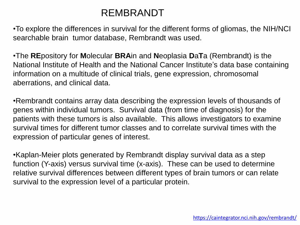
•To explore the differences in survival for the different forms of gliomas, the NIH/NCI
searchable brain tumor database, Rembrandt was used.
•The REpository for Molecular BRAin and Neoplasia DaTa (Rembrandt) is the
National Institute of Health and the National Cancer Institute’s data base containing
information on a multitude of clinical trials, gene expression, chromosomal
aberrations, and clinical data.
•Rembrandt contains array data describing the expression levels of thousands of
genes within individual tumors. Survival data (from time of diagnosis) for the
patients with these tumors is also available. This allows investigators to examine
survival times for different tumor classes and to correlate survival times with the
expression of particular genes of interest.
•Kaplan-Meier plots generated by Rembrandt display survival data as a step
function (Y-axis) versus survival time (x-axis). These can be used to determine
relative survival differences between different types of brain tumors or can relate
survival to the expression level of a particular protein.
https://caintegrator.nci.nih.gov/rembrandt/
REMBRANDT

Patients with astrocytomas (Grade I, II, and III) have a longer mean
survival time than the other patients with gliomas.
To examine the differences in survival between patients with all forms of glioma (blue line) and patients with Grade I, II, or III astrocytomas, the survival times of patients within these two categories were graphed as probability of survival (Y-axis) versus time from diagnosis (x-axis). Those patients with lower grade astrocytomas had significantly higher mean survival times (≥1250 days) than those with all forms of gliomas combined (≥ 600 days).
The all gliomas category represented by the blue line includes: astrocytomas, glioblastomas, mixed, oligodendrogliomas, non-invasive tumors (benign tumors) and brain tumors of unknown origin. https://caintegrator.nci.nih.gov/rembrandt/
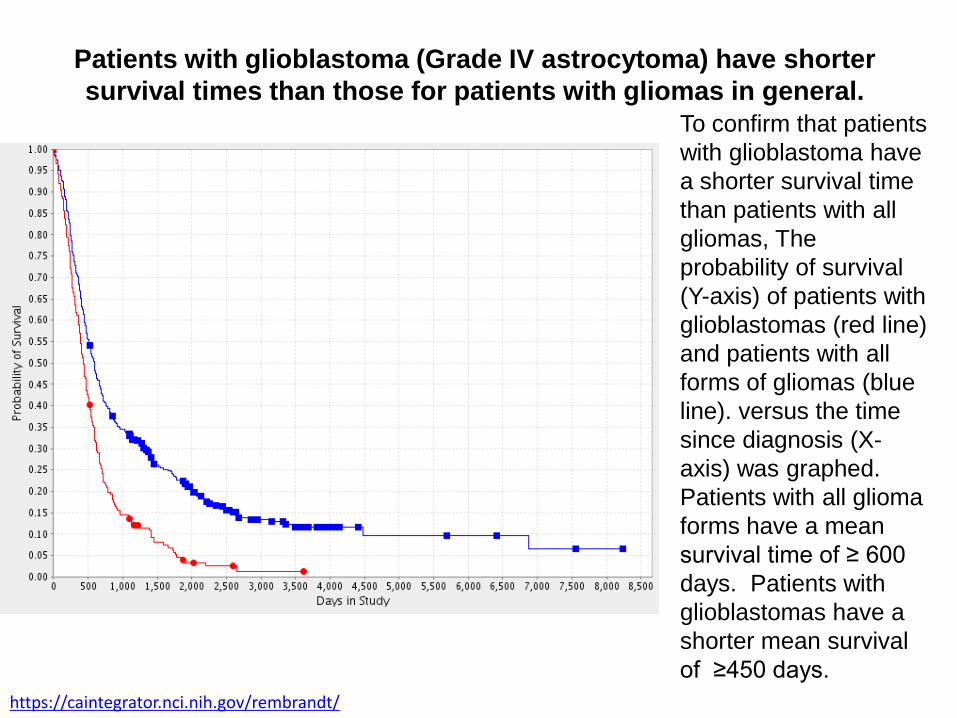
Patients with glioblastoma (Grade IV astrocytoma) have shorter
survival times than those for patients with gliomas in general.
To confirm that patients
with glioblastoma have
a shorter survival time
than patients with all
gliomas, The
probability of survival
(Y-axis) of patients with
glioblastomas (red line)
and patients with all
forms of gliomas (blue
line). versus the time
since diagnosis (X-
axis) was graphed.
Patients with all glioma
forms have a mean
survival time of ≥ 600
days. Patients with
glioblastomas have a
shorter mean survival
of ≥450 days.
https://caintegrator.nci.nih.gov/rembrandt/
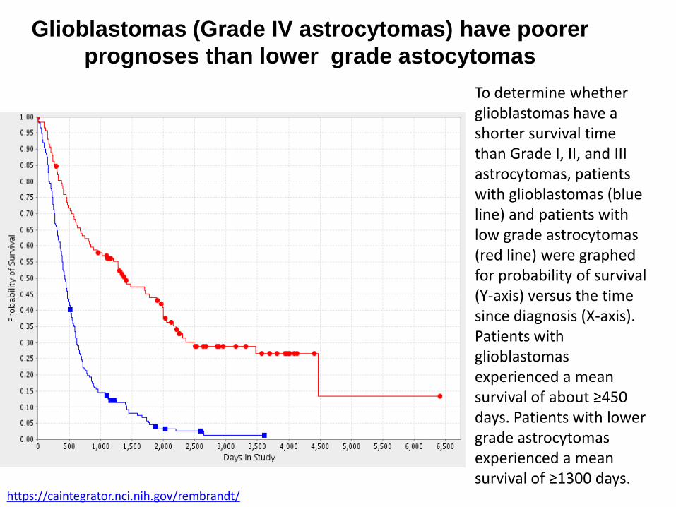
Glioblastomas (Grade IV astrocytomas) have poorer
prognoses than lower grade astocytomas
To determine whether glioblastomas have a shorter survival time than Grade I, II, and III astrocytomas, patients with glioblastomas (blue line) and patients with low grade astrocytomas (red line) were graphed for probability of survival (Y-axis) versus the time since diagnosis (X-axis). Patients with glioblastomas experienced a mean survival of about ≥450 days. Patients with lower grade astrocytomas experienced a mean survival of ≥1300 days.
https://caintegrator.nci.nih.gov/rembrandt/

Expression of the large Interferon-induced GTPase, hGBP-1, in different
tumor types has been correlated with prognosis. In some tumors the
expression of hGBP-1 is correlated with improved survival, while in others
it is correlated with poor prognosis.
To determine whether hGBP-1 expression could be predictive of
prognosis/survival in astrocytomas, Rembrandt was used to generate a
Kaplan-Meier curve correlating hGBP-1 expression levels with patient
survival.
https://caintegrator.nci.nih.gov/rembrandt/
Exploring a role for hGBP-1 in glioblastomas

Elevated hGBP-1 expression in gliomas correlates with
shorter survival
Patients with all forms of gliomas and all levels of hGBP-1 expression (blue) have a median survival at about 500 days. Patients with tumors that expressed low levels of hGBP-1 (yellow) had the longest mean survivals, at about 1500 days. Patients with tumors which over-expressed hGBP1 experienced the poorest survival prognosis, with a median survival of about 475 days.
https://caintegrator.nci.nih.gov/rembrandt/

Most glioblastomas over-express hGBP-1
Our data thus far indicates the:
– patients with glioblastomas have a shorter median survival than patients with other
gliomas.
– patients with tumors that express higher levels of hGBP1 have a poor median
survival than patients with tumors that express lower levels of hGBP1.
– a greater percentage of glioblastomas express high levels of hGBP1 than do low
grade astrocytomas.
Prevalence of elevated levels of hGBP-1 by tumor type
Tumor Type # Tumor Samples # of Tumors Expressingelevated levels of hGBP-1
Percentage of tumorswith elevated levels of hGBP-1
Grade I, II, IIIastrocytomas
105 65 62%
Glioblastomas 181 159 88%
To determine whether the tumors expressing the highest levels of hGBP-1
were glioblastomas, Rembrandt was searched for the tumor type and hGBP-1
level for each patient.

What are Guanylate-Binding Proteins
(GBPs) ?
• GBPs are a family of 67-69 kDa GTPases which are induced by INF- , IFN- , IFN- , TNF α, or IL-1 (reviewed in Vestal 2005).
• GBPs are unique in that unlike other GTPases they hydrolyzeGTP to both GDP and GMP (Neun,R et al 1996)
• Some of the functions of the family member, hGBP-1, are:– It inhibits the proliferation of endothelial cells. It also inhibits the
induction of matrix metalloproteinase-1 (MMP-1), resulting in reduced endothelial cell invasion and tube formation (Naschberger et al
2005)
– Has modest antiviral activity (Yin-ping LU, et al 2007)
– Has antimicrobial activity (Naschberger et al 2006)

hGBP-1 in glioblastomas
• Epidermal Growth Factor receptor (EGFR) amplification and/or mutation is one of the most common genetic changes in glioblastomas.
• EGFR signaling induces the expression of hGBP-1 and MMP-1.
• The induction of hGBP-1 by EGF is required for EGF-induction of MMP-1. The EGF-induced increased hGBP-1 and MMP-1 leads to greater invasiveness and proliferation in glioblastomas.
• However, IFN -induction of hGBP-1 will not induce MMP-1.
• This suggests that hGBP-1 behaves differently in the context of EGF signaling than IFN- signaling.
Ming, Li et al., 2011

Screening of glioblastoma cell lines for IFN-g
and EGF induction of hGBP-1 and MMP-1
Ming, Li et al., 2011
To identify glioblastoma cell
lines in which EGF treatment
induces the expression of both
hGBP-1 and MMP-1, multiple
glioblastoma cell lines were
serum starved for 24 hours
and then left untreated or
treated for 24 hours with either
500 units/ml IFN- or 50 ng/ml
EGF. Western blots examined
the expression of hGBP1 and
MMP-1. Actin was used as a
loading control.
SNB75 cells were the only cell
line in which EGF induced both
hGBP-1 and MMP-1.
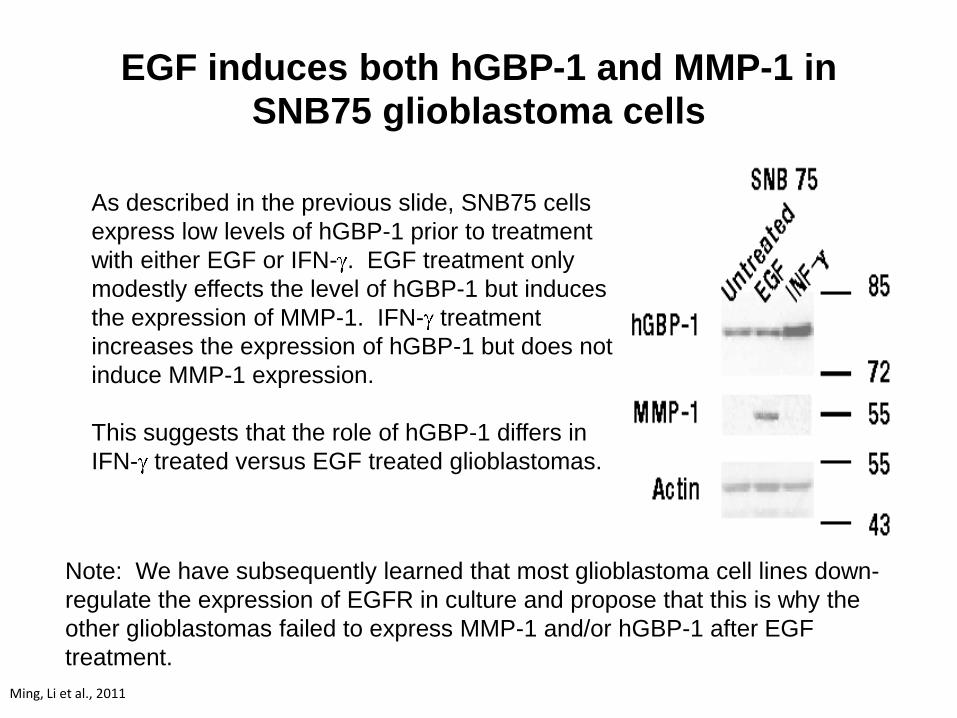
EGF induces both hGBP-1 and MMP-1 in SNB75 glioblastoma cells
Ming, Li et al., 2011
As described in the previous slide, SNB75 cells
express low levels of hGBP-1 prior to treatment
with either EGF or IFN- . EGF treatment only
modestly effects the level of hGBP-1 but induces
the expression of MMP-1. IFN- treatment
increases the expression of hGBP-1 but does not
induce MMP-1 expression.
This suggests that the role of hGBP-1 differs in
IFN- treated versus EGF treated glioblastomas.
Note: We have subsequently learned that most glioblastoma cell lines down-
regulate the expression of EGFR in culture and propose that this is why the
other glioblastomas failed to express MMP-1 and/or hGBP-1 after EGF
treatment.

Intracellular Distribution of hGBP-1 resulting from INF-
or EGF induction
To determine whether the different role for hGBP-1 in EGF signaling is reflected in a difference in its
intracellular distribution, SNB75 cells were plated onto cover slips, serum starved for 24 hours, and then
left untreated or treated for 24 hours with 5000 U/ml hIFN- or 50 ng/ml EGF. The distribution of hGBP-1
was determined with immuno-affinity purified rabbit anti-hGBP-1 and Alexa 488 conjugated goat anti-
rabbit secondary. Nuceli were localized by counter staining with DAPI.
hGBP-1 in IFN- -treated cells exhibits a typical cytosolic distribution with little or
none present in the nucleus (DAPI stain). However, in EGF treated cells the
protein is still found in the cytoplasm but, in addition, exhibits distinct nuclear
localization.

hGBP-1 does not contain a nuclear localization sequence (NLS) and is not
concentrated in the nucleus in IFN- treated cells.
The localization of hGBP-1 in the nucleus of EGF-treated cells suggests that it is
now part of a novel protein complex that moves hGBP-1 to the nucleus. One of
these interacting proteins should possess a NLS.
Future directions: Identification of the protein complex containing hGBP-1 is an
important step toward understanding how EGF induces the expression of MMP-1 in
glioblastomas. Understanding how MMP-1 is induced by EGF and hGBP-1 could
eventually lead to additional therapeutics that will inhibit MMP-1 induction and
improve the outcome for glioblastoma patients.
Significance of the different intracellular
localization of hGBP-1 in EGF-treated SNB75
cells

Up-regulation of hGBP1 in TMZ-resistant glioblastoma
cells • hGBP-1 is up-regulated in paclitaxel resistant
tumor cells, where it contributes to its resistance (ref).
• To determine whether hGBP-1 may also be up-regulated in glioblastomas cells that are resistance to TMZ, U251 glioblastoma cells that are TMZ sensitive or resistant (U251 TMZ) were examined for hGBP-1 by Western blot. Actin was used as a protein loading control.
• hGBP1 is up-regulated in U251 cells as they become resistant to TMZ, suggesting that hGBP-1 may be involved in TMZ resistance.
• Future studies will determine whether knocking down the expression of hGBP-1 in TMZ resistant cells with specific shRNA constructs will restore TMZ sensitivity.

Conclusions
• Patients with glioblastomas have poorer survival than patients with
lower grade astrocytomas or all gliomas.
• Patients with tumors that express high levels of hGBP-1 have poorer
survival than patients with low level expression.
• More glioblastomas express high levels of hGBP-1 than do lower
level astrocyotomas.
• Both IFN- and EGF are capable of inducing hGBP-1, but only EGF
induces MMP-1.
• We propose that hGBP-1 is found in a unique protein complex with a
nuclear localization in EGF treated cells.
• hGBP1 is expressed at much greater levels in TMZ-resistant glioblastomas.

Future Directions
• To determine if the nuclear hGBP-1 in EGF treated cells is associated with a
protein complex on the MMP-1 promoter? This can be done using ChIP
analysis of MMP-1 promoter occupancy.
• To identify the members of the protein complex binding to hGBP-1 in EGF
treated cells? For this hGBP-1 will be immunoprecipitated from EGF
treated cells and the associated proteins determined by mass spec
analysis.
• Determine if over-expressing hGBP-1 in TMZ-sensitive glioblastoma cells
will induce TMZ resistance. Cells can be transfected with an expression
vector for hGBP-1 driven by a powerful promoter and analysis of the
transfected cells for TMZ-mediated cell death.
• Determine if knocking down the expression of hGBP-1 in TMZ-resistant
glioblastomas will restore sensitivity. shRNA constructs against hGBP-1 will
be expressed in TMZ resistant cells and they will be analyzed for TMZ-
induced cell death.

References• Ming, L. et al (2011). Guanylate binding protein 1 is a novel effector of EGFR-driven invasion in
glioblastoma. Journal of Experimental Medicine 208; 2657-2673.
• American Brain Tumor Association. (n.d.). Glioblastoma and Malignant Astrocytoma. Retrieved February 2013, from American Brain Tumor Association: http://www.abta.org/secure/glioblastoma-brochure.pdf
• National Institute of Health. (2005). REMBRANDT. Retrieved February 23, 2013, from National Cancer Institute: <http://rembrandt.nci.nih.gov>
• Wolfgang, W. et al. (2008) New (alternative) temozolomide regimens for the treatment of glioma. Neuro-Oncology URL http://neuro-oncology.dukejournals.org;DOI: 10.1215/15228517-2008-078)
• Yin-ping LU, et al (2007) Antiviral Effect of Interferon-Induced Guanylate Binding Protein-1 against Coxackie Virus and Hepatitis B Virus B3 in Vitro, Virologica Sinica; Vol23, Article ID: 1003-5125(2007)03-0193-06
• Naschberger, E. et al (2006). Human guanylate binding protein-1 is secreted GTPase present in increased concentrations in the cerebrospinal fluic patients with bacterial meningitis. Am J. Pathol 169(3) 1088-1099
• Duan, Z et al (2005 Nov 5); GBP1 over expression is associated with a paciltaxel resistance phenotype. Cancer chemother Pharmacol: 57(1):25-33
• Naschberger, E et al (2005)Human guanylate Binding protein-1 (hGBP-1) characterizes and establishes non-angiogenic endothelial cell activation phenotype in inflammatory diseases, Adv Enzyme Regul 45: 215-227
• Neun R, et al, (1996) GTPase properties of the interferon-induced human guanylate-binding protein 2. FEBS Letters vol 390 issue 1 69-72
• Vestal D, et al. (2011), The Guanylate-Binding Proteins: Emerging insights into the Biochemical Properties and Funcitions of This Family of Large Interferon-Induced Guanosine Triphosphatase, J Interferon Cytokine Res 31(1) 89-97