Embed Size (px)
DESCRIPTION
o
Citation preview
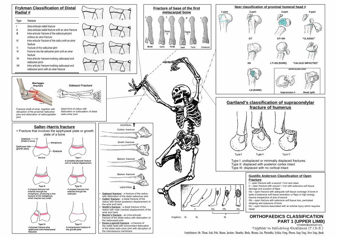
Frykman Classification of Distal Radial #
Galeazzi Fracture
distal third of radius with dislocation or subluxation of distal radio-ulnar joint
Fracture shaft of ulnar, together with disruption of the proximal radioulnar joint and dislocation of radiocapitallar joint
Fracture of base of the first metacarpal bone
Salter–Harris fracture = Fracture that involves the epiphyseal plate or growth
plate of a bone
Type I: undisplaced or minimally displaced fractures. Type II: displaced with posterior cortex intact Type III: displaced with no cortical intact
Gartland’s classification of supracondylar fracture of humerus
• Galeazzi fracture - a fracture of the radius with dislocation of the distal radioulnar joint
• Colles' fracture - a distal fracture of the radius with dorsal (posterior) displacement of the wrist and hand
• Smith's fracture - a distal fracture of the radius with volar (ventral) displacement of the wrist and hand
• Barton's fracture - an intra-articular fracture of the distal radius with dislocation of the radiocarpal joint
• Essex-Lopresti fracture - a fracture of the radial head with concomitant dislocation of the distal radio-ulnar joint with disruption of the interosseous membrane
ORTHOPAEDICS CLASSIFICATION PART 1 (UPPER LIMB)
HTARW5B/GKS2013/3-
Neer classification of proximal humeral head #
GT GT+SN “CLASSIC”
SN LT+SN (RARE) “VALGUS IMPACTED”
LN (RARE) Impression # Head split
1-part 2-part 3-part 4-part
Together In Delivering Excellence (T.I.D.E.) Contributors: Dr. Tham, Goh, Poh, Shaun, Justine, Shanthy, Huda, Miruna, Lin, Phrindha, Syikin, Fong, Phoon, Ling Ying, Siew Ling, Quah
Gustillo Anderson Classification of Open Fracture I – open fracture with a wound <1cm and clean II – open fracture with wound > 1cm with extensive soft tissue damage and avulsion of flaps IIIa – open fracture with adequate soft tissue coverage of bone in spite of extensive soft tissue laceration or flaps or high energy trauma irrespective of size of wound IIIb – open fracture with extensive soft tissue loss, periosteal stripping and exposure of bone IIIc – open fracture associated with an arterial injury which requires repair I II IIIa IIIb IIIc
3L 6L 9L Irrigation:

Letournel classification acetabular # Simple Types
Associated Types
Anterior column Anterior wall Posterior column Posterior wall Transverse
T-type Transverse Posterior column Anterior + posterior Both columns + posterior wall + posterior wall hemitransverse
Pipkin classification of femoral head fracture
Type I - # below fovea/ligamentum (small) Type II - # above fovea/ ligamentum (larger) Type III - type I or II with associated femoral neck # (high risk of AVN) Type IV - type I or II with associated acetabular #
Garden classification of femoral neck #
Evan classification of intertrochanteric #
Russel Taylor classification of subtrochanteric #
Winquist classification of femoral shaft fracture
I. Tiny cortical fragment II. Butterfly fragment is
large but there is still 50%of cortical intact between the main fragments
III. Butterfly fragment involves more than 50% of the bone width
IV. Segmental fractures
Schatzker classification of tibia plateau #
Lateral tibial plateau # w/o depression
Lateral tibial plateau # with depression
Focal depression with no associated split
Medial tibial plateau #, with or without depression
Bicondylar tibial plateau #
Tibial plateau fracture with diaphyseal discontinuity
Sanders classification of calcaneal fractures
I - # are non-displaced # (displacement < 2 mm). II - # consist of a single intrarticular # that divides the calcaneus into 2 pieces.
IIA: # occurs on lateral aspect of calcaneus. IIB: # occurs on central aspect of calcaneus. IIC: # occurs on medial aspect of calcaneus.
III # consist of 2 intrarticular fractures that divide the calcaneus into 3 articular pieces.
IIIAB: 2 # lines are present, 1 lateral and 1 central. IIIAC: 2 # lines are present, 1 lateral and 1 medial. IIIBC: 2 # lines are present, 1 central and 1 medial.
IV # consist of # with more than 3 intrarticular fractures.
ORTHOPAEDICS CLASSIFICATION PART 2 (PELVIC & LOWER LIMB)
HTARW5B/GKS2013/3b-
Lisfranc classification of tarsometatarsal injury
Homolateral Isolated Divergent
Garden I fracture incomplete and minimally displaced
Garden II fracture complete and nondisplaced
Garden III fracture complete and partially displaced
Garden IV fracture complete displaced with no engagement of the 2 principal fragment
Evan I Undisplaced 2 parts fracture
Evan 2 Displaced 2 parts fracture
Evan 3 Displaced 3 parts fracture with posteromedial comminution
Evan 4 Displaced 3 parts fracture with large posteromedial comminuted fragment
Evan 5 Displaced 4 parts fracture with comminution involving both trochanters
lateral
medial
Type IIA Type IIB Type IIC
Type IIIAB Type IIIAC Type IIIBC Type IV
Together In Delivering Excellence (T.I.D.E.) Contributors: Dr. Tham, Goh, Poh, Shaun, Justine, Shanthy, Huda, Miruna, Lin, Phrindha, Syikin, Fong, Phoon,
Ling Ying, Siew Ling, Quah
Together In Delivering Excellence (T.I.D.E.) Contributors: Dr. Tham, Goh, Poh, Shaun, Justine, Shanthy, Huda, Miruna, Lin, Phrindha, Syikin, Fong, Phoon, Ling Ying, Siew Ling, Quah
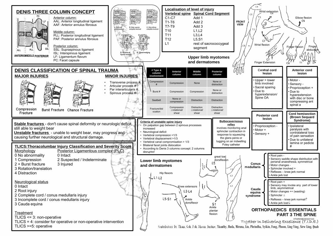
Anterior column: AAL: Anterior longitudinal ligament AAF: Anterior annulus fibrosus Middle column: PLL: Posterior longitudinal ligament PAF: Posterior annulus fibrosus Posterior column: SSL: Supraspinous ligament ISL: Interspinous ligament LF: Ligamentum flavum PC: Facet capsule
DENIS THREE COLUMN CONCEPT
DENIS CLASSIFICATION OF SPINAL TRAUMA MAJOR INJURIES MINOR INJURIES
• Transverse process # • Articular process # • Par interarticularis # • Spinous process #
Stable fractures - don't cause spinal deformity or neurologic deficit, still able to weight bear Unstable fractures - unable to weight bear, may progress and causing further neurological and structural damage.
TLICS:Thoracolumbar Injury Classification and Severity Score Morphology Posterior Ligamentous complex (PLC) 0 No abnormality 0 Intact 1 Compression 2 Suspected / Indeterminate 2 + Burst fracture 3 Injured 3 Rotation/translation 4 Distraction Neurological status 0 Intact 2 Root injury 2 Complete cord / conus medullaris injury 3 Incomplete cord / conus medullaris injury 3 Cauda equina Treatment TLICS <= 3: non-operative TLICS = 4: consider for operative or non-operative intervention TLICS >=5: operative
HTARW5B/GKS2013/4-
Criteria of unstable spine injury • On palpation gap between 2 spinous processes
increased • Neurological deficit • Vertebral compression >1/3 • Vertebral displacement >1/3 • Vertebral canal compromisation > 1/3 • Bilateral facet joints dislocation • According to Denis 3 columns concept: 2 columns
disrupted
Localisation of level of injury Vertebral spine Spinal Cord Segment C1-C7 Add 1 T1-T6 Add 2 T7-T9 Add 3 T10 L1,L2 T11 L3,L4 T12 L5,S1 L1 rest of sacrococcygeal segment
Central cord lesion
• Upper > lower limb involved
• Sacral sparing • Due to
hyperextension/ Spine OA
Anterior cord lesion
• Motor - • Sensory - • Proprioception + • Due to
hyperextension with disc or bone compressing ant spinal a
Posterior cord lesion
• Proprioception - • Motor + • Sensory +
Cord hemisection (Brown Sequard
Syndrome)
• Ipsilateral paralysis with contralateral loss of pain sensation
• Due to unilateral lamina or pedicle #
Conus medullaris
• Root pain - • Sensory saddle shape distribution with
perianal anaesthesia, symmetrical • Motor changes - • Sphincter involved + • Reflexes – knee jerk normal • Ankle jerk lost
Cauda equina
syndrome
• Root pain + • Sensory may invoke any part of lower
limb, asymmetrical • Motor changes ++ (wasting) • Sphincter ± • Reflexes – knee jerk normal/ • Ankle jerk lost/
ORTHOPAEDICS ESSENTIALS PART 3 THE SPINE
C6
T1
# Type & column
involvement
Anterior column
Middle column
Posterior column
Compression # Compression None None or
distraction
Burst # Compression Compression None or distraction
Seatbelt None or compression Distraction Distraction
Fracture/dislocation
Compression ±rotational/
shear
Distraction ±rotational/
shear
Distraction ±rotational/
shear
Lower limb myotomes and dermatomes
Upper limb myotomes and dermatomes
Elbow flexion
Wrist extension
Wrist flexion Finger flexion
Finger Extension
Finger abduction
Hip flexors
Knee extensors
Ankle dorsiflexor
great toe dorsiflexion
Ankle plantar flexion
Bulbocavernosus reflex
involves monitoring anal sphincter contraction in response to squeezing
the glans penis or tugging on an indwelling
Foley catheter

Together In Delivering Excellence (T.I.D.E.) Contributors: Dr. Tham, Goh, Poh, Shaun, Justine, Shanthy, Huda, Miruna, Lin, Phrindha, Syikin, Fong, Phoon, Ling Ying, Siew Ling, Quah
ORTHOPAEDICS ESSENTIALS PART 4 DRESSINGS
HTARW5B/GKS2013/4-
Name Active Ingredient Indication Contraindication Advantage Disadvantage 1. Opsite semi-permeable-thin, adhesive
transparent polyurethrane film superficial wounds as secondary dressing
highly exudative wounds • some moisture evaporation • reduce pain • barrier to external contamination • allows inspection
• exudate may pool • maybe traumatic to remove
2. Jelonet Bactigras PARAFFIN
non-adherent moist (Tulle Gras dressing) gauze impregnated with paraffin or maybe with antiseptics or antibiotics
burn wounds healing by secondary intention
allergy
• reduces adhesion to wound • moist environment aids healing
• does not absorb exudate • requires secondary dressingg • allergy • may delay healing when
impregnated
3. Kaltostat CALCIUM ALGINATE
Calcium alginate natural polysaccharide from seaweed
moderately/highly exudative wounds need for hemostasis
dry wound hard eschar
• forms gel on wound & hence moist • environment • reduces pain • can pack cavities • absorbent in exudative wounds • promotes hemostasis • low allergenic
• may require secondary dressing • not recommended in anearobic
infections • gel can be confused with slough • or pus in wound
4. Duoderm E HYDROCOLLOID
hydrocolloid dressing-hydrophilic colloid bound to polyurethrane film coated with adhesive mass
burn (small) abrasions mildly exudating ulcers donate moisture & absorb exudates
dry wound infection full thickness wound
• retains moisture • painless removal • facilitate autolytic debridement • thermal insulation • worn for 3-5days-fewer dressing
changes
• avoid on high exudate wounds, sinus tracts
• fragile skin
5. Duoderm Hydroactive HYDOGEL
hydogel - water or glycerin-based 80-99% water on a nonadherent, cross-linked polymer
pressure ulcer stage II-IV, partial & full thickness wound dermabrasion, painful wound dermal ulcer, radiation burn donor sites necrotic wounds
heavily draining wound
• rehydrate the wound bed • reduce pain • used on infected wound with • topical medication • promote autolytic debridement
• need 2ndary dressing • avoid heavily draining wound • absorptive properties may macerate • periwound skin
5. Aquacel HYDROFIBER SODIUM CARBOXY- METHYLCELLULOSE
soft, sterile, nonwoven pad or ribbon with sodium carboxymethylcellulose
moderate to heavily draining wound partial & fully thickness wound pressure ulcer (stage III & IV) surgical wound, donor site dehisced wound, cavity wound wounds with sinus tracts or tunnels
dry eschar non-exudating wound 3rd degree burn heavy bleeding
• retains moisture • absorb & retain exudate & harmful • components • do not damage tissues surrounding • exudating wound when dressing
changes • removal trauma free • reduce dead space • no frequent change
• dressing non-adherent, need • 2ndary dressing to secure it
6. Aquacel Ag SILVER
ionic silver for immediate and controlled release
infected/highly colonized wound partial thickness (2nd degree) burn DFU, leg ulcers traumatic wound wounds prone to bleeding oncology wounds with exudate
stage I pressure ulcers 3rd degree burn non-exudating wounds
• inhibit pathogen growth, especially • antibiotic-resistant strains • effective antimicrobial action up to 7
days
• 2ndary dressing to secure silver dressing
• allergy • not to use with topical medication • silver turns black when oxidizes, may • stain or discolor periwound tissue
7. Elase FIBRINOLYSIN DESOXYRIBONUCLEASE
fibrinolysin desoxyribonuclease
enzymatic debridement of necrotic tissue in wound & liquefaction & dissolution of exudates of injured skin & mucous membrane
allergic to bovine compound
• allergy
Together In Delivering Excellence (T.I.D.E.)
ORTHOPAEDICS ESSENTIALS PART 5 PLATINGS, NAILS AND SCREWS
HTARW5B/GKS2013/4-
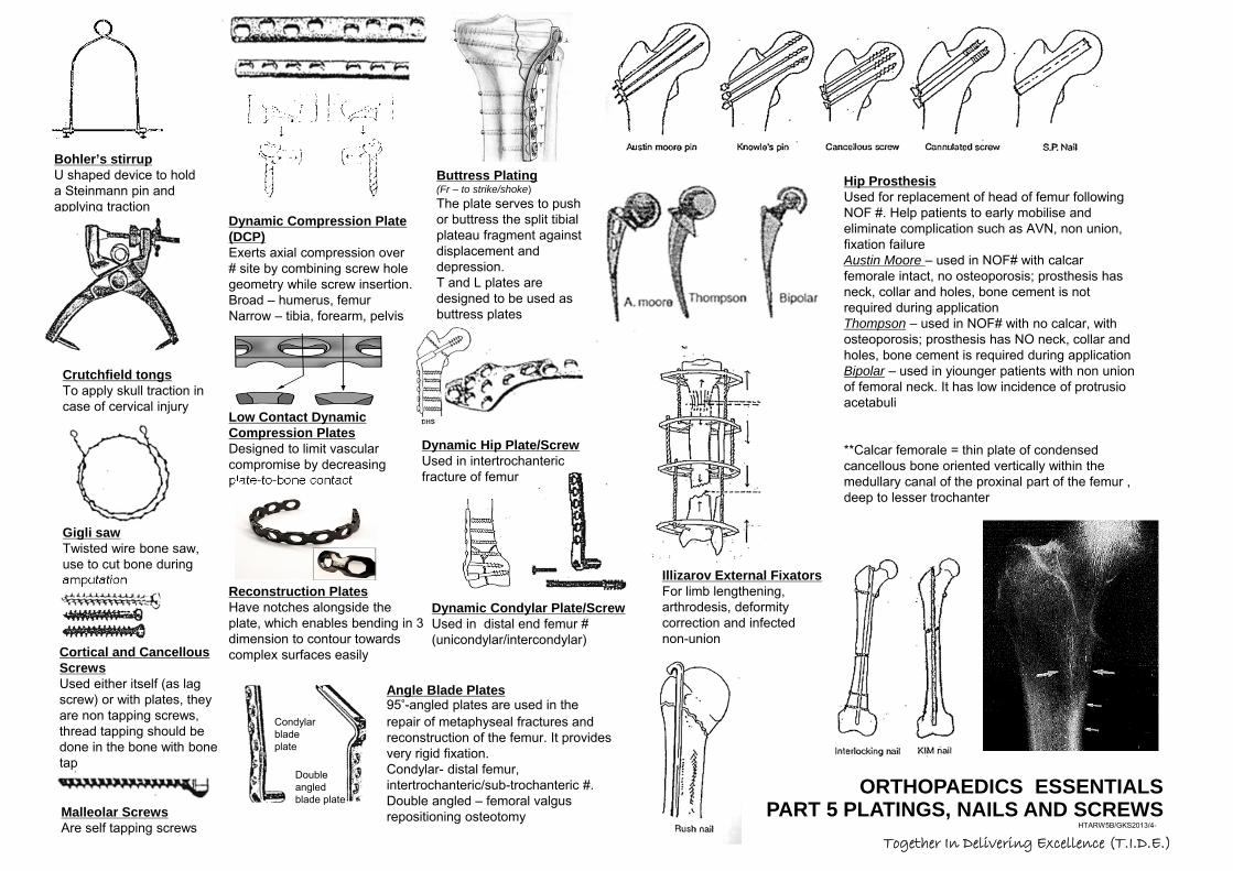
Bohler’s stirrup U shaped device to hold a Steinmann pin and applying traction
Gigli saw Twisted wire bone saw, use to cut bone during amputation
Crutchfield tongs To apply skull traction in case of cervical injury
Dynamic Compression Plate (DCP) Exerts axial compression over # site by combining screw hole geometry while screw insertion. Broad – humerus, femur Narrow – tibia, forearm, pelvis
Low Contact Dynamic Compression Plates Designed to limit vascular compromise by decreasing plate-to-bone contact
Reconstruction Plates Have notches alongside the plate, which enables bending in 3 dimension to contour towards complex surfaces easily
Buttress Plating (Fr – to strike/shoke) The plate serves to push or buttress the split tibial plateau fragment against displacement and depression. T and L plates are designed to be used as buttress plates
Angle Blade Plates 95°-angled plates are used in the repair of metaphyseal fractures and reconstruction of the femur. It provides very rigid fixation. Condylar- distal femur, intertrochanteric/sub-trochanteric #. Double angled – femoral valgus repositioning osteotomy
Condylar blade plate
Double angled blade plate
Dynamic Hip Plate/Screw Used in intertrochanteric fracture of femur
Dynamic Condylar Plate/Screw Used in distal end femur # (unicondylar/intercondylar)
Malleolar Screws Are self tapping screws
Hip Prosthesis Used for replacement of head of femur following NOF #. Help patients to early mobilise and eliminate complication such as AVN, non union, fixation failure Austin Moore – used in NOF# with calcar femorale intact, no osteoporosis; prosthesis has neck, collar and holes, bone cement is not required during application Thompson – used in NOF# with no calcar, with osteoporosis; prosthesis has NO neck, collar and holes, bone cement is required during application Bipolar – used in yiounger patients with non union of femoral neck. It has low incidence of protrusio acetabuli **Calcar femorale = thin plate of condensed cancellous bone oriented vertically within the medullary canal of the proxinal part of the femur , deep to lesser trochanter
Cortical and Cancellous Screws Used either itself (as lag screw) or with plates, they are non tapping screws, thread tapping should be done in the bone with bone tap
Illizarov External Fixators For limb lengthening, arthrodesis, deformity correction and infected non-union
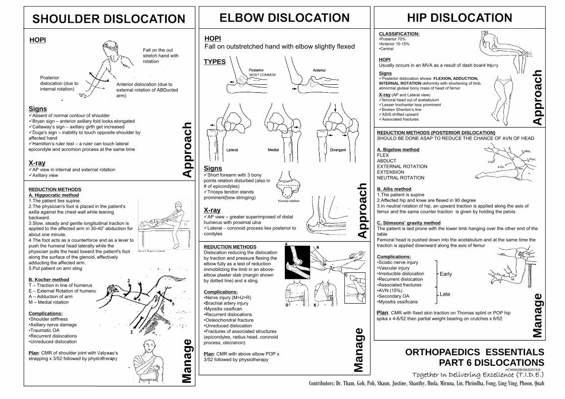
REDUCTION METHODS A. Hippocratic method 1.The patient lies supine. 2.The physician's foot is placed in the patient's axilla against the chest wall while leaning backward. 3.Slow, steady and gentle longitudinal traction is applied to the affected arm in 30-40° abduction for about one minute. 4.The foot acts as a counterforce and as a lever to push the humeral head laterally while the physician pulls the head toward the patient's foot along the surface of the glenoid, effectively adducting the affected arm. 5.Put patient on arm sling
B. Kocher method T – Traction in line of humerus E – External Rotation of humerus A – Adduction of arm M – Medial rotation Complications: •Shoulder stiffness •Axillary nerve damage •Traumatic OA •Recurrent dislocations •Unreduced dislocation
Plan: CMR of shoulder joint with Velpeau’s strapping x 3/52 followed by physiotherapy
Together In Delivering Excellence (T.I.D.E.) Contributors: Dr. Tham, Goh, Poh, Shaun, Justine, Shanthy, Huda, Miruna, Lin, Phrindha, Fong, Ling Ying, Phoon, Quah
ORTHOPAEDICS ESSENTIALS PART 6 DISLOCATIONS
HTARW5B/GKS2013/4-
App
roac
h M
anag
e
HOPI Fall on the out stretch hand with rotation
Anterior dislocation (due to external rotation of ABDucted arm)
Posterior dislocation (due to internal rotation)
Signs Absent of normal contour of shoulder Bryan sign – anterior axillary fold looks elongated Callaway’s sign – axillary girth get increased Duga’s sign – inability to touch opposite shoulder by affected hand Hamilton’s ruler test – a ruler can touch lateral epicondyle and acromion process at the same time
X-ray AP view in internal and external rotation Axillary view
HOPI Fall on outstretched hand with elbow slightly flexed
TYPES
MOST COMMON
Signs Short forearm with 3 bony points relation disturbed (also in # of epicondyles) Triceps tendon stands prominent(bow stringing)
X-ray AP view – greater superimposed of distal humerus with proximal ulna Lateral – coronoid process lies posterior to condyles A
ppro
ach
REDUCTION METHODS Dislocation reducing the dislocation by traction and pressure flexing the elbow fully as a test of reduction immobilizing the limb in an above-elbow plaster slab (margin shown by dotted line) and a sling. Complications: •Nerve injury (M>U>R) •Brachial artery injury •Myositis ossifican •Recurrent dislocations •Osteochondral fracture •Unreduced dislocation •Fractures of associated structures (epicondyles, radius head, coronoid process, olecranon)
Plan: CMR with above elbow POP x 3/52 followed by physiotherapy
Man
age
CLASSIFICATION: •Posterior 70% •Anterior 10-15% •Central HOPI Usually occurs in an MVA as a result of dash board injury
Signs Posterior dislocation shows: FLEXION, ADDUCTION, INTERNAL ROTATION deformity with shortening of limb, abnormal gluteal bony mass of head of femur
X-ray (AP and Lateral view) femoral head out of acetabulum Lesser trochanter less prominent Broken Shenton’s line ASIS shifted upward Associated fractures A
ppro
ach
REDUCTION METHODS (POSTERIOR DISLOCATION) SHOULD BE DONE ASAP TO REDUCE THE CHANCE OF AVN OF HEAD A. Bigelow method FLEX ABDUCT EXTERNAL ROTATION EXTENSION NEUTRAL ROTATION B. Allis method 1.The patient is supine 2.Affected hip and knee are flexed in 90 degree 3.In neutral rotation of hip, an upward traction is applied along the axis of femur and the same counter traction is given by holding the pelvis. C. Stimsons’ gravity method The patient is laid prone with the lower limb hanging over the other end of the table Femoral head is pushed down into the acetabulum and at the same time the traction is applied downward along the axis of femur Complications: •Sciatic nerve injury •Vascular injury •Irreducible dislocation •Recurrent dislocation •Associated fractures •AVN (15%) •Secondary OA •Myositis ossificans
Early Late
Man
age
Plan: CMR with fixed skin traction on Thomas splint or POP hip spika x 4-6/52 then partial weight bearing on crutches x 6/52

Together In Delivering Excellence (T.I.D.E.) Contributors: Dr. Tham, Goh, Poh, Shaun, Justine, Shanthy, Huda, Miruna, Lin, Phrindha, Fong, Phoon, Quah
ORTHOPAEDICS ESSENTIALS PART 7 (a) ANGLES IN ORTHOPAEDICS (b) DIABETIC FOOT
HTARW5B/GKS2013/4-
Calcaneum Fracture
Q angle Increased in genu valgum, external tibia torsion, lateral positioned tibial tuberosity, tight lateral retinaculum Norm: male= 14±3 females= 17±3
Baumann’s angle
Norm: 5-15 Excessive = cubitus valgus Decrease = gunstock deformity
Carrying angle
Supracondylar Fracture Wagner Classification of Diabetic Foot Ulcers: Grade 0: No ulcer in a high risk foot. Grade 1: Superficial ulcer involving the full skin thickness but not underlying tissues. Grade 2: Deep ulcer, penetrating down to ligaments and muscle, but no bone involvement or abscess formation. Grade 3: Deep ulcer with cellulitis or abscess formation, often with osteomyelitis. Grade 4: Localized gangrene (forefoot). Grade 5: Extensive gangrene involving the whole foot.
**Rays Amputation – Removal of toes with metatarsal from tarsometatarsal joint
Amputations
Cobb’s angle (Scoliosis)
x
1- Radial height 11mm (10-26)
2- Radial incline = 22 (12-28)
3- DRUJ space 4mm 4- Ulnar variance ±5mm
5-Volar tilt = 11 (3-16) 6- Step <2mm
7- Gap <2mm
Distal end radius fracture

Together In Delivering Excellence (T.I.D.E.)
ORTHOPAEDICS ESSENTIALS PART 8 SPLINTS, CASTS, PLASTERS, FRAMES
HTARW5B/GKS2013/4-
WELL LEG TRACTION Used in correction of abduction deformity of hip Traction is applied to the normal limb while deformed hip is stabilised by splint
90-90 TRACTION Used in subtrochanteric #, compound # of femur with posterior wound and shaft femur of children
GALLOW’S/BRYANT’S TRACTION Used in femur shaft # in children <2 yrs
HAMILTON-RUSSELL TRACTION Used in femur shaft # in adult, trochanteric #
PERKINS TRACTION Used in femur shaft # in adult
BUCK’S TRACTION Apply skin traction in femur shaft #, NOF #, acetabulum # after reduction of hip dislocation, to correct minor deformity of hip and knee
CALCANEAL TRACTION Used in open # of ankle joint/leg
BÖHLER BRAUN FRAME Apparatus used for application of skeletal traction of lower limb. It may be used with transcondylar, tibial or calcaneal pins
Uppe
r lim
b an
d Sp
ine
HEAD HALTER TRACTION Used in cervical spine injury
Low
er li
mb
and
Pelv
is
Mis
cella
neou
s
DUNLOP TRACTION Used in transcondylar or supracondylar fracture of the humerus in children
HALO-PELVIS TRACTION Used in scoliosis
HAMMOCK TRACTION Used in pelvis # with rotational instability e.g. open book, Malgaigne # (both pubic rami+ posterior SI complex/sacrum #), and bucket handle injury
CRUSH FIELD TRACTION Used in cervical spine injury
THOMAS SPLINT (with and without sling) Temporary stabilisation of femoral shaft fracture
BALKAN FRAME A frame employed in the treatment of fractured bones of extremities that provides overhead weights and pulleys for suspension, traction, and continuous extension of the splinted fracture limb.
PELVIC BINDER Used acutely in management of exsanguinating pelvic trauma, by applying large amount of compressive force to the pelvic ring to reduce the volume of the pelvis
Trapeze
ROBERT JONES DRESSING A tape stirrup is applied to the foot before the bandage is started Cotton roll is wrapped around a forelimb after the application of the tape stirrup. Elastic gauze is then applied firmly bind the cotton to the leg. Elastic tape is then use to complete the dressing.
POP CAST & SLAB Active ingredient of Plaster of Paris is Gypsum CaSO42H2O Slab only covers a part of circumferential of a limb whereas a cast covers whole of the limb circumference.
BROAD ARMSLING AND FIGURE ‘8’ STRAPPING Used in undisplaced and displaced clavicle fracture respectively.
Cast Slab
SCAPHOID CAST Applied from below elbow proximal to knuckle distally and incorporating proximal phalanx of thumb. The wrist is held in dorsiflexion (glass holding positiion)
90-90 HIP SPIKA Spika at 90 flexion at hip because in children proximal fragment flexes to 90 due to stronger pull by flexor muscle and illdevelopment of lumbar lordosis
MILWAUKEE BRACE For dorsal scoliosis
BOSTON BRACE For lumbar scoliosis
Together In Delivering Excellence (T.I.D.E.) Contributors: Dr. Goh, Poh, Shaun, Justine, Shanthy, Huda, Miruna, Lin, Maya, Fong, Ling Ying, Phoon, Quah
ORTHOPAEDICS ESSENTIALS PART 9 ORTHOPAEDIC EMERGENCIES
HTARW5B/GKS2013/4-
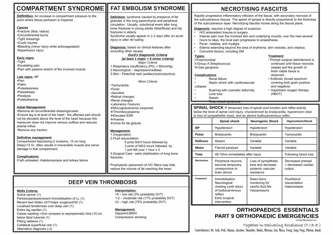
Definition: An increase in compartment pressure to the point where tissue perfusion is impaired. Causes •Fracture (tibia, radius) •Circumferential burns •Tight dressings •Crush injuries •Bleeding (minor injury while anticoagulated) •Reperfusion injury
Early signs •Tight •Escalating pain •Pain with passive stretch of the involved muscle Late signs -6P •Pain •Pallor •Pulselessness •Paresthesia •Paralysis •Poikilothermia Initial Management •Remove all circumferential dressings/casts •Ensure leg is at level of the heart - the affected part should not be elevated above the level of the heart because this maneuver does not improve venous outflow and reduces arterial inflow •Remove any traction Definitive management Compartment fasciotomy-2 incisions, 15 cm long Delay>12 hr. often results in irreversible muscle and nerve damage in that compartment Complications If left untreated: rhabdomyolysis and kidney failure
Definition: syndrome caused by presence of fat globules in the lung parenchyma and peripheral circulation. Usually subclinical event after long bone fractures in young adults (tibia/fibula) and hip fractures in elderly Syndrome usually appear in 1-2 days after an acute injury or after IM nailing. Diagnosis: based on clinical features after excluding other causes
Gurd's Diagnostic Criteria (at least 1 major + 4 minor criteria)
Major Criteria 1.Respiratory insufficiency (PO2 < 60mmHg) 2.Neurological – depression/restless 3.Skin - Petechial rash (axillary/subconjuctiva)
Minor Criteria •Tachycardia •Fever •Jaundice •Retinal changes •Renal changes •Laboratory Features Microglobulinemia (required) Thrombocytopenia Elevated ESR Anemia Urine for fat globule Management: 1.Oxygenation. 2.Fluid resuscitation
6 pints NS/3 hours followed by 3 pints of NS/2 hours followed by 1 pint NS over 1 hour x 3
3.Surgical Care - early stabilization of long bone fractures Prophylactic placement of IVC filters may help reduce the volume of fat reaching the heart.
Rapidly progressive inflammatory infection of the fascia, with secondary necrosis of the subcutaneous tissues. The speed of spread is directly proportional to the thickness of the subcutaneous layer. Necrotizing fasciitis moves along the fascial plane.
Diagnosis: requires a high degree of suspicion • H/O antecedent trauma or surgery • Intense pain over the involved skin and underlying muscle; over the next several
hours to days, the local pain progresses to anaesthesia. • Fever, malaise, and myalgia • Edema extending beyond the area of erythema, skin vesicles, and crepitus. • Comorbid factors, including DM Types: I-Polymicrobial II-Group A Streptococcus III-Gas gangrene Complications • Renal failure • Septic shock with cardiovascular collapse • Scarring with cosmetic deformity • Limb loss • Sepsis • Toxic shock syndrome
Treatment • Prompt surgical debridement is
continued until tissue necrosis ceases and the growth of fresh viable tissue is observed.
• Antibiotic (broad spectrum covering both gram positive and negative)
• Hyperbaric oxygen therapy (HBOT)
SPINAL SHOCK = temporary loss of spinal cord function and reflex activity below the level of spinal cord injury, characterised by bradycardia, hypotension (due to loss of sympathethic tone), and an absent bulbocarvenosus reflex Spinal shock Neurogenic Shock HypovolemicShock
BP Hypotension Hypotension Hypotension
Pulse Bradycardic Bradycardic Tachycardic
Reflexes Absent Variable Variable
Motor Flaccid paralysis Variable Variable
Time 48-72hrs immediately after injury Following blood loss
Mechanism Peripheral neurons become temporary unresponsive to brain stimuli
Loss of sympathetic tone and decrease systemic vascular resistance
Decreased preload = decreased cardiac output
Treatment Immobilisation Neurological charting (until return of bulbocarvenous reflex) Early surgical intervention
Swan-Ganz monitoring for careful fluid Mx Vasopressors
Fluid/blood resuscitation Haemostasis
Wells Criteria: Active cancer (1) Paresis/paralysis/recent immobilisation of LL (1) Recent bed ridden x3/7/major surgeryx4/52 (1) Localised tenderness over deep vein (1) Entire leg swollen (1) Calves swelling >3cm compare to asymptomatic limb (10 cm below tibial tubercle (1) Pitting oedema (1) Collateral superficial vein (1) Alternative diagnosis (-2)
Interpretation: <0 – low risk (3% probability DVT) 1-2 – moderate risk (17% probability DVT) ≥3 – high risk (75% probability DVT) Management: Heparin/LMWH Compression stocking