Embed Size (px)
DESCRIPTION
instability
Citation preview

SHOULDER INSTABILITY
Definition
Anatomy
Biomechanics of GH instability
Types of instability (Recurrent)
Evaluation of Recurrent
Treatment - brief

Shoulder Instability
• DEFINITION:– Glenohumeral instability is the inability to
maintain the humeral head in the glenoid fossa.

Bony Anatomy

Shoulder Girdle
• Sternum– pivot/anchor
• clavicle– strut
• scapula– lever & pulley
• humerus

Scapula
• Glenoid fossa– poorly shaped– 7 deg. of retroversion– 5 deg. of sup tilt
• Acromium
• Coracoid
• Plane of the scapula - 30-45 deg to coronal

Joints
• SC – only bony attachment to the axial skeleton
• AC– 3 degres of freedom
• scapular rotation
• scapular winging
• scapular tipping
– ligaments: AC and CC ligs

Glenohumeral joint
• Humeral head 3x larger than glenoid fossa
• Ball and socket with translation
• 3 degrees of freedom– flex/ext– abd/add– int/ext rot– plus
• horizontal flex/ext• horizontal abd/add

Scapulothoracic Articulation
• Elevation/Depression• Pro/retraction• up/downward rotation
– scapular rotation is necessary to keep GH joint in position of max stability

Musculature
Rotator cuff muscles (S.I.T.S.)
Biceps tendon, long head
- secondary stabilizer head depressor
Deltoid
- primary mover of shoulder in flexion and abduction
Periscapular muscles
- help position scapula and orient glenohumeral joint - contributes compressive force across joint
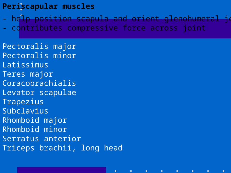
Periscapular muscles
- help position scapula and orient glenohumeral joint - contributes compressive force across joint
Pectoralis major Pectoralis minor Latissimus Teres major Coracobrachialis Levator scapulae Trapezius Subclavius Rhomboid major Rhomboid minor Serratus anterior Triceps brachii, long head

Ligaments and capsule
• Coracoacromial ligament– secondary stabilizer as it forms part of the
coracoacromial arch

• Coracohumeral ligament
• Origin: anterolateral coracoid process Insertion: greater and lesser tuberosities, blends with capsule in rotator interval
• Unclear role in providing stability; may contribute to restraining inferior subluxation with arm at side, Becomes taut with external rotation

• Capsule– attached medially to margin of glenoid fossa– laterally to circumfrence of anatomical neck of
humarus– ant cap thicker than post cap– 3 types
• anterior labrial attachment
• just medial to labrum
• further medially on glenoid

• Allows for 2-3 mm of distraction
• little contribution to joint stability
• strengthened by GH ligs and RC tendons
• rotator interval– between SGHL and MGHL

Glenohumeral ligaments (superior, middle , inferior)
• SGHL– O = tubercle on glenoid just post to long head
biceps– I = humeral head near upper end of lesser
tubercle– Resists inf subluxation and contributes to
stability in post and inf directions

• MGHL– O= sup glenoid and labrum– I= blends with subscapularis tendon– Limits ant excursion instability especially with
arm in 45 deg abd position – limits ext rotation

• IGHL– O= ant glenoid rim and labrum– I= inf aspect of humeral articular surface and
anatomic neck– 3 bands, anterior, axillary and posterior– IGHL complex acts like a sling and when is the
most important single ligamentous stabilizer in the shoulder.
– Primary restraint is at 45-90 deg abd.

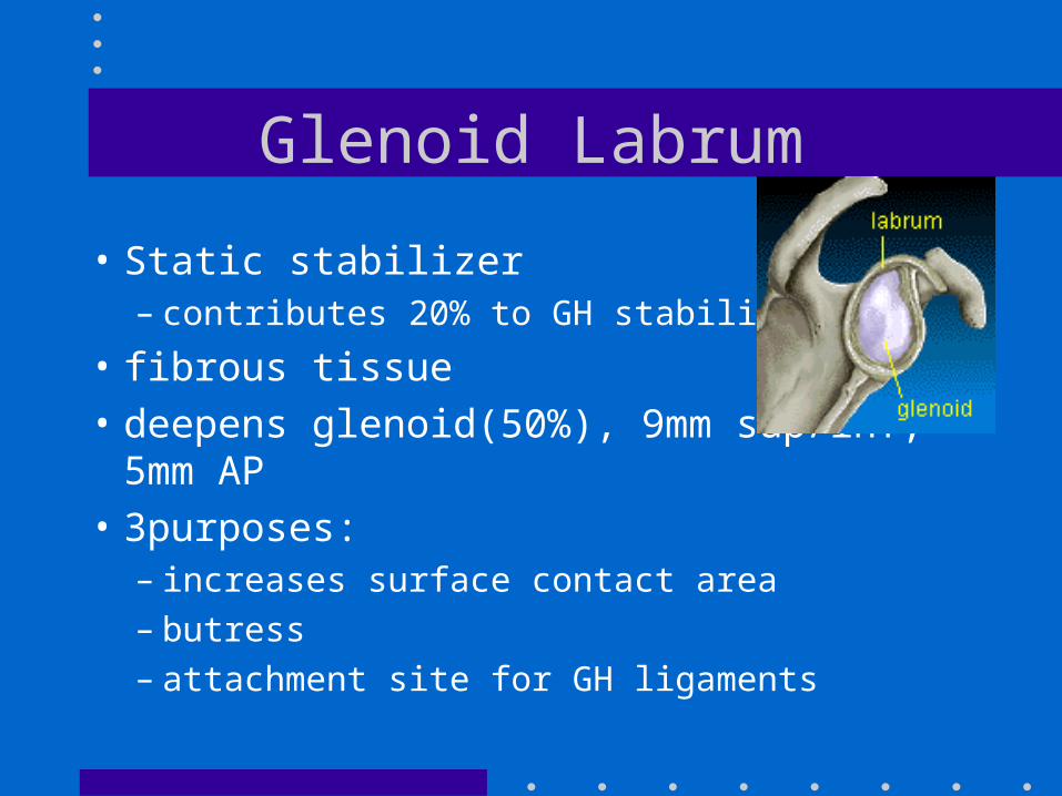
Glenoid Labrum
• Static stabilizer– contributes 20% to GH stability
• fibrous tissue
• deepens glenoid(50%), 9mm sup/inf, 5mm AP
• 3purposes:– increases surface contact area– butress– attachment site for GH ligaments

Biomechanics of GH stability
• the normal shoulder precisely constrains the humeral head to the center of the glenoid cavity throughout most of the arc of movement.

Static restraints
•negative intra-articular pressure
(venting capsule increases inferior translation at 0 degrees of abduction)
•ligaments and capsule
•labrum (increases concavity)
•articular surfaces/osseous anatomy
(very little because square area of humeral head is 3X glenoid)
•joint fluid adhesiveness
•suction cup
•limited joint volume

Dynamic restraints
• Rotator cuff muscles
• deltoid and biceps
• concavity compression

- The humeral head will remained centered in the glenoid fossa if
the glenoid and humeral joint surfaces are congruent and if the net
humeral joint reaction force is directed within the effective glenoid arc.

-The glenohumeral joint will not dislocate as long as the net
humeral joint reaction force is directed within the effective glenoid arc.
The maximal angle that the net humeral joint reaction force can make with
the glenoid center line in a given direction is the balance stability angle

Increasing the force of contraction of a muscle whose force direction
is close to the glenoid center line, the direction of the net humeral joint
reaction force can be aligned more closely with the glenoid fossa.
The elements of the rotator cuff are well positioned to contribute to this
muscle balance.

Stability ratio
• Maximal displacing force in a given direction(perpendicular to glenoid center line) that can be stabilized by compressive load.– Affected by
• Glenoid/labrum depth
• rim lesions
• Glenoid version
• dynamic stabilizer compromise– structural injury, paralysis, imbalance, atrophy etc..


Glenoid version

Scapular positioning

Ligamentous stabilization
• Check reins– ballanced force exceeds ballanced stability
angle.

Countervailing force-compresses humeral head into glenoid fosssa and resists displacement in direction of tight ligament

Types of instability
• Congenital
• Acute
• Cronic
• Recurrent
• Traumatic
• Atraumatic
Recurrent Instability
• Two groups:– TUBS
• Traumatic, unidirectional, Bankart, surgery
– AMBRII• Atraumatic(microtraumatic)
multidirectional,bilateral, rehab,inferior capsular shift,rotator interval.

Who?Atraumatic Traumatic

Combined Atraumatic and traumatic

Why?

Directions of instability
• Anterior– 97% of recurrent dislocations
• subcoracoid - abd, extension and external rotation
• subglenoid
• subclavicular
• intrathoracic

• Posterior– 3% of recurrent– Seizures, shock, fall on flexed + adducted arm
• subacromial• subglenoid• subspinous
• Inferior
• Superior
• Bilateral
Evaluation of recurrent atraumatic instability
• History– Trauma?– Sports– Throwing or overhead activitys– Voluntary subluxation– “Clunk” or knock– Fear– Hx of dislocations and energy associated

• Physical– Demonstrate dislocation/subluxation ?– Laxity tests– Stability tests

• Laxity tests– Drawer
– Sulcus
– Push - pull
• Stability tests– Fulcrum
– Apprehension (crank )
– Jerk
• Strength tests• X-ray, arthrogram,
MRI, arthroscope no help.

Evaluation of recurrent traumatic dislocations
• Injury to capsule, rot cuff, labrum, glenoid, humerus.
• Young (14-34)
• Male

• History– HX of 1st dislocation or injury– Subsequent dislocations/subluxations
• Physical– same tests– concentrate on area of capsule weakness– “fatigue tests”– Be prepared to reduce

• X-Rays– Identify Bankart, Hill-Sachs
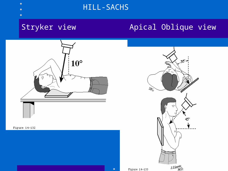
Stryker view Apical Oblique view
HILL-SACHS

West Point Axillary view
GLENOID

• Arthrogram
• MRI
• Ultrasound
• Arthroscopy - not necessary
TREATMENT
• Recurrent Traumatic Ant. Dislocation
– Surgical stabilization• Open or arthroscopic
– Poor response to non operative tx

• Recurrent Traumatic Posterior instability
– First line = non - operative (strengthening)
– Failure of surgical stabilization = 12 - 50%

• Atraumatic Instability
– 80% respond to physio
– Surgical stabilization - capsulorraphy if non - operative fails.

• Voluntary or Habitual
– Retrain muscles
– No surgery

• Multidirectional Instability– Surgery only if non-operative fails– Surgery - capsulorraphy (approach depends on
main direction of instability)• Latreral capsular shift (humoral side)- 91% success
• Medial capsular shift (glenoid side) fo associated BanKart

• Ant. Glenoid rim deficiency.
• Major glenoid bone loss.
• Impingement secondary to instability.
• Elderly
• Bristow.
• Bone butress procedure.
• Treat instability.
• Rot. Cuff repair.