Embed Size (px)
Citation preview
1
Short title: Translation recovery after heat shock 1 2 Corresponding authors: 3 Rémy MERRET 4 CNRS-LGDP UMR 5096, 58 av. Paul Alduy 66860 Perpignan, France 5 6 Yee-yung CHARNG 7 Agricultural Biotechnology Research Center, Academia Sinica, 128 Academia Rd. Sec. 2, 8 Taipei, Taiwan 11529, ROC 9 10 Article title: 11 Heat-shock protein HSP101 affects the release of ribosomal protein mRNAs for recovery 12 after heat shock 13 14 Authors: 15 Rémy MERRET1,2,3, Marie-Christine CARPENTIER2,3, Jean-Jacques FAVORY2,3, Claire 16 PICART2,3, Julie DESCOMBIN2,3, Cécile BOUSQUET-ANTONELLI2,3, Pascal TILLARD4, 17 Laurence LEJAY4, Jean-Marc DERAGON2,3,5, and Yee-yung CHARNG1 18 19 1Agricultural Biotechnology Research Center, Academia Sinica, 128 Academia Rd. Sec. 2, 20 Taipei, Taiwan 11529, ROC 21 2CNRS-LGDP UMR 5096, 58 av. Paul Alduy 66860 Perpignan, France 22 3Université de Perpignan Via Domitia, LGDP-UMR5096, 58 av. Paul Alduy, 66860 23 Perpignan, France 24 4Laboratoire de Biochimie et Physiologie Moléculaire des Plantes, Institut de Biologie 25 Intégrative des Plantes ‘Claude Grignon’, UMR CNRS/INRA/SupAgro/UM2, Place Viala, 26 34060 Montpellier cedex, France 27 5Institut Universitaire de France 28 29 30 One sentence summary: 31 mRNAs coding for ribosomal proteins are preferentially stored during heat shock and released 32 during recovery phase to enhance ribosome production in an HSP101-dependent manner. 33 List of author contributions: 34
Plant Physiology Preview. Published on April 5, 2017, as DOI:10.1104/pp.17.00269
Copyright 2017 by the American Society of Plant Biologists
www.plantphysiol.orgon October 8, 2018 - Published by Downloaded from Copyright © 2017 American Society of Plant Biologists. All rights reserved.
www.plantphysiol.orgon October 8, 2018 - Published by Downloaded from Copyright © 2017 American Society of Plant Biologists. All rights reserved.
www.plantphysiol.orgon October 8, 2018 - Published by Downloaded from Copyright © 2017 American Society of Plant Biologists. All rights reserved.
www.plantphysiol.orgon October 8, 2018 - Published by Downloaded from Copyright © 2017 American Society of Plant Biologists. All rights reserved.
www.plantphysiol.orgon October 8, 2018 - Published by Downloaded from Copyright © 2017 American Society of Plant Biologists. All rights reserved.
www.plantphysiol.orgon October 8, 2018 - Published by Downloaded from Copyright © 2017 American Society of Plant Biologists. All rights reserved.
www.plantphysiol.orgon October 8, 2018 - Published by Downloaded from Copyright © 2017 American Society of Plant Biologists. All rights reserved.
www.plantphysiol.orgon October 8, 2018 - Published by Downloaded from Copyright © 2017 American Society of Plant Biologists. All rights reserved.
www.plantphysiol.orgon October 8, 2018 - Published by Downloaded from Copyright © 2017 American Society of Plant Biologists. All rights reserved.
2
RM performed all the experiments except confocal microscopy, bioinformatic analysis and 35 elemental analysis. M-CC performed the bioinformatic analysis. CP and J-JF performed the 36 confocal microscope analysis. JD performed the construction of transgenic lines. PT 37 performed the elemental analysis under the supervision of LJ. RM, CB-A, J-MD, Y-YC 38 designed the experiments. RM, J-MD and Y-YC wrote the paper. 39 40 Funding: 41 Research was supported Ministry of Science and Technology, Taiwan, ROC (MOST 103-42 2311-B-001-011-MY3 and 104-2923-B-001-001-MY3) to Y-YC and ANR RNP-bodies 43 (ANR-2010 BLANC1707-01) and ANR Heat-Adapt (ANR-14-CE10-0015) to J-MD. 44 45 Present address: 46 RM is at CNRS-LGDP UMR 5096, 58 av. Paul Alduy 66860 Perpignan, France 47 48 Email corresponding authors: 49 [email protected] 50 [email protected] 51 52 53 54 55 56 57 58 59 60 61 62 63 64 65 66
www.plantphysiol.orgon October 8, 2018 - Published by Downloaded from Copyright © 2017 American Society of Plant Biologists. All rights reserved.

3
Abstract (203 words) 67 Heat shock (HS) is known to have a profound impact on gene expression at different levels, 68 such as inhibition of protein synthesis, in which HS blocks translation initiation and induces 69 the sequestration of mRNAs into stress granules (SGs) or P-bodies (PBs) for storage and/or 70 decay. SGs prevent the degradation of the stored mRNAs, which can be re-engaged into 71 translation in the recovery period. However, little is known on the mRNAs stored during the 72 stress, how these mRNAs are released from SGs afterward, and what is the functional 73 importance of this process. In this work, we report that Arabidopsis HEAT SHOCK 74 PROTEIN101 (HSP101) knockout mutant (hsp101) presented a defect in translation recovery 75 and SG dissociation after HS. Using RNA sequencing and RNA immunoprecipitation 76 approaches, we show that mRNAs encoding ribosomal proteins (RPs) were preferentially 77 stored during HS and that these mRNAs were released and translated in an HSP101-78 dependent manner during recovery. By 15N incorporation and polysome profile analyses, we 79 observed that these released mRNAs contributed to the production of new ribosomes to 80 enhance translation. We propose that, after HS, HSP101 is required for the efficient release of 81 RP mRNAs from SGs resulting in a rapid restoration of the translation machinery by 82 producing new RPs. 83
www.plantphysiol.orgon October 8, 2018 - Published by Downloaded from Copyright © 2017 American Society of Plant Biologists. All rights reserved.
4
Introduction 84 Plants are often exposed to elevated temperature and have developed different layers of 85 regulation to counteract adverse effects induced by heat stress. A well-defined plant response 86 is the heat shock response (HSR) resulting in the production of molecular chaperones, 87 including heat shock proteins (HSPs) (Kotak et al., 2007). The typical roles of these HSPs are 88 to maintain the cellular proteostasis in limiting the production and accumulation of protein 89 aggregates induced by HS and to confer thermotolerance (Kotak et al., 2007). Recently, the 90 functions of HSPs in eukaryotes are extended to a wider scope. It appears that some of these 91 proteins are also involved in the translational control of mRNAs under normal, stress 92 conditions or recovery after stress (Walters and Parker, 2015). 93 94 Especially, certain HSPs have been demonstrated to be involved in translation recovery after 95 stress. Recently in Arabidopsis, small heat shock proteins (sHSPs) and HSP101 were shown 96 to protect protein translation factors during HS (McLoughlin et al., 2016). sHSPs and HSP101 97 are involved in the resolubilization of translation factors like eEF1B and eIF4A during the 98 recovery phase. A similar mechanism was reported for human cells (Cuesta et al., 2000). 99 During HS, HSP27 binds eIF4G and traps it in aggregates to prevent assembly of the cap-100 initiation complex and to rapidly enhance translation recovery. During recovery phase, these 101 aggregates, called stress granules (SG), have to dissociate, and this mechanism implies also 102 HSP proteins. In yeast, it was reported that HSP104 (the homologue of Arabidopsis HSP101), 103 together with HSP70, is involved in disaggregation of SGs after HS (Cherkasov et al., 2013). 104 Yeast hsp104 knockout mutant presents some defect in SG dissociation resulting in a slower 105 translation recovery. In fact, the authors proposed that a slowest dissociation of SGs results in 106 a longer release of translation factors trapped in SG, and thus limiting translation initiation 107 recovery. 108 109 The assembly of SG, in the same time with P-bodies (PB), is generated by a massive 110 inhibition of translation induced by stress. This repression was observed under many stresses 111 in yeast (Kuhn et al., 2001; Melamed et al., 2008), mammals (Hamilton et al., 2006; Liu et al., 112 2013) as well as in plants like Arabidopsis (Juntawong and Bailey-Serres, 2012), Medicago 113 truncatula (Puckette et al., 2012), and rice (Park et al., 2012). HS is well known to repress 114 translation inducing massive translation repression (Ueda et al., 2012; Merret et al., 2013). 115 According to proteins composition, SG was attributed to mRNA storage while PBs are 116 involved in mRNA decay. Although proteomic composition and assembly of these aggregates 117
www.plantphysiol.orgon October 8, 2018 - Published by Downloaded from Copyright © 2017 American Society of Plant Biologists. All rights reserved.

5
are increasingly documented (Buchan, 2014; Protter and Parker, 2016), little is known about 118 the mRNAs stored in SGs, what is the functional role of the stored mRNAs during recovery, 119 and how these mRNAs are released. Nonetheless, it is now well accepted that the dynamics of 120 stress granules arise by an ATP-dependent mechanism and play a crucial role in mRNA 121 metabolism (Jain et al., 2016). 122 123 Recently, in Arabidopsis, formation and dissociation of SG were reported under hypoxia and 124 reoxygenation (Sorenson and Bailey-Serres, 2014). The authors demonstrated the role of 125 UBP1 in sequestration of poorly translated mRNAs during stress. They also showed that 126 UBP1 granules are dissociated during reoxygenation. The authors proposed that UBP1 127 orchestrates the translational control of mRNAs during and after stress. As in other 128 organisms, SGs play an important role in plant stress tolerance and development, as mutants 129 of SG components present some deficiency in acclimation to stress and/or development (Xu 130 et al., 2006; Xu and Chua, 2009; Merret et al., 2013; Sorenson and Bailey-Serres, 2014; 131 Perea-Resa et al., 2016) 132 133 Nonetheless, few analyses address the involvement of the stored mRNAs in translation 134 recovery. Here we show that the Arabidopsis hsp101 knockout mutant presents defects in SG 135 dissociation and polysome recovery. We took advantage of this defect to identify mRNAs 136 stored during HS and released at recovery. By RNA sequencing and RNA co-137 Immunoprecipitation (RIP) validation, we found that ribosomal protein (RP) mRNAs are 138 preferentially stored during stress and released during recovery phase, the latter of which 139 requires the chaperone function of HSP101. By 15N incorporation, we have further 140 demonstrated that new ribosomes can be produced independently of transcription, thus 141 contributing to revive translation during the recovery period. We postulate that this 142 mechanism could be a mean to rapidly adjust translational levels following successive 143 stresses. 144 145 Results 146 147 Polysome recovery is affected in hsp101 after HS 148
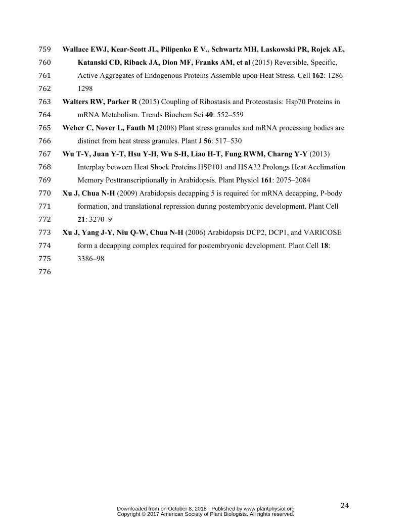
To check translational activity in wild type compared to hsp101 (hot1-3, Lee et al., 149 2005), we analyzed polysome profiles on 7-day-old seedlings under the normal condition at 150 22°C (control condition), after an HS treatment at 37°C for 1 h, and after 2 h of recovery at 151
www.plantphysiol.orgon October 8, 2018 - Published by Downloaded from Copyright © 2017 American Society of Plant Biologists. All rights reserved.
6
22°C following the HS (referred to as 2 h recovery hereafter, Fig. 1). Under the normal 152 condition and during HS, no obvious differences were observed between wild type and 153 hsp101 (Fig. 1A, D). After 1 h at 37°C, a massive polysome dissociation occurred that 154 induces accumulation of monosomes (80S) and free 40S/60S ribosomal subunits (Fig. 1B, E). 155 In wild type, after 2 h recovery, polysomes massively reappeared, suggesting a translational 156 restart (Fig. 1C). Nonetheless, polysome recovery in hsp101 is affected compared to wild type 157 (Fig. 1C, F). In fact, polysome increase is slower in hsp101, resulting in a higher amount of 158 monosomes (compare the 80S peaks). To confirm this result, polysomes area was quantified 159 for each condition using three biological replicates (Fig. 1G). A significant difference was 160 only observed between wild type and hsp101 after 2 h recovery. HSP101 is a molecular 161 chaperone protein; some missense mutants (dlt1-1 and dlt1-2) were shown to differentially 162 affect its chaperone activity (Wu et al., 2013). dlt1-1 contains a single mutation C-to-T in 163 HSP101 at position 599 near the N terminus of Nucleotide-Binding Domain2 causing the 164 replacement of Thr by Ile. In dlt1-2, a single mutation C-to-T appears at position 33 in the N-165 terminal domain resulting in a His-to-Tyr substitution (Wu et al., 2013). Both maintained a 166 normal level of HSP101 protein but dlt1-2 results in substantial loss of the chaperone function 167 of HSP101 as dlt1-1 mutation as little effect on HSP101 chaperone activity. Moreover dlt1-2 168 mutation induces thermotolerance defect, while dlt1-1 does not. Like hsp101, dlt1-2 seedlings 169 are not able to survive under short-acquired thermotolerance (Wu et al., 2013). To check if 170 HSP101 chaperone activity is involved in translation recovery, the polysome profiles of dlt1-1 171 and dlt1-2 were analyzed after 2 h recovery (Supplemental Fig. S1). Only dlt1-2 presented 172 defect in polysome recovery suggesting that HSP101 chaperone activity is involved in 173 translation recovery. We tried to correlate this defect with a defect in growth rate after HS. To 174 this end, the extension of root length was measured but no differences were observed between 175 the wild type and hsp101 during recovery (data not shown). 176 To check if this process is also correlated with a defect in SG dissociation, appearance 177 and disappearance of SGs were monitored (Fig. 2 and Supplemental Fig. S2) with the 178 Arabidopsis transgenic lines expressing the polyA binding protein 2 fused to the red 179 fluorescent protein (PABP2-RFP) under the PABP2 promotor in the wild type or hsp101 180 background. The levels of PABP2-RFP and HSP101 were checked by western blot 181 (Supplemental Fig. S2A). SG accumulation and dissociation were analyzed on different roots 182 by confocal microscope (Fig. 2 and Supplemental Fig. S2B). A defect in SG dissociation was 183 observed in hsp101 compared to wild type after 2 h recovery (Fig. 2C vs. 2I, and 184 Supplemental Fig. S2B), whereas no difference can be observed under normal and HS 185
www.plantphysiol.orgon October 8, 2018 - Published by Downloaded from Copyright © 2017 American Society of Plant Biologists. All rights reserved.

7
conditions (Fig. 2A vs. 2G and 2B vs. 2H). A SG quantification was performed in wild type 186 and hsp101 to confirm this observation (Fig. 2M). According to these data, we proposed that 187 HSP101 could be involved in translation recovery by regulating the dissociation of SG. 188 189 Identification of stored mRNAs released in an HSP101-dependent manner 190
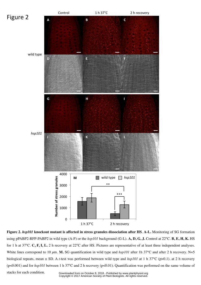
Although the involvement of the yeast HSP104 in SG dissociation is known 191 (Cherkasov et al., 2013), the nature of mRNA stored and released was not identified. As 192 stored mRNAs could play a role in translation recovery, we designed an RNA sequencing 193 strategy to identify them. Because SG purification could be difficult and quite challenging, we 194 decided to take advantage of the hsp101 translation defect to identity these mRNAs with the 195 idea that stored mRNAs will come back slower in translation in hsp101 compared to wild 196 type. To do that, we performed RNA sequencing on total and polysome-associated mRNAs 197 purified from wild type and hsp101 seedlings under the normal condition at 22°C, after 1 h at 198 37°C, and after 2 h recovery (Supplemental Table S1). Each condition was performed on two 199 biological repeats. 200 Prior to analysis, for each condition, reads per kilobase per million mapped 201 reads (RPKM) values obtained between replicates were compared. Each set of experiment 202 was found to be reproducible as demonstrated by plotting read counts pairwise between 203 replicates (with R2 ≈ 0.99) (Supplemental Table S1). Next, only transcripts with at least 1 204 RPKM in 1 library were kept, resulting in 19,599 transcripts. The aim of the RNA sequencing 205 is to identify mRNAs first stored during HS and next released in an HSP101-dependent 206 manner. For stored mRNAs, we reasoned as follows: a stored mRNA must be stable and 207 released from translation during HS, i.e. down regulated at polysomes level. To identify those 208 mRNAs, a fold change (FC) analysis was performed at total RNA level between HS and 209 control condition. The same analysis was also applied for polysomal RNA. A transcript 210 having an FC below 0.5 was considered as down-regulated and was considered as stable with 211 an FC between 0.5 and 2. To identify transcripts that were stored during HS (and not 212 degraded as in Merret et al., 2015; Merret et al., 2013), we selected from the total RNA 213 population heat “stable” mRNAs (having an FC between 0.5 and 2) that were clearly down 214 represented (having an FC below 0.5) in the polysomal fraction after HS. According to the 215 criteria, 11,558 and 11,779 transcripts are stable during HS in wild type and hsp101, 216 respectively, with an overlap of 90% between both genotypes (Fig. 3A). Then, only common 217 transcripts between wild type and hsp101 were kept, and their translation repression was 218
www.plantphysiol.orgon October 8, 2018 - Published by Downloaded from Copyright © 2017 American Society of Plant Biologists. All rights reserved.
8
analyzed. 3,309 transcripts were found to be downregulated at the polysomal level in both 219 genotypes (Fig. 3A and Supplemental Table S2). 220 To identify among the 3,309 transcripts, those released during recovery in an HSP101-221 dependent manner, we measured their translation efficiency (TE) (their ratio in polysomes vs. 222 total RNA), during and after HS, in wild type and hsp101. As shown in Fig. 3B and 3C, TEs 223 of these mRNAs were generally higher during recovery compared to HS, suggesting that most 224 of the stored mRNAs during HS can come back rapidly in translation after stress. As hsp101 225 presents a defect in polysome recovery, we decided to compare the TEs of these 3,309 226 mRNAs during recovery in wild type and hsp101. To do that, a scatter plot was performed 227 between wild type and hsp101 for the three conditions (Fig. 3D and Supplemental Fig. S3). 228 Next, a linear regression was calculated for each condition with the 3,309 dots. Although their 229 TEs are identical under control and HS conditions (Supplemental Fig. S3, linear regression 230 close to 1), during recovery, TEs in hsp101 appeared to be affected compared to wild type as 231 the linear regression decrease close to 0.8. Out of 3,309 transcripts, 2,103 showed TEs lower 232 in hsp101 compared to wild type (red dots on Fig. 3D and Supplemental Table S3). Since SG 233 dissociation was affected in hsp101, we propose that these 2,103 mRNAs are stored in SG 234 during HS and released in an HSP101-dependent manner. 235 236 mRNAs coding for RPs are stored during HS and released during recovery 237
To go further in the functional role of the stored mRNAs during recovery, we 238 performed a gene ontology (GO) analysis on the stored mRNAs released during recovery in 239 an HSP101-dependent manner. Two molecular functions were enriched in the 2,103 mRNA 240 population: “protein binding” (GO:0005515, p-value 0,000202) and “structural constituent of 241 ribosome” (GO:0003735, p-value 6,81e-54) (Supplemental Fig. S4). The second GO term is 242 highly enriched, and most of the mRNAs present in this group encode large or small subunits 243 ribosomal proteins (RPL or RPS). According to our RNAseq database (Supplemental Table 244 S1), 276 nuclear genes coding for RPS or RPL are expressed at seedlings stage. 219 245 correspond to cytoplasmic RPs, 50 to chloroplastic RPs and 7 to mitochondrial RPs. Thus, to 246 have a better overview of translation activity of mRNAs coding for RPs, TE was determined 247 for all of them in the three conditions (control, HS, and recovery) in wild type and hsp101 248 (Supplemental Table S4). TEs were clustered according to their behavior on three heatmaps 249 (Fig. 4 and Supplemental Fig. S5). According to the clustering, a major group can be 250 distinguished in the three class of RPs. This group is highly present for cytoplasmic RPs with 251 179 members (81.7 %) compared to chloroplastic and mitochondrial RPs with 27 (54 %) and 252
www.plantphysiol.orgon October 8, 2018 - Published by Downloaded from Copyright © 2017 American Society of Plant Biologists. All rights reserved.

9
4 (57 %) members, respectively. This group has members with low TE during HS compared 253 to the control, and their translation recovery seemed to be HSP101-dependent (Fig. 4 and 254 Supplemental Fig. S5). We propose that members of this group are stored during HS and 255 released directly or indirectly by HSP101 during the recovery phase. To confirm this 256 hypothesis, we performed qPCR validation on two candidate transcripts of RPS23B 257 (At5g02960) and RPS29B (At3g44010) (Fig. 5A, B). As expected, their TEs were 258 significantly and identically reduced during HS in wild type and hsp101 whereas translation 259 recovery was affected in hsp101 for both transcripts. 260
To confirm that these mRNAs are stored in SGs during HS, a RIP approach coupled to 261 RT-PCR was performed using a transgenic line expressing UBP1a fused to the green 262 fluorescent protein (GFP). UBP1a was previously reported to localize in SG during stress 263 (Weber et al., 2008; Sorenson and Bailey-Serres, 2014). The RIP experiment was performed 264 under the normal condition at 22°C, after 1 h at 37°C, and after 2 h recovery. After checking 265 immunoprecipitation efficiency (Fig. 6A), the association of RPS23B and RPS29B mRNAs 266 with UBP1a was analyzed. RPL37B mRNAs was used as a positive control as this transcript 267 was previously showed as an mRNA partner of UBP1 under hypoxia (Sorenson and Bailey-268 Serres, 2014). According to our data, this transcript seems to be stored during HS and 269 released in an HSP101-dependent manner. For all transcripts, association with UBP1a was 270 highly induced during HS (Fig. 6 B, C, D) and reduced or abolished during the recovery 271 phase, coincident with the model where RP mRNAs are stored during HS and released during 272 the recovery phase. Thus, we propose that HS induces a massive storage of mRNAs coding 273 RPs and these mRNAs are released during recovery to enhance translation. 274 275 Polysome recovery can be independent of transcription restart 276
The storage of mRNA coding for RPs during HS suggests that the production of new 277 ribosomes is a critical step to resume translation at recovery, meaning the new subunits have 278 to be produced. To test this hypothesis, polysome profiles were done at different time points 279 during recovery. We hypothesized that if pre-existing free ribosomal subunits (40S and 60S) 280 can be reused during recovery, their amount will be decreased with the concomitant increase 281 of 80S/polysomes. To test this hypothesis, the area of 40S/60S and 80S/polysomes were 282 determined for each condition and normalized according to the respective area for HS 283 condition. After 30 min recovery, monosomes (the 80S peak) decreased, and polysomes 284 increased as compared to the HS situation, suggesting a translation restart (Fig. 7A). 285 However, curiously, the amount of free ribosomal subunits did not change as compared to HS 286
www.plantphysiol.orgon October 8, 2018 - Published by Downloaded from Copyright © 2017 American Society of Plant Biologists. All rights reserved.
10
condition (ratio = 1.08 ± 0.03). After 1 h recovery, polysomes continued to increase and 287 reached a maximum level while the amount of free ribosomal subunits remained, again, 288 constant (ratio = 1.14 ± 0.09). After 2 h recovery, the amount of free ribosomal subunits 289 decreased, but the polysomes stopped to increase. These observations strongly suggest that, 290 up to 1 h recovery, pre-existing free ribosomal subunits could not fully account for the 291 translation restart and that new ribosomal subunits must come into play with the synthesis of 292 RPs. 293
The production of new RPs could result from the translation of either de novo 294 transcribed mRNAs or those released from SGs. To discriminate between these two 295 possibilities, seedlings in the recovery phase were incubated on liquid medium supplemented 296 or not with the transcription inhibitor, actinomycin D, to block both polymerase I and II 297 activities (Bensaude, 2011). After 2 h recovery, polysome profiles were performed. As shown 298 in Fig. 7B, although transcription is blocked, polysome recovery still took place, suggesting 299 that a least a portion of new polysomes can be formed without transcription. To prove that 300 new ribosomes can be formed in the time frame of the recovery period without necessarily 301 mobilizing pre-existing, heat-dissociated ribosomal subunits, a 15N incorporation experiment 302 was performed after stress. After 15N incorporation and polysome purification, the 15N 303 amount was determined for proteins present in the polysomal fractions (essentially composed 304 of ribosomes). These data were compared to a negative control (puromycin treatment) to be 305 sure that incorporated 15N signal only comes from proteins associated with polysomes. As 306 shown in Fig. 7C, 15N was detected in polysomes after 2 h recovery, suggesting the formation 307 of new ribosomes in this frame time. The same incorporation was performed with 308 actinomycin D treatment (Fig. 7C). Although transcription is blocked, 15N incorporation was 309 still detected in polysomes after 2 h recovery. 310
As ribosome biogenesis needs mature rRNAs produced by RNA polymerase I, we 311 investigated the effect of HS on nucleoli architecture. Nucleolus architecture is highly 312 dependent on the production of a proper amount of rRNAs. One of the major components of 313 the nucleolus is the dense fibrillar component (DFC) that consists of newly transcribed rRNA. 314 Fibrillarin is a nucleolar protein known to localize to the DFC, forming ring-like structures in 315 the nucleolus (Picart and Pontvianne, 2016) and can be used as a marker of nucleolus integrity 316 (Pontvianne et al., 2013). Using FIB2-YFP stable line, the nucleolus status was checked under 317 normal and HS conditions. As shown in Supplemental Fig. S6A and S6B, FIB2 formed ring-318 like structures both in normal and HS conditions, whereas actinomycin D treatment (that 319 inhibits polymerase I activity) disrupts FIB2 localization (Supplemental Fig. 6C). Thus, HS 320
www.plantphysiol.orgon October 8, 2018 - Published by Downloaded from Copyright © 2017 American Society of Plant Biologists. All rights reserved.
11
did not affect nucleolus architecture, suggesting that RNA polymerase I activity was not 321 significantly affected under this condition. All together, these data suggest that new 322 ribosomes can be formed shortly after HS by mobilizing stored RP-coding mRNAs. 323 324 Discussion 325 326 Translation repression and storage of RP mRNAs during stress 327 Although conditions leading to SG formation are now well documented, how specific 328 mRNAs are stored and released from these aggregates, and the functional consequences of 329 this process remain unclear. Here, using the hsp101 translation recovery defect, we observed 330 that 210 of 276 nuclear mRNAs coding for RPs are preferentially stored under HS and 331 released in the recovery period in an HSP101-dependent manner. Translation inhibition of RP 332 mRNAs was already observed for different stresses like hypoxia (Branco-Price et al., 2008), 333 sucrose deprivation (Nicolaï et al., 2006) or water deficit (Kawaguchi et al., 2004), suggesting 334 that inhibition of ribosome biogenesis is a conserved stress response. This dampening of 335 translation during stress was attributed to energy conservation (Branco-Price et al., 2008), as 336 ribosome biogenesis is highly ATP consuming. Recently, it was reported that the association 337 of RP mRNAs with UBP1 is enhanced under hypoxia (Sorenson and Bailey-Serres, 2014). 338 Our data suggest that the same phenomenon occurs under HS (Fig. 6). Thus, it appears that 339 RP mRNAs are translationally repressed and stored during stress. In yeast, the same 340 observation was reported under glucose deprivation, while adding back glucose led to RP 341 mRNAs translation recovery (Arribere et al., 2011). Interestingly, in this case, few genes can 342 be reactivated for translation upon glucose replenishing, and most of them were attributed to 343 ribosomal protein genes (Arribere et al., 2011). We observed the same mechanism in 344 Arabidopsis under HS, suggesting a common signalling pathway in regulating RP mRNAs 345 translation across eukaryotes. In yeast, it was suggested that hsp104 translational defect is due 346 to a defect in the release of preinitiation complexes stored in SG (Cherkasov et al., 2013). Our 347 data go further in this process and show that the nature of stored mRNAs could also influence 348 the translation recovery. 349 350 How to coordinate de novo ribosome synthesis after stress? 351 We show in this work that new ribosomes can be formed shortly after HS by mobilizing 352 stored RP-coding mRNAs. Ribosome biogenesis needs a fine-tuning between rRNA 353 production mediated by RNA polymerase I and RP production mediated by RNA polymerase 354
www.plantphysiol.orgon October 8, 2018 - Published by Downloaded from Copyright © 2017 American Society of Plant Biologists. All rights reserved.
12
II. Therefore, an important question is how to coordinate the timely production of rRNAs and 355 RPs to assemble new ribosomes shortly after a stress event. Previously it was reported that, in 356 tomato, the production of rRNA primary transcripts is maintained under HS, while the 357 maturation process is abolished, leading to the accumulation of primary transcripts in the 358 nucleolus (Nover et al., 1986). The same observation was recently proposed also for yeast 359 (Kos-braun et al., 2017). The authors showed that pre-rRNA continues to be synthesized upon 360 stress and suggested that continued transcription of rDNA allows maintenance of nucleolar 361 integrity to rapidly respond to environmental changes. This result agrees with our observation 362 that the nucleolus architecture (a proxy of polymerase I activity) is maintained during HS in 363 Arabidopsis (Supplemental Fig. S6A, B). The coordinated control during the stress of 364 essential molecules needed to generate new ribosomes (primary rRNA production in the 365 nucleolus and storage of RP-coding mRNAs in SGs) could be critical to synchronize 366 ribosome synthesis in the recovery period. This hypothesis is in agreement with our data 367 showing that RNA polymerase I and II activities are not needed to synthesize new ribosomes 368 early in the recovery period. The rapid restart of translation, only minutes after stress, could 369 therefore benefit from the HSP101-dependent release of RP-coding mRNAs from SGs, which 370 is to be synchronized with the maturation of primary rRNAs in the nucleolus, leading to new 371 ribosome formation. 372 373 Ribosomal subunits under stress and during recovery 374 The dynamics of ribosomal subunits we observed during and after the stress is supporting our 375 major conclusion that RP synthesis with stored mRNAs is needed to resume translation in the 376 recovery period. As shown in our polysome profiles (Fig. 7A), HS induces a massive 377 dissociation of polysomes and the accumulation of monosomes and free 40S and 60S 378 subunits. This is expected as the ribosomal subunits released following translation termination 379 cannot easily re-engage in new rounds of protein synthesis in a stress situation. Early in the 380 recovery period, one would expect that the re-activation of translation will increase the level 381 of polysomes with a corresponding decrease in monosomes and free subunits. However, this 382 is not what we observed as the level of free subunits was stable even 1 h in the recovery 383 period, when the polysome level is strongly increased. This phenomenon suggests that newly 384 synthetized ribosomal subunits, (and not only the pre-existing ones), are contributing to the 385 build up of polysome recovery. 386 387
www.plantphysiol.orgon October 8, 2018 - Published by Downloaded from Copyright © 2017 American Society of Plant Biologists. All rights reserved.
13
Why new ribosomes were produced instead of salvaging the pre-existing ones? One 388 possibility is that part of the pre-existing subunits have been damaged or irreversibly modified 389 by the stress and cannot be easily recycled. Indeed, under stress conditions that affect energy 390 status, alteration of ribosome subunits is one strategy that can preserve cellular energy 391 homeostasis. It was reported that under stress conditions or after translation inhibition, 392 modification of ribosomal subunits could occur through ubiquitination both for 40S and 60S 393 subunits (Kraft et al., 2008; Higgins et al., 2015). It was proposed that this modification not 394 only controls translational status under stress but also induces the ribosome degradation 395 pathway, called ribophagy (Kraft et al., 2008). To evade HS-inducing modifications, the 396 protein aggregation process was proposed to be a specific and reversible pathway to maintain 397 proteins activity for recovery phase (Wallace et al., 2015). This study suggests that free 398 ribosome subunits may not be able to efficiently aggregate under HS, making them more 399 sensitive to modification and/or degradation. This could be physiologically relevant as the 400 reduction in ribosome abundance was proposed to be a rapid and adaptive stress response 401 during proteotoxic stress in yeast (Guerra-Moreno et al., 2015). Ribosomal subunits 402 ubiquitination can occur rapidly upon stress (after 1 h DTT for human cells, Higgins et al., 403 2015), whereas ribosomal subunits cleavage increase only significantly after 4 hours (Kraft et 404 al., 2008). Thus we postulate that the same mechanisms could occur during and after HS in 405 Arabidopsis and one way to recover from these effects would be to store RP mRNAs in SG to 406 enhance ribosome production during recovery phase. 407 408 Another challenging question is how RP mRNAs could be specifically targeted for this 409 massive sequestration and release process. A common cis-element “TAGGGTTT” was 410 previously identify on Arabidopsis RP mRNAs and was shown to be important in the context 411 of translation efficiency during photomorphogenesis (Liu et al., 2012). However, this cis-412 element is also present on RP mRNAs that are apparently not stored and released following 413 HS. Therefore, it is unclear at the moment if this cis-element is involved or not and further 414 effort will be needed to determine the regulators, both cis and trans, required for sequestrate 415 and release of most RP mRNAs in a heat-stress situation. 416 417 In this study, we identified stress granule associated mRNAs using RNA sequencing 418 approach. According to our criteria, 210 of 276 nuclear RP mRNAs are stored during HS and 419 released in the recovery period in an HSP101-dependent manner (Supplemental Table S4). 420 This allows us to identify the functional role of stored RP mRNAs for recovery period. 421
www.plantphysiol.orgon October 8, 2018 - Published by Downloaded from Copyright © 2017 American Society of Plant Biologists. All rights reserved.
14
Moreover, another GO term was enriched in our analysis (“protein binding” GO:0005515). 422 As this term encompasses a large group of proteins with distinct functions, it is difficult to 423 draw a model around this term. Nonetheless, we cannot exclude that other mRNAs with 424 specific functions are stored during HS and also play a role during recovery. 425 426 Material Methods 427 428 Growth conditions 429 Analyses were carried out with 5- or 7-day-old whole plantlets grown on synthetic Murashige 430 and Skoog (MS½) medium (Duchefa) containing 1% sucrose and 0.8% plant agar at 22°C 431 under a 16h light/ 8h dark regime. For HS treatment, plates were immerged in a water bath at 432 37°C. For recovery, plates were transferred at 22°C under previous conditions. 433 434 Generation of transgenic plants 435 A vector containing pPABP2-tRFP-PABP2 was transformed into wild type (Col0) by floral 436 dip transformation. Next, a cross between this line and the hsp101 mutant (hot1-3, Lee et al., 437 2005) was realized to obtain pPABP2-tRFP-PABP2 in the hsp101 background. The 438 expression level of RFP-PAB2 and HSP101 was confirmed by western blot analysis. 439 Transgenic lines with levels similar to wild type were kept for confocal analysis. 440 441 Polysome profile analysis and quantification 442 Polysome profiles were performed as described previously (Merret et al., 2013). Briefly, 400 443 mg of tissue powder were homogenised with 1.2 mL of lysis buffer (200 mM Tris-HCl pH 444 9.0, 200 mM KCl, 25 mM EGTA, 35 mM MgCl2, 1% detergent mix (1% Tween20, 1% 445 Triton, 1% Brij35, 1% Igepal), 1% sodium deoxycholate, 1% polyoxyethylene (10) tridecyl 446 ether, 5 mM DTT, 50 μg.mL-1 cycloheximide, 50 μg.mL-1 chloramphenicol, 1% protease 447 inhibitor cocktail, Sigma). Crude extract was incubated 10 min on ice. After centrifugation, 448 the supernatant was clarified with a 0.2 μM filter. 360 μL (= 120 mg) of crude extract was 449 loaded on a 15%-60% sucrose gradient (9 mL). Ultracentrifugation was performed with an 450 SW41 rotor at 38,000 rpm (≈ 180 000g) for 3 h. Polysome profile analyses were performed 451 with an ISCO absorbance detector at 254 nm. For normalization and direct comparison of 452 profiles, the same volume of crude extract was loaded on the sucrose gradient for each 453 condition. For quantification, the percentage of polysomes for each condition was determined 454
www.plantphysiol.orgon October 8, 2018 - Published by Downloaded from Copyright © 2017 American Society of Plant Biologists. All rights reserved.
15
using Image J as the ratio between polysomes area and total area (including 40S, 60S, 455 monosomes and polysomes areas). 456 457 Confocal microscopy analysis and stress granules quantification 458 Confocal microscopy was performed on 5-day-old whole seedlings as described in Merret et 459 al., 2015. For SG monitoring, at least 5 different roots for each condition were analyzed. For 460 nucleoli analysis, FIB2-GFP line was used (Pontvianne et al., 2013). 25 nucleoli were 461 analyzed for each condition on 5 different seedlings. Stress granules were quantified using 462 plugins of Fidji (http://fiji.sc/, Schindelin et al., 2012) on 5 seedling roots per condition. First, 463 20 iterations of deconvolution were applied using Iterative Deconvolve 3D on 15-slide 464 (overlapping) stacks of 5.46 μm thick. Centre of granules were then counted using 3D Objects 465 Counter with a threshold of 1000, excluding signal from structures smaller than 50 voxels. 466 467 RNA sequencing 468 Total RNA extraction was performed by using RNeasy® Micro Kit (Qiagen) according to 469 manufacturer’s instructions. For polysomal RNA extraction, fractions corresponding to 470 polysomes were pooled. Two volumes of guanidium 8M and three volumes of absolute 471 ethanol were added. After overnight precipitation, centrifugation (16,000g, 45 min, 4°C) was 472 performed. Pellets were resuspended in RLT buffer (Qiagen). Next, RNA was extracted with 473 RNEasy® Micro Kit. RNA quality was assessed with Agilent 2100 bioanalyzer prior to RNA 474 sequencing. Libraries preparation was performed with TrueSeq Illumina kit. Libraries were 475 multiplexed and sequenced (PE 2x150) on an Illumina HiSeq 2500. For each library, at least 476 40 million reads were obtained. Reads filtering and mapping were performed as described in 477 Merret et al., 2015. For each condition, the mean value of RPKM (read per kilobase per 478 million mapped reads) was determined between replicates. Transcripts, which did not have at 479 least 1 RPKM in 1 condition, were removed from the analysis. FC was determined as the ratio 480 between transcript quantity during HS and normal condition or during recovery and normal 481 condition. TE was determined as the ratio between transcript quantity in polysomal RNA and 482 total RNA. Raw data are reported in Table S1. 483 484 GO and heatmap analysis 485 The GO analyses were performed with method available at the AgriGO website 486 (http://bioinfo.cau.edu.cn) (Du et al., 2010), with the following parameters: Hypergeometric 487 statistical test method, Yekutieli (FDR under dependency) multi-test adjustment method and a 488
www.plantphysiol.orgon October 8, 2018 - Published by Downloaded from Copyright © 2017 American Society of Plant Biologists. All rights reserved.
16
0.05 significance level. Heatmap was obtained using heatmap.2 function of gplots R package 489 (https://cran.r-project.org/web/packages/gplots/index.html) using Pearson distance and an 490 average method for the hierarchical clustering. 491 492 qPCR validation 493 For qPCR validation, 500 ng of total or polysomal RNA was reverse-transcribed with 494 SuperScript IV Kit (Life TechnologyTM) using Oligo dT18. Real-time PCR was performed in 495 an LC 480 384-well thermocycler. The following program was used: 5 min 95°C, 40 cycles of 496 15 s at 95°C, and 1 min at 60°C). The PCR mix contained TakyonTM qPCR master mix 497 (Eurogentec), 500 nM gene-specific primers, and 1.8 μL cDNA in a total volume of 9 μL. 498 Primer efficiencies were determined on standard curves. Primers used in this study are present 499 in Supplemental Table S5. 500 501 RNA co-Immunoprecipitation 502 RNA co-Immunoprecipitation was performed as described previously using the UBP1a-GFP 503 line or Col0 line as a negative control (Sorenson and Bailey-Serres, 2014). Briefly, 400 mg of 504 tissue powder were incubated in 3 mL of lysis buffer (200 mM Tris pH 9.0, 110 mM 505 Potassium Acetate, 0.5 % triton-X100, 0.1% Tween-20, 5 mM DTT, 1.5% Protease 506 Inhibitor). Lysate was clarified using Miracloth (MilliporeTM) layer and centrifuged at 1,500 g 507 during 2 min at 4°C. 1.5 mL of crude extract was incubated with 15 μL of GFP-trap® beads 508 (ChromoTek) during 1,5 h at 4°C under rotation. 0.75 mL of crude extract was incubated with 509 7.5 μL of GFP-trap beads for western blot analysis. Beads were washed five times with 0.75 510 mL of lysis buffer. For RIP experiments, elution was performed with 200 μL of 8M 511 guanidium for 5 min on ice and precipitated overnight with 300 μL of 100% ethanol before 512 RNA extraction as described above. For western blot, elution was performed by adding 20 μL 513 of Laemmli 2X to beads and incubated 5 min at 95°C. Reverse transcription was performed 514 on 10 μL of eluate or 500 ng of input as described above. 1 μL of cDNA was used for PCR 515 amplification with specific primers (Supplemental Table S5). 516 517 15N Incorporation and Elemental Analysis 518 After heat treatment (as described above), seedlings were transferred in a liquid medium 519 containing 1mM KH2PO4, 50 μM FeNaEDTA, 0.5 mM MgCl2, 0.5 mM CaCl2 2H2O, 10 mM 520 KNO3. For 15N incorporation, KNO3 was replaced by 99% K15NO3. Incorporation was 521 performed during 2 h recovery. For transcription inhibition, actinomycin D was added to the 522
www.plantphysiol.orgon October 8, 2018 - Published by Downloaded from Copyright © 2017 American Society of Plant Biologists. All rights reserved.

17
medium at 0.2 mg.mL-1 during recovery. After incorporation, polysomes were extracted as 523 described previously. Polysomal proteins were precipitated 6 h at 4°C by adding two volumes 524 of 100% ethanol. After centrifugation, pellets were dried at 70°C for 48 hours and analyzed 525 for total N content and atom % 15N abundance by Continuous-Flow Mass Spectrometry, using 526 a Euro-EA Elemental Analyzer (Eurovector SPA, Redavalle, Italy) coupled with an IsoPrime 527 mass spectrometer (Isoprime Ltd,Stockport, UK). 528 529 Accession number 530 The accession number for the RNAseq data reported in this paper is NCBI Bioproject 531 PRJNA345276. 532 533 Acknowledgments 534 We thank the Academia Sinica NGS Core Facility for libraries preparation and RNA 535 sequencing. RM was the recipient of an Academia Sinica Postdoctoral Research Fellowship. 536 We thank Dr. Frédéric Pontvianne and Pr. Julia Bailey-Serres for kindly giving us FIB2-YFP 537 and UBP1a-GFP lines respectively. This work was supported by CNRS and UVPD through 538 utilization of confocal microscope and qPCR devices at the Technoviv platform. 539 540 Figure legends 541 542 Figure 1. hsp101 knockout mutant is affected in polysome recovery. A-F. Polysome 543 profiles on 7 day-old seedlings in wild type (A, B, C) or hsp101 (D, E, F). A, D. Control 544 22°C. B, E. heat shock (HS) for 1 h at 37°C. C, F. 2 h of recovery at 22°C after HS. G. 545 Percentage of polysomes for each condition. N=3 biological repeats, mean ± SD. A t-test was 546 performed between wild type and hsp101 for control (p>0.1), at 1 h 37°C (p>0.1) and at 2 h 547 recovery (p<0.05). 548 549 Figure 2. hsp101 knockout mutant is affected in stress granules dissociation after HS. A-550 L. Monitoring of SG formation using pPABP2-RFP-PABP2 in wild type (A-F) or the hsp101 551 background (G-L). A, D, G, J. Control at 22°C. B, E, H, K. HS for 1 h at 37°C. C, F, I, L. 2 552 h recovery at 22°C after HS. Pictures are representative of at least three independent analyses. 553 White lines correspond to 10 μm. M. SG quantification in wild type and hsp101 after 1 h 554 37°C and after 2 h recovery. N=5 biological repeats, mean ± SD. A t-test was performed 555 between wild type and hsp101 at 1 h 37°C (p>0.1), at 2 h recovery (p<0.001) and for hsp101 556
www.plantphysiol.orgon October 8, 2018 - Published by Downloaded from Copyright © 2017 American Society of Plant Biologists. All rights reserved.
18
between 1 h 37°C and 2 h recovery (p<0.01). Quantification was performed on the same 557 volume of stacks for each condition. 558 559 Figure 3. Translation efficiency of mRNAs released from translation during HS is 560 affected in the hsp101 mutant during recovery phase. A. Workflow used to identify 561 transcripts stored in SG and released from translation during recovery. Only mRNAs with an 562 FC between 0.5 and 2 on total RNA population and FC below 0.5 on polysomal RNA 563 population in wt and hsp101 were kept. B, C. TE of the 3309 mRNAs during HS vs. recovery 564 period in wt (B) and hsp101 (C). D. The log2 value of TE of the 3309 mRNAs during 565 recovery period in wt vs. hsp101. Red dots correspond to mRNAs affected in translation 566 recovery in hsp101 (2103). The black line corresponds to a linear regression of the 3309 dots. 567 568 Figure 4. Translation efficiency of cytoplasmic RP mRNAs in wild type and hsp101 569 under normal condition, after 1 h at 37°C and after 2 h recovery. Heatmap of cytoplasmic 570 RP mRNAs translation efficiency in log2 value. The Heatmap was performed on the 219 571 cytoplasmic RP mRNAs identified in RNAseq database. 179 of them are grouped in the same 572 cluster. The numbers of transcripts and the mean value of TE of this cluster are marked. Red 573 values correspond to a high translation efficiency and green values to a low translation 574 efficiency. 575 576 Figure 5. Determination of RP mRNAs translation efficiency by qPCR. A, B. Translation 577 efficiency of the At5g02960 and At3g44010 genes determined by qPCR. Translation 578 efficiency was determined as the ratio of mRNAs quantity between polysomal and total RNA 579 values. Values are normalized to ACTIN7 level. N=3 biological repeats, Mean ± SD. 580 581 Figure 6. RP mRNAs are associated with UBP1 during HS and released after stress. A. 582 Western blot analysis of UBP1a-GFP immunoprecipitation efficiency using GFP antibody. B, 583 C, D, E. PCR amplification (30 cycles) of the At3g44010 (RPS29B), At5g02960 (RPS23B), 584 At1g52300 (RPL37B) and At5g09810 (ACTIN7) genes after RNA immunoprecipitation with 585 the 35S-UBP1a-GFP line. Wild type line was used as a negative control. 1. Input UBP1a. 2. 586 Input wild type. 3. Eluate UBP1a. 4. Eluate wild type. 587 588 Figure 7. New ribosomes can be produced during recovery independently of 40S and 589 60S reassembly. A. Polysome profiles analysis in wild type after HS 1 h at 37°C, and after 30 590
www.plantphysiol.orgon October 8, 2018 - Published by Downloaded from Copyright © 2017 American Society of Plant Biologists. All rights reserved.

19
min, 1 h and 2 h recovery at 22°C. Polysome profiles analysis was performed three times on 591 three biological repeats. The area of 40/60S and 80S/polysomes were determined for each 592 condition and normalized according to the respective area for HS condition. Data are 593 presented below each profile. N=3 biological repeats, Mean ± SD. B. Polysome recovery after 594 HS on liquid medium supplemented with actinomycin D. C. 15N incorporation in polysomal 595 proteins after 2 h recovery with or without actinomycin D treatment, N=3 biological repeats, 596 mean ± SD. Puromycin treatment was used as a negative control. Values were normalized by 597 15N natural abundance. 598 599 Supplemental data 600 601 Figure S1. Polysome profiles after 2 h of recovery in wild type (A), dlt1-1 (B), hsp101 (C) 602 and dlt1-2 (D). Data correspond to the same biological repeats. dlt1-1 is a missense mutant of 603 HSP101 with little effect on HSP101 chaperone activity whereas dlt1-2 is a missense of 604 HSP101 that impairs HSP101 chaperone activity. 605 606 Figure S2. A. Western blot analysis of RFP-PABP2 lines. Levels of HSP101, PABP, and 607 ACTIN were checked using specific antibodies. αPABP recognizes PABP2, PABP4, and 608 PABP8 proteins. 1. wild type. 2. hsp101. 3. pPABP2-RFP-PABP2 in wild type. 4. pPABP2-609 RFP-PABP2 in hsp101 background. B. Monitoring of SG formation using pPABP2-RFP-610 PABP2 in wild type or in hsp101 background after 2 h recovery. 5 different roots are 611 represented. White lines correspond to 10 μm. 612 613 Figure S3. Log2 value of the 3309 mRNAs in control (A) and after 1 h at 37°C (B) in wild 614 type vs. hsp101. Black line correspond to the linear regression of the gray dots. 615 616 Figure S4. Gene Ontology (GO) analysis of mRNAs poorly translated during HS and affected 617 in translation recovery in hsp101 (2103/3309). GO groups highly enriched are presented in 618 red. p-values are present in parentheses for all groups. 619 620 Figure S5. Translation efficiency of chloroplastic and mitochondrial RP mRNAs in wild type 621 and hsp101 under normal condition, after 1 h at 37°C and after 2 h recovery. A. Heatmap of 622 chloroplastic RP mRNAs translation efficiency in log2 value. The Heatmap was performed on 623 the 50 chloroplastic RP mRNAs identified in RNAseq database. 27 of them are grouped in the 624
www.plantphysiol.orgon October 8, 2018 - Published by Downloaded from Copyright © 2017 American Society of Plant Biologists. All rights reserved.
20
same cluster. B. Heatmap of mitochondrial RP mRNAs translation efficiency in log2 value. 625 The numbers of transcripts and the mean value of TE are marked. Red values correspond to a 626 high translation efficiency and green values to a low translation efficiency. 627 628 Figure S6. Heat stress did not affect nucleoli architecture. A, B, C. Confocal microscopy 629 analysis of nucleoli architecture using FIB2-YFP in normal condition (A), after 1 h at 37°C 630 (B), and after 1 h at 22°C in liquid medium supplemented with actinomycin D (C). White 631 lines correspond to 10 μm. Pictures are representative of 25 nucleoli analyzed on 5 different 632 seedlings (5 per seedlings) for each condition. 633 634 635 Figure S1. Polysome profiles after 2 h of recovery in wild type (A), dlt1-1 (B), hsp101 (C) 636 and dlt1-2 (D). 637 Figure S2. A. Western blot analysis of RFP-PABP2 lines. 638 Figure S3. Log2 value of the 3309 mRNAs in control (A) and after 1 h at 37°C (B) in wild 639 type vs. hsp101. 640 Figure S4. Gene Ontology (GO) analysis of mRNAs poorly translated during HS and affected 641 in translation recovery in hsp101 (2103/3309). 642 Figure S5. Translation efficiency of chloroplastic and mitochondrial RP mRNAs in wild type 643 and hsp101 under normal condition, after 1 h at 37°C and after 2 h recovery. 644 Figure S6. Heat stress did not affect nucleoli architecture. A, B, C. 645 646 Table S1. RNA sequencing raw data 647 648 Table S2. Transcripts stable and released from translation during HS in wt and hsp101 649 (3309) 650 651 Table S3. Transcripts affected in translation recovery in hsp101 (2103) 652 653 Table S4. Translation Efficiency of Ribosomal Proteins mRNAs 654 655 Table S5. Primers used in this study 656
www.plantphysiol.orgon October 8, 2018 - Published by Downloaded from Copyright © 2017 American Society of Plant Biologists. All rights reserved.

21
References 657 658 Arribere JA, Doudna JA, Gilbert W V. (2011) Reconsidering movement of eukaryotic 659
mRNAs between polysomes and P bodies. Mol Cell 44: 745–758 660 Bensaude O (2011) Inhibiting eukaryotic transcription: Which compound to choose? How to 661
evaluate its activity? Transcription 2: 103–108 662 Branco-Price C, Kaiser KA, Jang CJH, Larive CK, Bailey-Serres J (2008) Selective 663
mRNA translation coordinates energetic and metabolic adjustments to cellular oxygen 664 deprivation and reoxygenation in Arabidopsis thaliana. Plant J 56: 743–755 665
Buchan JR (2014) mRNP granules. RNA Biol 11: 1019–30 666 Cherkasov V, Hofmann S, Druffel-Augustin S, Mogk A, Tyedmers J, Stoecklin G, 667
Bukau B (2013) Coordination of translational control and protein homeostasis during 668 severe heat stress. Curr Biol 23: 2452–2462 669
Cuesta R, Laroia G, Schneider RJ (2000) Chaperone Hsp27 inhibits translation during heat 670 shock by binding eIF4G and facilitating dissociation of cap-initiation complexes. Genes 671 Dev 14: 1460–1470 672
Du Z, Zhou X, Ling Y, Zhang Z, Su Z (2010) agriGO: A GO analysis toolkit for the 673 agricultural community. Nucleic Acids Res 38: 64–70 674
Guerra-Moreno A, Isasa M, Bhanu MK, Waterman DP, Eapen V V., Gygi SP, Hanna J 675 (2015) Proteomic analysis identifies ribosome reduction as an effective proteotoxic 676 stress response. J Biol Chem 290: 29695–29706 677
Hamilton TL, Stoneley M, Spriggs K a, Bushell M (2006) TOPs and their regulation. 678 Biochem Soc Trans 34: 12–16 679
Higgins R, Gendron JM, Rising L, Mak R, Webb K, Kaiser SE, Zuzow N, Riviere P, 680 Yang B, Fenech E, et al (2015) The Unfolded Protein Response Triggers Site-Specific 681 Regulatory Ubiquitylation of 40S Ribosomal Proteins. Mol Cell 59: 35–49 682
Jain S, Wheeler JR, Walters RW, Agrawal A, Barsic A, Parker R (2016) ATPase-683 Modulated Stress Granules Contain a Diverse Proteome and Substructure. Cell 164: 684 487–498 685
Juntawong P, Bailey-Serres J (2012) Dynamic Light Regulation of Translation Status in 686 Arabidopsis thaliana. Front Plant Sci 3: 1–16 687
Kawaguchi R, Girke T, Bray EA, Bailey-Serres J (2004) Differential mRNA translation 688 contributes to gene regulation under non-stress and dehydration stress conditions in 689 Arabidopsis thaliana. Plant J 38: 823–839 690
www.plantphysiol.orgon October 8, 2018 - Published by Downloaded from Copyright © 2017 American Society of Plant Biologists. All rights reserved.
22
Kos-braun IC, Jung I, Kos M (2017) Tor1 and CK2 kinases control a switch between 691 alternative ribosome biogenesis pathways in a growth-dependent manner. doi: 692 10.1371/journal.pbio.2000245 693
Kotak S, Larkindale J, Lee U, von Koskull-Döring P, Vierling E, Scharf KD (2007) 694 Complexity of the heat stress response in plants. Curr Opin Plant Biol 10: 310–316 695
Kraft C, Deplazes A, Sohrmann M, Peter M (2008) Mature ribosomes are selectively 696 degraded upon starvation by an autophagy pathway requiring the Ubp3p/Bre5p ubiquitin 697 protease. Nat Cell Biol 10: 602–610 698
Kuhn KM, DeRisi JL, Brown PO, Sarnow P, Kuhn KM, DeRisi JL, Sarnow P (2001) 699 Global and specific translational regulation in the genomic response of Saccharomyces 700 cerevisiae to a rapid transfer from a fermentable to a nonfermentable carbon source. Mol 701 Cell Biol 21: 916–927 702
Lee U, Wie C, Escobar M, Williams B, Hong S, Vierling E (2005) Genetic Analysis 703 Reveals Domain Interactions of Arabidopsis Hsp100 / ClpB and Cooperation with the 704 Small Heat Shock Protein Chaperone System. Plant Cell 17: 559–571 705
Liu B, Han Y, Qian SB (2013) Cotranslational Response to Proteotoxic Stress by Elongation 706 Pausing of Ribosomes. Mol Cell 49: 453–463 707
Liu M-J, Wu S-H, Chen H-M, Wu S-H (2012) Widespread translational control contributes 708 to the regulation of Arabidopsis photomorphogenesis. Mol Syst Biol 8: 566 709
McLoughlin F, Basha E, Fowler ME, Kim M, Bordowitz J, Katiyar-Agarwal S, Vierling 710 E (2016) Class I and II small heat-shock proteins protect protein translation factors 711 during heat stress. Plant Physiol 172: 1221–1236 712
Melamed D, Pnueli L, Arava Y (2008) Yeast translational response to high salinity : Global 713 analysis reveals regulation at multiple levels Yeast translational response to high 714 salinity : Global analysis reveals regulation at multiple levels. Rna 14: 1337–1351 715
Merret R, Descombin J, Juan Y ting, Favory JJ, Carpentier MC, Chaparro C, Charng 716 Y yung, Deragon JM, Bousquet-Antonelli C (2013) XRN4 and LARP1 are required 717 for a heat-triggered mRNA decay pathway involved in plant acclimation and survival 718 during thermal stress. Cell Rep 5: 1279–1293 719
Merret R, Nagarajan VK, Carpentier MC, Park S, Favory JJ, Descombin J, Picart C, 720 Charng YY, Green PJ, Deragon JM, et al (2015) Heat-induced ribosome pausing 721 triggers mRNA co-translational decay in Arabidopsis thaliana. Nucleic Acids Res 43: 722 4121–4132 723
Nicolaï M, Roncato M a, Canoy a S, Rouquié D, Sarda X, Freyssinet G, Robaglia C 724 www.plantphysiol.orgon October 8, 2018 - Published by Downloaded from
Copyright © 2017 American Society of Plant Biologists. All rights reserved.
23
(2006) Large-scale analysis of mRNA translation states during sucrose starvation in 725 arabidopsis cells identifies cell proliferation and chromatin structure as targets of 726 translational control. Plant Physiol 141: 663–673 727
Nover L, Mumsche D, Neumann D, Ohme K, Scharf K (1986) Control of ribosome 728 biosynthesis in plant cell cultures under heat-shock conditions. Eur J Biochem 160: 297–729 304 730
Park S-H, Chung PJ, Juntawong P, Bailey-Serres J, Kim YS, Jung H, Bang SW, Kim Y-731 K, Do Choi Y, Kim J-K (2012) Posttranscriptional Control of Photosynthetic mRNA 732 Decay under Stress Conditions Requires 3’ and 5’ Untranslated Regions and Correlates 733 with Differential Polysome Association in Rice. Plant Physiol 159: 1111–1124 734
Perea-Resa C, Carrasco-López C, Catalá R, Turečková V, Novak O, Zhang W, Sieburth 735 L, Jiménez-Gómez JM, Salinas J (2016) The LSM1-7 Complex Differentially 736 Regulates Arabidopsis Tolerance to Abiotic Stress Conditions by Promoting Selective 737 mRNA Decapping. Plant Cell 28: 505–520 738
Picart C, Pontvianne F (2016) Plant nucleolar DNA: green light shed on the role of 739 Nucleolin in genome organization. Nucleus 0 740
Pontvianne F, Blevins T, Chandrasekhara C, Mozgová I, Hassel C, Pontes OMF, Tucker 741 S, Mokroš P, Muchová V, Fajkus J, et al (2013) Subnuclear partitioning of rRNA 742 genes between the nucleolus and nucleoplasm reflects alternative epiallelic states. Genes 743 Dev 27: 1545–1550 744
Protter DSW, Parker R (2016) Principles and Properties of Stress Granules. Trends Cell 745 Biol 26: 668–679 746
Puckette M, Iyer NJ, Tang Y, Dai X Bin, Zhao P, Mahalingam R (2012) Differential 747 mRNA translation in medicago truncatula accessions with contrasting responses to 748 ozone-induced oxidative stress. Mol Plant 5: 187–204 749
Schindelin J, Arganda-Carreras I, Frise E, Kaynig V, Longair M, Pietzsch T, Preibisch 750 S, Rueden C, Saalfeld S, Schmid B, et al (2012) Fiji: an open-source platform for 751 biological-image analysis. Nat Methods 9: 676–682 752
Sorenson R, Bailey-Serres J (2014) Selective mRNA sequestration by 753 OLIGOURIDYLATE-BINDING PROTEIN 1 contributes to translational control during 754 hypoxia in Arabidopsis. Proc Natl Acad Sci U S A 111: 2373–8 755
Ueda K, Matsuura H, Yamaguchi M, Demura T, Kato K (2012) Genome-wide analyses of 756 changes in translation state caused by elevated temperature in Oryza sativa. Plant Cell 757 Physiol 53: 1481–1491 758
www.plantphysiol.orgon October 8, 2018 - Published by Downloaded from Copyright © 2017 American Society of Plant Biologists. All rights reserved.
24
Wallace EWJ, Kear-Scott JL, Pilipenko E V., Schwartz MH, Laskowski PR, Rojek AE, 759 Katanski CD, Riback JA, Dion MF, Franks AM, et al (2015) Reversible, Specific, 760 Active Aggregates of Endogenous Proteins Assemble upon Heat Stress. Cell 162: 1286–761 1298 762
Walters RW, Parker R (2015) Coupling of Ribostasis and Proteostasis: Hsp70 Proteins in 763 mRNA Metabolism. Trends Biochem Sci 40: 552–559 764
Weber C, Nover L, Fauth M (2008) Plant stress granules and mRNA processing bodies are 765 distinct from heat stress granules. Plant J 56: 517–530 766
Wu T-Y, Juan Y-T, Hsu Y-H, Wu S-H, Liao H-T, Fung RWM, Charng Y-Y (2013) 767 Interplay between Heat Shock Proteins HSP101 and HSA32 Prolongs Heat Acclimation 768 Memory Posttranscriptionally in Arabidopsis. Plant Physiol 161: 2075–2084 769
Xu J, Chua N-H (2009) Arabidopsis decapping 5 is required for mRNA decapping, P-body 770 formation, and translational repression during postembryonic development. Plant Cell 771 21: 3270–9 772
Xu J, Yang J-Y, Niu Q-W, Chua N-H (2006) Arabidopsis DCP2, DCP1, and VARICOSE 773 form a decapping complex required for postembryonic development. Plant Cell 18: 774 3386–98 775
776
www.plantphysiol.orgon October 8, 2018 - Published by Downloaded from Copyright © 2017 American Society of Plant Biologists. All rights reserved.
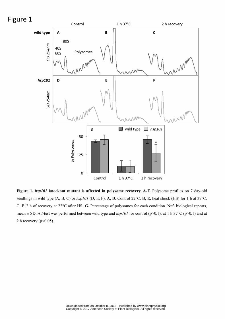
Figure1
Figure 1. hsp101 knockout mutant is affected in polysome recovery. A-F. Polysome profiles on 7 day-old
seedlings in wild type (A, B, C) or hsp101 (D, E, F). A, D. Control 22°C. B, E. heat shock (HS) for 1 h at 37°C.
C, F. 2 h of recovery at 22°C after HS. G. Percentage of polysomes for each condition. N=3 biological repeats,
mean ± SD. A t-test was performed between wild type and hsp101 for control (p>0.1), at 1 h 37°C (p>0.1) and at
2 h recovery (p<0.05).
wildtype
hsp101
A
D
B C
E F
IG H I
OD254n
m
OD254n
m
40S60S
80S
Polysomes
Control 1h37°C 2hrecovery
G
0
25
50
Control 1h37°C 2hrecovery
%Polysom
es
Control 1h37°C 2hrecovery
*
wildtype hsp101
www.plantphysiol.orgon October 8, 2018 - Published by Downloaded from Copyright © 2017 American Society of Plant Biologists. All rights reserved.
wildtype
hsp101J K L
Control 1h37°C 2hrecovery
A B C
G H I
D E F
Figure 2. hsp101 knockout mutant is affected in stress granules dissociation after HS. A-L. Monitoring of SG formation
using pPABP2-RFP-PABP2 in wild type (A-F) or the hsp101 background (G-L). A, D, G, J. Control at 22°C. B, E, H, K. HS
for 1 h at 37°C. C, F, I, L. 2 h recovery at 22°C after HS. Pictures are representative of at least three independent analyses.
White lines correspond to 10 µm. M. SG quantification in wild type and hsp101 after 1h 37°C and after 2 h recovery. N=5
biological repeats, mean ± SD. A t-test was performed between wild type and hsp101 at 1 h 37°C (p>0.1), at 2 h recovery
(p<0.001) and for hsp101 between 1 h 37°C and 2 h recovery (p<0.01). Quantification was performed on the same volume of
stacks for each condition.
Figure2
Hwildtype hsp101M
***
0
1000
2000
3000
4000
1h37°C 2hrecovery
Num
bero
fstressg
ranu
les
**
www.plantphysiol.orgon October 8, 2018 - Published by Downloaded from Copyright © 2017 American Society of Plant Biologists. All rights reserved.
Figure3
B
0
1
2
3
4
0 1 2 3 4TE2hre
covery
TE1h37°C
0
1
2
3
4
0 1 2 3 4
TE2hre
covery
TE1h37°C
C
wt
hsp101
Figure 3. Translation efficiency of mRNAs released from translation during HS is affected in the hsp101 mutant
during recovery phase. A. Workflow used to identify transcripts stored in SG and released from translation during
recovery. Only mRNAs with an FC between 0.5 and 2 on total RNA population and FC below 0.5 on polysomal RNA
population in wt and hsp101 were kept. B, C. TE of the 3309 mRNAs during HS vs. recovery period in wt (B) and hsp101
(C). D. The log2 value of TE of the 3309 mRNAs during recovery period in wt vs. hsp101. Red dots correspond to
mRNAs affected in translation recovery in hsp101 (2103). The black line corresponds to a linear regression of the 3309
dots.
10662 1117896
wt hsp101
FC37°C/ControlTotal[0.5;2]
3309 1188327
FC37°C/ControlPolysomes<0.5
A
wt hsp101
D
y=0.8041x
-3
-1,5
0
1,5
3
-3 -1,5 0 1,5 3TE2hrecoverywt(log2)
TE2hre
coveryhsp10
1(lo
g 2)
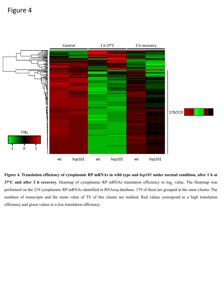
Figure4
Log2
0-1 1
179/219
Figure 4. Translation efficiency of cytoplasmic RP mRNAs in wild type and hsp101 under normal condition, after 1 h at
37°C and after 2 h recovery. Heatmap of cytoplasmic RP mRNAs translation efficiency in log2 value. The Heatmap was
performed on the 219 cytoplasmic RP mRNAs identified in RNAseq database. 179 of them are grouped in the same cluster. The
numbers of transcripts and the mean value of TE of this cluster are marked. Red values correspond to a high translation
efficiency and green values to a low translation efficiency.
Control 1h37°C 2hrecovery
wt hsp101 wt hsp101 wt hsp101

Figure5
0
2
4
6
Transla
2onEffi
cien
cy
0
2
4
6
Transla
2onEffi
cien
cy
At5g02960:RPS23B At3g44010:RPS29BA B
Figure 5. Determination of RP mRNAs translation efficiency by qPCR. A, B. Translation efficiency of the At5g02960
and At3g44010 genes determined by qPCR. Translation efficiency was determined as the ratio of mRNAs quantity between
polysomal and total RNA values. Values are normalized to ACTIN7 level. N=3 biological repeats, Mean ± SD.
Control 1h37°C 2hrecovery Control 1h37°C 2hrecovery
wildtype hsp101 wildtype hsp101
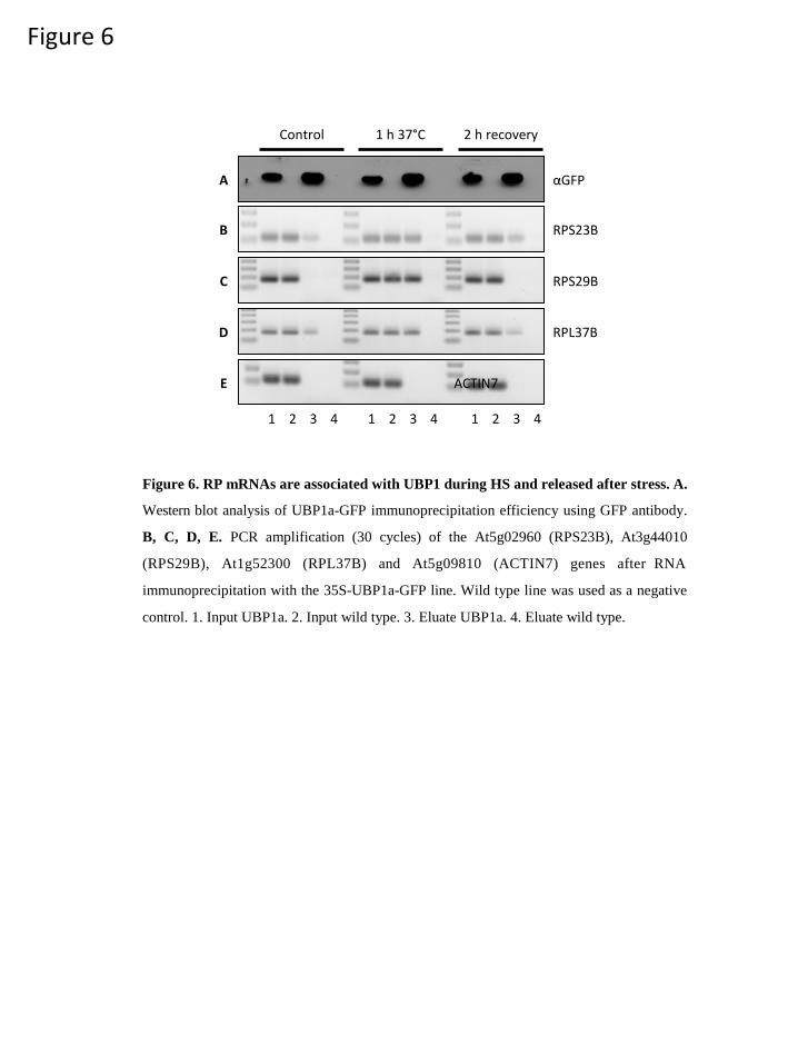
Figure 6
Control 1 h 37°C 2 h recovery
A αGFP
B RPS23B
C RPS29B
D RPL37B
E ACTIN7
1 2 3 4 1 2 3 4 1 2 3 4
Figure 6. RP mRNAs are associated with UBP1 during HS and released after stress. A.
Western blot analysis of UBP1a-GFP immunoprecipitation efficiency using GFP antibody.
B, C, D, E. PCR amplification (30 cycles) of the At5g02960 (RPS23B), At3g44010
(RPS29B), At1g52300 (RPL37B) and At5g09810 (ACTIN7) genes after RNA
immunoprecipitation with the 35S-UBP1a-GFP line. Wild type line was used as a negative
control. 1. Input UBP1a. 2. Input wild type. 3. Eluate UBP1a. 4. Eluate wild type.

A
Figure 7. New ribosomes can be produced during recovery independently of 40S and 60S reassembly. A.
Polysome profiles analysis in wild type after HS 1 h at 37°C, and after 30 min, 1 h and 2 h recovery at 22°C.
Polysome profiles analysis was performed three times on three biological repeats. The area of 40/60S and 80S/
polysomes were determined for each condition and normalized according to the respective area for HS condition.
Data are presented bellow each profile. N=3 biological repeats, Mean ± SD. B. Polysome recovery after HS on liquid
medium supplemented with actinomycin D. C. 15N incorporation in polysomal proteins after 2 h recovery with or
without actinomycin D treatment, N=3 biological repeats, mean ± SD. Puromycin treatment was used as a negative
control. Values were normalized by 15N natural abundance.
0
2
4
6
8
15N ActD+15NPuromycin
15NAmou
nt(μ
moles/gDW
)D
E
F
OD254nm
B C
Figure7
1h37°C 30’recovery 1hrecovery 2hrecovery
OD254nm
1 1 1.08±
0.03
1.1±
0.01
1.14±
0.09
1.65±0.2
0.94±
0.06
1.6±0.2
1h37°C 1hrecoveryActD 2hrecoveryActD 15N 15N+ActD
Puromycin
Parsed CitationsArribere JA, Doudna JA, Gilbert W V. (2011) Reconsidering movement of eukaryotic mRNAs between polysomes and P bodies. MolCell 44: 745-758
Pubmed: Author and TitleCrossRef: Author and TitleGoogle Scholar: Author Only Title Only Author and Title
Bensaude O (2011) Inhibiting eukaryotic transcription: Which compound to choose? How to evaluate its activity? Transcription 2:103-108
Pubmed: Author and TitleCrossRef: Author and TitleGoogle Scholar: Author Only Title Only Author and Title
Branco-Price C, Kaiser KA, Jang CJH, Larive CK, Bailey-Serres J (2008) Selective mRNA translation coordinates energetic andmetabolic adjustments to cellular oxygen deprivation and reoxygenation in Arabidopsis thaliana. Plant J 56: 743-755
Pubmed: Author and TitleCrossRef: Author and TitleGoogle Scholar: Author Only Title Only Author and Title
Buchan JR (2014) mRNP granules. RNA Biol 11: 1019-30Pubmed: Author and TitleCrossRef: Author and TitleGoogle Scholar: Author Only Title Only Author and Title
Cherkasov V, Hofmann S, Druffel-Augustin S, Mogk A, Tyedmers J, Stoecklin G, Bukau B (2013) Coordination of translationalcontrol and protein homeostasis during severe heat stress. Curr Biol 23: 2452-2462
Pubmed: Author and TitleCrossRef: Author and TitleGoogle Scholar: Author Only Title Only Author and Title
Cuesta R, Laroia G, Schneider RJ (2000) Chaperone Hsp27 inhibits translation during heat shock by binding eIF4G and facilitatingdissociation of cap-initiation complexes. Genes Dev 14: 1460-1470
Pubmed: Author and TitleCrossRef: Author and TitleGoogle Scholar: Author Only Title Only Author and Title
Du Z, Zhou X, Ling Y, Zhang Z, Su Z (2010) agriGO: A GO analysis toolkit for the agricultural community. Nucleic Acids Res 38: 64-70Pubmed: Author and TitleCrossRef: Author and TitleGoogle Scholar: Author Only Title Only Author and Title
Guerra-Moreno A, Isasa M, Bhanu MK, Waterman DP, Eapen V V., Gygi SP, Hanna J (2015) Proteomic analysis identifies ribosomereduction as an effective proteotoxic stress response. J Biol Chem 290: 29695-29706
Pubmed: Author and TitleCrossRef: Author and TitleGoogle Scholar: Author Only Title Only Author and Title
Hamilton TL, Stoneley M, Spriggs K a, Bushell M (2006) TOPs and their regulation. Biochem Soc Trans 34: 12-16Pubmed: Author and TitleCrossRef: Author and TitleGoogle Scholar: Author Only Title Only Author and Title
Higgins R, Gendron JM, Rising L, Mak R, Webb K, Kaiser SE, Zuzow N, Riviere P, Yang B, Fenech E, et al (2015) The UnfoldedProtein Response Triggers Site-Specific Regulatory Ubiquitylation of 40S Ribosomal Proteins. Mol Cell 59: 35-49
Pubmed: Author and TitleCrossRef: Author and TitleGoogle Scholar: Author Only Title Only Author and Title
Jain S, Wheeler JR, Walters RW, Agrawal A, Barsic A, Parker R (2016) ATPase-Modulated Stress Granules Contain a DiverseProteome and Substructure. Cell 164: 487-498
Pubmed: Author and TitleCrossRef: Author and TitleGoogle Scholar: Author Only Title Only Author and Title
Juntawong P, Bailey-Serres J (2012) Dynamic Light Regulation of Translation Status in Arabidopsis thaliana. Front Plant Sci 3: 1-16Pubmed: Author and TitleCrossRef: Author and TitleGoogle Scholar: Author Only Title Only Author and Title
Kawaguchi R, Girke T, Bray EA, Bailey-Serres J (2004) Differential mRNA translation contributes to gene regulation under non-stress and dehydration stress conditions in Arabidopsis thaliana. Plant J 38: 823-839
Pubmed: Author and TitleCrossRef: Author and TitleGoogle Scholar: Author Only Title Only Author and Title
Kos-braun IC, Jung I, Kos M (2017) Tor1 and CK2 kinases control a switch between alternative ribosome biogenesis pathways in agrowth-dependent manner. doi: 10.1371/journal.pbio.2000245
Pubmed: Author and TitleCrossRef: Author and Title

Google Scholar: Author Only Title Only Author and Title
Kotak S, Larkindale J, Lee U, von Koskull-Döring P, Vierling E, Scharf KD (2007) Complexity of the heat stress response in plants.Curr Opin Plant Biol 10: 310-316
Pubmed: Author and TitleCrossRef: Author and TitleGoogle Scholar: Author Only Title Only Author and Title
Kraft C, Deplazes A, Sohrmann M, Peter M (2008) Mature ribosomes are selectively degraded upon starvation by an autophagypathway requiring the Ubp3p/Bre5p ubiquitin protease. Nat Cell Biol 10: 602-610
Pubmed: Author and TitleCrossRef: Author and TitleGoogle Scholar: Author Only Title Only Author and Title
Kuhn KM, DeRisi JL, Brown PO, Sarnow P, Kuhn KM, DeRisi JL, Sarnow P (2001) Global and specific translational regulation in thegenomic response of Saccharomyces cerevisiae to a rapid transfer from a fermentable to a nonfermentable carbon source. MolCell Biol 21: 916-927
Pubmed: Author and TitleCrossRef: Author and TitleGoogle Scholar: Author Only Title Only Author and Title
Lee U, Wie C, Escobar M, Williams B, Hong S, Vierling E (2005) Genetic Analysis Reveals Domain Interactions of ArabidopsisHsp100 / ClpB and Cooperation with the Small Heat Shock Protein Chaperone System. Plant Cell 17: 559-571
Pubmed: Author and TitleCrossRef: Author and TitleGoogle Scholar: Author Only Title Only Author and Title
Liu B, Han Y, Qian SB (2013) Cotranslational Response to Proteotoxic Stress by Elongation Pausing of Ribosomes. Mol Cell 49:453-463
Pubmed: Author and TitleCrossRef: Author and TitleGoogle Scholar: Author Only Title Only Author and Title
Liu M-J, Wu S-H, Chen H-M, Wu S-H (2012) Widespread translational control contributes to the regulation of Arabidopsisphotomorphogenesis. Mol Syst Biol 8: 566
Pubmed: Author and TitleCrossRef: Author and TitleGoogle Scholar: Author Only Title Only Author and Title
McLoughlin F, Basha E, Fowler ME, Kim M, Bordowitz J, Katiyar-Agarwal S, Vierling E (2016) Class I and II small heat-shockproteins protect protein translation factors during heat stress. Plant Physiol 172: 1221-1236
Pubmed: Author and TitleCrossRef: Author and TitleGoogle Scholar: Author Only Title Only Author and Title
Melamed D, Pnueli L, Arava Y (2008) Yeast translational response to high salinity?: Global analysis reveals regulation at multiplelevels Yeast translational response to high salinity?: Global analysis reveals regulation at multiple levels. Rna 14: 1337-1351
Pubmed: Author and TitleCrossRef: Author and TitleGoogle Scholar: Author Only Title Only Author and Title
Merret R, Descombin J, Juan Y ting, Favory JJ, Carpentier MC, Chaparro C, Charng Y yung, Deragon JM, Bousquet-Antonelli C(2013) XRN4 and LARP1 are required for a heat-triggered mRNA decay pathway involved in plant acclimation and survival duringthermal stress. Cell Rep 5: 1279-1293
Pubmed: Author and TitleCrossRef: Author and TitleGoogle Scholar: Author Only Title Only Author and Title
Merret R, Nagarajan VK, Carpentier MC, Park S, Favory JJ, Descombin J, Picart C, Charng YY, Green PJ, Deragon JM, et al (2015)Heat-induced ribosome pausing triggers mRNA co-translational decay in Arabidopsis thaliana. Nucleic Acids Res 43: 4121-4132
Pubmed: Author and TitleCrossRef: Author and TitleGoogle Scholar: Author Only Title Only Author and Title
Nicolaï M, Roncato M a, Canoy a S, Rouquié D, Sarda X, Freyssinet G, Robaglia C (2006) Large-scale analysis of mRNA translationstates during sucrose starvation in arabidopsis cells identifies cell proliferation and chromatin structure as targets of translationalcontrol. Plant Physiol 141: 663-673
Pubmed: Author and TitleCrossRef: Author and TitleGoogle Scholar: Author Only Title Only Author and Title
Nover L, Mumsche D, Neumann D, Ohme K, Scharf K (1986) Control of ribosome biosynthesis in plant cell cultures under heat-shock conditions. Eur J Biochem 160: 297-304
Pubmed: Author and TitleCrossRef: Author and TitleGoogle Scholar: Author Only Title Only Author and Title
Park S-H, Chung PJ, Juntawong P, Bailey-Serres J, Kim YS, Jung H, Bang SW, Kim Y-K, Do Choi Y, Kim J-K (2012)Posttranscriptional Control of Photosynthetic mRNA Decay under Stress Conditions Requires 3' and 5' Untranslated Regions andCorrelates with Differential Polysome Association in Rice. Plant Physiol 159: 1111-1124
Pubmed: Author and TitleCrossRef: Author and TitleGoogle Scholar: Author Only Title Only Author and Title
Perea-Resa C, Carrasco-López C, Catalá R, Turecková V, Novak O, Zhang W, Sieburth L, Jiménez-Gómez JM, Salinas J (2016) TheLSM1-7 Complex Differentially Regulates Arabidopsis Tolerance to Abiotic Stress Conditions by Promoting Selective mRNADecapping. Plant Cell 28: 505-520
Pubmed: Author and TitleCrossRef: Author and TitleGoogle Scholar: Author Only Title Only Author and Title
Picart C, Pontvianne F (2016) Plant nucleolar DNA: green light shed on the role of Nucleolin in genome organization. Nucleus 0Pubmed: Author and TitleCrossRef: Author and TitleGoogle Scholar: Author Only Title Only Author and Title
Pontvianne F, Blevins T, Chandrasekhara C, Mozgová I, Hassel C, Pontes OMF, Tucker S, Mokroš P, Muchová V, Fajkus J, et al(2013) Subnuclear partitioning of rRNA genes between the nucleolus and nucleoplasm reflects alternative epiallelic states. GenesDev 27: 1545-1550
Pubmed: Author and TitleCrossRef: Author and TitleGoogle Scholar: Author Only Title Only Author and Title
Protter DSW, Parker R (2016) Principles and Properties of Stress Granules. Trends Cell Biol 26: 668-679Pubmed: Author and TitleCrossRef: Author and TitleGoogle Scholar: Author Only Title Only Author and Title
Puckette M, Iyer NJ, Tang Y, Dai X Bin, Zhao P, Mahalingam R (2012) Differential mRNA translation in medicago truncatulaaccessions with contrasting responses to ozone-induced oxidative stress. Mol Plant 5: 187-204
Pubmed: Author and TitleCrossRef: Author and TitleGoogle Scholar: Author Only Title Only Author and Title
Schindelin J, Arganda-Carreras I, Frise E, Kaynig V, Longair M, Pietzsch T, Preibisch S, Rueden C, Saalfeld S, Schmid B, et al(2012) Fiji: an open-source platform for biological-image analysis. Nat Methods 9: 676-682
Pubmed: Author and TitleCrossRef: Author and TitleGoogle Scholar: Author Only Title Only Author and Title
Sorenson R, Bailey-Serres J (2014) Selective mRNA sequestration by OLIGOURIDYLATE-BINDING PROTEIN 1 contributes totranslational control during hypoxia in Arabidopsis. Proc Natl Acad Sci U S A 111: 2373-8
Pubmed: Author and TitleCrossRef: Author and TitleGoogle Scholar: Author Only Title Only Author and Title
Ueda K, Matsuura H, Yamaguchi M, Demura T, Kato K (2012) Genome-wide analyses of changes in translation state caused byelevated temperature in Oryza sativa. Plant Cell Physiol 53: 1481-1491
Pubmed: Author and TitleCrossRef: Author and TitleGoogle Scholar: Author Only Title Only Author and Title
Wallace EWJ, Kear-Scott JL, Pilipenko E V., Schwartz MH, Laskowski PR, Rojek AE, Katanski CD, Riback JA, Dion MF, Franks AM,et al (2015) Reversible, Specific, Active Aggregates of Endogenous Proteins Assemble upon Heat Stress. Cell 162: 1286-1298
Pubmed: Author and TitleCrossRef: Author and TitleGoogle Scholar: Author Only Title Only Author and Title
Walters RW, Parker R (2015) Coupling of Ribostasis and Proteostasis: Hsp70 Proteins in mRNA Metabolism. Trends Biochem Sci40: 552-559
Pubmed: Author and TitleCrossRef: Author and TitleGoogle Scholar: Author Only Title Only Author and Title
Weber C, Nover L, Fauth M (2008) Plant stress granules and mRNA processing bodies are distinct from heat stress granules. PlantJ 56: 517-530
Pubmed: Author and TitleCrossRef: Author and TitleGoogle Scholar: Author Only Title Only Author and Title
Wu T-Y, Juan Y-T, Hsu Y-H, Wu S-H, Liao H-T, Fung RWM, Charng Y-Y (2013) Interplay between Heat Shock Proteins HSP101 andHSA32 Prolongs Heat Acclimation Memory Posttranscriptionally in Arabidopsis. Plant Physiol 161: 2075-2084
Pubmed: Author and TitleCrossRef: Author and TitleGoogle Scholar: Author Only Title Only Author and Title
Xu J, Chua N-H (2009) Arabidopsis decapping 5 is required for mRNA decapping, P-body formation, and translational repressionduring postembryonic development. Plant Cell 21: 3270-9
Pubmed: Author and TitleCrossRef: Author and TitleGoogle Scholar: Author Only Title Only Author and Title

Xu J, Yang J-Y, Niu Q-W, Chua N-H (2006) Arabidopsis DCP2, DCP1, and VARICOSE form a decapping complex required forpostembryonic development. Plant Cell 18: 3386-98
Pubmed: Author and TitleCrossRef: Author and TitleGoogle Scholar: Author Only Title Only Author and Title