Embed Size (px)
Citation preview

The Journal of Immunology
Short Leucine-Rich Proteoglycans Modulate ComplementActivity and Increase Killing of the Respiratory PathogenMoraxella catarrhalis
Maisem Laabei,*,1 Guanghui Liu,*,1,2 David Ermert,* John D. Lambris,† Kristian Riesbeck,‡
and Anna M. Blom*
The respiratory pathogen Moraxella catarrhalis is a human-specific commensal that frequently causes acute otitis media in
children and stimulates acute exacerbations in chronic obstructive pulmonary disease patients. The exact molecular mechanisms
defining host–pathogen interactions promoting pathogenesis are not clearly understood. Limited knowledge hampers vaccine and
immunotherapeutic development required to treat this emerging pathogen. In this study, we reveal in detail a novel antibacterial
role displayed by short leucine-rich proteoglycans (SLRPs) in concert with complement. We show that fibromodulin (FMOD),
osteoadherin (OSAD), and biglycan (BGN) but not decorin (DCN) enhance serum killing of M. catarrhalis. Our results suggest
thatM. catarrhalis binding to SLRPs is a conserved feature, as the overwhelming majority of clinical and laboratory strains bound
all four SLRPs. Furthermore, we resolve the binding mechanism responsible for this interaction and highlight the role of the
ubiquitous surface protein (Usp) A2/A2H in mediating binding to host SLRPs. A conserved immune evasive strategy used by
M. catarrhalis and other pathogens is the surface acquisition of host complement inhibitors such as C4b-binding protein (C4BP).
We observed that FMOD, OSAD, and BGN competitively inhibit binding of C4BP to the surface of M. catarrhalis, resulting in
increased C3b/iC3b deposition, membrane attack complex (MAC) formation, and subsequently decreased bacterial survival.
Furthermore, both OSAD and BGN promote enhanced neutrophil killing in vitro, both in a complement-dependent and inde-
pendent fashion. In summary, our results illustrate that SLRPs, FMOD, OSAD, and BGN portray complement-modulating
activity enhancing M. catarrhalis killing, defining a new antibacterial role supplied by SLRPs. The Journal of Immunology,
2018, 201: 000–000.
Evolutionary pressure has dictated the development ofseveral key features to protect the mammalian host frominfection from the billions of endogenous and exogenous
microflora. The innate immune system governs the first response toany potentially infectious agent. Physical barriers lined with in-tricate detection and signaling systems, ancient elaborate effectorpathways, and responder phagocytic and APCs mediate overallprotection. One critical element of innate immunity in mediatingthis detection, response, and subsequent elimination of foreignspecies is complement.The complement system is composed of a multitude of soluble or
surface-expressed proteins with defined activators and inhibitorsembroiled in a constant flux to maintain homeostasis. Comple-ment components circulate in the blood and extracellular fluids.Microbial activation of complement occurs through various meansbut converges at the level of C3 activated through the formation ofC3 convertases. These complexes instigate the cleavage of C3 into
the anaphylatoxin and antimicrobial C3a peptide and major op-sonin C3b/iC3b responsible for mediating phagocytosis of foreignbodies by professional phagocytes. The next major step in com-plement activation is the formation of C5 convertases via binding ofC3b to C3 convertases, resulting in a new enzymatic platformdirecting the cleavage of C5 into C5a and C5b. Whereas C5a is apotent anaphylatoxin, C5b deposits onto the bacterial membraneand initiates the formation of the membrane attack complex(MAC), resulting in lysis of susceptible cells, such as Gram-negative bacteria (1). To prevent host cell attack, complementinhibitors regulate complement activation in a strict manner. Twosoluble inhibitors, factor H (FH) and C4b-binding protein (C4BP)(2) prevent formation of C3 convertase through binding of C3band C4b, respectively, and serve as cofactors for the serine pro-tease factor I.Microbes, particularly bacteria, have evolved several mecha-
nisms to inhibit complement activation, and examples of bacteria
*Division of Medical Protein Chemistry, Lund University, 21428 Malmo, Sweden;†Department of Pathology and Laboratory Medicine, University of Pennsylvania,Philadelphia, PA 19104; and ‡Division of Clinical Microbiology, Department of Trans-lational Medicine, Faculty of Medicine, Lund University, 21428 Malmo, Sweden
1M.L. and G.L. contributed equally to this work.
2Current address: Department of Respiratory, Inflammation, and Autoimmune Dis-ease Safety, Innovative Medicines and Early Development Biotech Unit, AstraZenecaResearch and Development Gothenburg, Molndal, Sweden
ORCIDs: 0000-0002-8425-3704 (M.L.); 0000-0003-4600-9070 (D.E.); 0000-0002-9370-5776 (J.D.L.); 0000-0001-6274-6965 (K.R.); 0000-0002-1348-1734 (A.M.B.).
Received for publication May 24, 2018. Accepted for publication August 30, 2018.
This work was supported by a Swedish Research Council grant (2016-01142), theKing Gustav V 80 Years Anniversary Foundation, the Osterlunds Foundation (toA.M.B.), the Lars Hierta Memorial Foundation, the Tore Nilssons Foundation, the
Royal Physiographic Society of Lund (to M.L.), and a National Institutes of Healthgrant (AI 068730) (to J.D.L.).
Address correspondence and reprint requests to Prof. Anna M. Blom, Division ofMedical Protein Chemistry, Lund University, The Wallenberg Laboratory, Floor 4,Inga Marie Nilssons Street 53, Skane University Hospital, Malmo, Sweden. E-mailaddress: [email protected]
The online version of this article contains supplemental material.
Abbreviations used in this article: AF647, Alexa Fluor 647; BGN, biglycan; BHI,brain–heart infusion; C4BP, C4b-binding protein; C4BP-dpl, C4BP-depleted NHS;CCP, complement control protein; DCN, decorin; ECM, extracellular matrix; FH,factor H; FMOD, fibromodulin; MAC, membrane attack complex; NHS, normalhuman serum; OSAD, osteoadherin; SLRP, short leucine-rich proteoglycan; Usp,ubiquitous surface protein.
Copyright� 2018 by TheAmerican Association of Immunologists, Inc. 0022-1767/18/$37.50
www.jimmunol.org/cgi/doi/10.4049/jimmunol.1800734
Published September 28, 2018, doi:10.4049/jimmunol.1800734 by guest on O
ctober 1, 2018http://w
ww
.jimm
unol.org/D
ownloaded from

targeting every feature of complement have been reported (3).The Gram-negative opportunistic respiratory pathogen Moraxellacatarrhalis is no exception. M. catarrhalis is a human-specificcommensal and a recognized respiratory pathogen (4, 5).M. catarrhalis causes significant morbidity and economic burdenas a common etiological agent of otitis media and exacerbations inpatients with chronic obstructive pulmonary disease (4, 5). Onemajor immune evasion strategy employed by M. catarrhalis is therecruitment of the complement inhibitor C4BP (6). InhibitingC4BP acquisition by M. catarrhalis may provide a novel thera-peutic avenue to treat infections, which is urgently required, giventhe increasing problem of failed therapy because of antibioticresistance.Short leucine-rich proteoglycans (SLRPs), such as fibromodulin
(FMOD), osteoadherin (OSAD), biglycan (BGN), and decorin(DCN), are extracellular matrix (ECM) components containing adistinct central leucine-rich repeat region flanked by disulphidebridges at the N- and C-termini (7). SLRPs are highly versatilemolecules displaying differences in glycosylation of the core re-gion and amino acid sequence and charge at the terminal ends.Classically, SLRPs function as important components in main-taining and regulating the ECM structure and cellular adhesionthrough interaction with integrins (7). More recently, the role ofSLRPs, specifically BGN and DCN, as regulators of the innateimmune system in response to tissue injury or cellular stress hasbeen illustrated. Under normal physiological conditions, matrix-bound SLRPs are not capable of immune activation; however, insoluble form, following limited proteolysis of the ECM or se-cretion from macrophages, SLRPs act as endogenous ligands ofTLR, triggering a rapid sterile inflammatory response (8, 9).SLRPs also function as complement modulators, both as acti-
vators and inhibitors (10). Both FMOD and OSAD interact withthe globular head domain of C1q, stimulating activation of theclassical complement pathway (11). In contrast, both BGN andDCN bind primarily to the stalk region of C1q, inhibiting classicalpathway activation, presumably through inhibition of C1s/C1ractivity (11, 12). Additionally, both FMOD and OSAD captureC4BP and FH and, therefore, may limit complement activation atearly stages of the classical pathway (11, 13). Whether theseSLRPs interact with M. catarrhalis and alter complement activityand bacterial elimination is currently unknown and provided themotivation for the current study.
Materials and MethodsBacteria and culture conditions
A list of bacterial strains used in this study is shown in Table I.M. catarrhalis clinical and laboratory strains, Haemophilus influenzaetype b strain RM804 and nontypeable H. influenzae strain 3655 weregrown on chocolate agar plates for 24 h at 37˚C with 5% CO2. Bacteriawere subsequently streaked onto new chocolate agar plates for 6 h,scraped from plates, resuspended in 25% (v/v) brain–heart infusion(BHI) broth/glycerol, and stored in aliquots at 280˚C. Pseudomonasaeruginosa ATCC 27853 and KR601 were grown in Luria–Bertani brothfor 24 h at 37˚C with shaking.
Proteins, Abs, and sera
Human recombinant SLRPs, including FMOD, OSAD, BGN, and DCN,were expressed with a hexa histidine tag from the pCEP4 vector inFreeStyle 293-F cells (Invitrogen) and purified using a similar protocol asdescribed (14). The pCEP4 vector containing FMOD was a gift fromDr. S. Kalamajski (15) (Uppsala University, Uppsala, Sweden). Briefly,FreeStyle 293 Expression Medium (Invitrogen) containing secreted SLRPswas collected and adjusted to 0.3 M NaCl and 50 mM Tris–HCl (pH 8).Medium was then filtered through a 0.45-mm membrane and concentratedusing a 10-kDa cellulose membrane in a stirred ultrafiltration system(Amicon). The concentrated medium was then applied to a Ni2+–NTAcolumn equilibrated with 50 mM Tris–HCl (pH 8) with 0.3 M NaCl. After
washing with 5 vol of 50 mM Tris–HCl (pH 8), the protein in the columnwas eluted with a linear gradient of 0–500 mM imidazole in 50 mM Tris–HCl (pH 8). The eluted proteins were analyzed by SDS-PAGE, dialyzedagainst PBS, and stored at 280˚C in aliquots. SLRPs were confirmed byWestern blotting with polyclonal rabbit anti-bovine SLRPs Abs (home-made). The yield of protein from 1 l of conditioned medium was 17 mg forFMOD, 10 mg for OSAD, 7 mg for BGN, and 14 mg for DCN. C4BP waspurified from human plasma as described previously (16). Biotinylationof SLRPs was achieved using the EZ-Link Sulfo-NHS-LC-BiotinylationKit (Thermo Fisher Scientific) as per manufacturers’ instructions. BSA(A8806; Sigma-Aldrich) was used as control protein.
The following primary Abs were used for flow cytometric analysis ofcomplement deposition on the surface of M. catarrhalis: polyclonalrabbit anti-human C1q (A0136; Dako), monoclonal mouse anti-humanC4BP MK104 [homemade (17)], mouse anti-human MAC (aE11; HycultBiotech), and polyclonal rabbit anti-human C3d (A0063; Dako). PrimaryAbs were detected using fluorescently labeled secondary F(ab9)2 goatanti-rabbit Alexa Fluor 647 (AF647) (A21246; Invitrogen) or goat anti-mouse AF647 (A21235; Invitrogen). For the detection of biotinylatedproteins, streptavidin–AF647 conjugate (S21374; Thermo Fisher Scien-tific) was used.
Normal human serum (NHS) was prepared from freshly drawn bloodobtained from at least 10 healthy volunteers. Blood was allowed to clot for30 min at room temperature and then incubated on ice for 1 h. Following tworounds of centrifugations at 7003 g at 4˚C for 8 min, serum fractions werecollected, pooled, and stored immediately at 280˚C. All healthy volun-teers provided written informed consent according to the recommendationsof the local ethical committee in Lund, Sweden (permit 2017/582) and theDeclaration of Helsinki (18). To prepare C4BP-depleted NHS (C4BP-dpl),freshly pooled human serum from four donors was passed througha HiTrap affinity column coupled with the monoclonal C4BP Ab MK104.Resulting serum samples were verified to be C4BP-dpl through ELISAanalysis as described previously (19). Plasma-purified C1q was added(20 mg/ml) to restore C1q concentration to normal levels, as C1q is par-tially lost during C4BP depletion because of C1q binding to the Ab col-umn. C4BP, purified from the serum from which it was depleted, wasreplenished at physiological concentrations (200 mg/ml).
Binding of SLRPs to bacteria
To screen binding of SLRPs to pathogenic bacteria, bacteria were grown oncorresponding agar plates, washed, and suspended in PBS. After stainingwith 10 mM CFSE (Sigma-Aldrich), bacteria were resuspended into 1%(w/v) BSA/PBS. Bacterial suspension with 53 106 CFU in 50 ml was thenmixed with an equal volume of 1% (w/v) BSA/PBS containing 100 mg/mlbiotinylated FMOD (2.3 mM), OSAD (1.94 mM), BGN (2.35 mM), and200 mg/ml biotinylated DCN (4.94 mM). After an incubation at 37˚C for1 h, bacteria were centrifuged at 5000 3 g for 10 min, washed once with1% (w/v) BSA/PBS, and incubated with streptavidin–AF647 at roomtemperature. After incubation for 1 h in the dark, bacteria were centrifugedand washed once with 1% (w/v) BSA/PBS, and bound SLRPs on bacteriawere detected using a CyFlow Space flow cytometer (Partec). To examinebinding of SLRPs to clinical isolates of bacteria, binding assays wereperformed as described above, and bound SLRPs on bacteria were detectedin a 96-well plate using a CytoFLEX flow cytometer (Beckman Coulter).Bacteria directly incubated with streptavidin–AF647 conjugate were usedas treatment control for background binding. CFSE+ bacteria were detectedbased on their fluorescence signal, and a gating region was set to excludedebris. Geometric mean fluorescence intensity was used to determine theamount of SLRPs binding to bacteria.
To assess bacterial cell surface proteins responsible for binding ofSLRPs, wild-type (RH4) and isogenic mutants of uspA1, uspA2, and midand double mutant uspA1uspA2 (Table I) were grown and stained withCFSE, and binding was performed in identical fashion to the above con-ditions using a CytoFLEX flow cytometer.
To assess direct binding of SLRPs with ubiquitous surface protein (Usp)A2, MaxiSorp microtiter plates (Nunc) were coated overnight at 4˚C withrecombinant UspA2 (0.14 mM; 10 mg/ml) cloned from M. catarrhalisstrain RH4 and expressed in Escherichia coli as described previously (20).Plates were washed three times with 300 ml of wash buffer (50 mM Tris,150 mM NaCl, and 0.1% Tween 20 [pH 8]). Plates were blocked to preventnonspecific binding by using 250 ml of quench (wash buffer containing 3%fish gelatin) and incubated at room temperature for 2 h. Plates were fur-ther washed three times in wash buffer, and biotinylated SLRPs wereadded at increasing amounts (0.012–1.98 mM; 0.6–80 mg/ml) in bind-ing buffer (50 mM HEPES, 150 mM NaCl, and 2 mM CaCl2 [pH 7.4])for 30 min at room temperature. Following incubation, plates werewashed three times in wash buffer, and wells were incubated with 50 ml
2 SLRPs ENHANCE KILLING OF M. CATARRHALIS
by guest on October 1, 2018
http://ww
w.jim
munol.org/
Dow
nloaded from

streptavidin–HRP (1:200) in quench for 1 h at room temperature. Plateswere further washed three times in wash buffer and developed using TMBsubstrate solution (Thermo Fisher Scientific), and the reaction was stoppedusing 0.5 M H2SO4. Binding of SLRPs was detected using a Cytation5 Cell Imaging Multi-Mode Reader (BioTek) at 450 nm.
Serum bactericidal assay
Serum bactericidal assay was performed as described previously (21).Briefly, M. catarrhalis (2.5 3 105 CFU) was incubated with either 5 or 50mg/ml SLRPs or BSA at 37˚C for 30 min in GVB++ buffer (5 mM veronalbuffer [pH 7.3], 0.1% [w/v] gelatin, 140 mM NaCl, 1 mM MgCl2, and0.15 mM CaCl2). Following incubation, SLRPs/bacteria solution waswashed in PBS or not and further incubated with pooled NHS to a finalconcentration of 10% (strain RH4) or 20% (strain Bc5) in GVB++ buffer.For calculation of bacterial survival, aliquots of bacteria were removed attime 0 and, following incubation at 37˚C for 30 min, diluted in PBS andspread onto BHI agar for colony enumeration. Serum treated with com-plement C5 inhibitor OmCI (10 mg/ml; 0.625 mM) (Swedish OrphanBiovitrum) (22) on ice for 30 min or compstatin CP40 (20 mM) (23) wereused as serum controls. BSA at 50 mg/ml (0.75 mM), which has no effecton complement activation, was used as negative protein control. Bacteriawere incubated with SLRPs alone at 37˚C for 30 min in GVB++ buffer todetermine whether SLRPs have antimicrobial activity.
Complement deposition assay
CFSE-labeled M. catarrhalis was incubated with pooled NHS in a 96-wellplate in the presence of SLRPs, as described in the serum bactericidalassay. After incubation, bacteria were washed once with 1% BSA/PBS, anddeposited complement components were detected with primary Abs in-cubated at room temperature for 30 min at a dilution of 1:1000 in 1%BSA/PBS. Bacteria were centrifuged and washed once in 1% BSA/PBSfollowed by fluorescently labeled secondary Ab staining for 30 min atroom temperature in the dark using a dilution of 1:1000. Bacteria wereagain centrifuged and washed once in 1% BSA/PBS and finallyresuspended in 150 ml of 1% BSA/PBS. Deposited complement com-ponents were assessed using a CytoFLEX flow cytometer. Geometricmean fluorescence intensity was used to determine the amount ofcomplement deposition. Heat-inactivated serum, primary isotype Ab,and secondary Ab only controls were used to assess specificity of Absused. Stained and unstained bacteria were used for gating bacteria, anda minimum of 20,000 events were examined.
Neutrophil bactericidal assays
Human neutrophils were isolated using, first, a Histopaque-1119 (Sigma-Aldrich) separation of peripheral venous blood drawn from healthyvolunteers and, second, a Percoll-based gradient method as previouslydescribed (24). Neutrophils were resuspended in RPMI 1640 plus 10 mMHEPES, and viability was assessed by trypan blue staining, typicallyyielding .95%. For neutrophil bactericidal assays, neutrophils (5 3 105)were incubated with M. catarrhalis (5 3 106 CFU) (multiplicity of in-fection 10) in the presence of 200 mg/ml SLRPs (4.5 mM FMOD, 3.9 mMOSAD, 4.7 mM BGN, and 4.9 mM DCN) or BSA with either 5% OmCI-treated or compstatin-treated serum in a final volume of 300 ml. Plateswere incubated at 37˚C and 5% CO2, and at time 30 and 60 min neu-trophils were lysed using 1% saponin/PBS for 15 min on ice. Bacteriawere diluted in PBS and plated onto BHI agar plates and incubated for24 h at 37˚C with 5% CO2. Colonies were counted, and intra- and ex-tracellular bacterial survival was assessed by dividing CFU at time 30 or60 min by CFU at time 0.
Statistical analysis
A one-way or two-wayANOVAwas used to examine the difference betweenexperimental results (GraphPad Prism v7.0) in which a p value ,0.05 wasconsidered to be statistically significant. The p values reported in figurelegends represent the post hoc tests.
ResultsSLRPs specifically bind M. catarrhalis
SLRPs have been shown to regulate various ECMs and modulatecellular functions and innate immunity via interaction withcell surface receptors (7–9). We previously reported that SLRPsFMOD, OSAD, BGN, and DCN could regulate complementactivity through interaction with C1q, C4BP, and FH (11, 13).
However, whether these SLRPs play a role in modulating innateimmune responses directed against pathogenic bacteria remainsunclear. To understand the role of SLRPs in innate immunity, weexpressed recombinant human SLRPs in eukaryotic cells andpurified them using affinity chromatography. The purified SLRPswere estimated with a purity of$90% by SDS-PAGE under reducingconditions (Fig. 1A) and confirmed by Western blotting using our in-house rabbit anti-bovine SLPRs, which are highly similar to humanSLRPs (Fig. 1B). Recombinant his-tagged FMOD, OSAD, BGN, andDCN are predicted be 44.0, 50.4, 40.6, and 38.7 kDa, respectively.However, all proteins are larger than the predicted molecular mass inSDS-PAGE gel because of glycosylation. Next, we determined thebinding of biotinylated SLRPs to major Gram-negative bacterialspecies important in respiratory infections, namely P. aerugi-nosa, H. influenzae, and M. catarrhalis. We found that of thesepathogens only M. catarrhalis (laboratory strains Bc5 and RH4)bound the four SLRPs (Fig. 1C–F).
FMOD, OSAD, and BGN enhance complement-mediatedkilling of M. catarrhalis
As SLRPs can both regulate complement activity and bindM. catarrhalis, we aimed to determine whether SLRPs affectsurvival of M. catarrhalis in pooled NHS. We found that SLRPsFMOD, OSAD, and BGN, when supplemented at 50 mg/ml, sig-nificantly decreased survival of both M. catarrhalis RH4 (Fig. 2A)and Bc5 (Fig. 2B) in NHS. Despite being not statistically signif-icant, DCN led to a slight reduction in survival in the Bc5 straincompared with BSA, but no difference was observed in strainRH4, suggesting that DCN does not enhance complement-mediated killing of M. catarrhalis. Furthermore, inhibition ofMAC formation by previous treatment of serum with the C5
Table I. List of strains used in this study
Clinical Isolate/Strain Description ReferenceM. catarrhalis
KR529 Clinical isolate (14)KR485 Clinical isolate (14)O35E Clinical isolate (14)KR516 Clinical isolate (14)KR531 Clinical isolate (14)KR540 Clinical isolate (14)KR503 Clinical isolate (14)KR488 Clinical isolate (14)KR509 Clinical isolate (14)KR484 Clinical isolate (14)KR480 Clinical isolate (14)BBH18 Clinical isolate (14)O46E Clinical isolate (14)CCUG353 Clinical isolate (14)KR483 Clinical isolate (14)Bc5 Laboratory strain (34)RH4 Laboratory strain (35)RH4DuspA1 RH4 devoid of Usp A1 (6)RH4DuspA2 RH4 devoid of Usp A2 (6)RH4DuspA1DuspA2 RH4 devoid of both
Usp A1 and A2(6)
RH4Dmid RH4 devoid of IgD-binding protein (MID)
(36)
P. aeruginosaATCC27853 Laboratory strain ATCCKR601 Clinical isolate This study
H. influenzaeType b strain RM804 Clinical isolate,
capsule-deficient(37)
Nontypeable (NTHi)strain 3655
Clinical isolate,encapsulated
CCUG
ATCC, American Type Culture Collection; CCUG, Culture Collection Universityof Gothenburg.
The Journal of Immunology 3
by guest on October 1, 2018
http://ww
w.jim
munol.org/
Dow
nloaded from

inhibitor OmCI prevented killing of M. catarrhalis under anySLRP condition, illustrating that SLRPs enhance killing throughcomplement-mediated lysis (Fig. 2A, 2B). Finally, no antimicro-bial activity was observed when SLRPs were incubated withM. catarrhalis in GVB++ buffer in the absence of serum, con-firming that the enhanced killing was mediated by complement.To verify that excess unbound SLRPs were not causing a by-stander complement activation effect and contributing to enhancedkilling, we also measured the effect of washing bacteria followingSLRP binding prior to incubation with serum (Supplemental Fig.1). As in the above results, a significant decrease in survival wasobserved for FMOD, OSAD, and BGN but not DCN, indicatingthe SLRPs bound to the bacterial surface promoted enhancedbacterial killing in the presence of serum.
SLRPs interact directly with UspA2/2H of M. catarrhalis
M. catarrhalis interacts with human proteins via major surfaceproteins such as UspA1/A2, MID, and outer membrane porins,
such as OmpCD and Mha (4). Given that previous work has shownthat UspA1, UspA2, and MID can interact with soluble ECMproteins, we investigated the interaction of wild-type (RH4) andisogenic mutants lacking the above surface proteins with bio-tinylated SLRPs through flow cytometry (Fig. 3A–D). We foundthat deletion of the uspA2 gene resulted in a significant decrease inbinding of all SLRPs in question, highlighting the importance ofUspA2 as a ligand for SLRP binding. No difference in bindingwas observed with either the uspA1 nor mid mutants.To further elucidate the interaction between SLRPs and
M. catarrhalis, we employed a direct biochemical binding assay usingimmobilized recombinant UspA2, derived from strain RH4, and in-creasing concentrations of biotinylated SLRPs and BSA (Fig. 3E, 3F).In accordance with our binding results above employing wild-type anduspA2 mutant, all four SLRPs bound UspA2, with the highest affinityobserved for BGN (Kd = 896 11 nM); similar affinities were seen forFMOD (Kd = 2026 21 nM) and OSAD (Kd = 2316 28 nM), and thelowest affinity was seen for DCN (Kd = 293 6 32 nM).
FIGURE 1. SLRPs interact with M.
catarrhalis. Recombinant human SLRPs
detected by (A) reducing SDS-PAGE (5 mg
of each protein) and (B) Western blotting
(0.5 mg). Biotinylated (C) FMOD, (D)
OSAD, (E) BGN, and (F) DCN were incu-
bated with major respiratory pathogens P.
aeruginosa, H. influenzae, and M. catar-
rhalis, and bound SLRPs were detected
with fluorescently labeled streptavidin by
measuring fluorescence intensity using a
CyFlow Space flow cytometer. Mean values
and SD of at least three individual experi-
ments are shown. Statistical differences
were calculated using a one-way ANOVA
analysis with Bonferroni posttest in com-
parison with control without SLRPs. **p ,0.01, ***p , 0.001, ****p , 0.0001.
4 SLRPs ENHANCE KILLING OF M. CATARRHALIS
by guest on October 1, 2018
http://ww
w.jim
munol.org/
Dow
nloaded from

SLRPs bind to the majority of clinical isolates of M. catarrhalis
To determine the clinical relevance of M. catarrhalis interaction withSLRPs, we evaluated the binding capacity of a panel of clinicalisolates (n = 16) to all four SLRPs (Fig. 4A–D). These clinical isolateswere chosen based on their respective differences in the N-terminalsequence motif of the UspA2 protein to capture a significant diversityof important clinicalM. catarrhalis strains. This domain is classifiedinto the different groups 2A, 2B, 2C, and “nontypeable” basedon the domain distribution and sequence similarity (25). Wefound that the overwhelming majority of clinical isolates boundall four SLRPs whereby there was a general trend for increasedbinding in the order of FMOD $ OSAD . BGN . DCN.However, isolates that express UspA2/2H with differentN-terminal repeats of head domains showed no significantdifference in binding of SLRPs (Supplemental Fig. 2).
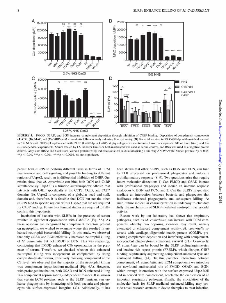
FMOD, OSAD, and BGN increase C3b and MAC deposition bypreventing C4BP binding to M. catarrhalis
To further understand how SLRPs regulate complement leading to theenhanced serum sensitivity of M. catarrhalis, we measured deposi-tion of complement components on the bacterial surface in thepresence of SLRPs, BSA, or no added protein using flow cytometry.In agreement with decreased survival of M. catarrhalis in serum,FMOD, OSAD, and BGN significantly increased C3b depositioncompared with BSA (gray stars), whereas only OSAD significantlyincreased C3b deposition compared with no protein control(Fig. 5A). Next, we looked at MAC deposition following incubationfor 20 min in serum, a shortened time to prevent significant lysis.
Complementing the serum killing and C3b deposition results,FMOD, OSAD, and BGN had significantly more MAC depositedon the bacterial surface compared with BSA or DCN (Fig. 5B).As acquisition of C4BP by M. catarrhalis is an efficient
strategy to prevent complement-mediated lysis and is facilitatedthrough interaction with UspA1 and UspA2 (6), we hypothesizedthat SLRPs FMOD, OSAD, and BGN may competitively inhibitbinding of C4BP and, thus, render M. catarrhalis more suscep-tible to serum killing. We measured C4BP binding followingincubation in OmCI-treated serum and showed a significant de-crease when bacteria were previously incubated with FMOD,OSAD, and BGN, with again no difference observed with DCNor BSA (Fig. 5C).To confirm our results that SLRPs inhibit binding of C4BP and,
thus, disrupt a major immune evasive strategy of M. catarrhalis,
we depleted C4BP from NHS using an anti-C4BP mAb MK104-coupled column, which interacts with high affinity to the a-chain
complement control protein (CCP) domain 1 of C4BP (17).
C4BP-dpl resulted in increased killing of M. catarrhalis RH4compared with NHS in the presence of both BSA and DCN
(Fig. 5D). Increased survival comparable to NHS was observed
following replenishment of purified C4BP to physiological levels(200 mg/ml) when BSA and DCN were present. In comparison,
FMOD, OSAD, and BGN enhanced serum bactericidal activity in
NHS compared with both BSA and DCN as observed previously
(Fig. 2A, 2B). Importantly, no significant change in serum killingwas observed between BSA/DCN and FMOD/OSAD/BGN in
C4BP-dpl (Fig. 5D), confirming that prevention of C4BP binding
by FMOD, OSAD, and BGN to the bacterial surface is responsiblefor the increased complement-mediated killing of M. catarrhalis.
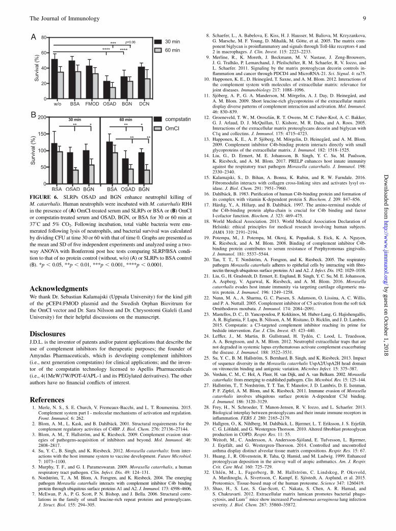
OSAD and BGN enhance neutrophil killing of M. catarrhalis ina complement- dependent and independent manner
Neutrophils represent a critical phagocytic cell type in innateimmunity, central to host defense against invading pathogens.Additionally, complement-mediated opsonization acceleratesphagocytosis and removal of pathogenic bacteria. Considering thatSLRP-bound bacteria had increased complement deposition inthe presence of serum, we wanted to investigate whether thistranslated into increased killing in a neutrophil bactericidal assay.Interestingly, in the presence of human neutrophils and OmCI-treated serum, both OSAD and BGN significantly enhancedM. catarrhalis RH4 killing observed at both 30 and 60 min in-cubation periods (Fig. 6A). Incubation with FMOD or DCN didnot significantly increase bacterial killing compared with BSA.Next, we wanted to investigate whether this enhanced neutro-phil killing was dependent on complement opsonization orwhether SLRPs themselves could serve as mediators of enhancedneutrophil killing. Using OSAD and BGN, we repeated the neu-trophil bactericidal assays with either OmCI-treated serum (inhib-iting complement at the C5 level) or compstatin-treated serum(inhibiting complement at the C3 level). At 30 min, we observedonly a decrease in bacterial survival in the OmCI-treated serumconditions and not in the presence of compstatin (Fig. 6B). Sur-prisingly, after 60 min we observed a statistically significant de-crease in survival both with the OmCI- and compstatin-treated seracompared with BSA. This suggests that the main mechanism ofSLRP-dependently enhanced neutrophil killing is via complementactivation. After a prolonged incubation time, however, SLRPspromote a bactericidal killing effect in concert with neutrophils thatis independent of complement.As compstatin-treated serum still contains C1q, which can act as
an opsonin and promote phagocytosis, and as previous work hasshown that SLRPs can interact with C1q (11, 12), we investigated
FIGURE 2. FMOD, OSAD, and BGN enhance serum killing of
M. catarrhalis strains (A) RH4 and (B) Bc5. Bacterial survival in human
serum was defined as the ratio (percentage) of the CFUs at 30 min to time
0. Error bars represent SD of three independent experiments. Serum treated
by C5 inhibitor OmCI was used as serum control, and 50 mg/ml BSA was
used as a negative protein control. Statistical differences were calculated
using a one-way ANOVA with Dunnett posttest versus bacteria without
SLRPs. *p , 0.05, **p , 0.01, ***p , 0.001, ****p , 0.0001.
The Journal of Immunology 5
by guest on October 1, 2018
http://ww
w.jim
munol.org/
Dow
nloaded from

the binding of C1q from serum in the presence of SLRPs(Supplemental Fig. 3). We observed no difference in binding ofC1q to the bacterial surface when bacteria were incubated withFMOD or DCN compared with BSA. In contrast, a significantreduction in C1q binding was shown when bacteria were boundwith OSAD and BGN. Therefore, these results indicate that en-hanced neutrophil killing under compstatin-treated serum condi-tions in the presence of OSAD and BGN was not due to increasedC1q binding.
DiscussionM. catarrhalis causes significant morbidity in children and chronicobstructive pulmonary disease patients and is responsible for aplethora of respiratory infections and occasionally systemic diseases(26). The exact molecular mechanisms governing M. catarrhalispathogenicity are not fully understood. However, mounting evidencesuggests that immune evasion, directed primarily at circumventing
the complement system, is an essential feature of pathogenic strains(4–6, 14, 21, 27). Therefore, future treatment intervention directed athampering complement inhibitor recruitment is a promising avenueof research. In this study, we highlight a novel antimicrobial roledisplayed by specific SLRPs, namely FMOD, OSAD, and BGN, andreveal in detail the molecular mechanisms resulting in enhanced in-nate immunity against M. catarrhalis.SLRPs, such as BGN and DCN, are considered bifunctional
proteoglycans, acting both as central structural components of theECM and danger-associated molecular patterns stimulating im-mune reactions (28). SLRPs are abundantly present in the ECMand distributed in numerous tissues throughout the body (7).Previous immumohistochemical analysis has shown that BGN andDCN are expressed in the human lung and bronchial tissue (29–31). Furthermore, mining of the Human Protein Atlas (www.proteinatlas.org) (32), a genome-wide analysis of RNA and pro-tein expression from samples representing major tissues and
FIGURE 3. SLRPs interact directly with UspA2/2H of M. catarrhalis. Biotinylated SLRPs were incubated with M. catarrhalis mutants devoid of se-
lected surface proteins UspA1, UspA2/2H, and MID that interact with various host proteins. Bound SLRPs were detected with fluorescently labeled
streptavidin by measuring fluorescence intensity using a CytoFLEX flow cytometer. UspA2/2H mutant showed significantly reduced binding of all SLRPs
(A–D). Biotinylated SLRPs (E) (FMOD and OSAD) and (F) (BGN and DCN) bind immobilized recombinant UspA2 with differing affinities. Error bars
represent SD of three independent experiments. Statistical differences were calculated using a one-way ANOVA with Dunnett posttest versus wild-type
(RH4) bacteria. **p , 0.01, ***p , 0.001, ****p , 0.0001.
6 SLRPs ENHANCE KILLING OF M. CATARRHALIS
by guest on October 1, 2018
http://ww
w.jim
munol.org/
Dow
nloaded from

organs, confirmed the expression of all SLRPs used in this study inlung tissue. RNA expression data generated from 320 individualtissue samples showed that for this set of SLRPs, BGN and DCNhad the highest expression, followed by FMOD, with OSADhaving the lowest expression (Supplemental Fig. 4). Combined,these expression data and previous immunohistochemical analysisindicate that these SLRPs are present in sites anatomically im-portant for M. catarrhalis infection and, therefore, may play a rolein host innate immune defense. The exact concentrations ofSLRPs present in human tissues and/or plasma are difficult toestimate. One reason for this is that SLRPs are present in higherconcentrations following trauma, proteolysis of the ECM, andunder sterile and nonsterile inflammatory conditions. Previouswork in the field has shown that both BGN and DCN expression isenhanced during experimental sepsis in murine models followingLPS challenge (8, 9). Macrophages were observed to be the mainsecretory cell responsible for enhanced expression. Followingstimulation with IL-1b and IL-6, macrophage increased BGNsecretion, which in turn induced expression of TNF-a and MIP-2,contributing to the overall proinflammatory environment and in-creased SLRP expression (8). Furthermore, DCN expression washigher in cohorts of septic patients compared with healthy controls(9). In this study, DCN concentration in plasma was estimated tobe at 10 ng/ml in septic patients. Previous studies by this groupalso estimated another SLRP, proline/arginine-rich end leucine-rich repeat protein (PRELP), to be present at a similar range inbronchoalveolar lavage fluid (14). It is tempting to speculate thatduring infection and particularly sepsis, SLRP expression is in-creased as a result of secretion of proinflammatory cytokinesstimulating macrophages and other immune and nonimmune cells,
while at the same time the highly inflamed environment contrib-utes to increased SLRP proteolysis from the ECM. Therefore,during infection, the local concentration of SLRPs may be higherthan the surrounding environment, which could influence com-plement and innate immune activity and bacterial survival.Of the three main Gram-negative respiratory pathogens screened in
this study, only M. catarrhalis was bound by SLRPs. M. catarrhalisexpresses numerous surface proteins, which bind an array ofECM proteins, plasma, and complement components, permittingcolonization and evasion of the host innate and adaptive immunity(4, 26). The major surface proteins of M. catarrhalis are the UspA1and UspA2/2H. Both UspA1 and A2/2H interact with C4BP; how-ever, UspA2/2H is more strongly expressed than UspA1 and,therefore, plays a more prominent role in C4BP binding and inconferring a complement resistant phenotype (6). Through muta-tional analysis, we determined that all four SLRPs bound toM. catarrhalis predominantly through UspA2/2H. This sug-gested that SLRP binding to UspA2/2H could competitivelyinhibit C4BP binding, resulting in reduced complement inhi-bition. Using flow cytometry, we illustrated that prior bindingof FMOD, OSAD, and BGN but not DCN to M. catarrhaliseffectively reduced C4BP binding, thus explaining the in-creased serum sensitivity.Given the similarity between BGN and DCN, it is surprising that
BGN and not DCN competitively inhibits C4BP binding. BothBGN and DCN are members of the class I SLRP family, possessingsignificant homology at both the protein and genetic level. BGNcontains two N-terminal tissue-specific chondroitin/dermatansulfate side chains, whereas DCN contains one, and both differin the pattern and level of glycosylation (7). These differences
FIGURE 4. SLRPs bind to M. catarrhalis clinical isolates expressing UspA2/2H. The highly diverse head domains of UspA2/2H are classified into
N-terminal repeats (NTER) 2A, 2B, 2C, and 2H, and nontypeable. All tested clinical strains bound SLRPs (A) FMOD, (B) OSAD, (C) BGN, and (D) DCN
at various degrees. Negative control consisted of bacterial straining with streptavidin–AF647 in the absence of biotinylated proteins. Error bars represent the
SD of three individual experiments.
The Journal of Immunology 7
by guest on October 1, 2018
http://ww
w.jim
munol.org/
Dow
nloaded from
permit both SLRPs to perform different tasks in terms of ECMmaintenance and cell signaling and possibly binding to differentregions of UspA2, resulting in differential inhibition of C4BP. Ourresults show that M. catarrhalis can bind both DCN and C4BPsimultaneously. UspA2 is a trimeric autotransporter adhesin thatinteracts with C4BP specifically at the CCP2, CCP5, and CCP7domains (6). UspA2 is composed of a globular head and stalkdomain and, therefore, it is feasible that DCN but not the otherSLRPs bind to specific regions within UspA2 that are not requiredfor C4BP binding. Future biochemical studies are required to fullyconfirm this hypothesis.Incubation of bacteria with SLRPs in the presence of serum
resulted in significant opsonization with C3b/iC3b (Fig. 5A). Asthese opsonins are recognized by complement receptors presenton neutrophils, we wished to examine where this resulted in en-hanced neutrophil bactericidal killing. In this study, we observedthat only OSAD and BGN effectively increased neutrophil killingof M. catarrhalis but not FMOD or DCN. This was surprising,considering that FMOD enhanced C3b opsonization in the pres-ence of serum. Therefore, we checked whether this enhancedneutrophil killing was independent of complement by usingcompstatin-treated serum, effectively blocking complement at theC3 level. We observed that the majority of the neutrophil killingwas complement (opsonization)-mediated (Fig. 6A). However,with prolonged incubation, both OSAD and BGN enhanced killingin a complement (opsonization)-independent manner. It is knownthat certain ECM proteins, such as the SLRP lumican, can en-hance phagocytosis by interacting with both bacteria and phago-cytes via surface-expressed integrins (33). Additionally, it has
been shown that other SLRPs, such as BGN and DCN, can bindto TLR expressed on professional phagocytes and induce aproinflammatory response (8, 9). Two questions arise that requirefuture molecular dissection: 1) Can FMOD and OSAD interactwith professional phagocytes and induce an immune responseanalogous to BGN and DCN; and 2) Can the SLRPs in questionmediate an interaction between bacteria and phagocytes thatfacilitates enhanced phagocytosis and subsequent killing. Assuch, future molecular characterization is underway to elucidatefully the mechanisms of SLRP-mediated neutrophil bactericidalactivity.Recent work by our laboratory has shown that respiratory
pathogens, such as M. catarrhalis, can interact with ECM com-ponents whereby two opposing scenarios may result, namelyattenuated or enhanced complement activity. M. catarrhalis in-teracts with cartilage oligomeric matrix protein (COMP), pre-venting complement deposition and interfering with complement-independent phagocytosis, enhancing survival (21). Conversely,M. catarrhalis can be bound by the SLRP proline/arginine-richend leucine-rich repeat protein (PRELP), which disrupts C4BPbinding, significantly augmenting complement-mediated lysis andneutrophil killing (14). To this complex interaction betweencomplement, M. catarrhalis, and ECM components we introducethe newfound antibacterial role of FMOD, OSAD, and BGN,which through interaction with the surface-expressed UspA2/2Hand in concert with complement, accelerate the eradication of animportant respiratory pathogen. Finally, the elucidation of themolecular basis for SLRP-mediated–enhanced killing may pro-vide novel research avenues to devise therapies to treat infection.
FIGURE 5. FMOD, OSAD, and BGN increase complement deposition through inhibition of C4BP binding. Deposition of complement components
(A) C3b, (B), MAC, and (C) C4BP onM. catarrhalis RH4 was analyzed using flow cytometry. (D) Bacterial survival in 5% C4BP-dpl with matched survival
in 5% NHS and C4BP-dpl replenished with C4BP (C4BP-dpl + C4BP) at physiological concentrations. Error bars represent SD of three (A–C) and five
(D) independent experiments. Serum treated by C5 inhibitor OmCI or heat-inactivated was used as serum control, and BSAwas used as a negative protein
control. Gray stars (BSA) and black stars (without protein [w/o]) indicate statistical calculations using a one-way ANOVAwith Dunnett posttest. *p, 0.05,
**p , 0.01, ***p , 0.001, ****p , 0.0001. ns, not significant.
8 SLRPs ENHANCE KILLING OF M. CATARRHALIS
by guest on October 1, 2018
http://ww
w.jim
munol.org/
Dow
nloaded from
AcknowledgmentsWe thank Dr. Sebastian Kalamajski (Uppsala University) for the kind gift
of the pCEP4:FMOD plasmid and the Swedish Orphan Biovitrum for
the OmCI vector and Dr. Sara Nilsson and Dr. Chrysostomi Gialeli (Lund
University) for their helpful discussions on the manuscript.
DisclosuresJ.D.L. is the inventor of patents and/or patent applications that describe the
use of complement inhibitors for therapeutic purposes; the founder of
Amyndas Pharmaceuticals, which is developing complement inhibitors
(i.e., next generation compstatins) for clinical applications; and the inven-
tor of the compstatin technology licensed to Apellis Pharmaceuticals
(i.e., 4(1MeW)7W/POT-4/APL-1 and its PEGylated derivatives). The other
authors have no financial conflicts of interest.
References1. Merle, N. S., S. E. Church, V. Fremeaux-Bacchi, and L. T. Roumenina. 2015.
Complement system part I - molecular mechanisms of activation and regulation.Front. Immunol. 6: 262.
2. Blom, A. M., L. Kask, and B. Dahlback. 2001. Structural requirements for thecomplement regulatory activities of C4BP. J. Biol. Chem. 276: 27136–27144.
3. Blom, A. M., T. Hallstrom, and K. Riesbeck. 2009. Complement evasion strat-egies of pathogens-acquisition of inhibitors and beyond. Mol. Immunol. 46:2808–2817.
4. Su, Y. C., B. Singh, and K. Riesbeck. 2012. Moraxella catarrhalis: from inter-actions with the host immune system to vaccine development. Future Microbiol.7: 1073–1100.
5. Murphy, T. F., and G. I. Parameswaran. 2009. Moraxella catarrhalis, a humanrespiratory tract pathogen. Clin. Infect. Dis. 49: 124–131.
6. Nordstrom, T., A. M. Blom, A. Forsgren, and K. Riesbeck. 2004. The emergingpathogen Moraxella catarrhalis interacts with complement inhibitor C4b bindingprotein through ubiquitous surface proteins A1 and A2. J. Immunol. 173: 4598–4606.
7. McEwan, P. A., P. G. Scott, P. N. Bishop, and J. Bella. 2006. Structural corre-lations in the family of small leucine-rich repeat proteins and proteoglycans.J. Struct. Biol. 155: 294–305.
8. Schaefer, L., A. Babelova, E. Kiss, H. J. Hausser, M. Baliova, M. Krzyzankova,G. Marsche, M. F. Young, D. Mihalik, M. Gotte, et al. 2005. The matrix com-ponent biglycan is proinflammatory and signals through Toll-like receptors 4 and2 in macrophages. J. Clin. Invest. 115: 2223–2233.
9. Merline, R., K. Moreth, J. Beckmann, M. V. Nastase, J. Zeng-Brouwers,J. G. Tralhao, P. Lemarchand, J. Pfeilschifter, R. M. Schaefer, R. V. Iozzo, andL. Schaefer. 2011. Signaling by the matrix proteoglycan decorin controls in-flammation and cancer through PDCD4 and MicroRNA-21. Sci. Signal. 4: ra75.
10. Happonen, K. E., D. Heinegard, T. Saxne, and A. M. Blom. 2012. Interactions ofthe complement system with molecules of extracellular matrix: relevance forjoint diseases. Immunobiology 217: 1088–1096.
11. Sjoberg, A. P., G. A. Manderson, M. Morgelin, A. J. Day, D. Heinegard, andA. M. Blom. 2009. Short leucine-rich glycoproteins of the extracellular matrixdisplay diverse patterns of complement interaction and activation.Mol. Immunol.46: 830–839.
12. Groeneveld, T. W., M. Oroszlan, R. T. Owens, M. C. Faber-Krol, A. C. Bakker,G. J. Arlaud, D. J. McQuillan, U. Kishore, M. R. Daha, and A. Roos. 2005.Interactions of the extracellular matrix proteoglycans decorin and biglycan withC1q and collectins. J. Immunol. 175: 4715–4723.
13. Happonen, K. E., A. P. Sjoberg, M. Morgelin, D. Heinegard, and A. M. Blom.2009. Complement inhibitor C4b-binding protein interacts directly with smallglycoproteins of the extracellular matrix. J. Immunol. 182: 1518–1525.
14. Liu, G., D. Ermert, M. E. Johansson, B. Singh, Y. C. Su, M. Paulsson,K. Riesbeck, and A. M. Blom. 2017. PRELP enhances host innate immunityagainst the respiratory tract pathogen Moraxella catarrhalis. J. Immunol. 198:2330–2340.
15. Kalamajski, S., D. Bihan, A. Bonna, K. Rubin, and R. W. Farndale. 2016.Fibromodulin interacts with collagen cross-linking sites and activates lysyl ox-idase. J. Biol. Chem. 291: 7951–7960.
16. Dahlback, B. 1983. Purification of human C4b-binding protein and formation ofits complex with vitamin K-dependent protein S. Biochem. J. 209: 847–856.
17. Hardig, Y., A. Hillarp, and B. Dahlback. 1997. The amino-terminal module ofthe C4b-binding protein alpha-chain is crucial for C4b binding and factorI-cofactor function. Biochem. J. 323: 469–475.
18. World Medical Association. 2013. World Medical Association Declaration ofHelsinki: ethical principles for medical research involving human subjects.JAMA 310: 2191–2194.
19. Potempa, M., J. Potempa, M. Okroj, K. Popadiak, S. Eick, K. A. Nguyen,K. Riesbeck, and A. M. Blom. 2008. Binding of complement inhibitor C4b-binding protein contributes to serum resistance of Porphyromonas gingivalis.J. Immunol. 181: 5537–5544.
20. Tan, T. T., T. Nordstrom, A. Forsgren, and K. Riesbeck. 2005. The respiratorypathogen Moraxella catarrhalis adheres to epithelial cells by interacting with fibro-nectin through ubiquitous surface proteins A1 and A2. J. Infect. Dis. 192: 1029–1038.
21. Liu, G., H. Gradstedt, D. Ermert, E. Englund, B. Singh, Y. C. Su, M. E. Johansson,A. Aspberg, V. Agarwal, K. Riesbeck, and A. M. Blom. 2016. Moraxellacatarrhalis evades host innate immunity via targeting cartilage oligomeric ma-trix protein. J. Immunol. 196: 1249–1258.
22. Nunn, M. A., A. Sharma, G. C. Paesen, S. Adamson, O. Lissina, A. C. Willis,and P. A. Nuttall. 2005. Complement inhibitor of C5 activation from the soft tickOrnithodoros moubata. J. Immunol. 174: 2084–2091.
23. Mastellos, D. C., D. Yancopoulou, P. Kokkinos, M. Huber-Lang, G. Hajishengallis,A. R. Biglarnia, F. Lupu, B. Nilsson, A. M. Risitano, D. Ricklin, and J. D. Lambris.2015. Compstatin: a C3-targeted complement inhibitor reaching its prime forbedside intervention. Eur. J. Clin. Invest. 45: 423–440.
24. Leffler, J., M. Martin, B. Gullstrand, H. Tyden, C. Lood, L. Truedsson,A. A. Bengtsson, and A. M. Blom. 2012. Neutrophil extracellular traps that arenot degraded in systemic lupus erythematosus activate complement exacerbatingthe disease. J. Immunol. 188: 3522–3531.
25. Su, Y. C., B. M. Hallstrom, S. Bernhard, B. Singh, and K. Riesbeck. 2013. Impactof sequence diversity in the Moraxella catarrhalis UspA2/UspA2H head domainon vitronectin binding and antigenic variation. Microbes Infect. 15: 375–387.
26. Verduin, C. M., C. Hol, A. Fleer, H. van Dijk, and A. van Belkum. 2002. Moraxellacatarrhalis: from emerging to established pathogen. Clin. Microbiol. Rev. 15: 125–144.
27. Hallstrom, T., T. Nordstrom, T. T. Tan, T. Manolov, J. D. Lambris, D. E. Isenman,P. F. Zipfel, A. M. Blom, and K. Riesbeck. 2011. Immune evasion of Moraxellacatarrhalis involves ubiquitous surface protein A-dependent C3d binding.J. Immunol. 186: 3120–3129.
28. Frey, H., N. Schroeder, T. Manon-Jensen, R. V. Iozzo, and L. Schaefer. 2013.Biological interplay between proteoglycans and their innate immune receptors ininflammation. FEBS J. 280: 2165–2179.
29. Hallgren, O., K. Nihlberg, M. Dahlback, L. Bjermer, L. T. Eriksson, J. S. Erjefalt,C. G. Lofdahl, and G. Westergren-Thorsson. 2010. Altered fibroblast proteoglycanproduction in COPD. Respir. Res. 11: 55.
30. Weitoft, M., C. Andersson, A. Andersson-Sjoland, E. Tufvesson, L. Bjermer,J. Erjefalt, and G. Westergren-Thorsson. 2014. Controlled and uncontrolledasthma display distinct alveolar tissue matrix compositions. Respir. Res. 15: 67.
31. Huang, J., R. Olivenstein, R. Taha, Q. Hamid, and M. Ludwig. 1999. Enhancedproteoglycan deposition in the airway wall of atopic asthmatics. Am. J. Respir.Crit. Care Med. 160: 725–729.
32. Uhlen, M., L. Fagerberg, B. M. Hallstrom, C. Lindskog, P. Oksvold,A. Mardinoglu, A. Sivertsson, C. Kampf, E. Sjostedt, A. Asplund, et al. 2015.Proteomics. Tissue-based map of the human proteome. Science 347: 1260419.
33. Shao, H., S. Lee, S. Gae-Scott, C. Nakata, S. Chen, A. R. Hamad, andS. Chakravarti. 2012. Extracellular matrix lumican promotes bacterial phago-cytosis, and Lum-/- mice show increased Pseudomonas aeruginosa lung infectionseverity. J. Biol. Chem. 287: 35860–35872.
FIGURE 6. SLRPs OSAD and BGN enhance neutrophil killing of
M. catarrhalis. Human neutrophils were incubated with M. catarrhalis RH4
in the presence of (A) OmCI-treated serum and SLRPs or BSA or (B) OmCI
or compstatin-treated serum and OSAD, BGN, or BSA for 30 or 60 min at
37˚C and 5% CO2. Following incubation, total viable bacteria were enu-
merated following lysis of neutrophils, and bacterial survival was calculated
by dividing CFU at time 30 or 60 with that of time 0. Graphs are presented as
the mean and SD of five independent experiments and analyzed using a two-
way ANOVA with Bonferroni post hoc tests comparing SLRP/BSA condi-
tion to that of no protein control (without, w/o) (A) or SLRPs to BSA control
(B). *p , 0.05, **p , 0.01, ***p , 0.001, ****p , 0.0001.
The Journal of Immunology 9
by guest on October 1, 2018
http://ww
w.jim
munol.org/
Dow
nloaded from

34. Forsgren, A., M. Brant, A. Mollenkvist, A. Muyombwe, H. Janson, N. Woin, andK. Riesbeck. 2001. Isolation and characterization of a novel IgD-binding proteinfrom Moraxella catarrhalis. J. Immunol. 167: 2112–2120.
35. Christensen, J. J., J. Ursing, and B. Bruun. 1994. Genotypic and phenotypicrelatedness of 80 strains of Branhamella catarrhalis of worldwide origin. FEMSMicrobiol. Lett. 119: 155–159.
36. Nordstrom, T., J. Jendholm, M. Samuelsson, A. Forsgren, and K. Riesbeck.2006. The IgD-binding domain of the Moraxella IgD-binding protein MID(MID962-1200) activates human B cells in the presence of T cell cytokines.J. Leukoc. Biol. 79: 319–329.
37. Kroll, J. S., and E. R. Moxon. 1988. Capsulation and gene copy number at thecap locus of Haemophilus influenzae type b. J. Bacteriol. 170: 859–864.
10 SLRPs ENHANCE KILLING OF M. CATARRHALIS
by guest on October 1, 2018
http://ww
w.jim
munol.org/
Dow
nloaded from