Embed Size (px)
Citation preview
1346
Short Communications
Sequential Cerebral Blood Flow Changes inShort-term Cerebral Ischemia in GerbilsHiroyuki Kato, MD, DMSc, Tsutomu Araki, PhD, Kyuya Kogure, MD, DMSc,
Matsutaro Murakami, PhD, and Kazuo Uemura, MD, DMSc
Using quantitative autoradiography, we studied sequential changes in regional cerebral bloodflow during and after 2 minutes of bilateral common carotid artery occlusion in 18 gerbils.Occlusion (n=4) led to severe ischemia in the forebrain (regional cerebral blood flow <5% ofcontrol [ft=4]) and midbrain (regional cerebral blood flow <10% of control), but wasmorphologically nonlethal. Reperfusion of the brain was complete, and regional cerebral bloodflow was not different from control 1 minute after ischemia (ft=4), but hypoperfusion (regionalcerebral blood flow 30-50% of control) occurred at 5 minutes (ft=3) and was pronounced at 1hour (n=4); at this stage blood flow was inhomogeneous. Hypoperfusion had disappeared at 4hours (n=3). Our results indicate that the well-documented sequence of cerebral blood flowchanges (i.e., ischemia, initial recovery of blood flow, and delayed hypoperfusion) takes placeeven after nonlethal cerebral ischemia. (Stroke 1990;21:1346-1349)
Bilateral common carotid artery occlusion ingerbils leads to severe forebrain ischemia in>90% of animals because of a unique anom-
aly of the circle of Willis.1-3 Five minutes of ischemiadamages only neurons in the hippocampus,2-4'5
whereas 10-15 minutes of ischemia causes extensiveneuronal damage to selectively vulnerable regions.5
This damage occurs not only in forebrain regions(such as the hippocampal CA1 subfield, the striatum,thalamus, and neocortical layers 3 and 5) but also inmidbrain structures (i.e., the medial geniculate body,substantia nigra, and inferior colliculus).5
On the other hand, repeated brief bilateral carotidartery occlusions in gerbils produce cumulative dam-age to the brain. Tomida et al6 found that damagefollowing three 5-minute ischemic insults at 1-hourintervals is greater than that following a single 15-minute ischemic insult and concluded that postisch-emic hypoperfusion plays an important role. We havereported that repeated 2-minute ischemic insults,each of which is nonlethal to the brain when givensingly, produce severe neuronal damage.78 However,whether hypoperfusion takes place after nonlethal2-minute ischemic insults is not known.
From the Department of Neurology (H.K., T.A., K.K.), Instituteof Brain Diseases, Tohoku University School of Medicine, Sendai,and the Department of Radiology and Nuclear Medicine (M.M.,K.U.), Research Institute of Brain and Blood Vessels, Akita,Japan.
Address for correspondence: Hiroyuki Kato, MD, Department ofNeurology, Institute of Brain Diseases, Tohoku University School ofMedicine, 1-1 Seiryo-machi, Aoba-ku, Sendai 980, Japan.
Received February 23, 1990; accepted May 1, 1990.
The purpose of this study was to visualize andquantify using autoradiography the extent anddegree of regional cerebral blood flow (rCBF)changes during and after 2 minutes of cerebralischemia in gerbils.
Materials and MethodsWe used a total of 33 adult male Mongolian gerbils
weighing 70-90 g. Anesthesia was induced with 2%halothane and maintained with 1% halothane in 30%O2 and 70% N2O. A midcervical skin incision wasmade, and both common carotid arteries were gentlyexposed. Nine control gerbils received no occlusion,but in the remaining 24 experimental gerbils thearteries were occluded with aneurysm clips for 2minutes. In 14 gerbils for autoradiography thecarotid arteries were reperfused by removing theclips for 1 (n=4) or 5 («=3) minutes or 1 (w=4) or 4(n=3) hours. In all gerbils rectal temperature wasmaintained at around 37° C using a heating pad anda lamp.
For the autoradiographic determination of rCBF,a femoral artery and vein were cannulated for arte-rial blood sampling and radioisotope injection,respectively, in four control and 18 experimentalgerbils. At the indicated time, 10 fxCi of [14C]iodo-antipyrine (Amersham, Tokyo, Japan) dissolved inapproximately 0.7 ml saline was injected intrave-nously over 30 seconds, and six to eight-ju.1 timedarterial blood samples were collected. The gerbilswere then decapitated, and the brains were quicklyremoved and frozen in powdered dry ice. Frozensections 20 fim thick were cut in a cryostat at -18° C,
by guest on June 25, 2018http://stroke.ahajournals.org/
Dow
nloaded from
Kato et al CBF Changes After Brief Cerebral Ischemia 1347
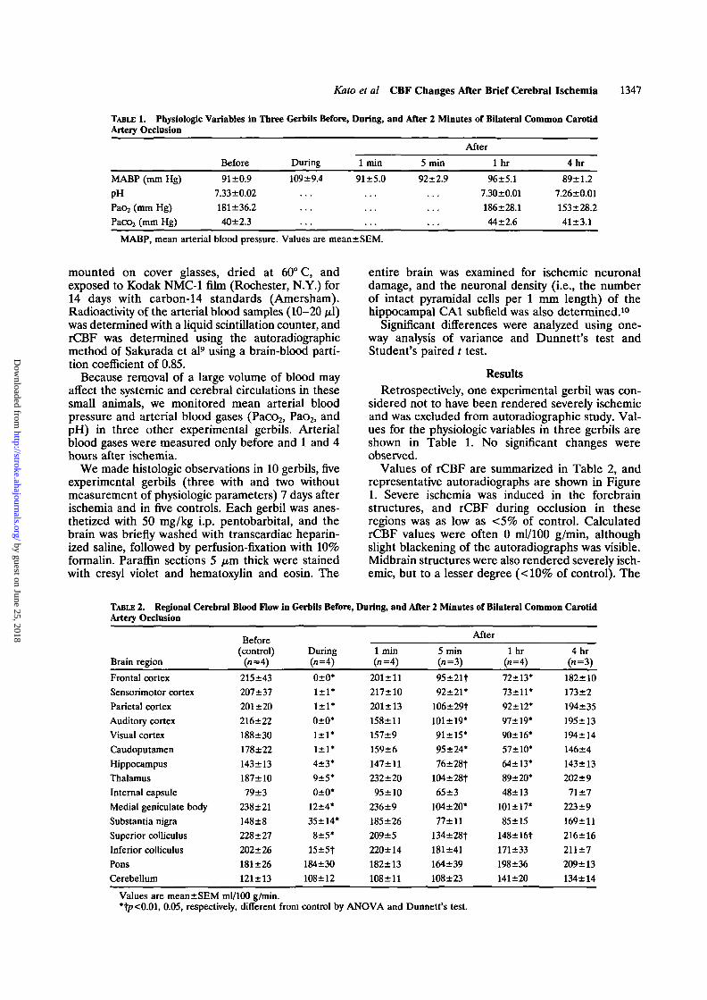
TABLE 1. Physiologic Variables in Three Gerbils Before,Artery Occlusion
Before During
During,
1 min
and After 2 Minutes of Bilateral
After
5 min 1 hr
Common Carotid
4hr
MABP (mm Hg)PHPa(>2 (mm Hg)Pacc>2 (mm Hg)
91 ±0.97.33±0.02181±36.240±2.3
109+9.4 91 ±5.0 92±2.9 92±2.97.30±0.01186±28.144±2.6
92±2.97.26±0.01153±28.241±3.1
MABP, mean arterial blood pressure. Values are mean±SEM.
mounted on cover glasses, dried at 60° C, andexposed to Kodak NMC-1 film (Rochester, N.Y.) for14 days with carbon-14 standards (Amersham).Radioactivity of the arterial blood samples (10-20 /xl)was determined with a liquid scintillation counter, andrCBF was determined using the autoradiographicmethod of Sakurada et al9 using a brain-blood parti-tion coefficient of 0.85.
Because removal of a large volume of blood mayaffect the systemic and cerebral circulations in thesesmall animals, we monitored mean arterial bloodpressure and arterial blood gases (Paco2, Pao2, andpH) in three other experimental gerbils. Arterialblood gases were measured only before and 1 and 4hours after ischemia.
We made histologic observations in 10 gerbils, fiveexperimental gerbils (three with and two withoutmeasurement of physiologic parameters) 7 days afterischemia and in five controls. Each gerbil was anes-thetized with 50 mg/kg i.p. pentobarbital, and thebrain was briefly washed with transcardiac heparin-ized saline, followed by perfusion-fixation with 10%formalin. Paraffin sections 5 pra thick were stainedwith cresyl violet and hematoxylin and eosin. The
entire brain was examined for ischemic neuronaldamage, and the neuronal density (i.e., the numberof intact pyramidal cells per 1 mm length) of thehippocampal CA1 subfield was also determined.10
Significant differences were analyzed using one-way analysis of variance and Dunnett's test andStudent's paired t test.
ResultsRetrospectively, one experimental gerbil was con-
sidered not to have been rendered severely ischemicand was excluded from autoradiographic study. Val-ues for the physiologic variables in three gerbils areshown in Table 1. No significant changes wereobserved.
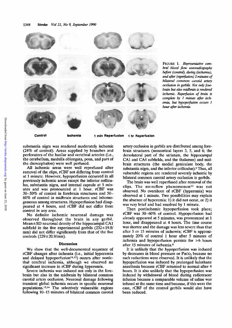
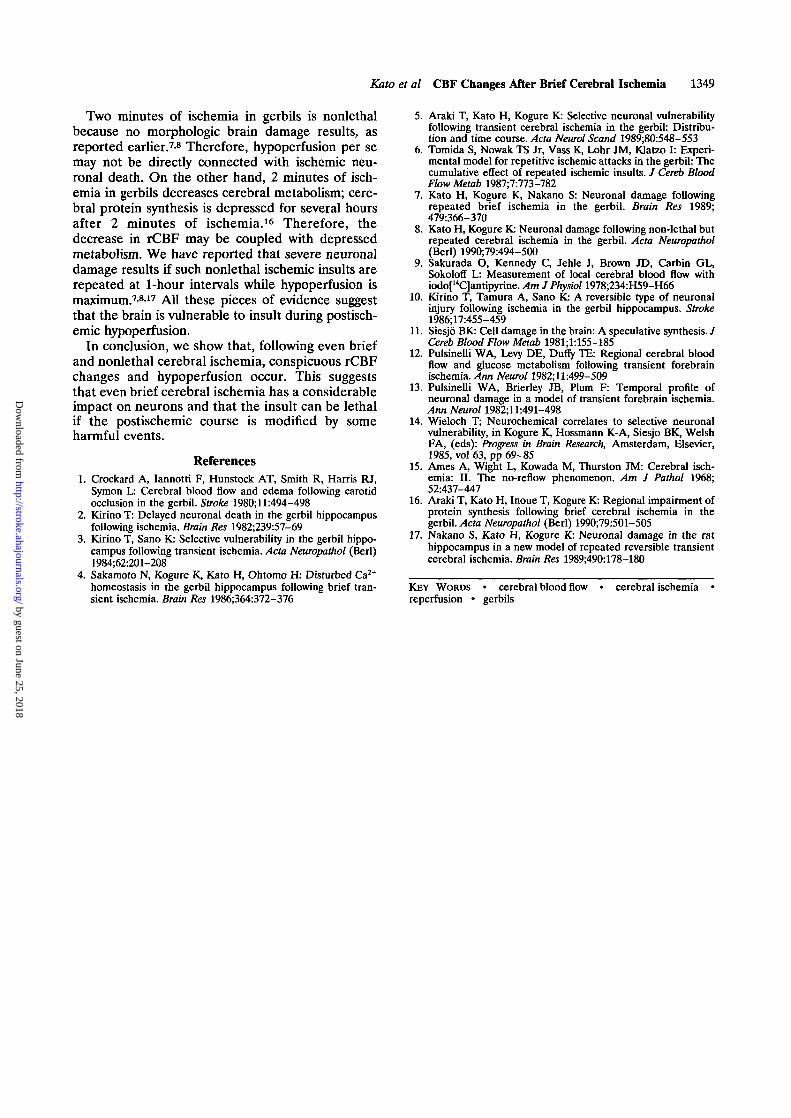
Values of rCBF are summarized in Table 2, andrepresentative autoradiographs are shown in Figure1. Severe ischemia was induced in the forebrainstructures, and rCBF during occlusion in theseregions was as low as <5% of control. CalculatedrCBF values were often 0 ml/100 g/min, althoughslight blackening of the autoradiographs was visible.Midbrain structures were also rendered severely isch-emic, but to a lesser degree (<10% of control). The
TABLE 2. Regional Cerebral Blood Flow in Gerbils Before, During, and After 2 Minutes of Bilateral Common CarotidArtery Occlusion
Brain regionFrontal cortexSensorimotor cortexParietal cortexAuditory cortexVisual cortexCaudoputamenHippocampusThalamusInternal capsuleMedial geniculate bodySubstantia nigraSuperior colliculusInferior colliculusPonsCerebellum
Before(control)
(«-4)215±43207 ±37201 ±20216±22188±30178±22143±13187±1079±3
238±21148±8228 ±27202±26181±26121 ±13
During(n=4)0±0*1±1*1±1*0±0*1±1*1±1*4±3*9±5*0±0*
12±4*35+14*
8±5*15±5t
184±30108±12
1 min(»=4)
201±ll217±10201 ±13158±11157±9159±6147±11232±2095 ±10
236±9185±26209 ±5220±14182±13108±ll
5 min(n=3)
95±21|92±21*
106±29t101+19*91±15*95 ±24*
76±28t104±28t65±3
104±20*77+11
134±28t181 ±41164±39108±23
After
lhr(n=4)
72±13*73±11*92±12*97±19*90±16*57±10*64±13*89±20*48±13
101±17*85 ±15
148±16t171±33198±36141 ±20
4hr(n=3)
182±10173±2194±35195±13194±14146±4143 ±13202±971 ±7
223±9169±11216±16211±7209±13134±14
Values are mean±SEM ml/100 g/min.*t/><0.01, 0.05, respectively, different from control by ANOVA and Dunnett's test.
by guest on June 25, 2018http://stroke.ahajournals.org/
Dow
nloaded from
1348 Stroke Vol 21, No 9, September 1990
i • •
FIGURE 1. Representative cere-bral blood flow autoradiographsbefore (control), during (ischemia),and after (reperfusion) 2 minutes ofbilateral common carotid arteryocclusion in gerbils. Not only fore-brain but also midbrain is renderedischemic. Reperfusion of brain iscomplete by 1 minute after isch-emia, but hypoperfusion occurs 1hour after ischemia.
Control Ischemia 1 mln Reperfusion 1 hr Reperfusion
substantia nigra was rendered moderately ischemic(24% of control). Areas supplied by branches andperforators of the basilar and vertebral arteries (i.e.,the cerebellum, medulla oblongata, pons, and part ofthe diencephalon) were well perfused.
All ischemic areas were well reperfused afterremoval of the clips, rCBF not differing from controlat 1 minute. However, hypoperfusion occurred in allpreviously ischemic areas except the inferior collicu-lus, substantia nigra, and internal capsule at 5 min-utes and was pronounced at 1 hour. rCBF was30-50% of control in forebrain structures and 50-60% of control in midbrain structures and inhomo-geneous among structures. Hypoperfusion had disap-peared at 4 hours, and rCBF did not differ fromcontrol in any area.
No definite ischemic neuronal damage wasobserved throughout the brain in any gerbil.Mean±SD neuronal density of the hippocampal CA1subfield in the five experimental gerbils (232±19.8/mm) did not differ significantly from that of the fivecontrols (229±20.9/mm).
DiscussionWe show that the well-documented sequence of
rCBF changes after ischemia (i.e., initial hyperemiaand delayed hypoperfusion11'12) occurs after nonle-thal cerebral ischemia, although we observed nosignificant increase in rCBF during hyperemia.
Severe ischemia was induced not only in the fore-brain but also in the midbrain by bilateral commoncarotid artery occlusion. Neuronal damage followingtransient global ischemia occurs in specific neuronalpopulations.13'14 The selectively vulnerable regionsfollowing 10-15 minutes of bilateral common carotid
artery occlusion in gerbils are distributed among fore-brain structures (neocortical layers 3, 5, and 6; thedorsolateral part of the striatum, the hippocampalCA1 and CA4 subfields, and the thalamus) and mid-brain structures (the medial geniculate body, thesubstantia nigra, and the inferior colliculus).5 Thus, allvulnerable regions are rendered severely ischemic bybilateral common carotid artery occlusion in gerbils.
The brain was well reperfused after removal of theclips. The no-reflow phenomenon15 was notobserved. No overshoot of rCBF (hyperemia) wasobserved at 1 minute. Two possibilities may explainthe absence of hyperemia: 1) it did not occur, or 2) itwas very brief and had resolved by 1 minute.
Then postischemic hypoperfusion took place;rCBF was 30-60% of control. Hypoperfusion hadalready appeared at 5 minutes, was pronounced at 1hour, and disappeared at 4 hours. The time coursewas shorter and the damage was less severe than thatafter 5 or 15 minutes of ischemia; rCBF is approxi-mately 20% of control 1 hour after 5 minutes ofischemia and hypoperfusion persists for >6 hoursafter 15 minutes of ischemia.6
It is unlikely that the hypoperfusion was inducedby decreases in blood pressure or Paco2 because nosuch reductions were observed. It is unlikely that thehypoperfusion was induced by prolonged halothaneanesthesia because rCBF returned to normal after 4hours. It is also unlikely that the hypoperfusion wasinduced by withdrawal of blood during radiotracerinfusion because a comparable volume of saline wasinfused at the same time and because, if this were thecase, rCBF of the control gerbils would also havebeen reduced.
by guest on June 25, 2018http://stroke.ahajournals.org/
Dow
nloaded from
Kato et al CBF Changes After Brief Cerebral Ischemia 1349
Two minutes of ischemia in gerbils is nonlethalbecause no morphologic brain damage results, asreported earlier.78 Therefore, hypoperfusion per semay not be directly connected with ischemic neu-ronal death. On the other hand, 2 minutes of isch-emia in gerbils decreases cerebral metabolism; cere-bral protein synthesis is depressed for several hoursafter 2 minutes of ischemia.16 Therefore, thedecrease in rCBF may be coupled with depressedmetabolism. We have reported that severe neuronaldamage results if such nonlethal ischemic insults arerepeated at 1-hour intervals while hypoperfusion ismaximum.'' All these pieces of evidence suggestthat the brain is vulnerable to insult during postisch-emic hypoperfusion.
In conclusion, we show that, following even briefand nonlethal cerebral ischemia, conspicuous rCBFchanges and hypoperfusion occur. This suggeststhat even brief cerebral ischemia has a considerableimpact on neurons and that the insult can be lethalif the postischemic course is modified by someharmful events.
References1. Crockard A, Iannotti F, Hunstock AT, Smith R, Harris RJ,
Symon L: Cerebral blood flow and edema following carotidocclusion in the gerbil. Stroke 1980;ll:494-498
2. Kirino T: Delayed neuronal death in the gerbil hippocampusfollowing ischemia. Brain Res 1982;239:57-69
3. Kirino T, Sano K: Selective vulnerability in the gerbil hippo-campus following transient ischemia. Acta Neuropathol (Berl)1984;62:201-208
4. Sakamoto N, Kogure K, Kato H, Ohtomo H: Disturbed Ca2+
homeostasis in the gerbil hippocampus following brief tran-sient ischemia. Brain Res 1986;364:372-376
5. Araki T, Kato H, Kogure K: Selective neuronal vulnerabilityfollowing transient cerebral ischemia in the gerbil: Distribu-tion and time course. Acta Neurol Scand 1989;80:548-553
6. Tomida S, Nowak TS Jr, Vass K, Lohr JM, Klatzo I: Experi-mental model for repetitive ischemic attacks in the gerbil: Thecumulative effect of repeated ischemic insults. / Cereb BloodFlow Metab 1987;7:773-782
7. Kato H, Kogure K, Nakano S: Neuronal damage followingrepeated brief ischemia in the gerbil. Brain Res 1989;479:366-370
8. Kato H, Kogure K: Neuronal damage following non-lethal butrepeated cerebral ischemia in the gerbil. Acta Neuropathol(Berl) 1990;79:494-500
9. Sakurada O, Kennedy C, Jehle J, Brown JD, Carbin GL,Sokoloff L: Measurement of local cerebral blood flow withiodo[MC]antipyrine. Am J Physiol 1978;234:H59-H66
10. Kirino T, Tamura A, Sano K: A reversible type of neuronalinjury following ischemia in the gerbil hippocampus. Stroke1986;17:455-459
11. Siesjo BK: Cell damage in the brain: A speculative synthesis./Cereb Blood Flow Metab 1981;1:155-185
12. Pulsinelli WA, Levy DE, Duffy TE: Regional cerebral bloodflow and glucose metabolism following transient forebrainischemia. Ann Neurol 1982; ll:499-509
13. Pulsinelli WA, Brierley JB, Plum F: Temporal profile ofneuronal damage in a model of transient forebrain ischemia.Ann Neurol 1982;ll:491-498
14. Wieloch T; Neurochemical correlates to selective neuronalvulnerability, in Kogure K, Hossmann K-A, Siesjo BK, WelshFA, (eds): Progress in Brain Research, Amsterdam, Elsevier,1985, vol 63, pp 69-85
15. Ames A, Wight L, Kowada M, Thurston JM: Cerebral isch-emia: II. The no-reflow phenomenon. Am J Pathol 1968;52:437-447
16. Araki T, Kato H, Inoue T, Kogure K: Regional impairment ofprotein synthesis following brief cerebral ischemia in thegerbil. Acta Neuropathol (Berl) 1990;79:501-505
17. Nakano S, Kato H, Kogure K: Neuronal damage in the rathippocampus in a new model of repeated reversible transientcerebral ischemia. Brain Res 1989;490:178-180
KEY WORDS • cerebral blood flowreperfusion • gerbils
cerebral ischemia
by guest on June 25, 2018http://stroke.ahajournals.org/
Dow
nloaded from
H Kato, T Araki, K Kogure, M Murakami and K UemuraSequential cerebral blood flow changes in short-term cerebral ischemia in gerbils.
Print ISSN: 0039-2499. Online ISSN: 1524-4628 Copyright © 1990 American Heart Association, Inc. All rights reserved.
is published by the American Heart Association, 7272 Greenville Avenue, Dallas, TX 75231Stroke doi: 10.1161/01.STR.21.9.1346
1990;21:1346-1349Stroke.
http://stroke.ahajournals.org/content/21/9/1346World Wide Web at:
The online version of this article, along with updated information and services, is located on the
http://stroke.ahajournals.org//subscriptions/
is online at: Stroke Information about subscribing to Subscriptions:
http://www.lww.com/reprints Information about reprints can be found online at: Reprints:
document. Permissions and Rights Question and Answer available in the
Permissions in the middle column of the Web page under Services. Further information about this process isOnce the online version of the published article for which permission is being requested is located, click Request
can be obtained via RightsLink, a service of the Copyright Clearance Center, not the Editorial Office.Stroke Requests for permissions to reproduce figures, tables, or portions of articles originally published inPermissions:
by guest on June 25, 2018http://stroke.ahajournals.org/
Dow
nloaded from