Embed Size (px)
Citation preview
International Journal of
Molecular Sciences
Review
Short-Chain Fatty Acids and Their Association withSignalling Pathways in Inflammation, Glucose andLipid Metabolism
Jin He 1,2,† , Peiwen Zhang 1,2,†, Linyuan Shen 1,2 , Lili Niu 1,2, Ya Tan 1,2,3, Lei Chen 1,2,Ye Zhao 1,2, Lin Bai 1,2, Xiaoxia Hao 1,2, Xuewei Li 1,2, Shunhua Zhang 1,2,* and Li Zhu 1,2,*
1 College of Animal Science and Technology, Sichuan Agricultural University, Chengdu 611130, China;[email protected] (J.H.); [email protected] (P.Z.); [email protected] (L.S.);[email protected] (L.N.); [email protected] (Y.T.); [email protected] (L.C.);[email protected] (Y.Z.); [email protected] (L.B.); [email protected] (X.H.); [email protected] (X.L.)
2 Farm Animal Genetic Resource Exploration and Innovation Key Laboratory of Sichuan Province,Sichuan Agricultural University, Chengdu 611130, China
3 Institute of Animal Husbandry and Veterinary, Guizhou Academy of Agricultural Science,Guiyang 550005, China
* Correspondence: [email protected] (S.Z.); [email protected] (L.Z.);Tel.: +86-28-8629-1133 (S.Z. & L.Z.)
† These authors contributed equally to this work.
Received: 11 July 2020; Accepted: 27 August 2020; Published: 2 September 2020�����������������
Abstract: Short-chain fatty acids (SCFAs), particularly acetate, propionate and butyrate, are mainlyproduced by anaerobic fermentation of gut microbes. SCFAs play an important role in regulatingenergy metabolism and energy supply, as well as maintaining the homeostasis of the intestinalenvironment. In recent years, many studies have shown that SCFAs demonstrate physiologicallybeneficial effects, and the signalling pathways related to SCFA production, absorption, metabolism,and intestinal effects have been discovered. Two major signalling pathways concerning SCFAs,G-protein-coupled receptors (GPRCs) and histone deacetylases (HDACs), are well recognized. In thisreview, we summarize the recent advances concerning the biological properties of SCFAs and thesignalling pathways in inflammation and glucose and lipid metabolism.
Keywords: short-chain fatty acids; inflammation; glycose and lipid metabolism; signalling pathways
1. Introduction
Short-chain fatty acids (SCFAs), also called volatile fatty acids, are organic linear carboxylic acidswith fewer than six carbons, including acetic acid, propionic acid, butyric acid, and valeric acid. Amongthem, acetate (C2), propionate (C3), and butyrate (C4) are the most abundant (≥95%) [1], and the C2, C3,and C4 are in an approximate molar ratio of 60:20:20, respectively [2,3]. Except for a small portion ofSCFAs obtained directly from food, most are produced by intestinal microbial anaerobic fermentation.Furthermore, around 500–600 mmol of SCFAs are produced in the intestinal tract per day, dependingon the diet, type and number of microbiomes, and residence time in the intestinal tract [4,5].
Dietary fibre is the main food component that affects the production of SCFAs, which are mainlyderived from plant foods. Humans lack enzymes that breakdown dietary fibre. Therefore, dietaryfibre passes through the upper digestive tract largely undigested and is fermented in the caecumand large intestine by anaerobic microorganisms. A key mechanism of metabolic regulation bythe gut microbiota is the production of SCFAs. Different intestinal microbes will produce differentamounts of SCFAs. Bacteroidetes (Gram negative) mainly produce acetate and propionate, whereas
Int. J. Mol. Sci. 2020, 21, 6356; doi:10.3390/ijms21176356 www.mdpi.com/journal/ijms
Int. J. Mol. Sci. 2020, 21, 6356 2 of 16
Firmicutes (Gram positive) use butyrate as the primary metabolic end product [5]. Human-derivedBifidobacterium breve UCC2003 and Bifidobacterium longum NCIMB 8809 use novel oligosaccharides toproduce acetate [6], and Bifidobacterium animalis subsp. Lactis GCL2505 can also increase the productionof acetate [7]. Although anaerobic fermentation of fibre by intestinal microorganisms is the largestsource of SCFAs, SCFAs are also formed as products from peptide and amino acid fermentation(less than 1%) [8,9]. Although diet and the microbiome are the main factors affecting the production ofSCFAs, species evolution and colonic environment have important effects [10,11].
Colonocytes absorb SCFAs after they are produced mainly via H+-dependent or sodium-dependentmonocarboxylate transporters [12]. After supplying colonocytes, the remaining SCFAs are transportedthrough the blood to various parts of the body. These SCFAs can be used as substrates to synthesizesugars or lipids and can also be used as cytokines to regulate metabolism [13–16]. These results showthat SCFAs are carried from the intestinal cavity into the blood vessels of the host and finally to organsas substrates or signalling molecules.
Additionally, a growing number of functions are attributed to SCFAs. For a long time, many studieshave argued that dietary fibre and resistant starch have many benefits, such as reducing cholesterollevels and maintaining normal blood glucose levels [17–19], and these benefits of high-fibre diets areat least a part put down to SCFAs. When SCFAs are produced in the gut, many of them are used asenergy sources. For humans, SCFAs provide approximately 10% of the daily calorie requirement [20].SCFAs also improve gut health through several partial effects, including maintaining the integrity ofthe intestinal barrier, producing mucus, preventing inflammation and reducing the risk of colorectalcancer [21–25]. Furthermore, the effects on activating brown adipose tissue [26], regulating livermitochondrial function [27], maintaining body energy homeostasis [28], controlling appetite [26] andsleep [29] are all related to SCFAs.
Among the many physiological functions of SCFAs, the regulation of inflammation and glucoseand lipid metabolism, has concerned most researchers. SCFAs play a very important role in thesephysiological processes, but how do SCFAs work? This mini review aims to summarize the currentknowledge concerning the biological properties of SCFAs and their signalling pathways in inflammationand glycose and lipid metabolism.
2. G Protein-Coupled Receptors (GPCRs)
G protein-coupled receptors (GPCRs), involving seven transmembrane domains, are the biggestreceptor family in mammals and participate in regulating almost all cell and physiological functions inthe body. GPCRs can bind chemicals in the extracellular environment, such as odourants, hormones,neurotransmitters, chemokines, sugars, lipids, and proteins. After being activated by a ligand, GPCRscan bind to four different heterotrimeric G proteins (Gs, Gi/o, Gq/11 and G12/13), which can influence theactivity of single or multiple effectors, such as second-messenger-producing enzymes or ion channels.Presently, GPR41 and GPR43 have been identified as the most important receptors of SCFAs in theGPCR family [30,31]. After the discovery of SCFA receptors, GPR41 was renamed free fatty acidreceptor 3 (FFAR3) and GPR43 was renamed FFAR2. Different SCFAs and receptors have differentaffinities. Specifically, in humans, the affinity ranking of FFAR2 is C2 = C3 > C4 > C5 = C1 and that ofFFAR3 is C3 = C4 = C5 > C2 > C1 [32,33]. Both FFAR2 and FFAR3 are associated with metabolic diseases,and they have become effective targets for the treatment of type 2 diabetes, asthma, cardiovasculardisease, as well as metabolic syndrome.
FFAR2 is widely expressed in vivo. FFAR2 is present in pancreatic islet α and β cells [34,35] andintestinal enteroendocrine cells (I and L cells) (Table 1) [36,37]. FFAR2 expression has been identifiedalong the entire gastrointestinal tract and white adipocytes [38,39]. Moreover, FFAR2 is present inmonocytes, neutrophils, eosinophils, intestinal Treg cells and other immune cells [40–42]. An increasingnumber of studies have confirmed the function of FFAR2. Attilio et al. found that FFAR2 stimulatesinsulin secretion and reduces apoptosis in mouse and human islets in vitro [43]. Kendle et al. foundthat FFAR2 gene knockout mice showed significant aggravation in colitis, arthritis and asthma [40].
Int. J. Mol. Sci. 2020, 21, 6356 3 of 16
FFAR2 significantly inhibits the lipolysis of primary human fat cells and stimulates the secretion ofGlucagon-Like Peptide-1 (GLP-1) by mouse enterocrine tumour (STC-1) cells [44]. However, in anotherstudy, FFAR2 inhibited intestinal transport, intestinal function, and food intake through the peptideYY (PYY, an enteroendocrine hormone that reduces gut motility) pathway, along with limiting thefunction of GLP-1 [45]. Thus, although SCFA may alter the function of some proteins, further analysisis needed to determine the specific role of SCFA in different situations.
Additionally, SCFAs can activate Gi/o protein through FFAR2 and inhibit adenylate cyclase,reducing the production of cAMP from ATP [46]. Activation of FFAR2 can phosphorylate ERK1/2,activate mitogen-activated protein kinase (MAPK) and increase Ca2+ concentration [47]. C3 canpromote the release of anti-inflammatory interleukin 10 (IL-10) from Treg cells, a process that occurs ina manner specific to FFAR2 [42]. Moreover, SCFAs can inhibit the expression of IL-6, IL-1β and tumournecrosis factor α (TNFα) to exert anti-inflammatory effects by FFAR2 [48–50].
In addition to intestinal enteroendocrine cells, FFAR3 was found in K cells, enteric neurons andsympathetic ganglia (Table 1) [51,52]. Consistent with FFAR2 function, FFAR3 also plays importantroles in inflammation. C3 can inhibit the expression of IL-4, IL-5, and IL-17A through FFAR3,and C4 can inhibit the expression of induced nitric oxide synthase (iNOS), TNFα, IL-6, and monocytechemoattractant protein-1 (MCP-1) [53–55]. Both FFAR2 and FFAR3 can be combined with Gi/o
receptor [31,56,57]. But FFAR3 can only function through Gi/o receptor, while FFAR2 is pleiotropic,and it can also perform its functions through the Gq/11 pathway [30].
Certainly, FFAR3 also has different functions from FFAR2. FFAR3 is mainly expressed inneurons with a vasoconstrictor phenotype [58]. Many studies have investigated the role of FFAR3in vasoconstriction. For example, C3 causes protective effects on allergic airway inflammationthrough FFAR3 [59]. FFAR3 activation induces vasodilation and lowers systemic blood pressure invascular smooth muscle cells [60,61]. In human airway smooth muscle (ASM), FFAR3 promotes ASMcontraction by reducing cAMP and increasing intracellular Ca2+ [62]. FFAR3 does not only play arole in vasoconstriction. Ørgaard et al. found that SCFA treatment did not affect the secretion ofglucagon in vitro, and the inhibitory effects on insulin secretion were weak but induced a strongincrease in somatostatin secretion [63]. In another study, FFAR3 signalling mediated glucose-stimulatedinsulin secretion through the Gi/o-sensitive pathway, and FFAR3 signalling negatively mediates insulinsecretion [64]. Moreover, exogenous supplementation of SCFAs can reduce liver fat content andimprove liver metabolism by inhibiting the expression of lipid synthesis genes in FFAR3-deficient miceliver but not FFAR2-deficient [65].
Table 1. Short-chain fatty acids (SCFAs) receptor and its main expression site.
SCFAs Receptors Alternative Names G-Protein Coupling Affinity Expression References
FFAR2 FFA2; GPR43 Gq/G11; Gi C2 = C3 > C4 > C5 = C1 white adipocytes, immune cells, islet α andβ cells, intestinal enteroendocrine cells [34–42]
FFAR3 FFA3; GPR41 Gi C3 = C4 = C5 > C2 > C1immune cells, enteric neurons and
sympathetic ganglia, intestinalenteroendocrine cells, islet α and β cells,
[34–37,51,52]
GPR109A HCA2 Gi C4, niacinMacrophages, immune cells, Adipocytes, β
cells microvascular endothelial cells,microglial cells
[66–73]
In initial reports, GPR109A was found in the intestinal tract, partial immune cells, and adipocytes(Table 1) [66,67]. Interestingly, these researchers found that GPR109A is not activated by SCFAs butniacin, a famous lipid-lowering substances [68]. Almost simultaneously, the receptor GPR109b, which ishomologous to the GPR109A sequence, was also found, but it had a lower affinity for niacin [69].Over time, researchers found that SCFAs also activate GPR109A. Unlike FFAR2 and FFAR3, GPR109Ais activated by longer SCFAs, mainly C4 [70]. GPR109A is also widely expressed in vivo. GPR109A wasdetected in the bone marrow, lymph node, prostate and spleen by qRT-PCR and plays an important rolein these organs [71]. Similarly, GPR109A is expressed in both mouse and human islet β cells, althoughvery rarely. And GPR109A, also expressed in breast tissue, inhibits the progression of breast tumours
Int. J. Mol. Sci. 2020, 21, 6356 4 of 16
by promoting apoptosis [72]. In addition, the intestinal epithelial cells are also the site of GPR109Aexpression [67]. Moreover, GPR109A is also expressed in the brain, especially at the ventrolateral endof the rostral medulla, and is related to blood pressure regulation [73]. Li et al. found that GPR109Afunctions as an anti-inflammatory effector by inhibiting the Akt/mTOR signalling pathway in MIN6pancreatic β cells [74].
The GPR109A receptor has many functions. For example, Kaye et al. found that SCFAs canexert cardiovascular protection through GPR43/GPR109A receptors and can affect DNA methylationto increase the number of Treg cells [75]. Additionally, GPR109A inhibits insulin secretion and isdownregulated in pancreatic beta cells of type 2 diabetes [76]. Activation of niacin receptor HCA2 cansuppress macrophage migration induced by chemical kinases [77]. These effects were mainly causedby the effect of activated GPR109A on the intracellular CAMP content. In islets, GPR109A activationreduces insulin secretion. Interestingly, GPR109A expression was significantly reduced in the islets ofdb/db mice in the same study [76]. In adipose tissue, lipase activity, plasma triglyceride levels and freefatty acid levels were reduced following GPR109A activation. Moreover, in the gut, GPR109A was usedas a nutrient signal by enhancing the expression of MCT-1 through C4 treatment. GPR109A KO micewere more susceptible to dss-induced colitis than the control group, and GPR109A was found to bedownregulated in human colon cancer cell lines (along with mouse models of colon cancer) [42,67,78].In summary, GPR109A is expressed in many organs and cells and is beneficial to metabolism.
3. Histone Deacetylases (HDACs)
Histone deacetylase (HDAC) is a type of protease that plays an important role in chromosomestructure modification and gene expression regulation. In general, histone acetylation is conducive tothe dissociation of DNA from histone octamers and the relaxation of nucleosome structure, causingvarious transcription factors and synergistic transcription factors to specifically bind to DNA bindingsites and activate gene transcription. Within the nucleus, histone acetylation and histone deacetylationare in dynamic equilibrium and are jointly regulated by histone acetyltransferase (HAT) and histonedeacetylase (HDAC). HAT transfers the acetyl group of acetyl-CoA to a specific lysine residue at theamino terminus of histones. HDAC deacetylates histones, making them bind to negatively chargedDNA, curling chromatin, and inhibiting gene transcription.
SCFAs are natural inhibitors of HDACs. The inhibitory effect of SCFAs on HDACs depends on theSCFA concentration, and the inhibitory effect of a high SCFA concentration is more obvious. Among allSCFAs, butyrate is the most potent inhibitor of HDAC activity. Studies dating back to 1978 have shownthat SCFAs can increase histone acetylation. Sealy and Chalkley found that treatment of hepatomatissue with acetate, propionate, or butyrate leads to a global increase in histone acetylation [79]. In thesame year, Boffa et al. found that butyrate had stronger HDAC inhibitory activity than propionate inHeLa cells and colon cancer cell lines [80]. Similarly, in the follow-up study, Waldecker et al. found thatHDAC was inhibited at concentrations ≥ 10 mM in most SCFAs tests, except for acetate, while butyrateshowed significant inhibition at 2 mM and almost 40% inhibition at 1 mM; butyrate also showed betterinhibition in nuclear extracts [81]. However, in both studies and some other studies, acetate had littleor no effect on HDAC. However, this lack of effect on HDACs by acetate may be tissue dependentsince others have shown that acetate can inhibit HDACs. In 2011, Soliman and Rosenberger found thatexogenous supplementation significantly reduced HDAC levels in rat brain and liver without affectingHAT levels [82]. On the other hand, Bulusu et al. found that acetate can be converted to acetyl-coadirectly providing the acetyl group for histone acetylation but in a very small proportion [83]. The roleof acetate in histone acetylation is complex because it can supply acetyl units for HAT and act as anHDAC inhibitor.
Although the mechanism by which SCFA inhibits HDACs is still unclear, SCFAs may directlyact on HDACs through transporters into the cell, or may indirectly act on HDACs through GPCRactivation. For example, C4 inhibits the production of nitric oxide and inflammatory cytokines such asIL-6 and IL-12 induced by lipopolysaccharide, and the way does not depend on GPCRs, presumably
Int. J. Mol. Sci. 2020, 21, 6356 5 of 16
by inhibiting HDAC [84]. In this study, the author believes that C4 acts directly on HDAC. SCFAs canenter cells through the transporter sodium coupled to monocarboxylic acid transporter 1 (SMCT-1)without passing through the membrane receptor, occupying the active site of HDACs and causinginhibition [85]. In another report, Wu et al. found that activation of GPR41 can inhibit histoneacetylation in Chinese hamster ovary cell lines by inhibiting HDAC [86]. Additionally, GPR41, GPR43and GPR109 may be involved in SCFA-mediated HDAC inhibition. How SCFAs directly or indirectlyinhibit HDAC activity remains unclear, and extensive research is needed to answer these questions.
Generally, HDAC inhibition promotes chromatin acetylation and target gene transcription, therebyaffecting cell function, although it is not clear how cell and gene selectivity is achieved. Additionally,HDAC inhibition has numerous downstream consequences. Our understanding of how SCFAs inhibitsHDAC is still in its preliminary stages and will require us to study and explain this question inthe future.
4. SCFAs and Inflammation
LPS (lipopolysaccharide), which comprises lipids and polysaccharides, is a major component of thecell wall of Gram-negative bacteria. As a classical pattern-recognition molecule, LPS plays an importantrole in the dynamic process of natural immunity. The LPS levels are one of the diagnostic markers ininflammatory diseases [87]. After LPS falls off the bacterial cell wall, it is detected by lipopolysaccharidebinding protein (LBP) in the serum and applied to the TLR on macrophages or neutral cells. Thesecells then secrete pro-inflammatory factors such as TNF, IL-1β and IL-6, leading to an inflammatoryresponse [88,89]. The two major inflammation-related signalling pathways in cells, NF-κB and MAPKpathways, are activated after TLR activation. NF-κB belongs to a family of nuclear transcription factorscomprising p50, p52, REL, REL-a, and REL-B, and it is the primary response to harmful cellular stimuli.Currently, several downstream mediators have been identified to activate the NF-κB pathway: TNF-α,IL-1, IL-2, IL-6, IL-8, IL-12, iNOS, COX2, chemokines, adhesion molecules, and colony stimulatingfactors [90–93]. The MAPK signalling pathway is involved in the regulation of various key functionsin the body and plays an important role in cell proliferation, differentiation and apoptosis. Studieshave shown that inhibition of the MAPK pathway in the lung can abolish the LPS-induced TNFproduction, inhibit the recruitment of neutrophils in pulmonary bronchi, and completely disable theendotoxin-induced inflammatory response [94]. Presently, three downstream MAPK pathways havebeen identified: ERK1/2, JNK/ASPK and P38 MAPK. Activation of the LPS-induced ERK pathwayresults in the secretion of inflammatory cytokines, such as TNF-α and IL-6, and increased expression ofiNOS and nitric oxide (NO) [95]. Similarly, the JNK and P38 MAPK pathways also play importantroles in inflammation. When the body has an inflammatory response, JNK can induce apoptosis andpromote the activation of inflammatory factors TNF-α and IL-1β [96]. Inhibition of the p38/MAPKsignalling pathway in airway smooth muscle plays an anti-inflammatory role by reducing the secretionof the inflammatory factor TNF-α.
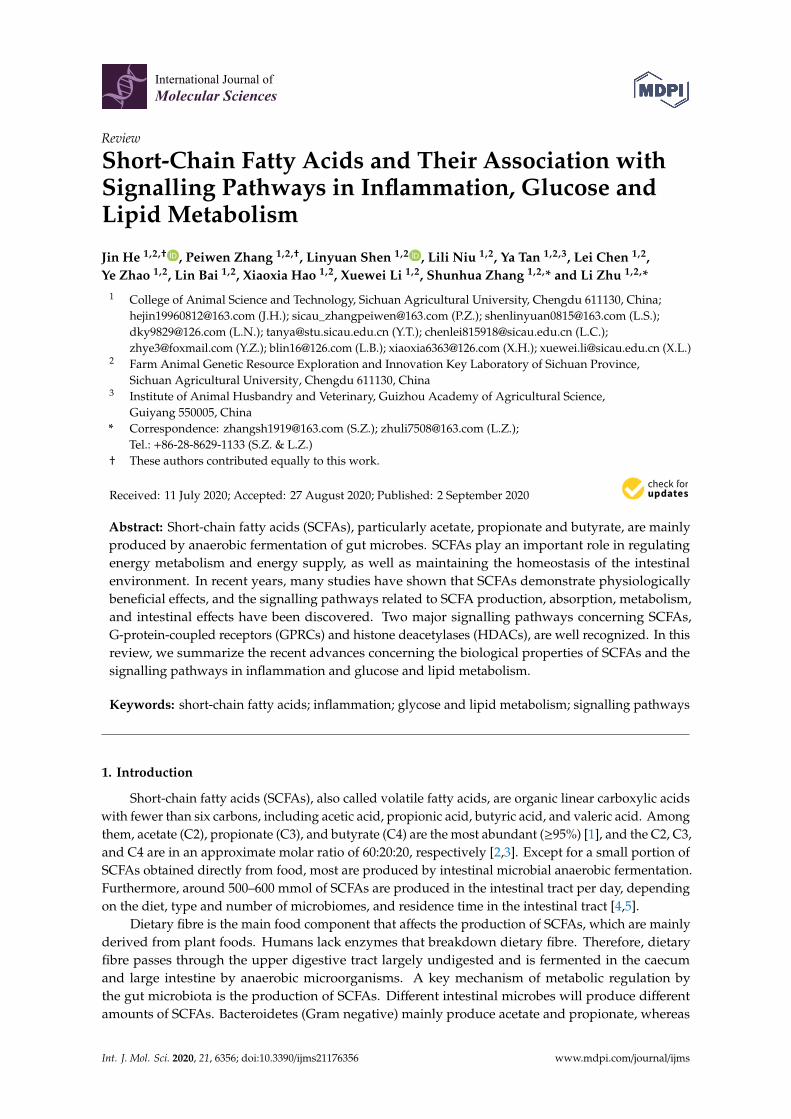
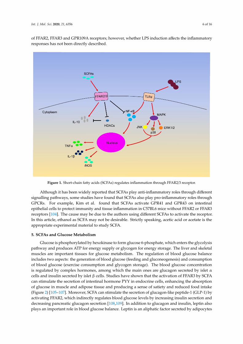
Interestingly, although LPS can induce inflammation through different signalling pathways, manystudies have shown that SCFAs can inhibit LPS-induced inflammation through the above GPCRsand HDAC (Figure 1). For example, butyrate and propionate reduce the expression of TNF andnitric oxide synthase (NOS) in LPS-induced monocytes [97]. Furthermore, butyrate treatment caninduce the phosphorylation of ERK, p38, JNK and NF-κB p65 through TLR4 in colon cancer cells [98].Acetate restrains LPS-induced TNFα secretion from mice and human mononuclear cells by activatingFFAR receptors [99]. In other studies, butyrate and propionate treatment can inhibit the secretion ofTNFα and the activity of NF-κB and up-regulate the expression of anti-inflammatory factors IL-10in LPS-activated mononuclear cells and neutrophils by HDAC inhibition [84,100,101]. Additionally,SCFAs can down-regulate the expression of IL-8 in airway inflammation by activating FFAR2 andFFAR3 receptors [102]. In macrophages, butyrate exerts anti-inflammatory effects by reducing theproduction of iNOS, TNFα, MCP-1 and IL-6 through FFAR3 receptor [53], and cytokines such as IFN-γcan increase GPR109A expression [103]. These anti-inflammatory effects are attributed to the activation
Int. J. Mol. Sci. 2020, 21, 6356 6 of 16
of FFAR2, FFAR3 and GPR109A receptors; however, whether LPS induction affects the inflammatoryresponses has not been directly described.
Int. J. Mol. Sci. 2020, 21, x FOR PEER REVIEW 6 of 16
such as IFN-γ can increase GPR109A expression [103]. These anti-inflammatory effects are attributed to the activation of FFAR2, FFAR3 and GPR109A receptors; however, whether LPS induction affects the inflammatory responses has not been directly described.
Although it has been widely reported that SCFAs play anti-inflammatory roles through different signalling pathways, some studies have found that SCFAs also play pro-inflammatory roles through GPCRs. For example, Kim et al. found that SCFAs activate GPR41 and GPR43 on intestinal epithelial cells to protect immunity and tissue inflammation in C57BL6 mice without FFAR2 or FFAR3 receptors [104]. The cause may be due to the authors using different SCFAs to activate the receptor. In this article, ethanol as SCFA may not be desirable. Strictly speaking, acetic acid or acetate is the appropriate experimental material to study SCFA.
Figure 1. Short-chain fatty acids (SCFAs) regulates inflammation through FFAR2/3 receptor.
5. SCFAs and Glucose Metabolism
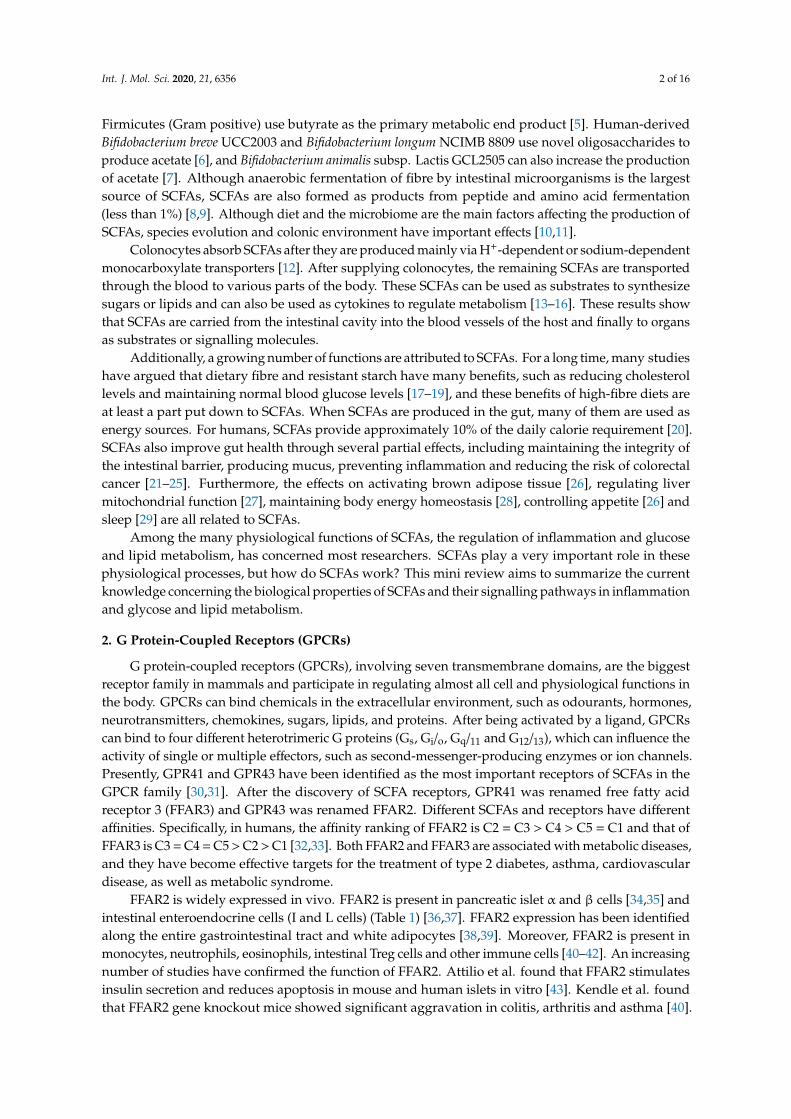
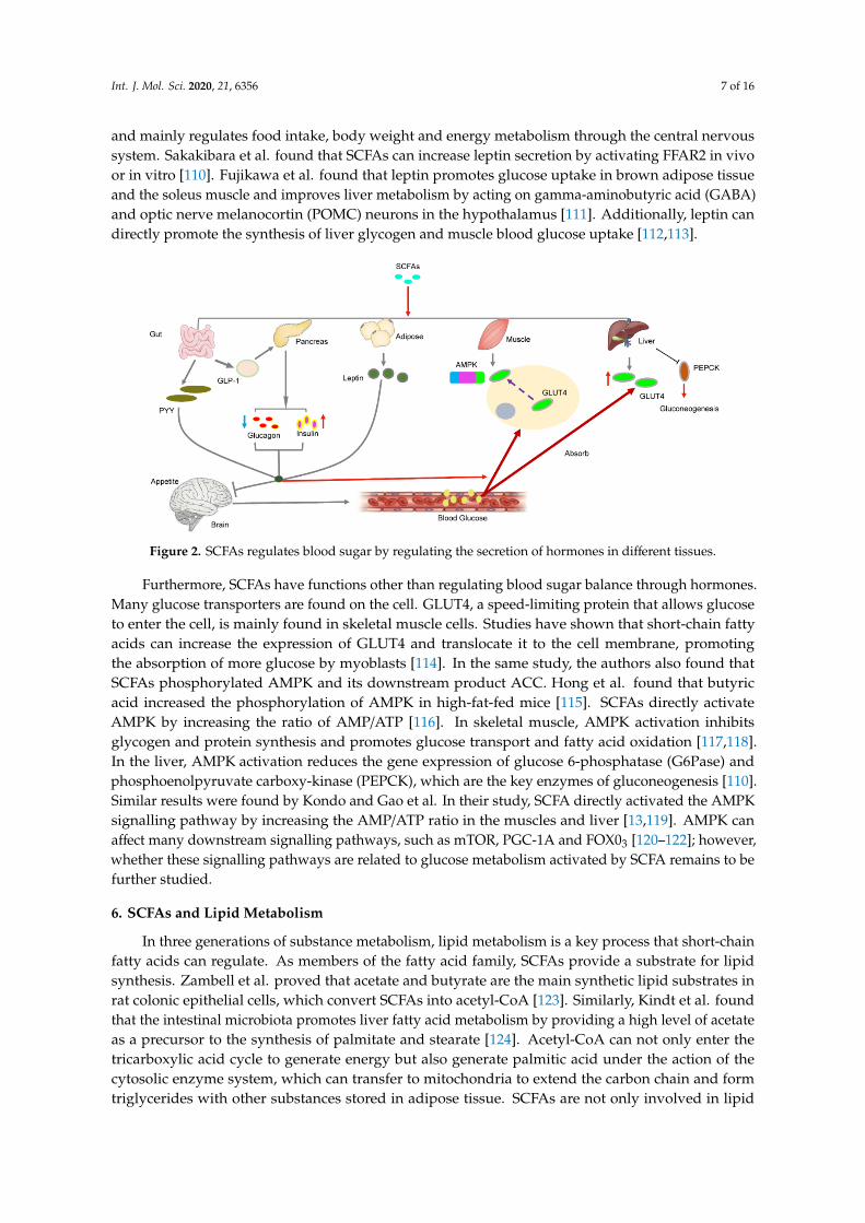
Glucose is phosphorylated by hexokinase to form glucose 6-phosphate, which enters the glycolysis pathway and produces ATP for energy supply or glycogen for energy storage. The liver and skeletal muscles are important tissues for glucose metabolism. The regulation of blood glucose balance includes two aspects: the generation of blood glucose (feeding and gluconeogenesis) and consumption of blood glucose (exercise consumption and glycogen storage). The blood glucose concentration is regulated by complex hormones, among which the main ones are glucagon secreted by islet α cells and insulin secreted by islet β cells. Studies have shown that the activation of FFAR3 by SCFA can stimulate the secretion of intestinal hormone PYY in endocrine cells, enhancing the absorption of glucose in muscle and adipose tissue and producing a sense of satiety and reduced food intake (Figure 2) [105–107]. Moreover, SCFA can stimulate the secretion of glucagon-like peptide-1 (GLP-1) by activating FFAR2, which indirectly regulates blood glucose levels by increasing insulin secretion and decreasing pancreatic glucagon secretion [108,109]. In addition to glucagon and insulin, leptin also plays an important role in blood glucose balance. Leptin is an aliphatic factor secreted by adipocytes and mainly regulates food intake, body weight and energy metabolism through the central nervous system. Sakakibara et al. found that SCFAs can increase leptin secretion by activating
Figure 1. Short-chain fatty acids (SCFAs) regulates inflammation through FFAR2/3 receptor.
Although it has been widely reported that SCFAs play anti-inflammatory roles through differentsignalling pathways, some studies have found that SCFAs also play pro-inflammatory roles throughGPCRs. For example, Kim et al. found that SCFAs activate GPR41 and GPR43 on intestinalepithelial cells to protect immunity and tissue inflammation in C57BL6 mice without FFAR2 or FFAR3receptors [104]. The cause may be due to the authors using different SCFAs to activate the receptor.In this article, ethanol as SCFA may not be desirable. Strictly speaking, acetic acid or acetate is theappropriate experimental material to study SCFA.
5. SCFAs and Glucose Metabolism
Glucose is phosphorylated by hexokinase to form glucose 6-phosphate, which enters the glycolysispathway and produces ATP for energy supply or glycogen for energy storage. The liver and skeletalmuscles are important tissues for glucose metabolism. The regulation of blood glucose balanceincludes two aspects: the generation of blood glucose (feeding and gluconeogenesis) and consumptionof blood glucose (exercise consumption and glycogen storage). The blood glucose concentrationis regulated by complex hormones, among which the main ones are glucagon secreted by islet αcells and insulin secreted by islet β cells. Studies have shown that the activation of FFAR3 by SCFAcan stimulate the secretion of intestinal hormone PYY in endocrine cells, enhancing the absorptionof glucose in muscle and adipose tissue and producing a sense of satiety and reduced food intake(Figure 2) [105–107]. Moreover, SCFA can stimulate the secretion of glucagon-like peptide-1 (GLP-1) byactivating FFAR2, which indirectly regulates blood glucose levels by increasing insulin secretion anddecreasing pancreatic glucagon secretion [108,109]. In addition to glucagon and insulin, leptin alsoplays an important role in blood glucose balance. Leptin is an aliphatic factor secreted by adipocytes
Int. J. Mol. Sci. 2020, 21, 6356 7 of 16
and mainly regulates food intake, body weight and energy metabolism through the central nervoussystem. Sakakibara et al. found that SCFAs can increase leptin secretion by activating FFAR2 in vivoor in vitro [110]. Fujikawa et al. found that leptin promotes glucose uptake in brown adipose tissueand the soleus muscle and improves liver metabolism by acting on gamma-aminobutyric acid (GABA)and optic nerve melanocortin (POMC) neurons in the hypothalamus [111]. Additionally, leptin candirectly promote the synthesis of liver glycogen and muscle blood glucose uptake [112,113].
Int. J. Mol. Sci. 2020, 21, x FOR PEER REVIEW 7 of 16
FFAR2 in vivo or in vitro [110]. Fujikawa et al. found that leptin promotes glucose uptake in brown adipose tissue and the soleus muscle and improves liver metabolism by acting on gamma-aminobutyric acid (GABA) and optic nerve melanocortin (POMC) neurons in the hypothalamus [111]. Additionally, leptin can directly promote the synthesis of liver glycogen and muscle blood glucose uptake [112,113].
Furthermore, SCFAs have functions other than regulating blood sugar balance through hormones. Many glucose transporters are found on the cell. GLUT4, a speed-limiting protein that allows glucose to enter the cell, is mainly found in skeletal muscle cells. Studies have shown that short-chain fatty acids can increase the expression of GLUT4 and translocate it to the cell membrane, promoting the absorption of more glucose by myoblasts [114]. In the same study, the authors also found that SCFAs phosphorylated AMPK and its downstream product ACC. Hong et al. found that butyric acid increased the phosphorylation of AMPK in high-fat-fed mice [115]. SCFAs directly activate AMPK by increasing the ratio of AMP/ATP [116]. In skeletal muscle, AMPK activation inhibits glycogen and protein synthesis and promotes glucose transport and fatty acid oxidation [117,118]. In the liver, AMPK activation reduces the gene expression of glucose 6-phosphatase (G6Pase) and phosphoenolpyruvate carboxy-kinase (PEPCK), which are the key enzymes of gluconeogenesis [110]. Similar results were found by Kondo and Gao et al. In their study, SCFA directly activated the AMPK signalling pathway by increasing the AMP/ATP ratio in the muscles and liver [13,119]. AMPK can affect many downstream signalling pathways, such as mTOR, PGC-1A and FOX03 [120–122]; however, whether these signalling pathways are related to glucose metabolism activated by SCFA remains to be further studied.
Figure 2. SCFAs regulates blood sugar by regulating the secretion of hormones in different tissues.
6. SCFAs and Lipid Metabolism
In three generations of substance metabolism, lipid metabolism is a key process that short-chain fatty acids can regulate. As members of the fatty acid family, SCFAs provide a substrate for lipid synthesis. Zambell et al. proved that acetate and butyrate are the main synthetic lipid substrates in rat colonic epithelial cells, which convert SCFAs into acetyl-CoA [123]. Similarly, Kindt et al. found that the intestinal microbiota promotes liver fatty acid metabolism by providing a high level of acetate as a precursor to the synthesis of palmitate and stearate [124]. Acetyl-CoA can not only enter the tricarboxylic acid cycle to generate energy but also generate palmitic acid under the action of the cytosolic enzyme system, which can transfer to mitochondria to extend the carbon chain and form triglycerides with other substances stored in adipose tissue. SCFAs are not only involved in lipid metabolism as a substrate but also can be used as a regulatory factor to regulate lipid metabolism. The experiment of Li et al. showed that butyric acid increased the oxidation of fatty acids in brown adipose tissue and improved the obesity and insulin resistance caused by diet [26]. Butyric acid can
Figure 2. SCFAs regulates blood sugar by regulating the secretion of hormones in different tissues.
Furthermore, SCFAs have functions other than regulating blood sugar balance through hormones.Many glucose transporters are found on the cell. GLUT4, a speed-limiting protein that allows glucoseto enter the cell, is mainly found in skeletal muscle cells. Studies have shown that short-chain fattyacids can increase the expression of GLUT4 and translocate it to the cell membrane, promotingthe absorption of more glucose by myoblasts [114]. In the same study, the authors also found thatSCFAs phosphorylated AMPK and its downstream product ACC. Hong et al. found that butyricacid increased the phosphorylation of AMPK in high-fat-fed mice [115]. SCFAs directly activateAMPK by increasing the ratio of AMP/ATP [116]. In skeletal muscle, AMPK activation inhibitsglycogen and protein synthesis and promotes glucose transport and fatty acid oxidation [117,118].In the liver, AMPK activation reduces the gene expression of glucose 6-phosphatase (G6Pase) andphosphoenolpyruvate carboxy-kinase (PEPCK), which are the key enzymes of gluconeogenesis [110].Similar results were found by Kondo and Gao et al. In their study, SCFA directly activated the AMPKsignalling pathway by increasing the AMP/ATP ratio in the muscles and liver [13,119]. AMPK canaffect many downstream signalling pathways, such as mTOR, PGC-1A and FOX03 [120–122]; however,whether these signalling pathways are related to glucose metabolism activated by SCFA remains to befurther studied.
6. SCFAs and Lipid Metabolism
In three generations of substance metabolism, lipid metabolism is a key process that short-chainfatty acids can regulate. As members of the fatty acid family, SCFAs provide a substrate for lipidsynthesis. Zambell et al. proved that acetate and butyrate are the main synthetic lipid substrates inrat colonic epithelial cells, which convert SCFAs into acetyl-CoA [123]. Similarly, Kindt et al. foundthat the intestinal microbiota promotes liver fatty acid metabolism by providing a high level of acetateas a precursor to the synthesis of palmitate and stearate [124]. Acetyl-CoA can not only enter thetricarboxylic acid cycle to generate energy but also generate palmitic acid under the action of thecytosolic enzyme system, which can transfer to mitochondria to extend the carbon chain and formtriglycerides with other substances stored in adipose tissue. SCFAs are not only involved in lipid
Int. J. Mol. Sci. 2020, 21, 6356 8 of 16
metabolism as a substrate but also can be used as a regulatory factor to regulate lipid metabolism.The experiment of Li et al. showed that butyric acid increased the oxidation of fatty acids in brownadipose tissue and improved the obesity and insulin resistance caused by diet [26]. Butyric acid can alsopromote the browning of white tissue, reduce the size of adipose cells morphologically, and increasethe number of multicellular adipose cells [13].
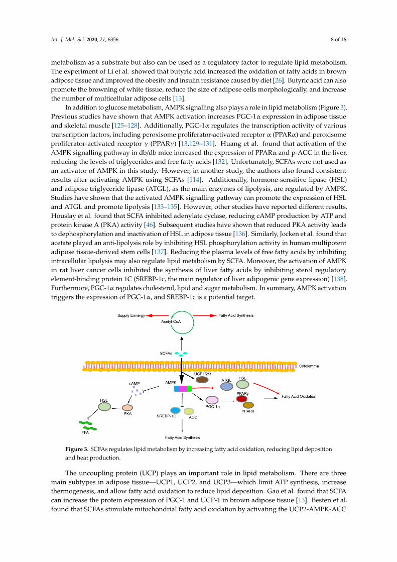
In addition to glucose metabolism, AMPK signalling also plays a role in lipid metabolism (Figure 3).Previous studies have shown that AMPK activation increases PGC-1α expression in adipose tissueand skeletal muscle [125–128]. Additionally, PGC-1α regulates the transcription activity of varioustranscription factors, including peroxisome proliferator-activated receptor α (PPARα) and peroxisomeproliferator-activated receptor γ (PPARγ) [13,129–131]. Huang et al. found that activation of theAMPK signalling pathway in db/db mice increased the expression of PPARα and p-ACC in the liver,reducing the levels of triglycerides and free fatty acids [132]. Unfortunately, SCFAs were not used asan activator of AMPK in this study. However, in another study, the authors also found consistentresults after activating AMPK using SCFAs [114]. Additionally, hormone-sensitive lipase (HSL)and adipose triglyceride lipase (ATGL), as the main enzymes of lipolysis, are regulated by AMPK.Studies have shown that the activated AMPK signalling pathway can promote the expression of HSLand ATGL and promote lipolysis [133–135]. However, other studies have reported different results.Houslay et al. found that SCFA inhibited adenylate cyclase, reducing cAMP production by ATP andprotein kinase A (PKA) activity [46]. Subsequent studies have shown that reduced PKA activity leadsto dephosphorylation and inactivation of HSL in adipose tissue [136]. Similarly, Jocken et al. found thatacetate played an anti-lipolysis role by inhibiting HSL phosphorylation activity in human multipotentadipose tissue-derived stem cells [137]. Reducing the plasma levels of free fatty acids by inhibitingintracellular lipolysis may also regulate lipid metabolism by SCFA. Moreover, the activation of AMPKin rat liver cancer cells inhibited the synthesis of liver fatty acids by inhibiting sterol regulatoryelement-binding protein 1C (SREBP-1c, the main regulator of liver adipogenic gene expression) [138].Furthermore, PGC-1α regulates cholesterol, lipid and sugar metabolism. In summary, AMPK activationtriggers the expression of PGC-1α, and SREBP-1c is a potential target.Int. J. Mol. Sci. 2020, 21, x FOR PEER REVIEW 9 of 16
Figure 3. SCFAs regulates lipid metabolism by increasing fatty acid oxidation, reducing lipid deposition and heat production.
7. Summary
Overall, SCFAs have been shown to benefit many aspects of the body’s metabolism. SCFAs have become a hot topic among researchers in terms of their initial food intake, intestinal flora composition and subsequent regulation of metabolism. Although many studies have investigated the role of SCFAs in inflammation, glucose metabolism and lipid metabolism, no systematic review exists to elucidate the SCFA-related signalling pathway in inflammation, glucose metabolism and lipid metabolism. Thus, we hope that this review increases awareness of the dominant roles of SCFAs in inflammation, glucose and lipid metabolism.
In studies of inflammation, although some studies have shown that SCFA is detrimental, most studies have shown that SCFA can reduce inflammation. However, in the existing studies, we found that butyrate was favoured by most researchers in the studies on SCFA-associated inflammation; few reports concerned other SCFAs. Thus, the inhibitory effect of different SCFAs in the same inflammatory model must be studied. The role of SCFAs in inflammation requires further study. Additionally, the regulation of glucose metabolism by SCFAs is mainly achieved by maintaining blood glucose stability. The effects of insulin and glucagon secretion are crucial for regulating glucose metabolism. Regarding lipid metabolism, SCFAs can increase fatty acid oxidation, inhibit fatty acid synthesis, increase heat production and reduce fat storage.
SCFAs have demonstrated a strong ability to regulate metabolism, but the regulatory network of SCFAs is very complex and the underlying molecular mechanism remains unclear. And the current study still has limitations. For example, although mice and other laboratory animals are used as model animals to study whether it is applicable to humans, but whether these animal data are fully applicable to humans is still a question worth exploring. However, studies in humans are mainly carried out using available mature cell lines or analysing SCFAs residue in faeces, and the inability to obtain data in vivo is still a big problem. Most of the current studies have focused on butyrate; whether it fully represents SCFAs is questionable. Although exogenous supplements can improve metabolism, could they be used in humans? Currently, there are no mature SCFA products on the market. Follow-up studies should focus on the development of effective SCFAs for clinical use.
Funding: This study was supported by the National Natural Science Foundation of China (No. 31972524, No. 31530073), the Sichuan Science and Technology Support Program (No. 2016NYZ0050; No. SCCXTD-2020-08), the earmarked fund for China Agriculture Research System (No. CARS-36-05B).
Conflicts of Interest: The authors declare no conflict of interest.
References
Figure 3. SCFAs regulates lipid metabolism by increasing fatty acid oxidation, reducing lipid depositionand heat production.
The uncoupling protein (UCP) plays an important role in lipid metabolism. There are threemain subtypes in adipose tissue—UCP1, UCP2, and UCP3—which limit ATP synthesis, increasethermogenesis, and allow fatty acid oxidation to reduce lipid deposition. Gao et al. found that SCFAcan increase the protein expression of PGC-1 and UCP-1 in brown adipose tissue [13]. Besten et al.found that SCFAs stimulate mitochondrial fatty acid oxidation by activating the UCP2-AMPK-ACC
Int. J. Mol. Sci. 2020, 21, 6356 9 of 16
signalling pathway in human HepG2 hepatocytes and mouse 3T3L1 adipocytes [139]. Hong et al.found that butyrate upregulated the expression of UCP2, UCP3 and fatty acid oxidase in skeletal muscle.After butyrate administration, histone markers H3K9Ac with high expression of gene activation weredetected in the promoter regions of adiponectin receptor 1/2, Ucp2 and Ucp3 in the muscle of obesemice [115]. Thus, butyrate not only increases heat production and lipid consumption through UCP butalso improves lipid metabolism by activating adiponectin. Other genes, such as leptin, may also play arole in the regulation of lipid metabolism by SCFA, a topic that is not discussed too much here.
7. Summary
Overall, SCFAs have been shown to benefit many aspects of the body’s metabolism. SCFAs havebecome a hot topic among researchers in terms of their initial food intake, intestinal flora compositionand subsequent regulation of metabolism. Although many studies have investigated the role of SCFAsin inflammation, glucose metabolism and lipid metabolism, no systematic review exists to elucidate theSCFA-related signalling pathway in inflammation, glucose metabolism and lipid metabolism. Thus,we hope that this review increases awareness of the dominant roles of SCFAs in inflammation, glucoseand lipid metabolism.
In studies of inflammation, although some studies have shown that SCFA is detrimental, moststudies have shown that SCFA can reduce inflammation. However, in the existing studies, we found thatbutyrate was favoured by most researchers in the studies on SCFA-associated inflammation; few reportsconcerned other SCFAs. Thus, the inhibitory effect of different SCFAs in the same inflammatorymodel must be studied. The role of SCFAs in inflammation requires further study. Additionally,the regulation of glucose metabolism by SCFAs is mainly achieved by maintaining blood glucosestability. The effects of insulin and glucagon secretion are crucial for regulating glucose metabolism.Regarding lipid metabolism, SCFAs can increase fatty acid oxidation, inhibit fatty acid synthesis,increase heat production and reduce fat storage.
SCFAs have demonstrated a strong ability to regulate metabolism, but the regulatory network ofSCFAs is very complex and the underlying molecular mechanism remains unclear. And the currentstudy still has limitations. For example, although mice and other laboratory animals are used as modelanimals to study whether it is applicable to humans, but whether these animal data are fully applicableto humans is still a question worth exploring. However, studies in humans are mainly carried out usingavailable mature cell lines or analysing SCFAs residue in faeces, and the inability to obtain data in vivois still a big problem. Most of the current studies have focused on butyrate; whether it fully representsSCFAs is questionable. Although exogenous supplements can improve metabolism, could they beused in humans? Currently, there are no mature SCFA products on the market. Follow-up studiesshould focus on the development of effective SCFAs for clinical use.
Funding: This study was supported by the National Natural Science Foundation of China (No. 31972524,No. 31530073), the Sichuan Science and Technology Support Program (No. 2016NYZ0050; No. SCCXTD-2020-08),the earmarked fund for China Agriculture Research System (No. CARS-36-05B).
Conflicts of Interest: The authors declare no conflict of interest.
References
1. Cook, S.I.; Sellin, J.H. Review Article: Short Chain Fatty Acids in Health and Disease. Aliment. Pharmacol Ther.1998, 12, 499–507. [CrossRef]
2. Cummings, J.H.; Pomare, E.W.; Branch, W.J.; Naylor, C.P.; Macfarlane, G.T. Short Chain Fatty Acids inHuman Large Intestine, Portal, Hepatic and Venous Blood. Gut 1987, 28, 1221–1227. [CrossRef] [PubMed]
3. Louis, P.; Flint, H.J. Formation of Propionate and Butyrate by the Human Colonic Microbiota.Environ. Microbiol. 2017, 19, 29–41. [CrossRef] [PubMed]
4. Bergman, E.N. Energy Contributions of Volatile Fatty Acids from the Gastrointestinal Tract in Various Species.Physiol. Rev. 1990, 70, 567–590. [CrossRef]
Int. J. Mol. Sci. 2020, 21, 6356 10 of 16
5. Macfarlane, S.; Macfarlane, G.T. Regulation of Short-Chain Fatty Acid Production. Proc. Nutr. Soc. 2003, 62,67–72. [CrossRef] [PubMed]
6. Ruiz-Aceituno, L.; Esteban-Torres, M.; James, K.; Moreno, F.J.; Sinderen, D.V. Metabolism of BiosyntheticOligosaccharides by Human-Derived Bifidobacterium Breve Ucc2003 and Bifidobacterium LongumNcimb 8809. Int. J. Food Microbiol. 2020, 316, 108476. [CrossRef]
7. Horiuchi, H.; Kamikado, K.; Aoki, R.; Suganuma, N.; Nishijima, T.; Nakatani, A.; Kimura, I. BifidobacteriumAnimalis Subsp. Lactis Gcl2505 Modulates Host Energy Metabolism Via the Short-Chain Fatty Acid ReceptorGpr43. Sci. Rep. 2020, 10, 4158. [CrossRef]
8. Smith, E.A.; Macfarlane, G.T. Enumeration of amino acid fermenting bacteria in the human large intestine:Effects of pH and starch on peptide metabolism and dissimilation of amino acids. FEMS Microbiol. Ecol.1998, 25, 355–368. [CrossRef]
9. Dai, Z.L.; Wu, G.; Zhu, W.Y. Amino acid metabolism in intestinal bacteria: Links between gut ecology andhost health. Front. Biosci. 2011, 16, 1768–1786. [CrossRef]
10. Duncan, S.H.; Louis, P.; Thomson, J.M.; Flint, H.J. The role of pH in determining the species composition ofthe human colonic microbiota. Environ. Microbiol. 2009, 11, 2112–2122. [CrossRef]
11. Youngblut, N.D.; Reischer, G.H.; Walters, W.A.; Schuster, N.; Walzer, C.; Stalder, G.; Ley, R.E.; Farnleitner, A.H.Host diet and evolutionary history explain different aspects of gut microbiome diversity amongvertebrate clades. Nat. Commun. 2019, 10, 2200. [CrossRef] [PubMed]
12. Besten, G.D.; Eunen, K.V.; Groen, A.K.; Venema, K.; Reijngoud, D.J.; Bakker, B.M. The role of short-chainfatty acids in the interplay between diet, gut microbiota, and host energy metabolism. J. Lipid Res. 2013, 54,2325–2340. [CrossRef] [PubMed]
13. Gao, Z.; Yin, J.; Zhang, J.; Ward, R.E.; Martin, R.J.; Lefevre, M.; Cefalu, W.T.; Ye, J. Butyrate Improves InsulinSensitivity and Increases Energy Expenditure in Mice. Diabetes 2009, 58, 1509–1517. [CrossRef] [PubMed]
14. Fushimi, T.; Suruga, K.; Oshima, Y.; Fukiharu, M.; Tsukamoto, Y.; Goda, T. Dietary Acetic Acid ReducesSerum Cholesterol and Triacylglycerols in Rats Fed a Cholesterol-Rich Diet. Br. J. Nutr. 2006, 95, 916–924.[CrossRef]
15. Demigné, C.; Morand, C.; Levrat, M.A.; Besson, C.; Moundras, C.; Rémésy, C. Effect of Propionate on FattyAcid and Cholesterol Synthesis and on Acetate Metabolism in Isolated Rat Hepatocytes. Br. J. Nutr. 1995, 74,209–219. [CrossRef] [PubMed]
16. Todesco, T.; Rao, A.V.; Bosello, O.; Jenkins, D.J. Propionate Lowers Blood Glucose and Alters Lipid Metabolismin Healthy Subjects. Am. J. Clin. Nutr. 1991, 54, 860–865. [CrossRef]
17. Brown, L.; Rosner, B.; Willett, W.W.; Sacks, F.M. Cholesterol-Lowering Effects of Dietary Fiber:A Meta-Analysis. Am. J. Clin. Nutr. 1999, 69, 30–42. [CrossRef]
18. Causey, J.L.; Feirtag, J.M.; Gallaher, D.D.; Tungland, B.C.; Slavin, J.L. Effects of Dietary Inulin on SerumLipids, Blood Glucose and the Gastrointestinal Environment in Hypercholesterolemic Men. Nutr. Res. 2000,20, 191–201. [CrossRef]
19. Canfora, E.E.; Jocken, J.W.; Blaak, E.E. Short-Chain Fatty Acids in Control of Body Weight and InsulinSensitivity. Nat. Rev. Endocrinol. 2015, 11, 577–591. [CrossRef]
20. Vadder, F.D.; Kovatcheva-Datchary, P.; Zitoun, C.; Duchampt, A.; Backhed, F.; Mithieux, G.Microbiota-Produced Succinate Improves Glucose Homeostasis Via Intestinal Gluconeogenesis. Cell Metab.2016, 24, 151–157. [CrossRef]
21. Lewis, K.; Lutgendorff, F.; Phan, V.; Söderholm, J.D.; Sherman, P.M.; McKay, D.M. Enhanced Translocationof Bacteria across Metabolically Stressed Epithelia Is Reduced by Butyrate. Inflamm. Bowel Dis. 2010, 16,1138–1148. [CrossRef] [PubMed]
22. Peng, L.; Li, Z.; Green, R.S.; Holzman, I.R.; Lin, J. Butyrate Enhances the Intestinal Barrier by FacilitatingTight Junction Assembly Via Activation of Amp-Activated Protein Kinase in Caco-2 Cell Monolayers. J. Nutr.2009, 139, 1619–1625. [CrossRef] [PubMed]
23. Gaudier, E.; Rival, M.; Buisine, M.P.; Robineau, I.; Hoebler, C. Butyrate Enemas Upregulate Muc GenesExpression but Decrease Adherent Mucus Thickness in Mice Colon. Physiol. Res. 2009, 58, 111–119. [PubMed]
24. O’Keefe, S.J.D. Diet, Microorganisms and Their Metabolites, and Colon Cancer. Nat. Rev.Gastroenterol. Hepatol. 2016, 13, 691–706. [CrossRef]
25. Brown, E.M.; Sadarangani, M.; Finlay, B.B. The Role of the Immune System in Governing Host-MicrobeInteractions in the Intestine. Nat. Immunol. 2013, 14, 660–667. [CrossRef]
Int. J. Mol. Sci. 2020, 21, 6356 11 of 16
26. Li, Z.; Yi, C.; Katiraei, S.; Kooijman, S.; Zhou, E.; Chung, C.K.; Gao, Y.; Heuvel, J.V.D.; Meijer, O.C.;Berbée, J.F.P.; et al. Butyrate Reduces Appetite and Activates Brown Adipose Tissue Via the Gut-Brain NeuralCircuit. Gut 2018, 67, 1269–1279. [CrossRef]
27. Mollica, M.P.; Raso, G.M.; Cavaliere, G. Butyrate Regulates Liver Mitochondrial Function, Efficiency,and Dynamics in Insulin-Resistant Obese Mice. Diabetes 2017, 66, 1405–1418. [CrossRef]
28. Vadder, F.D.; Kovatcheva-Datchary, P.; Goncalve, D.; Vinera, J.; Zitoun, C.; Duchampt, A.; Bäckhed, F.;Mithieux, G. Microbiota-Generated Metabolites Promote Metabolic Benefits Via Gut-Brain Neural Circuits.Cell 2014, 156, 84–96. [CrossRef]
29. Szentirmai, É.; Millican, N.S.; Massie, A.R.; Kapás, L. Butyrate, a Metabolite of Intestinal Bacteria, EnhancesSleep. Sci. Rep. 2019, 9, 7035. [CrossRef]
30. Brown, A.J.; Goldsworthy, S.M.; Barnes, A.A.; Eilert, M.M.; Tcheang, L.; Daniels, D.; Muir, A.I.;Wigglesworth, M.J.; Kinghorn, I.; Fraser, N.J.; et al. The Orphan G Protein-Coupled Receptors Gpr41and Gpr43 Are Activated by Propionate and Other Short Chain Carboxylic Acids. J. Biol. Chem. 2003, 278,11312–11319. [CrossRef]
31. Poul, E.L.; Loison, C.; Struyf, S.; Springael, J.; Lannoy, V.; Decobecq, M.; Brezillon, S.; Dupriez, V.; Vassart, G.;Damme, J.V.; et al. Functional Characterization of Human Receptors for Short Chain Fatty Acids and TheirRole in Polymorphonuclear Cell Activation. J. Biol. Chem. 2003, 278, 25481–25489. [CrossRef] [PubMed]
32. Milligan, G.; Stoddart, L.A.; Smith, N.J. Agonism and Allosterism: The Pharmacology of the Free Fatty AcidReceptors Ffa2 and Ffa3. Br. J. Pharmacol. 2009, 158, 146–153. [CrossRef] [PubMed]
33. Brown, A.J.; Jupe, S.; Briscoe, C.P. A Family of Fatty Acid Binding Receptors. DNA Cell Biol. 2005, 24, 54–61.[CrossRef] [PubMed]
34. Kebede, M.A.; Alquier, T.; Latour, M.G.; Poitout, V. Lipid Receptors and Islet Function: TherapeuticImplications? Diabetes Obes. Metab. 2009, 4, 10–20. [CrossRef] [PubMed]
35. Halpern, K.B.; Veprik, A.; Rubins, N.; Naaman, O.; Walker, M.D. Gpr41 Gene Expression Is Mediated byInternal Ribosome Entry Site (Ires)-Dependent Translation of Bicistronic Mrna Encoding Gpr40 and Gpr41Proteins. J. Biol. Chem. 2012, 287, 20154–20163. [CrossRef]
36. Tazoe, H.; Otomo, Y.; Karaki, S.; Kato, I.; Fukami, Y.; Terasaki, M.; Kuwahara, A. Expression of Short-ChainFatty Acid Receptor Gpr41 in the Human Colon. Biomed. Res. 2009, 30, 149–156. [CrossRef]
37. Sykaras, A.G.; Demenis, C.; Case, R.M.; McLaughlin, J.T.; Smith, C.P. Duodenal Enteroendocrine I-CellsContain Mrna Transcripts Encoding Key Endocannabinoid and Fatty Acid Receptors. PLoS ONE 2012,7, e42373. [CrossRef]
38. Karaki, S.; Mitsui, R.; Hayashi, H.; Kato, I.; Sugiya, H.; Iwanaga, T.; Furness, J.B.; Kuwahara, A. Short-ChainFatty Acid Receptor, Gpr43, Is Expressed by Enteroendocrine Cells and Mucosal Mast Cells in Rat Intestine.Cell Tissue Res. 2006, 324, 353–360. [CrossRef]
39. Hong, Y.; Nishimura, Y.; Hishikawa, D.; Tsuzuki, H.; Miyahara, H.; Gotoh, C.; Choi, K.; Feng, D.D.;Chen, C.; Lee, H.; et al. Acetate and Propionate Short Chain Fatty Acids Stimulate Adipogenesis Via Gpcr43.Endocrinology 2005, 146, 5092–5099. [CrossRef]
40. Maslowski, K.; Vieira, A.T.; Ng, A.; Kranich, J.; Sierro, F.; Yu, D.; Schilter, H.C.; Rolph, M.S.; Mackay, F.;Artis, D.; et al. Regulation of Inflammatory Responses by Gut Microbiota and Chemoattractant ReceptorGpr43. Nature 2009, 461, 1282–1286. [CrossRef]
41. Nilsson, C.; Swolin-Eide, D.; Ohlsso, C.; Eriksson, E.; Ho, H.; Björntorp, P.; Holmäng, A. Reductions inAdipose Tissue and Skeletal Growth in Rat Adult Offspring after Prenatal Leptin Exposure. J. Endocrinol.2003, 176, 13–21. [CrossRef] [PubMed]
42. Smith, P.M.; Howitt, M.R.; Panikov, N.; Michaud, M.; Gallini, C.A.; Bohlooly-Y, M.; Glickman, J.N.;Garrett, W.S. The Microbial Metabolites, Short Chain Fatty Acids, Regulate Colonic Treg Cell Homeostasis.Science 2013, 341, 569–573. [CrossRef] [PubMed]
43. Pingitore, A.; Gonzalez-Abuin, N.; Ruz-Maldonado, I.; Huang, G.C.; Frost, G.; Persaud, S.J. Short ChainFatty Acids Stimulate Insulin Secretion and Reduce Apoptosis in Mouse and Human Islets in Vitro: Role ofFree Fatty Acid Receptor 2. Diabetes Obes. Metab. 2019, 21, 330–339. [CrossRef] [PubMed]
44. Brown, A.J.; Tsoulou, C.; Ward, E.; Gower, E.; Bhudia, N.; Chowdhury, F.; Dean, T.W.; Faucher, N.; Gangar, A.;Dowell, S.J. Pharmacological Properties of Acid N-Thiazolylamide Ffa2 Agonists. Pharmacol. Res. Perspect.2015, 3, e00141. [CrossRef] [PubMed]
Int. J. Mol. Sci. 2020, 21, 6356 12 of 16
45. Forbes, S.; Stafford, S.; Coope, G.; Heffron, H.; Real, K.; Newman, R.; Davenport, R.; Barnes, M.; Grosse, J.;Cox, H. Selective Ffa2 Agonism Appears to Act Via Intestinal Pyy to Reduce Transit and Food Intake butDoes Not Improve Glucose Tolerance in Mouse Models. Diabetes 2015, 64, 3763–3771. [CrossRef] [PubMed]
46. Houslay, M.D.; Milligan, G. Tailoring Camp-Signalling Responses through Isoform Multiplicity.Trends Biochem. Sci. 1997, 22, 217–224. [CrossRef]
47. Kimura, I.; Inoue, D.; Hirano, K.; Tsujimoto, G. The SCFA Receptor Gpr43 and Energy Metabolism.Front. Endocrinol. 2014, 5, 85. [CrossRef] [PubMed]
48. Pirozzi, C.; Francisco, V.; Guida, F.D.; Gómez, R.; Lago, F.; Pino, J.; Meli, R.; Gualillo, O. Butyrate ModulatesInflammation in Chondrocytes Via Gpr43 Receptor. Cell Physiol. Biochem. 2018, 51, 228–243. [CrossRef]
49. Nakajima, A.; Nakatani, A.; Hasegawa, S.; Irie, J.; Ozawa, K.; Tsujimoto, G.; Suganami, T.; Itoh, H.; Kimura, I.The Short Chain Fatty Acid Receptor Gpr43 Regulates Inflammatory Signals in Adipose Tissue M2-TypeMacrophages. PLoS ONE 2017, 12, e0179696. [CrossRef]
50. Mizuta, K.; Matoba, A.; Shibata, S.; Masaki, E.; Sr, C.W.E. Obesity-Induced Asthma: Role of Free Fatty AcidReceptors. Jpn. Dent. Sci. Rev. 2019, 55, 103–107. [CrossRef]
51. Samuel, B.S.; Shaito, A.; Motoike, T.; Rey, F.E.; Backhed, F.; Manchester, J.K.; Hammer, R.E.; Williams, S.C.;Crowley, J.; Yanagisawa, M.; et al. Effects of the Gut Microbiota on Host Adiposity Are Modulated by theShort-Chain Fatty-Acid Binding G Protein-Coupled Receptor, Gpr41. Proc. Natl. Acad. Sci. USA 2008, 105,16767–16772. [CrossRef] [PubMed]
52. Kimura, I.; Inoue, D.; Maeda, T.; Hara, T.; Ichimura, A.; Miyauchi, S.; Kobayashi, M.; Hirasawa, A.;Tsujimoto, G. Short-Chain Fatty Acids and Ketones Directly Regulate Sympathetic Nervous System Via GProtein-Coupled Receptor 41 (Gpr41). Proc. Natl. Acad. Sci. USA 2011, 108, 8030–8035. [CrossRef] [PubMed]
53. Ohira, H.; Fujioka, Y.; Katagiri, C.; Mamoto, R.; Aoyama-Ishikawa, M.; Amako, K.; Izumi, Y.; Nishiumi, S.;Yoshida, M.; Usami, M.; et al. Butyrate Attenuates Inflammation and Lipolysis Generated by the Interactionof Adipocytes and Macrophages. J. Atheroscler. Thromb. 2013, 20, 425–442. [CrossRef] [PubMed]
54. Li, M.; Esch, B.C.A.M.; Wagenaar, G.T.M.; Garssen, J.; Folkerts, G.; Henricks, P.A.J. Pro- and Anti-InflammatoryEffects of Short Chain Fatty Acids on Immune and Endothelial Cells. Eur. J. Pharmacol. 2018, 831, 52–59.[CrossRef]
55. Miyamoto, J.; Kasubuchi, M.; Nakajima, A.; Kimura, I. Anti-Inflammatory and Insulin-Sensitizing Effects ofFree Fatty Acid Receptors. Handb. Exp. Pharmacol. 2016, 236, 221–231.
56. Nilsson, N.E.; Kotarsky, K.; Owman, C.; Olde, B. Identification of a Free Fatty Acid Receptor, Ffa2r, Expressedon Leukocytes and Activated by Short-Chain Fatty Acids. Biochem. Biophys. Res. Commun. 2003, 303,1047–1052. [CrossRef]
57. Stoddart, L.A.; Smith, N.J.; Jenkins, L.; Brown, A.J.; Milligan, G. Conserved Polar Residues in TransmembraneDomains V, Vi, and Vii of Free Fatty Acid Receptor 2 and Free Fatty Acid Receptor 3 Are Required for theBinding and Function of Short Chain Fatty Acids. J. Biol. Chem. 2008, 283, 32913–32924. [CrossRef]
58. Colina, C.; Puhl, H.L.; Ikeda, S.R. Selective Tracking of Ffar3-Expressing Neurons Supports Receptor Couplingto N-Type Calcium Channels in Mouse Sympathetic Neurons. Sci. Rep. 2018, 8, 17379. [CrossRef]
59. Trompette, A.; Gollwitzer, E.S.; Yadava, K.; Sichelstiel, A.K.; Sprenger, N.; Ngom-Bru, C.; Blanchard, C.;Junt, T.; Nicod, L.P.; Harris, N.L.; et al. Gut Microbiota Metabolism of Dietary Fiber Influences AllergicAirway Disease and Hematopoiesis. Nat. Med. 2014, 20, 159–166. [CrossRef]
60. Pluznick, J.L.; Protzko, R.J.; Gevorgyan, H.; Peterlin, Z.; Sipos, A.; Han, J.; Brunet, I.; Wan, L.; Rey, F.;Wang, T.; et al. Olfactory Receptor Responding to Gut Microbiota-Derived Signals Plays a Role in ReninSecretion and Blood Pressure Regulation. Proc. Natl. Acad. Sci. USA 2013, 110, 4410–4415. [CrossRef]
61. Pluznick, J. A Novel SCFA Receptor, the Microbiota, and Blood Pressure Regulation. Gut Microb. 2014, 5,202–207. [CrossRef] [PubMed]
62. Mizuta, K.; Sasaki, H.; Zhang, Y.; Matoba, A.; Sr, C.W.E. The Short-Chain Free Fatty Acid Receptor Ffar3Is Expressed and Potentiates Contraction in Human Airway Smooth Muscle. Am. J. Physiol. Lung CellMol. Physiol. 2020, 318, L1248–L1260. [CrossRef] [PubMed]
63. Ørgaard, A.; Jepsen, S.L.; Holst, J.J. Short-Chain Fatty Acids and Regulation of Pancreatic Endocrine Secretionin Mice. Islets 2019, 11, 103–111. [CrossRef]
64. Priyadarshini, M.; Layden, B.T. Ffar3 Modulates Insulin Secretion and Global Gene Expression in MouseIslets. Islets 2015, 7, e1045182. [CrossRef]
Int. J. Mol. Sci. 2020, 21, 6356 13 of 16
65. Shimizu, H.; Masujima, Y.; Ushiroda, C.; Mizushima, R.; Taira, S.; Ohue-Kitano, R.; Kimura, I. DietaryShort-Chain Fatty Acid Intake Improves the Hepatic Metabolic Condition Via Ffar3. Sci. Rep. 2019, 9, 16574.[CrossRef] [PubMed]
66. Wanders, D.; Graff, E.C.; Judd, R.L. Effects of High Fat Diet on Gpr109A and Gpr81 Gene Expression.Biochem. Biophys. Res. Commun. 2012, 425, 278–283. [CrossRef]
67. Thangaraju, M.; Cresci, G.A.; Liu, K.; Ananth, S.; Gnanaprakasam, J.P.; Browning, D.D.; Mellinger, J.D.;Smith, S.B.; Digby, G.J.; Lambert, N.A.; et al. Gpr109A Is a G-Protein-Coupled Receptor for the BacterialFermentation Product Butyrate and Functions as a Tumor Suppressor in Colon. Cancer Res. 2009, 69,2826–2832. [CrossRef]
68. Benyó, Z.; Gille, A.; Kero, J.; Csiky, M.; Suchánková, M.C.; Nüsing, R.M.; Moers, A.; Pfeffer, K.; Offermanns, S.Gpr109A (Puma-G/Hm74a) Mediates Nicotinic Acid-Induced Flushing. J. Clin. Investig. 2005, 115, 3634–3640.[CrossRef]
69. Zellner, C.; Pullinger, C.R.; Aouizerat, B.E.; Frost, P.H.; Kwok, P.; Malloy, M.J.; Kane, J.P. Variations in HumanHm74 (Gpr109b) and Hm74a (Gpr109A) Niacin Receptors. Human Mutat. 2005, 25, 18–21. [CrossRef]
70. Offermanns, S. Hydroxy-Carboxylic Acid Receptor Actions in Metabolism. Trends Endocrinol. Metab. 2017,28, 227–236. [CrossRef]
71. Rezq, S.; Abdel-Rahman, A.A. Central Gpr109A Activation Mediates Glutamate-Dependent Pressor Responsein Conscious Rats. J. Pharmacol. Exp. Ther. 2016, 356, 456–465. [CrossRef] [PubMed]
72. Li, Z.; Li, X.; Lin, S.; Chen, Y.; Ma, S.; Fu, Y.; Wei, C.; Xu, W. Nicotinic Acid Receptor Gpr109A ExertsAnti-Inflammatory Effects through Inhibiting the Akt/mTOR Signaling Pathway in Min6 Pancreatic B Cells.Ann. Clin. Lab. Sci. 2017, 47, 729–737. [PubMed]
73. Elangovan, S.; Pathania, R.; Ramachandran, S.; Ananth, S.; Padia, R.N.; Lan, L.; Singh, N.; Martin, P.M.;Hawthorn, L.; Prasad, P.D.; et al. The Niacin/Butyrate Receptor Gpr109A Suppresses Mammary Tumorigenesisby Inhibiting Cell Survival. Cancer Res. 2014, 74, 1166–1178. [CrossRef] [PubMed]
74. Priyadarshini, M.; Kotlo, K.U.; Dudeja, P.K.; Layden, B.T. Role of Short Chain Fatty Acid Receptors inIntestinal Physiology and Pathophysiology. Compr. Physiol. 2018, 8, 1091–1115. [PubMed]
75. Kaye, D.M.; Shihata, W.A.; Jama, H.A.; Tsyganov, K.; Ziemann, M.; Kiriazis, H.; Horlock, D.; Vijay, A.; Giam, B.;Vinh, A.; et al. Deficiency of Prebiotic Fiber and Insufficient Signaling through Gut Metabolite-SensingReceptors Leads to Cardiovascular Disease. Circulation 2020, 141, 1393–1403. [CrossRef]
76. Wang, N.; Guo, D.; Tian, X.; Lin, H.; Li, Y.; Chen, S.; Fu, Y.; Xu, W.; Wei, C. Niacin Receptor Gpr109A InhibitsInsulin Secretion and Is Down-Regulated in Type 2 Diabetic Islet Beta-Cells. Gen. Comp. Endocrinol. 2016,237, 98–108. [CrossRef]
77. Shi, Y.; Lai, X.; Ye, L.; Chen, K.; Cao, Z.; Gong, W.; Jin, L.; Wang, C.; Liu, M.; Liao, Y.; et al. Activated NiacinReceptor Hca2 Inhibits Chemoattractant-Mediated Macrophage Migration Via Gβγ/PKC/ERK1/2 Pathwayand Heterologous Receptor Desensitization. Sci. Rep. 2017, 7, 42279. [CrossRef]
78. Borthakur, A.; Priyamvada, S.; Kumar, A.; Natarajan, A.A.; Gill, R.K.; Alrefai, W.A.; Dudeja, P.K. A NovelNutrient Sensing Mechanism Underlies Substrate-Induced Regulation of Monocarboxylate Transporter-1.Am. J. Physiol. Gastrointest. Liver Physiol. 2012, 303, G1126–G1133. [CrossRef]
79. Linda Sealy, R.C. The Effect of Sodium Butyrate on Histone Modification. Cell 1978, 14, 115–121. [CrossRef]80. Boffa, L.C.; Vidali, G.; Mann, R.S.; Allfrey, V.G. Suppression of Histone Deacetylation in Vivo and in Vitro by
Sodium Butyrate. J. Biol. Chem. 1978, 253, 3364–3366.81. Waldecker, M.; Kautenburger, T.; Daumann, H.; Busch, C.; Schrenk, D. Inhibition of Histone-Deacetylase
Activity by Short-Chain Fatty Acids and Some Polyphenol Metabolites Formed in the Colon. J. Nutr. Biochem.2008, 19, 587–593. [CrossRef] [PubMed]
82. Soliman, M.L.; Rosenberger, T.A. Acetate Supplementation Increases Brain Histone Acetylation and InhibitsHistone Deacetylase Activity and Expression. Mol. Cell Biochem. 2011, 352, 173–180. [CrossRef] [PubMed]
83. Bulusu, V.; Tumanov, S.; Michalopoulou, E.; Broek, N.J.; MacKay, G.; Nixon, C.; Dhayade, S.; Schug, Z.T.;Voorde, J.V.; Blyth, K.; et al. Acetate Recapturing by Nuclear Acetyl-CoA Synthetase 2 Prevents Loss ofHistone Acetylation during Oxygen and Serum Limitation. Cell Rep. 2017, 18, 647–658. [CrossRef] [PubMed]
84. Chang, P.V.; Hao, L.; Offermanns, S.; Medzhitov, R. The Microbial Metabolite Butyrate Regulates IntestinalMacrophage Function Via Histone Deacetylase Inhibition. Proc. Natl. Acad. Sci. USA 2014, 111, 2247–2252.[CrossRef] [PubMed]
Int. J. Mol. Sci. 2020, 21, 6356 14 of 16
85. Sun, M.; Wu, W.; Liu, Z.; Cong, Y. Microbiota Metabolite Short Chain Fatty Acids, Gpcr, and InflammatoryBowel Diseases. J. Gastroenterol. 2017, 52, 1–8. [CrossRef]
86. Wu, J.; Zhou, Z.; Hu, Y.; Dong, S. Butyrate-Induced Gpr41 Activation Inhibits Histone Acetylation and CellGrowth. J. Genet. Genom. 2012, 39, 375–384. [CrossRef]
87. Czepiel, J.; Biesiada, G.; Brzozowski, T.; Ptak-Belowska, A.; Perucki, W.; Birczynska, M.; Jurczyszyn, A.;Strzalka, M.; Targosz, A.; Garlicki, A. The Role of Local and Systemic Cytokines in Patients Infected withClostridium Difficile. J. Physiol. Pharmacol. 2014, 65, 695–703.
88. Park, B.S.; Lee, J. Recognition of Lipopolysaccharide Pattern by TLR4 Complexes. Exp. Mol. Med. 2013,45, e66. [CrossRef]
89. Hagar, J.A.; Powell, D.A.; Aachoui, Y.; Ernst, R.K.; Miao, E.A. Cytoplasmic LPS Activates Caspase-11:Implications in TLR4-Independent Endotoxic shock. Science 2013, 341, 1250–1253. [CrossRef]
90. Adrie, C.; Pinsky, M.R. The Inflammatory Balance in Human Sepsis. Intensive Care Med. 2000, 26, 364–375.[CrossRef]
91. Baeuerle, P.A.; Baichwal, V.R. NF-Kappa B as a Frequent Target for Immunosuppressive andAnti-Inflammatory Molecules. Adv. Immunol. 1997, 65, 111–137.
92. Pahl, H.L. Activators and Target Genes of Rel/NF-Kappab Transcription Factors. Oncogene 1999, 18, 6853–6866.[CrossRef] [PubMed]
93. Liu, S.F.; Malik, A.B. NF-Kappa B Activation as a Pathological Mechanism of Septic Shock and Inflammation.Am. J. Physiol. Lung Cell. Mol. Physiol. 2006, 290, L622–L645. [CrossRef] [PubMed]
94. Liu, S.; Feng, G.; Wang, G.; Liu, G. p38MAPK Inhibition Attenuates LPS-induced Acute Lung InjuryInvolvement of NF-kappaB Pathway. Eur. J. Pharmacol. 2008, 584, 159–165. [CrossRef] [PubMed]
95. Jeong, Y.; Du, R.; Zhu, X.; Yin, S.; Wang, J.; Cui, H.; Cao, W.; Lowenstein, C.J. Histone Deacetylase IsoformsRegulate Innate Immune Responses by Deacetylating Mitogen-Activated Protein Kinase Phosphatase-1.J. Leukoc. Biol. 2014, 95, 651–659. [CrossRef] [PubMed]
96. Subedi, L.; Venkatesan, R.; Kim, S.Y. Neuroprotective and Anti-Inflammatory Activities of Allyl IsothiocyanateThrough Attenuation of JNK/NF-κB/TNF-α Signaling. Int. J. Mol. Sci. 2017, 18, 1423. [CrossRef]
97. Vinolo, M.A.R.; Rodrigues, H.G.; Hatanaka, E.; Sato, F.T.; Sampaio, S.C.; Curi, R. Suppressive Effect ofShort-Chain Fatty Acids on Production of Proinflammatory Mediators by Neutrophils. J. Nutr. Biochem.2011, 22, 849–855. [CrossRef]
98. Xiao, T.; Wu, S.; Yan, C.; Zhao, C.; Jin, H.; Yan, N.; Xu, J.; Wu, Y.; Li, C.; Shao, Q.; et al. Butyrate Upregulates theTLR4 Expression and the Phosphorylation of MAPKs and NK-κB in Colon Cancer Cell in vitro. Oncol. Lett.2018, 16, 4439–4447. [CrossRef]
99. Masui, R.; Sasaki, M.; Funaki, Y.; Ogasawara, N.; Mizuno, M.; Iida, A.; Izawa, S.; Kondo, Y.; Ito, Y.;Tamura, Y.; et al. G Protein-Coupled Receptor 43 Moderates Gut Inflammation through Cytokine Regulationfrom Mononuclear Cells. Inflamm. Bowel Dis. 2013, 19, 2848–2856. [CrossRef]
100. Aoyama, M.; Kotani, J.; Usami, M. Butyrate and Propionate Induced Activated or Non-Activated NeutrophilApoptosis via HDAC Inhibitor Activity but Without Activating GPR-41/GPR-43 Pathways. Nutrition 2010,26, 653–661. [CrossRef]
101. Vinolo, M.A.R.; Rodrigues, H.G.; Nachbar, R.T.; Curi, R. Regulation of Inflammation by Short ChainFatty Acids. Nutrients 2011, 3, 858–876. [CrossRef] [PubMed]
102. Halnes, I.; Baines, K.J.; Berthon, B.S.; MacDonald-Wicks, L.K.; Gibson, P.G.; Wood, L.G. Soluble Fibre MealChallenge Reduces Airway Inflammation and Expression of GPR43 and GPR41 in Asthma. Nutrients 2017,9, 57. [CrossRef] [PubMed]
103. Schaub, A.; Fütterer, A.; Pfeffer, K. PUMA-G, an IFN-gamma-inducible Gene in Macrophages Is a NovelMember of the Seven Transmembrane Spanning Receptor Superfamily. Eur. J. Immunol. 2001, 31, 3714–3739.[CrossRef]
104. Kim, M.H.; Kang, S.G.; Park, J.H.; Yanagisawa, M.; Kim, C.H. Short-chain Fatty Acids Activate GPR41 andGPR43 on Intestinal Epithelial Cells to Promote Inflammatory Responses in Mice. Gastroenterology 2013, 145,396–406. [CrossRef] [PubMed]
105. Ribola, F.A.; Cançado, F.B.; Schoueri, J.H.M.; Toni, V.F.; Medeiros, V.H.; Feder, D. Effects of SGLT2 Inhibitorson Weight Loss in Patients With Type 2 Diabetes Mellitus. Eur. Rev. Med. Pharmacol. Sci. 2017, 21, 199–211.
Int. J. Mol. Sci. 2020, 21, 6356 15 of 16
106. Batterham, R.L.; Cowley, M.A.; Small, C.J.; Herzog, H.; Cohen, M.A.; Dakin, C.L.; Wren, A.M.; Brynes, A.E.;Low, M.J.; Ghatei, M.A.; et al. Gut Hormone PYY(3–36) Physiologically Inhibits Food Intake. Nature 2002,418, 650–654. [CrossRef]
107. Hoek, A.M.; Heijboer, A.C.; Corssmit, E.P.M.; Voshol, P.J.; Romijn, J.A.; Havekes, L.M.; Pijl, H. PYY3–36Reinforces Insulin Action on Glucose Disposal in Mice Fed a High-Fat Diet. Diabetes 2004, 53, 1949–1952.[CrossRef]
108. Mazibuko, S.E.; Muller, C.J.F.; Joubert, E.; Beer, D.; Johnson, R.; Opoku, A.R.; Louw, J. Amelioration ofPalmitate-Induced Insulin Resistance in C2C12 Muscle Cells by Rooibos (Aspalathus Linearis). Phytomedicine2013, 20, 813–822. [CrossRef]
109. Barrera, J.G.; Sandoval, D.A.; D’Alessio, D.A.; Seeley, R.J. GLP-1 and Energy Balance: An Integrated Modelof Short-Term and Long-Term Control. Nat. Rev. Endocrinol. 2011, 7, 507–516. [CrossRef]
110. Sakakibara, S.; Yamauchi, T.; Oshima, Y.; Tsukamoto, Y.; Kadowaki, T. Acetic acid activates hepatic AMPKand reduces hyperglycemia in diabetic KK-A(y) mice. Biochem. Biophys. Res. Commun. 2006, 344, 597–604.[CrossRef]
111. Fujikawa, T.; Berglund, E.D.; Patel, V.R.; Ramadori, G.; Vianna, C.R.; Vong, L.; Thorel, F.; Chera, S.;Herrera, P.L.; Lowell, B.B.; et al. Leptin Engages a Hypothalamic Neurocircuitry to Permit Survival in theAbsence of Insulin. Cell Metab. 2013, 18, 431–444. [CrossRef] [PubMed]
112. Fujikawa, T.; Chuang, J.; Sakata, I.; Ramadori, G.; Coppari, R. Leptin Therapy Improves Insulin-DeficientType 1 Diabetes by Cns-Dependent Mechanisms in Mice. Proc. Natl. Acad. Sci. USA 2010, 107, 17391–17396.[CrossRef] [PubMed]
113. Minokoshi, Y.; Kim, Y.; Peroni, O.D.; Fryer, L.G.; Müller, C.; Carling, D.; Kahn, B.B. Leptin StimulatesFatty-Acid Oxidation by Activating Amp-Activated Protein Kinase. Nature 2002, 415, 339–343. [CrossRef][PubMed]
114. Deng, B. Study the Mechanism of Calcium Signal- and Short Chain Fatty Acids-Regulated Glucose Metabolism.Ph.D. Thesis, Tianjin Medical University, Tianjin, China, 2018.
115. Hong, J.; Yimin Jia, Y.; Pan, S.; Jia, L.; Li, H.; Han, Z.; Cai, D.; Zhao, R. Butyrate Alleviates High Fat Diet-InducedObesity through Activation of Adiponectin-Mediated Pathway and Stimulation of Mitochondrial Functionin the Skeletal Muscle of Mice. Oncotarget 2016, 7, 56071–56082. [CrossRef]
116. Besten, G.; Gerding, A.; Dijk, T.H.; Ciapaite, J.; Bleeker, A.; Eunen, K.; Havinga, R.; Groen, A.K.; Reijngoud, D.J.;Bakker, B.M. Protection against the Metabolic Syndrome by Guar Gum-Derived Short-Chain Fatty AcidsDepends on Peroxisome Proliferator-Activated Receptor Γ and Glucagon-Like Peptide-1. PLoS ONE 2015,10, e0136364. [CrossRef]
117. Merrill, G.F.; Kurth, E.J.; Hardie, D.G.; Winder, W.W. Aica Riboside Increases Amp Activated Protein Kinase,Fatty Acid Oxidati on, and Glucose Uptake in Rat Muscle. Am. J. Physiol. 1997, 273, E1107–E1119.
118. Hardie, D.G.; Hawley, S.A.; Scott, J.W. AMP-activated Protein Kinase—Development of the Energy SensorConcept. J. Physiol. 2006, 574, 7–15. [CrossRef]
119. Kondo, T.; Kishi, M.; Fushimi, T.; Kaga, T. Acetic Acid Upregulates the Expression of Genes for Fatty AcidOxidation Enzymes in Liver to Suppress Body Fat Accumulation. J. Agric. Food Chem. 2009, 57, 5982–5986.[CrossRef]
120. Kim, G.T.; Lee, S.H.; Kim, Y.M. Quercetin Regulates Sestrin 2-Ampk-Mtor Signaling Pathway and InducesApoptosis Via Increased Intracellular Ros in Hct116 Colon Cancer Cells. J. Cancer Prev. 2013, 18, 264–270.[CrossRef]
121. Liu, H.; Peng, H.; Xiang, H.; Guo, L.; Chen, R.; Zhao, S.; Chen, W.; Chen, P.; Lu, H.; Chen, S. TWEAK/Fn14Promotes Oxidative Stress Through AMPK/PGC-1α/MnSOD Signaling Pathway in Endothelial Cells.Mol. Med. Rep. 2017, 17, 1998–2004. [CrossRef]
122. Ma, W.; Wang, J.; Bu, X.; Zhang, H.; Zhang, J.; Zhang, X.; He, Y.; Wang, D.; Zhang, Z.; Meng, F. Effects ofPolygonum Cuspidatum on AMPK-FOXO3α Signaling Pathway in Rat Model of Uric Acid-Induced RenalDamage. Chin. J. Integr. Med. 2019, 25, 182–189. [CrossRef] [PubMed]
123. Zambell, K.L.; Fitch, M.D.; Fleming, S.E. Acetate and Butyrate Are the Major Substrates for De NovoLipogenesis in Rat Colonic Epithelial Cells. J. Nutr. 2003, 133, 3509–3515. [CrossRef] [PubMed]
124. Kindt, A.; Liebisch, G.; Clacel, T.; Haller, D.; Hörmannsperger, G.; Yoon, H.; Kolmeder, D.; Sigruener, A.;Krautbauer, S.; Seeliger, C.; et al. The gut microbiota promotes hepatic fatty acid desaturation and elongationin mice. Nat. Commun. 2018, 9, 3760. [CrossRef] [PubMed]
Int. J. Mol. Sci. 2020, 21, 6356 16 of 16
125. Taylor, E.B.; Lamb, J.D.; Hurst, R.W.; Chesser, D.G.; Ellingson, W.J.; Greenwood, L.J.; Porter, B.B.; Herway, S.T.;Winder, W.W. Endurance Training Increases Skeletal Muscle LKB1 and PGC-1alpha Protein Abundance:Effects of Time and Intensity. Am. J. Physiol. Endocrinol. Metab. 2005, 289, E960–E968. [CrossRef]
126. Terada, S.; Goto, M.; Kato, M.; Kawanaka, K.; Shimokawa, T.; Tabata, I. Effects of Low-Intensity ProlongedExercise on PGC-1 mRNA Expression in Rat Epitrochlearis Muscle. Biochem. Biophys. Res. Commun. 2002,296, 350–354. [CrossRef]
127. Wan, Z.; Root-McCaig, J.; Castellani, L.; Kemp, B.E.; Steinberg, G.R.; Wright, D.C. Evidence for the Role ofAMPK in Regulating PGC-1 Alpha Expression and Mitochondrial Proteins in Mouse Epididymal AdiposeTissue. Obesity 2014, 22, 730–738. [CrossRef]
128. Yan, M.; Audet-Walsh, É.; Manteghi, S.; Dufour, C.R.; Walker, B.; Baba, M.; St-Pierre, J.; Giguère, V.;Pause, A. Chronic AMPK Activation via Loss of FLCN Induces Functional Beige Adipose Tissue ThroughPGC-1α/ERRα. Genes Dev. 2016, 30, 1034–1046. [CrossRef]
129. Muoio, D.M.; Way, J.M.; Tanner, C.J.; Winegar, D.A.; Kliewer, S.A.; Houmard, J.A.; Kraus, W.E.; Dohm, G.L.Peroxisome Proliferator-Activated Receptor-Alpha Regulates Fatty Acid Utilization in Primary HumanSkeletal Muscle Cells. Diabetes 2002, 51, 901–909. [CrossRef]
130. Lin, J.; Handschin, C.; Spiegelman, B.M. Metabolic Control through the PGC-1 Family of TranscriptionCoactivators. Cell Metab. 2005, 1, 361–370. [CrossRef]
131. Jäger, S.; Handschin, C.; St-Pierre, J.; Spiegelman, B.M. AMP-activated Protein Kinase (AMPK) Action inSkeletal Muscle via Direct Phosphorylation of PGC-1alpha. Proc. Natl. Acad. Sci. USA 2007, 104, 12017–12022.[CrossRef]
132. Huang, M.; Zhou, C.; Zhang, Y.; Zhang, X.; Xu, W.; Lin, J.; Wang, P. Salvianolic Acid B AmelioratesHyperglycemia and Dyslipidemia in db/db Mice Through the AMPK Pathway. Cell Physiol. Biochem. 2016,40, 933–943. [CrossRef] [PubMed]
133. Cantó, C.; Auwerx, J. AMP-activated Protein Kinase and Its Downstream Transcriptional Pathways. Cell Mol.Life Sci. 2010, 67, 3407–3423. [CrossRef] [PubMed]
134. Tang, T.; Song, J.; Li, J.; Wang, H.; Zhang, Y.; Suo, H. Synbiotic Consisting of Lactobacillus Plantarum S58 andHull-Less Barley β-glucan Ameliorates Lipid Accumulation in Mice Fed With a High-Fat Diet by ActivatingAMPK Signaling and Modulating the Gut Microbiota. Carbohydr. Polym. 2020, 243, 116398. [CrossRef][PubMed]
135. Guo, L.; Kang, J.S.; Park, Y.H.; Je, B.; Lee, Y.; Kang, N.; Park, S.; Hwang, D.Y.; Choi, Y.W. S-petasin InhibitsLipid Accumulation in Oleic Acid-Induced HepG2 Cells Through Activation of the AMPK Signaling Pathway.Food Funct. 2020, 11, 5664–5673. [CrossRef] [PubMed]
136. Carmen, G.; Víctor, S. Signalling Mechanisms Regulating Lipolysis. Cell Signal. 2006, 18, 401–408. [CrossRef]137. Jocken, J.W.E.; Hernández, M.A.G.; Hoebers, N.T.H.; Beek, C.M.; Essers, Y.P.G.; Blaak, E.E.; Canfora, E.E.
Short-Chain Fatty Acids Differentially Affect Intracellular Lipolysis in a Human White Adipocyte Model.Front. Endocrinol. 2018, 8, 372. [CrossRef]
138. Yap, F.; Craddock, L.; Yang, J. Mechanism of AMPK Suppression of LXR-dependent Srebp-1c Transcription.Int. J. Biol. Sci. 2011, 7, 645–650. [CrossRef]
139. Besten, G.; Bleeker, A.; Gerding, A.; Eunenet, K.; Havinga, R.; Dijk, T.; Oosterveer, M.; Jonker, J.; Groen, A.;Reijngoud, D.; et al. Short-Chain Fatty Acids Protect Against High-Fat Diet-Induced Obesity via aPPARγ-Dependent Switch From Lipogenesis to Fat Oxidation. Diabetes 2015, 64, 2398–2408. [CrossRef]
© 2020 by the authors. Licensee MDPI, Basel, Switzerland. This article is an open accessarticle distributed under the terms and conditions of the Creative Commons Attribution(CC BY) license (http://creativecommons.org/licenses/by/4.0/).