Embed Size (px)
Citation preview
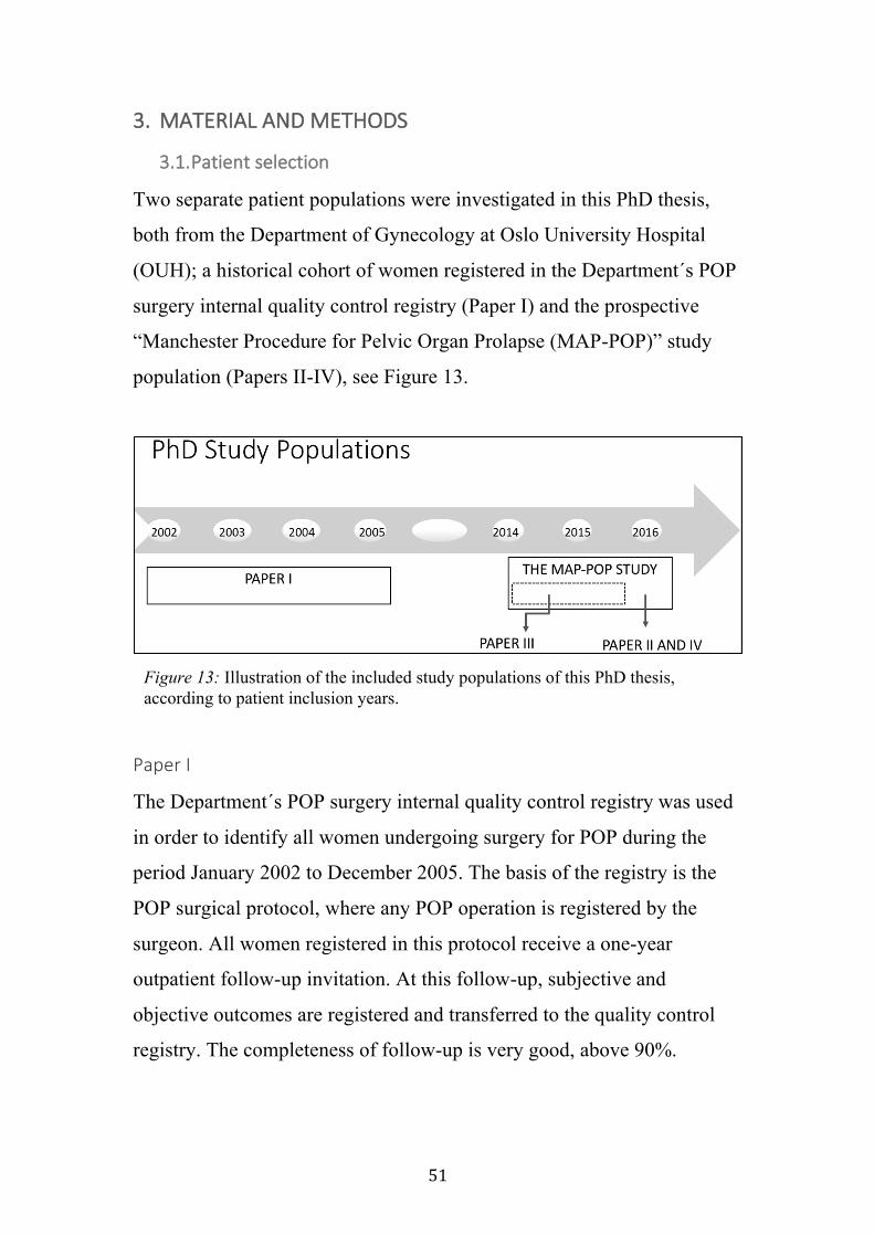
Short and long-term outcomes of the Manchester
Procedure for Pelvic Organ Prolapse and the impact
of major Levator Ani Muscle defects
PhD thesis
by
Sissel Hegdahl Oversand, MD
2018
Department of Gynaecology,
Oslo University Hospital, Ullevål
and
University of Oslo,
Faculty of Medicine,
Norway

© Sissel Hegdahl Oversand, 2018
Series of dissertations submitted to the Faculty of Medicine, University of Oslo
ISBN 978-82-8377-335-4
All rights reserved. No part of this publication may be reproduced or transmitted, in any form or by any means, without permission.
Cover: Hanne Baadsgaard Utigard. Print production: Reprosentralen, University of Oslo.


[ ]


≥


22
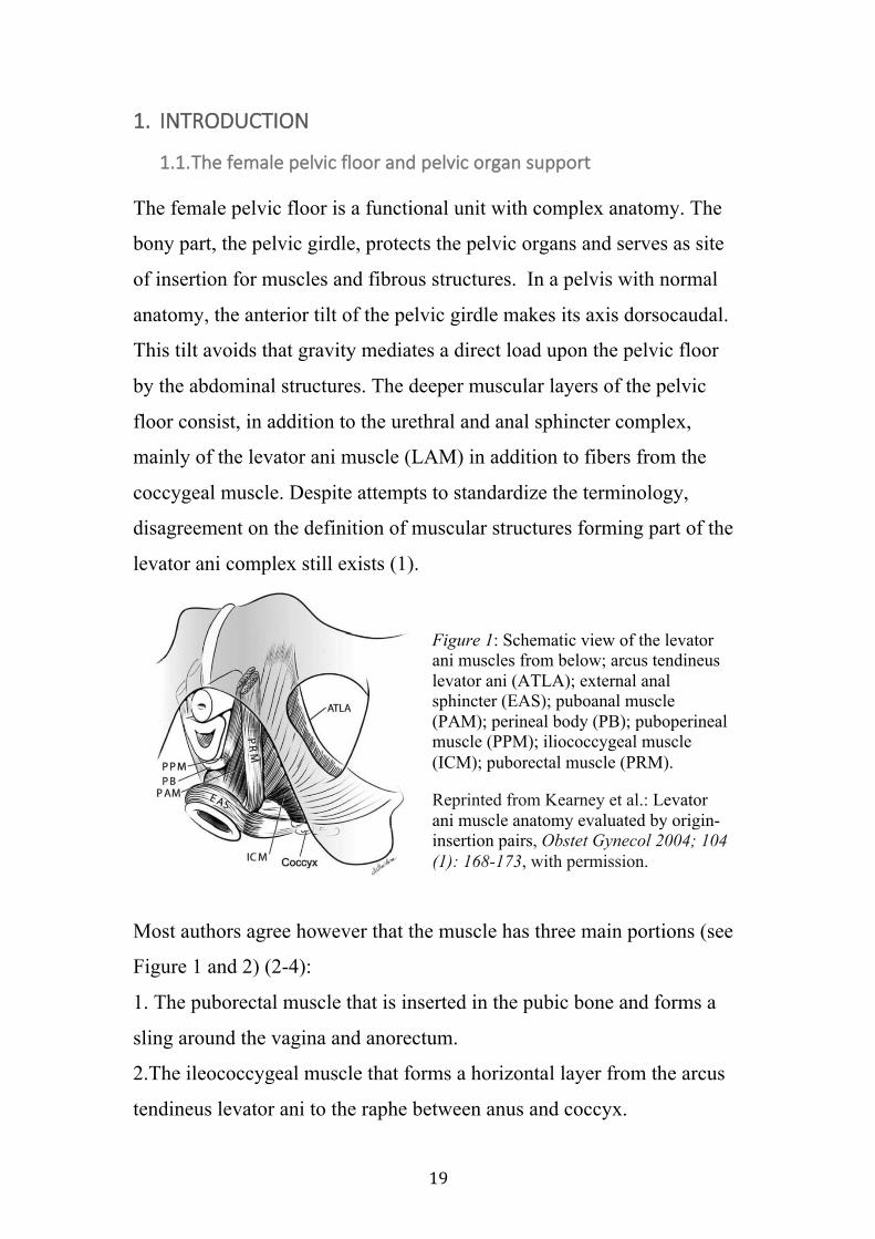
The pudendal nerves inervate the voluntary parts of the urethral and anal
sphincters whereas the levator ani muscle complex receives its nerve
supply from both the pudendal and direct sacral nerves. During the
second stage of labor, the nerves to the anal sphincter undergo the most
strain (8).
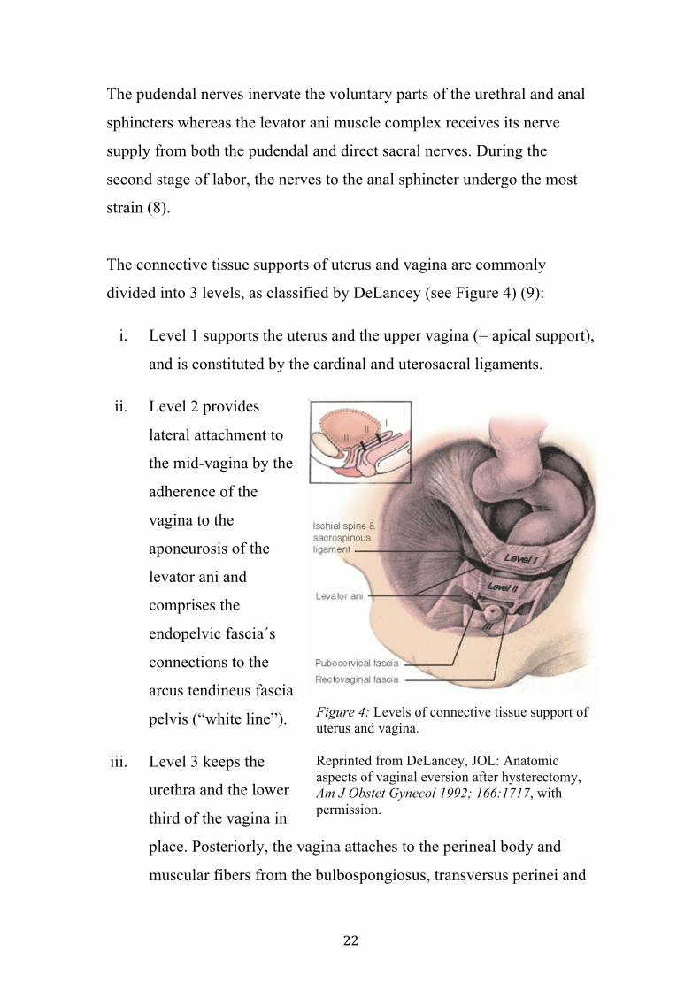
The connective tissue supports of uterus and vagina are commonly
divided into 3 levels, as classified by DeLancey (see Figure 4) (9):
i. Level 1 supports the uterus and the upper vagina (= apical support),
and is constituted by the cardinal and uterosacral ligaments.
ii. Level 2 provides
lateral attachment to
the mid-vagina by the
adherence of the
vagina to the
aponeurosis of the
levator ani and
comprises the
endopelvic fascia´s
connections to the
arcus tendineus fascia
pelvis (“white line”).
iii. Level 3 keeps the
urethra and the lower
third of the vagina in
place. Posteriorly, the vagina attaches to the perineal body and
muscular fibers from the bulbospongiosus, transversus perinei and
Figure 4: Levels of connective tissue support of uterus and vagina. Reprinted from DeLancey, JOL: Anatomic aspects of vaginal eversion after hysterectomy, Am J Obstet Gynecol 1992; 166:1717, with permission.


29
“The boat in dry dock” theory postulated by Norton provides a simple
structural understanding of POP, particularly of uterine descent (46). The
boat represents the pelvic organs, the water represents the pelvic floor
muscles (PFM), and the moorings represent their fascia and ligamentous
attachments to the pelvic sidewall. If the ligaments (“moorings”) are cut
or the PFM tone (“water”) is reduced, increased strain will be put on the
remaining structures and thereby increase the risk for POP (see Figure 7)
(46).
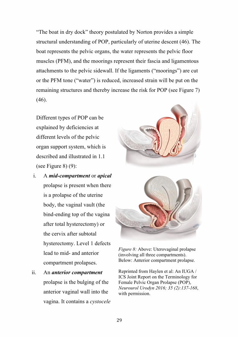
Different types of POP can be
explained by deficiencies at
different levels of the pelvic
organ support system, which is
described and illustrated in 1.1
(see Figure 8) (9):
i. A mid-compartment or apical
prolapse is present when there
is a prolapse of the uterine
body, the vaginal vault (the
bind-ending top of the vagina
after total hysterectomy) or
the cervix after subtotal
hysterectomy. Level 1 defects
lead to mid- and anterior
compartment prolapses.
ii. An anterior compartment
prolapse is the bulging of the
anterior vaginal wall into the
vagina. It contains a cystocele
Figure 8: Above: Uterovaginal prolapse (involving all three compartments). Below: Anterior compartment prolapse. Reprinted from Haylen et al: An IUGA / ICS Joint Report on the Terminology for Female Pelvic Organ Prolapse (POP), Neurourol Urodyn 2016; 35 (2):137-168, with permission.


Mid-compartment
POPPrevious
hysterectomy
Sacrospinousvault fixation
ManchesterProcedure
Sacrocolpopexy
Uterus-sparingsurgery
Uterosacralligament
suspension
Intactuterus
Hysterectomy
VaginalAbdominal/ laparoscopic/robotic
Sacrospinousligament
suspension
Sacrospinous hystero-(cervico)pexy
McCallsCuldoplasty
Obliterativeprocedures
LeFort´sColpocleisis
PartialColpocleisis
VaginalAbdominal/
laparoscopic/robotic
Sacrohysteropexy
Native tissuerepairs
Synthetic vaginalmesh procedures
TotalSubtotal


≥


≥
55
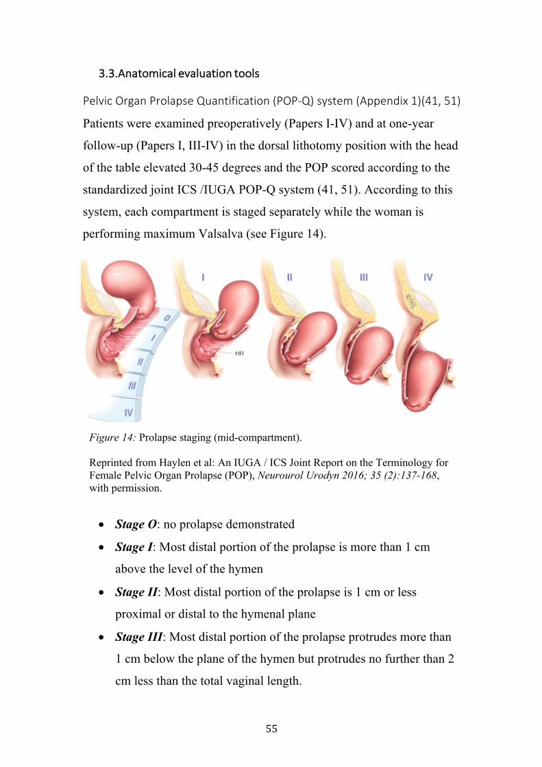
3.3. Anatomical evaluation tools
Pelvic Organ Prolapse Quantification (POP-Q) system (Appendix 1)(41, 51)
Patients were examined preoperatively (Papers I-IV) and at one-year
follow-up (Papers I, III-IV) in the dorsal lithotomy position with the head
of the table elevated 30-45 degrees and the POP scored according to the
standardized joint ICS /IUGA POP-Q system (41, 51). According to this
system, each compartment is staged separately while the woman is
performing maximum Valsalva (see Figure 14).
• Stage O: no prolapse demonstrated
• Stage I: Most distal portion of the prolapse is more than 1 cm
above the level of the hymen
• Stage II: Most distal portion of the prolapse is 1 cm or less
proximal or distal to the hymenal plane
• Stage III: Most distal portion of the prolapse protrudes more than
1 cm below the plane of the hymen but protrudes no further than 2
cm less than the total vaginal length.
Figure 14: Prolapse staging (mid-compartment).
Reprinted from Haylen et al: An IUGA / ICS Joint Report on the Terminology for Female Pelvic Organ Prolapse (POP), Neurourol Urodyn 2016; 35 (2):137-168, with permission.
56
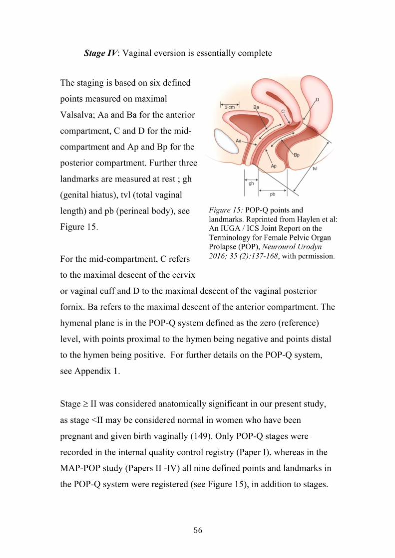
Stage IV: Vaginal eversion is essentially complete
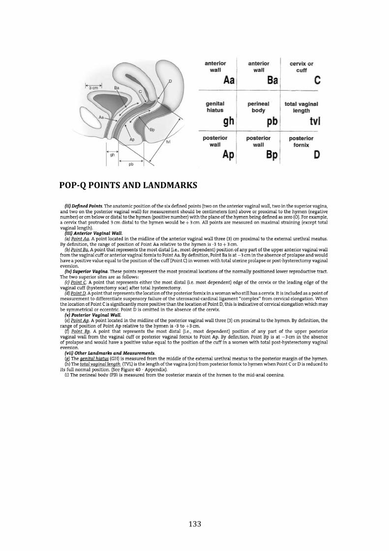
The staging is based on six defined
points measured on maximal
Valsalva; Aa and Ba for the anterior
compartment, C and D for the mid-
compartment and Ap and Bp for the
posterior compartment. Further three
landmarks are measured at rest ; gh
(genital hiatus), tvl (total vaginal
length) and pb (perineal body), see
Figure 15.
For the mid-compartment, C refers
to the maximal descent of the cervix
or vaginal cuff and D to the maximal descent of the vaginal posterior
fornix. Ba refers to the maximal descent of the anterior compartment. The
hymenal plane is in the POP-Q system defined as the zero (reference)
level, with points proximal to the hymen being negative and points distal
to the hymen being positive. For further details on the POP-Q system,
see Appendix 1.
Stage ³ II was considered anatomically significant in our present study,
as stage <II may be considered normal in women who have been
pregnant and given birth vaginally (149). Only POP-Q stages were
recorded in the internal quality control registry (Paper I), whereas in the
MAP-POP study (Papers II -IV) all nine defined points and landmarks in
the POP-Q system were registered (see Figure 15), in addition to stages.
Figure 15: POP-Q points and landmarks. Reprinted from Haylen et al: An IUGA / ICS Joint Report on the Terminology for Female Pelvic Organ Prolapse (POP), Neurourol Urodyn 2016; 35 (2):137-168, with permission.


≥


≥
≥
≥


α
β


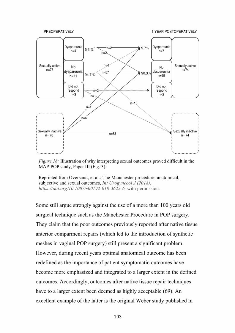
Sexually activen=78
Sexually inactiven= 74
Nodyspareunia
n=71
Dyspareunian=4
Did notrespond
n=3
5.3 %*
94.7 %*
PREOPERATIVELY
Sexually inactiven= 70
Sexually activen=74
1 YEAR POSTOPERATIVELY
Nodyspareunia
n=65
Did notrespond
n=2
Dyspareunian=7
n=10
n=4
n=57
n=2n=2
n=6
n=1
n=63
9.7%
90.3%
n=2
n=1

≥


≥