Embed Size (px)
Citation preview

Case Report
Short- and Long-Term Evaluation of a Condylar
Hyperplasia: A Case Report
Sxirin Nevzatoglu, DDS, PhD;1,* Evin Koc, DDS;2 Toros Alcan, DDS, PhD;3 and Zeki Guzel, MD4
ABSTRACT
Objective: This case report presents the diagnosis and short- and long-term treatment results of an orthodontically and surgicallytreated patient with condylar hyperplasia.Materials and Methods: Condylar hyperplasia was diagnosed in an 18-year-old patient. In the clinical and radiologicexamination, asymmetrical face, overdevelopment of the head and lengthening of the neck of the right condyle, deviation of thementon to the opposite side, temporomandibular joint pain, occlusal canting, slight open bite in the affected side, and an impactedupper right premolar were detected. Technetium-99 radioisotope scanning was made in 6-month intervals, and surgery wasplanned and performed. Changes were measured on the cephalograms taken at the beginning of orthodontic treatment, beforeand after surgery, at the end of fixed treatment, and in 4 years 6 months follow-up period.Results: Skeletal and dental Class I relationship was established and the profile improved. Slight increase of the verticaldimension was observed in the finishing and follow-up cephalograms.Conclusion: In order to decide the appropriate time for surgery for condylar hyperplasia patients in which osteoblastic activitycontinues, isotope-scanning examination is a must. Short-term results achieved with the orthodontic and surgical treatment weresatisfactory and were maintained in the long term. (Turkish J Orthod 2013;26:103–113)
KEY WORDS: Condylar Hyperplasia, High Condylectomy, Long-term Stability, Orthognathic surgery
INTRODUCTION
Condylar hyperplasia (CH) of the mandible is a
rare condition that causes overdevelopment of the
condylar head and leads to facial asymmetry,
deviation of the skeletal menton to the unaffected
side, malocclusion, pain, and articular dysfunction.
Prominent features include an enlarged mandibular
condyle, outward bowing, and downward growth of
the body and ramus of the mandible on the affected
side, causing fullness of the face on that side and
flattening of the face on the contralateral side.1
Condylar hyperplasia was first described by
Adams2 in 1836 as a complication of rheumatic
arthritis. The cause is unknown, but circulatory
problems, endocrine disturbances, trauma, inflam-
mation, and intrauterine influences (incorrect uterine
position causing squeezing of the mandible) can be
the possible etiologic factors.3–5 Epidemiologically,
the incidence between men and women seems
similar. CH manifests itself in patients with ages
ranging from 11–30 years, showing no predilection
for the left or right side.6–8
Differential diagnosis includes hemifacial hyper-
trophy (enlargement of all soft and hard tissues on
one side), unilateral macrognathia (includes con-
dyle, ramus, and ends at the midline), laterognathia,
chondroma, and osteochondroma. Conditions that
initiate after the age of 20 are most often related to
some type of proliferative pathology. Anamnesis,
previous medical and dental history, clinical and
radiologic examination, and bone scintigraphy are
1Assistant Professor, University of Marmara, School ofDentistry, Department of Orthodontics, Istanbul, Turkey
2PhD student, University of Marmara, School of Dentistry,Department of Orthodontics, Istanbul, Turkey
3Associate Professor, private practice, Istanbul, Turkey4Professor, Plastic and Reconstructive Surgeon, private
practice Istanbul, Turkey
*Corresponding author: Marmara Universitesi, Disx Hekim-
ligi Fakultesi, Ortodonti AD, Tesxvikiye mah. Buyukciftlik sok.No:6 k.3, 34365, Nisxantasxı, Sxisxli, Istanbul, Turkiye. Tel: þ90-212-231 9120 – 418 E-mail: [email protected], [email protected]
To cite this article: Nevzatoglu S, Koc E, Alcan T, Guzel Z.Short and long term evaluation of a condylar hyperplasia: a
case report. Turkish J Orthod. 2013;26:103–113 (DOI: http://dx.doi.org/10.13076/j.tjo.2013.26.01_103)
Date Submitted: October 2012. Date Accepted: January 2013.
Copyright 2013 by Turkish Orthodontic Society
103
important tools for correct diagnosis. The radioactive
isotope in scintigraphy is technetium-99 methylene
bisphosphonate. Increased radionuclide uptake by
the hyperplastic condyle can be an indication of
continued abnormal growth. It has been reported
that a difference in uptake of 55% : 45% or more
between the affected and healthy condyles can be
indicative of CH because the affected condyles had
a relative uptake of 55% or more.9
Treatment depends on the degree of severity and
the status of condylar growth. Different surgical
options have been proposed such as high condyl-
ectomy, orthognathic surgery, or both. There is also
controversy with respect to the time of surgery, either
performing the surgeries as soon as possible or
waiting for cessation of growth.
In this case report, the combined surgical treat-
ment of a patient with severe facial asymmetry
secondary to CH and 4-year follow-up is presented.
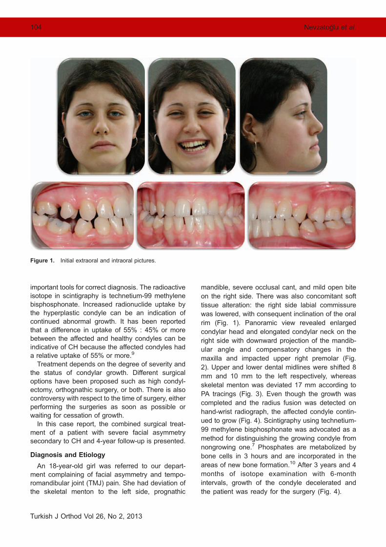
Diagnosis and Etiology
An 18-year-old girl was referred to our depart-
ment complaining of facial asymmetry and tempo-
romandibular joint (TMJ) pain. She had deviation of
the skeletal menton to the left side, prognathic
mandible, severe occlusal cant, and mild open bite
on the right side. There was also concomitant soft
tissue alteration: the right side labial commissure
was lowered, with consequent inclination of the oral
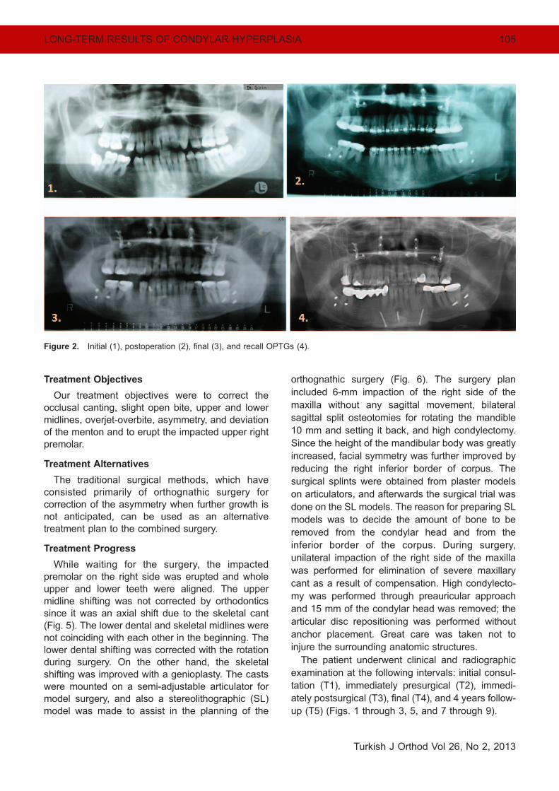
rim (Fig. 1). Panoramic view revealed enlarged
condylar head and elongated condylar neck on the
right side with downward projection of the mandib-
ular angle and compensatory changes in the
maxilla and impacted upper right premolar (Fig.
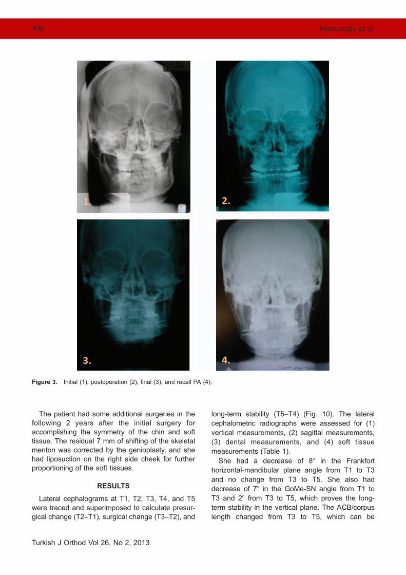
2). Upper and lower dental midlines were shifted 8
mm and 10 mm to the left respectively, whereas
skeletal menton was deviated 17 mm according to
PA tracings (Fig. 3). Even though the growth was
completed and the radius fusion was detected on
hand-wrist radiograph, the affected condyle contin-
ued to grow (Fig. 4). Scintigraphy using technetium-
99 methylene bisphosphonate was advocated as a
method for distinguishing the growing condyle from
nongrowing one.7 Phosphates are metabolized by
bone cells in 3 hours and are incorporated in the
areas of new bone formation.10 After 3 years and 4
months of isotope examination with 6-month
intervals, growth of the condyle decelerated and
the patient was ready for the surgery (Fig. 4).
Figure 1. Initial extraoral and intraoral pictures.
104 Nevzatoglu et al.
Turkish J Orthod Vol 26, No 2, 2013
Treatment Objectives
Our treatment objectives were to correct the
occlusal canting, slight open bite, upper and lower
midlines, overjet-overbite, asymmetry, and deviation
of the menton and to erupt the impacted upper right
premolar.
Treatment Alternatives
The traditional surgical methods, which have
consisted primarily of orthognathic surgery for
correction of the asymmetry when further growth is
not anticipated, can be used as an alternative
treatment plan to the combined surgery.
Treatment Progress
While waiting for the surgery, the impacted
premolar on the right side was erupted and whole
upper and lower teeth were aligned. The upper
midline shifting was not corrected by orthodontics
since it was an axial shift due to the skeletal cant
(Fig. 5). The lower dental and skeletal midlines were
not coinciding with each other in the beginning. The
lower dental shifting was corrected with the rotation
during surgery. On the other hand, the skeletal
shifting was improved with a genioplasty. The casts
were mounted on a semi-adjustable articulator for
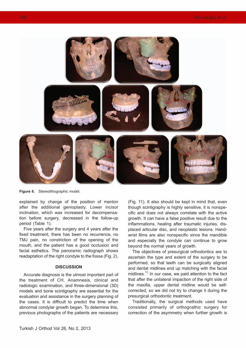
model surgery, and also a stereolithographic (SL)
model was made to assist in the planning of the
orthognathic surgery (Fig. 6). The surgery plan
included 6-mm impaction of the right side of the
maxilla without any sagittal movement, bilateral
sagittal split osteotomies for rotating the mandible
10 mm and setting it back, and high condylectomy.
Since the height of the mandibular body was greatly
increased, facial symmetry was further improved by
reducing the right inferior border of corpus. The
surgical splints were obtained from plaster models
on articulators, and afterwards the surgical trial was
done on the SL models. The reason for preparing SL
models was to decide the amount of bone to be
removed from the condylar head and from the
inferior border of the corpus. During surgery,
unilateral impaction of the right side of the maxilla
was performed for elimination of severe maxillary
cant as a result of compensation. High condylecto-
my was performed through preauricular approach
and 15 mm of the condylar head was removed; the
articular disc repositioning was performed without
anchor placement. Great care was taken not to
injure the surrounding anatomic structures.
The patient underwent clinical and radiographic
examination at the following intervals: initial consul-
tation (T1), immediately presurgical (T2), immedi-
ately postsurgical (T3), final (T4), and 4 years follow-
up (T5) (Figs. 1 through 3, 5, and 7 through 9).
Figure 2. Initial (1), postoperation (2), final (3), and recall OPTGs (4).
LONG-TERM RESULTS OF CONDYLAR HYPERPLASIA 105
Turkish J Orthod Vol 26, No 2, 2013
The patient had some additional surgeries in the
following 2 years after the initial surgery for
accomplishing the symmetry of the chin and soft
tissue. The residual 7 mm of shifting of the skeletal
menton was corrected by the genioplasty, and she
had liposuction on the right side cheek for further
proportioning of the soft tissues.
RESULTS
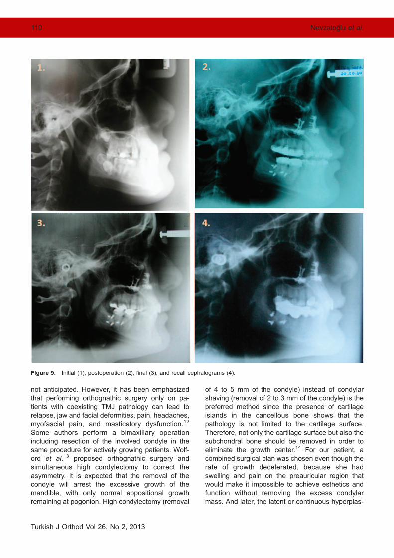
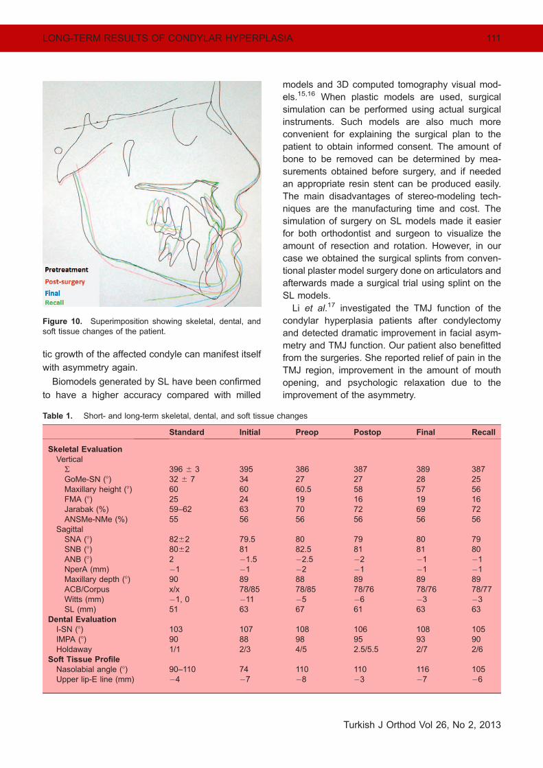
Lateral cephalograms at T1, T2, T3, T4, and T5
were traced and superimposed to calculate presur-
gical change (T2–T1), surgical change (T3–T2), and
long-term stability (T5–T4) (Fig. 10). The lateral
cephalometric radiographs were assessed for (1)
vertical measurements, (2) sagittal measurements,
(3) dental measurements, and (4) soft tissue
measurements (Table 1).
She had a decrease of 88 in the Frankfort
horizontal-mandibular plane angle from T1 to T3
and no change from T3 to T5. She also had
decrease of 78 in the GoMe-SN angle from T1 to
T3 and 28 from T3 to T5, which proves the long-
term stability in the vertical plane. The ACB/corpus
length changed from T3 to T5, which can be
Figure 3. Initial (1), postoperation (2), final (3), and recall PA (4).
106 Nevzatoglu et al.
Turkish J Orthod Vol 26, No 2, 2013
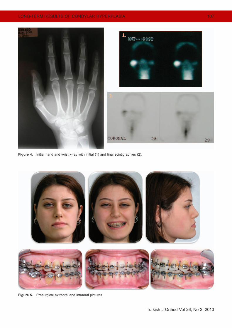
Figure 4. Initial hand and wrist x-ray with initial (1) and final scintigraphies (2).
Figure 5. Presurgical extraoral and intraoral pictures.
LONG-TERM RESULTS OF CONDYLAR HYPERPLASIA 107
Turkish J Orthod Vol 26, No 2, 2013
explained by change of the position of menton
after the additional genioplasty. Lower incisor
inclination, which was increased for decompensa-
tion before surgery, decreased in the follow-up
period (Table 1).
Five years after the surgery and 4 years after the
fixed treatment, there has been no recurrence, no
TMJ pain, no constriction of the opening of the
mouth, and the patient has a good occlusion and
facial esthetics. The panoramic radiograph shows
readaptation of the right condyle to the fossa (Fig. 2).
DISCUSSION
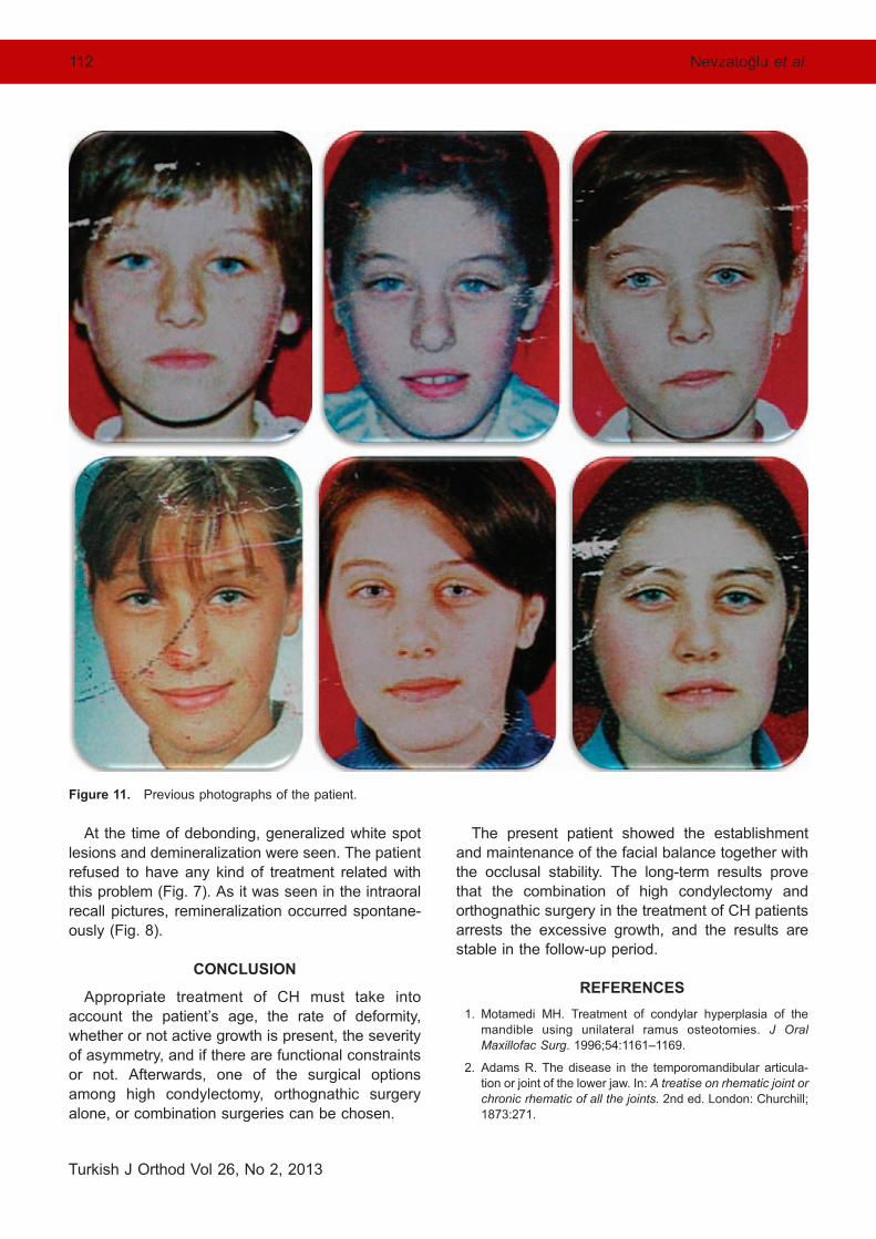
Accurate diagnosis is the utmost important part of
the treatment of CH. Anamnesis, clinical and
radiologic examination, and three-dimensional (3D)
models and bone scintigraphy are essential for the
evaluation and assistance in the surgery planning of
the cases. It is difficult to predict the time when
abnormal condylar growth began. To determine this,
previous photographs of the patients are necessary
(Fig. 11). It also should be kept in mind that, even
though scintigraphy is highly sensitive, it is nonspe-
cific and does not always correlate with the active
growth. It can have a false positive result due to the
inflammations, healing after traumatic injuries, dis-
placed articular disc, and neoplastic lesions. Hand-
wrist films are also nonspecific since the mandible
and especially the condyle can continue to grow
beyond the normal years of growth.
The objectives of presurgical orthodontics are to
ascertain the type and extent of the surgery to be
performed, so that teeth can be surgically aligned
and dental midlines end up matching with the facial
midlines.11 In our case, we paid attention to the fact
that after the unilateral impaction of the right side of
the maxilla, upper dental midline would be self-
corrected, so we did not try to change it during the
presurgical orthodontic treatment.
Traditionally, the surgical methods used have
consisted primarily of orthognathic surgery for
correction of the asymmetry when further growth is
Figure 6. Stereolithographic model.
108 Nevzatoglu et al.
Turkish J Orthod Vol 26, No 2, 2013
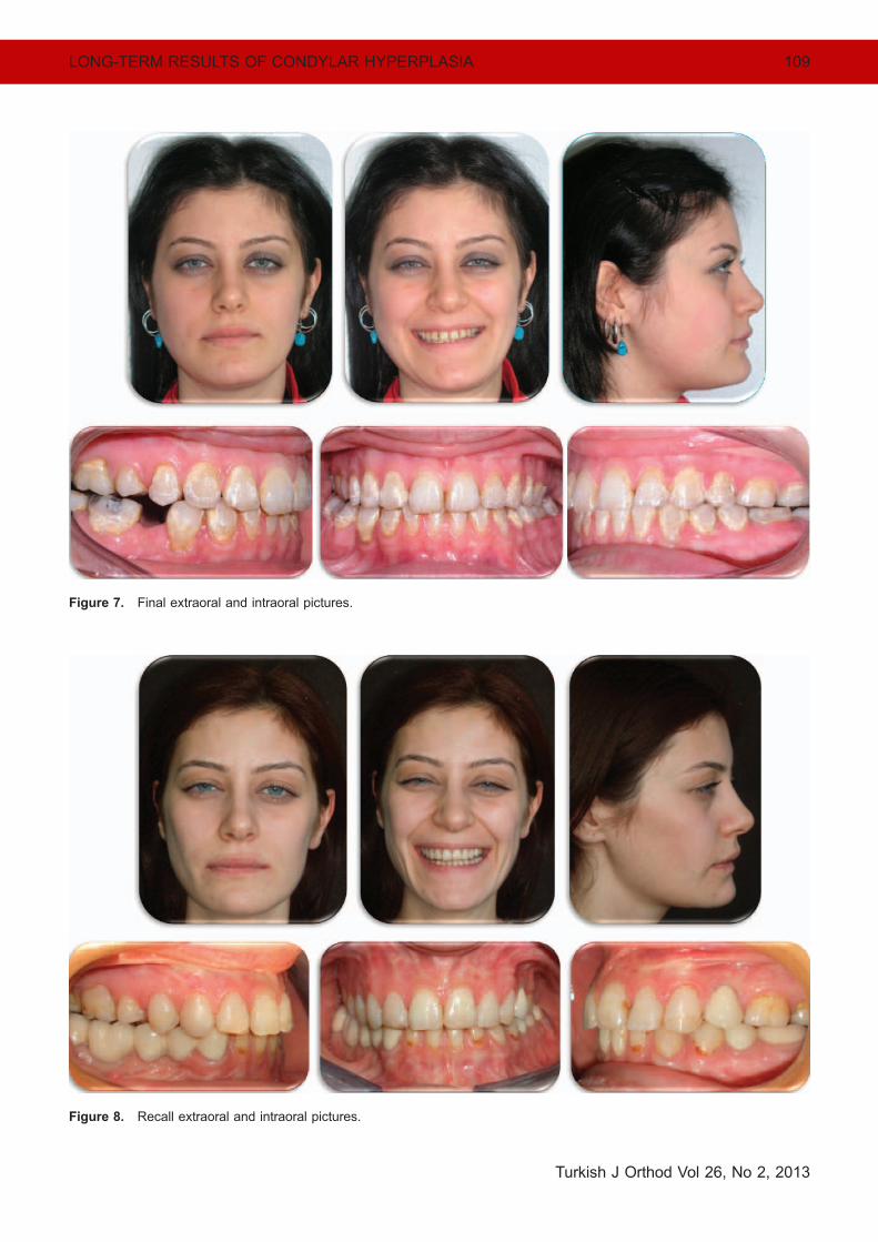
Figure 8. Recall extraoral and intraoral pictures.
Figure 7. Final extraoral and intraoral pictures.
LONG-TERM RESULTS OF CONDYLAR HYPERPLASIA 109
Turkish J Orthod Vol 26, No 2, 2013
not anticipated. However, it has been emphasized
that performing orthognathic surgery only on pa-
tients with coexisting TMJ pathology can lead torelapse, jaw and facial deformities, pain, headaches,
myofascial pain, and masticatory dysfunction.12
Some authors perform a bimaxillary operation
including resection of the involved condyle in thesame procedure for actively growing patients. Wolf-
ord et al.13 proposed orthognathic surgery and
simultaneous high condylectomy to correct the
asymmetry. It is expected that the removal of thecondyle will arrest the excessive growth of the
mandible, with only normal appositional growth
remaining at pogonion. High condylectomy (removal
of 4 to 5 mm of the condyle) instead of condylar
shaving (removal of 2 to 3 mm of the condyle) is the
preferred method since the presence of cartilageislands in the cancellous bone shows that the
pathology is not limited to the cartilage surface.
Therefore, not only the cartilage surface but also the
subchondral bone should be removed in order toeliminate the growth center.14 For our patient, a
combined surgical plan was chosen even though the
rate of growth decelerated, because she had
swelling and pain on the preauricular region thatwould make it impossible to achieve esthetics and
function without removing the excess condylar
mass. And later, the latent or continuous hyperplas-
Figure 9. Initial (1), postoperation (2), final (3), and recall cephalograms (4).
110 Nevzatoglu et al.
Turkish J Orthod Vol 26, No 2, 2013
tic growth of the affected condyle can manifest itself
with asymmetry again.
Biomodels generated by SL have been confirmed
to have a higher accuracy compared with milled
models and 3D computed tomography visual mod-
els.15,16 When plastic models are used, surgical
simulation can be performed using actual surgical
instruments. Such models are also much more
convenient for explaining the surgical plan to the
patient to obtain informed consent. The amount of
bone to be removed can be determined by mea-
surements obtained before surgery, and if needed
an appropriate resin stent can be produced easily.
The main disadvantages of stereo-modeling tech-
niques are the manufacturing time and cost. The
simulation of surgery on SL models made it easier
for both orthodontist and surgeon to visualize the
amount of resection and rotation. However, in our
case we obtained the surgical splints from conven-
tional plaster model surgery done on articulators and
afterwards made a surgical trial using splint on the
SL models.
Li et al.17 investigated the TMJ function of the
condylar hyperplasia patients after condylectomy
and detected dramatic improvement in facial asym-
metry and TMJ function. Our patient also benefitted
from the surgeries. She reported relief of pain in the
TMJ region, improvement in the amount of mouth
opening, and psychologic relaxation due to the
improvement of the asymmetry.
Figure 10. Superimposition showing skeletal, dental, andsoft tissue changes of the patient.
Table 1. Short- and long-term skeletal, dental, and soft tissue changes
Standard Initial Preop Postop Final Recall
Skeletal Evaluation
VerticalR 396 6 3 395 386 387 389 387GoMe-SN (8) 32 6 7 34 27 27 28 25Maxillary height (8) 60 60 60.5 58 57 56FMA (8) 25 24 19 16 19 16Jarabak (%) 59–62 63 70 72 69 72ANSMe-NMe (%) 55 56 56 56 56 56
SagittalSNA (8) 8262 79.5 80 79 80 79SNB (8) 8062 81 82.5 81 81 80ANB (8) 2 �1.5 �2.5 �2 �1 �1NperA (mm) �1 �1 �2 �1 �1 �1Maxillary depth (8) 90 89 88 89 89 89ACB/Corpus x/x 78/85 78/85 78/76 78/76 78/77Witts (mm) �1, 0 �11 �5 �6 �3 �3SL (mm) 51 63 67 61 63 63
Dental EvaluationI-SN (8) 103 107 108 106 108 105IMPA (8) 90 88 98 95 93 90Holdaway 1/1 2/3 4/5 2.5/5.5 2/7 2/6
Soft Tissue ProfileNasolabial angle (8) 90–110 74 110 110 116 105Upper lip-E line (mm) �4 �7 �8 �3 �7 �6
LONG-TERM RESULTS OF CONDYLAR HYPERPLASIA 111
Turkish J Orthod Vol 26, No 2, 2013
At the time of debonding, generalized white spot
lesions and demineralization were seen. The patient
refused to have any kind of treatment related with
this problem (Fig. 7). As it was seen in the intraoral
recall pictures, remineralization occurred spontane-
ously (Fig. 8).
CONCLUSION
Appropriate treatment of CH must take into
account the patient’s age, the rate of deformity,
whether or not active growth is present, the severity
of asymmetry, and if there are functional constraints
or not. Afterwards, one of the surgical options
among high condylectomy, orthognathic surgery
alone, or combination surgeries can be chosen.
The present patient showed the establishment
and maintenance of the facial balance together with
the occlusal stability. The long-term results prove
that the combination of high condylectomy and
orthognathic surgery in the treatment of CH patients
arrests the excessive growth, and the results are
stable in the follow-up period.
REFERENCES
1. Motamedi MH. Treatment of condylar hyperplasia of the
mandible using unilateral ramus osteotomies. J Oral
Maxillofac Surg. 1996;54:1161–1169.
2. Adams R. The disease in the temporomandibular articula-
tion or joint of the lower jaw. In: A treatise on rhematic joint or
chronic rhematic of all the joints. 2nd ed. London: Churchill;
1873:271.
Figure 11. Previous photographs of the patient.
112 Nevzatoglu et al.
Turkish J Orthod Vol 26, No 2, 2013
3. Cervelli V, Bottini DJ, Arpino A, Trimarco A, Cervelli G,
Mugnaini F. Hypercondylia: problems in diagnosis andtherapeutic indications. J Craniofac Surg. 2008;19:406–410.
4. Egyedi P. Aetiology of condylar hyperplasia. Aust Dent J.
1969;14:12–17.
5. Munoz MF, Monje F, Goizueta C, Rodrıguez-Campo F.
Active condylar hyperplasia treated by high condylectomy:report of case. J Oral Maxillofac Surg. 1999;57:1455–1459.
6. Nitzan DW, Katsnelson A, Bermanis I, Brin I, Casap N. Theclinical characteristics of condylar hyperplasia: experience
with 61 patients. J Oral Maxillofac Surg. 2008;66:312–318.
7. Saridin CP, Raijmakers P, Becking AG. Quantitative analysis
of planar bone scintigraphy in patients with unilateralcondylar hyperplasia. Oral Surg Oral Med Oral Pathol Oral
Radiol Endod. 2007;104:259–263.
8. Eslami B, Behnia H, Javadi H, Khiabani KS, Saffar AS.
Histopathologic comparison of normal and hyperplastic
condyles. Oral Surg Oral Med Oral Pathol Oral Radiol
Endod. 2003;96:711–717.
9. Bohuslavizki KH, Brenner W, Kerscher A, Fleiner B,Tinnemeyer S, et al. The value of bone scanning in the
pre-operative decision-making in patients with progressivefacial asymmetry. Nucl Med Commun. 1996;17:562–567.
10. Robinson PD, Harris K, Coghlan KC, Altman K. Bone scansand the timing of treatment for condylar hyperplasia. Int J
Oral Maxillofac Surg. 1990;19:243–246.
11. Slootweg PJ, Muller H. Condylar hyperplasia. A clinico-
pathological analysis of 22 cases. J Maxillofac Surg. 1986;
14:209–214.
12. Yamada K, Hanada K, Fukui T, Satou Y, Ochi K, et al.
Condylar bony change and self-reported parafunctional
habits in prospective orthognathic surgery patients with
temporomandibular disorders. Oral Surg Oral Med Oral
Pathol Oral Radiol Endod. 2001;92:265–271.
13. Wolford LM, Mehra P, Reiche-Fischel O, Morales-Ryn CA,
Garcia-Morales P. Efficacy of high condylectomy for
management of condylar hyperplasia. Am J Orthod Dento-
facial Orthop. 2002;121:136–150.
14. Villanueva-Alcojol L, Monje F, Gonzalez-Garcıa R. Hyper-
plasia of the mandibular condyle: clinical, histopathologic,
and treatment considerations in a series of 36 patients. J
Oral Maxillofac Surg. 2011;69:447–455.
15. Lill W, Solar P, Ulm C, Watzek G, Blahout R, Matejka M.
Reproducibility of three-dimensional CT-assisted model
production in the maxillofacial area. Br J Oral Maxillofac
Surg. 1992;30:233–236.
16. Klein HM, Schneider W, Alzen G, Voy ED, Gunter RW.
Pediatric craniofacial surgery: comparison of milling and
stereolithography 3D model manufacturing. Pediatr Radiol.
1992;22:458–460.
17. Li J, Long X, Yang XW, Li XD, Cheng Y, Deng MH.
Evaluation of temporomandibular joint function after condyl-
ectomy for condylar hyperplasia [in Chinese]. Zhonghua
Zheng Xing Wai Ke Za Zhi. 2006;22:175–179.
LONG-TERM RESULTS OF CONDYLAR HYPERPLASIA 113
Turkish J Orthod Vol 26, No 2, 2013

![Conservative Approach to Unilateral Condylar Fracture in a … · 2016-10-09 · of condylar fractures [7]. It appears that pediatric condylar fractures could be managed by closed](https://img.pdfslide.us/doc/110x75/5f48360e47a39a42e102f2f1/conservative-approach-to-unilateral-condylar-fracture-in-a-2016-10-09-of-condylar.jpg)


![Endometrium presentation - Dr Wright[1] · Endometrial Hyperplasia Simple hyperplasia Complex hyperplasia (adenomatous) Simple atypical hyperplasia ... Progression of Hyperplasia](https://img.pdfslide.us/doc/110x75/5b8a421e7f8b9a50388bc13d/endometrium-presentation-dr-wright1-endometrial-hyperplasia-simple-hyperplasia.jpg)