Embed Size (px)
Citation preview
Page 1/18
Evaluation of the Effectiveness of ContinuousVenovenous Hemodia�ltration Appli̇ed With Oxirisand AN69 Membranes in Patients With SepticShock-Related Acute Kidney InjuryCem Kıvılcım Kaçar
Diyarbakir Gazi Yasargil Egitim ve Arastirma HastanesiOsman Uzundere
Diyarbakir Gazi Yasargil Egitim ve Arastirma HastanesiEnver Yüksel
Diyarbakir Gazi Yasargil Egitim ve Arastirma HastanesiDeniz Kandemir
Diyarbakir Gazi Yasargil Egitim ve Arastirma HastanesiEsra Akiz Bıçak
Diyarbakir Gazi Yasargil Egitim ve Arastirma HastanesiAbdulkadir Yektaş ( [email protected] )
Diyarbakir Gazi Yasargil Egitim ve Arastirma Hastanesi https://orcid.org/0000-0003-4400-548X
Research article
Keywords: Septic shock, Acute kidney injury, Continuous renal replacement therapy, Continuousvenovenous hemodia�ltration, AN69, Oxiris
Posted Date: January 22nd, 2020
DOI: https://doi.org/10.21203/rs.2.21569/v1
License: This work is licensed under a Creative Commons Attribution 4.0 International License. Read Full License
Page 2/18
AbstractBackground and objectives: AN69 and Oxiris are �lters used in continuous renal replacement therapy. Inthis study, we aimed to research the effects of these �lters on blood cell counts, blood biochemistry,in�ammation indicators, clinical status and mortality of patients diagnosed with septic shock-relatedacute kidney injury.
Method: Between March 2019 and October 2019, 42 adult patients (Group 1: Oxiris (n = 21) or Group 2:AN69 (n=21)) with septic shock-related acute kidney injury and received continuous venovenoushemodia�ltration (CVVHDF) in the intensive care unit were included in the study and their results wereprospectively observed and compared. The data at the begining of CVVHDF (pre-CVVHDF) and 24 hoursafter the onset of CVVHDF (post-CVVHDF) were recorded.
Results: In the comparison of the pre- and post-CVVHDF values in Group 1, there was a statisticallysigni�cant decrease detected in the procalcitonin (p = 0.04) and noradrenaline infusion rate (p = 0.02)levels. In terms of the other data there was no statistically signi�cant difference between pre- and post-CVVHDF values in Group 1. In the comparison of the pre- and post-CVVHDF values in Group 2, there wasa statistically signi�cant decrease detected in the urea (p = 0.04), platelet count (p = 0.02) andprocalcitonin (p = 0.002) levels. There was no statistically signi�cant difference between pre- and post-CVVHDF values in terms of the other data in Group 2. There was no statistically signi�cant differencebetween the groups in terms of mortality.
Conclusions: CVVHDF with Oxiris �lter causes a statistically signi�cant decrease in noradrenalineinfusion rate. Therefore, we think that the use of CVVHDF with Oxiris �lter applied for septic shock-relatedacute kidney injury will save us time and increase the improvement in the treatment.
IntroductionSeptic shock is a subset of sepsis with circulatory, cellular and metabolic abnormalities. Patients withseptic shock may be clinically de�ned by the need for vasopressors to maintain mean arterial pressure≥65 mmHg and serum lactate levels> 2 mmol / L (> 18 mg / dL) in the absence of hypovolemia (1).
Lipoteichoic acid (LTA) in the structure of gram-positive bacteria and lipopolysaccharide (LPS) -endotoxin- in the structure of gram-negative bacteria are proin�ammatory bacterial lipids. These bacteriallipids induce proin�ammatory cytokine synthesis by signaling monocytes, macrophages, neutrophils andother immune cell types (2). Proin�ammatory cytokines induce a cytokine storm, causing endothelial celldisorder. Large molecules and liquid are extravasated into the interstitium (3). As a result, organdysfunction develops (4). One of the organ dysfunctions that may develop in sepsis is Acute KidneyInjury (AKI). In septic AKI, microcirculatory disorder develops in renal parenchyma as a result ofderegulation of in�ammatory mediators, immune cell in�ltration and nitric oxide synthase (5).
Page 3/18
Treatment of AKI involves treating the underlying disease and supporting renal function with renalreplacement therapy (RRT). However, The Surviving Sepsis Campaign Guidelines (6) contain littleexplanation regarding AKI treatment.
Many studies, with con�icting results, have evaluated the capacity of extracorporeal devices to adsorbendotoxins and cytokines (7,8), but The Surviving Sepsis Campaign Guidelines (6) have not made anyrecommendations on the use of blood puri�cation techniques.
The size of the endotoxin molecules is about 10 kDa. However, it can form aggregates up to 1,000 kDaconsisting of covalently bound lipid and polysaccharide (9). Cytokines are molecules that dissolve inwater and are in free form in circulation. Their molecular weight is between 0.5-60 kDa (10). RRT �ltersare semi-permeable membranes with approximately 35 kDa pores (11). Two of these �lters, AN69 (M100,Gambro, France) and Oxiris (Baxter, France), are widely used.
AN69 adsorbs cytokines but does not adsorb endotoxins (12). Oxiris adsorbs endotoxins (negativelycharged) thanks to the positive charge on the surface in addition to cytokine elimination (13). Moststudies of Oxiris include patients with sepsis/septic shock due to gram-negative bacteria. Becauseendotoxins are a component of gram-negative bacteria, rather than gram-positive bacteria. However,Oxiris may also be useful in the case of sepsis/septic shock due to gram-positive bacteria, as intestinalhypoperfusion usually causes gram-negative bacteria to pass through the digestive lumen into the blood(14).
We think that the use of Oxiris �lter in patients with septic shock-related AKI will improve hemodynamicsto save time for treatment, reducing the need for noradrenaline.
In this study, we aimed to research the effects of AN69 and Oxiris membranes on blood cell counts, bloodbiochemistry, in�ammation indicators, clinical conditions and mortality of patients with septic shock-related AKI.
MethodsThis prospective observational study was performed between March 2019 and October 2019 in theAnesthesiology and Reanimation Intensive Care Unit of the Health Sciences University of Diyarbakır GaziYaşargil Training and Research Hospital. Our study protocol was approved by the ethics committee of ourhospital (234/2019). This study was conducted in accordance with the 2008 Declaration of Helsinki andwritten informed consent was obtained from all patients or their relatives. G-Power version 3.1.9.4(Universität Kiel, Germany) was used to calculate the sample size with reference to the proportionsspeci�ed in a previous study (4). The minimum number of patients to be included in the study was 42with a two-tailed alpha error of 0.05, a power of 0.80, an allocation ratio of N2 / N1 = 1 and an effect sizeof 0.8. Forty-two patients with septic shock in our intensive care unit (1) who underwent continuousvenovenous hemodia�ltration (CVVHDF) due to AKI (15) were included in the study and the results werecompared.
Page 4/18
Inclusion criteria
Age ≥18 years
Presence of septic shock (1)
Development of AKI (15)
Exclusion criteria
Documented Stage 5 chronic kidney disease (glomerular �ltration rate (GFR) <15 mL / min / 1.73m2)
End-stage renal failure in long-term dialysis
Patients receiving RRT before admission to the ICU
Patients with an inferior vena cava collabability index (IVCCI) that cannot be measured or havecomorbidities that may affect outcomes (16).
Electrocardiography (ECG), pulse oximetry (SpO2) assessment and continuous invasive arterial pressuremeasurement after intraarterial cannulation were performed in all patients using the BSM-9101K monitor(Nihon Kohden Europe GmbH, Germany).
For vascular access, a double-lumen hemodialysis catheter (11.5 Fr, Scw medicath, China) was insertedinto the femoral or internal jugular vein with ultrasonography (USG) guidance (GE Vivid device, UnitedMedical Instruments, USA). In addition to standard treatment, all patients received CVVHDF for 24 hoursusing Prisma�ex CRRT system (Gambro, Sweden) with the adsorbing Oxiris or AN69 �lter. Daily dialysisdose was maintained between 35-45 mL / kg / h, blood �ow between 100-150 mL / min, and �ltrationfraction was between 35-45%. DIALISAN CVVHD BG 2D (Baxter, Italy) was used as dialysis andpostdilution solution. The content of dialysis solution was Na: 140, K: 2, Ca: 1.75, Mg: 0.5, Cl: 111.5,HCO3: 32, Lactate: 3, Glucose: 6.1 mmol / l.
For the anticoagulation of the circuit, continuous infusion of nonfractionated heparin was used inpatients with hemorrhagic pro�le at physiological margin, and citrat solution was used in patients whosehemorrhage pro�le was not at physiological margin and / or who had bleeding risk. Heparin was usedwith an infusion rate of 5–15 IU / kg / h. The dosage of heparin was set for 45-60 seconds with activatedpartial thromboplastin time [aPTT] (ACL TOP500 and ACL TOP700, Instrumentation Laboratory, Bedford,MA, USA). Prismocitrate 10/2 (Gambro, Italy), a calcium-free but sterile citrate-containing solution, wasinfused in pretreatment mode in patients undergoing citrated anticoagulation. Content of prismocitratewas 10/2 Citrate: 10, Na: 136, Cl: 106, Citric acid: 2 mmol / l. In postdilution, 10% calcium chloride wasinfused and after �ltration was Ca ++ 0.6-0.8 meq / L and arterial Ca ++ 1-1.5 meq / L.
The �uid balance of the patients was calculated from the inferior vena cava by USG every 4 hoursand IVCCI was calculated to keep the IVCCI within the range of 30-40% (17).
Page 5/18
The researchers who made the diagnosis, applied the CRTT, calculated the USG and IVCCI,collected and evaluated the results were different. The data were collected from the electronic medicalrecord system and patient �les of our institution.
Demographic data of the patients, Acute Physiology and Chronic Health Interrogation (APACHE) IIscore, number of organs with failure, mortality after ARF, duration of ICU stay, source of sepsis, isolatedmicroorganisms in blood culture, KDIGO stage, the type of �lter used (AN69 or Oxiris), anticoagulationmethod (heparin / citrate) was recorded. Sequential Organ Failure Assessment (SOFA) score wererecorded at the beginning of CVVHDF (pre-CVVHDF) and 24 hours after the onset of CVVHDF (post-CVVHDF).
Furthermore the values of hemoglobin, hematocrit (BC-6800 auto hematology analyzer, Mindray,China), blood cell count (white blood cells [WBC], platelets: BC-6800 auto hematology analyzer, Mindray,China), blood biochemistry (urea, creatinine, GFR, albumin: c702-502 autoanalyser, Roche, Germany),blood gas (lactate: Rapid Point 500 blood gas analyzer, Siemens, Germany), in�ammation indicators (C-reactive protein [CRP]: Cobas c702 autoanalyser, Roche, Germany; procalcitonin [PRC]: Cobas e601 andCOBAS e602 analyzers, Roche, Germany, erythrocyte sedimentation rate [ESR]: Vision-C automatic ESRanalyzer, YHLO Biotech, China) and noradrenaline infusion rate (NIR) were recorded at pre- and post-CVVHDF. The e�cacy of AN69 or Oxiris �lters were evaluated by comparing the parameters at pre- andpost-CVVHDF.
While writing the article, necessary checks were made with strobe statement checklist used forobservational studies.
Statistical Analysis
SPSS 16.0 for Windows program was used for statistical analysis. Statistical data were expressed asmean and standard deviation, and categorical data were expressed as frequency and percentage. Thecomparison of categorical data in the groups was made with chi-square test and the results were givenas n%. The Kolmogorov-Smirnov test was used to determine whether the numerical data matched thenormality distribution. Student's T-test was used for the evaluation of the numerical data matching thenormal distribution between the groups, and Mann-Whitney U test was used for the non-normaldistribution. Paired T-test and Wilcoxon Signed Rank test were used for comparison of two normaldistribution measurements. Results regarding numerical data were given as mean ± standard deviation. P<0.05 was accepted as statistically signi�cant.
ResultsThe mean age of the patients included in the study was 61.33 ± 20.01; Apache II scores were 29.26 ± 7.75and SOFA 1 scores were 10,07 ± 2,78. At least 2 and at most 4 organ failure were detected in the patientsincluded in the study. Fifty percent of the patients were female and 50% were male. During the studyperiod twenty-�ve of the patients were died. Mortality rate was 59.5%. Heparin was used as anticoagulant
Page 6/18
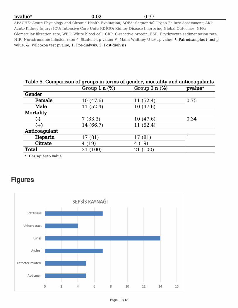
in 81% (n = 34) and citrate was used in 19% (n = 8). Twenty-one patients had KDIGO stage 3, 13 hadKDIGO stage 2, and 8 had KDIGO stage 1. There was no statistically signi�cant difference between thetwo groups in terms of KDIGO stages. The most common source of sepsis was the lungs (33.3%). Thedistribution of the patients in terms of the source of sepsis is shown in Figure 1.
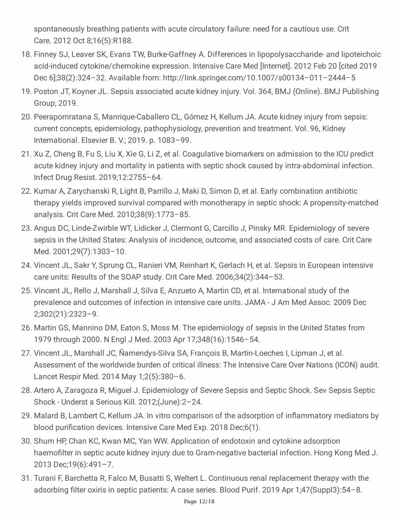
The source of sepsis, reproduction in blood culture and the type of �lter used are shown in Table 1.Among the gram-negative bacteria P. aeruginosa was found in the blood culture of 7 patients, A.Baumannii in 7, K. Pneumoniae in 4 and Enterobacter spp. in 3 patients; while among the gram-positivebacteria S. epidermidis was found in the blood culture of 3 patients, S. aureus in 3 and S. hominis ssphominis in 3 patients (Table 1).
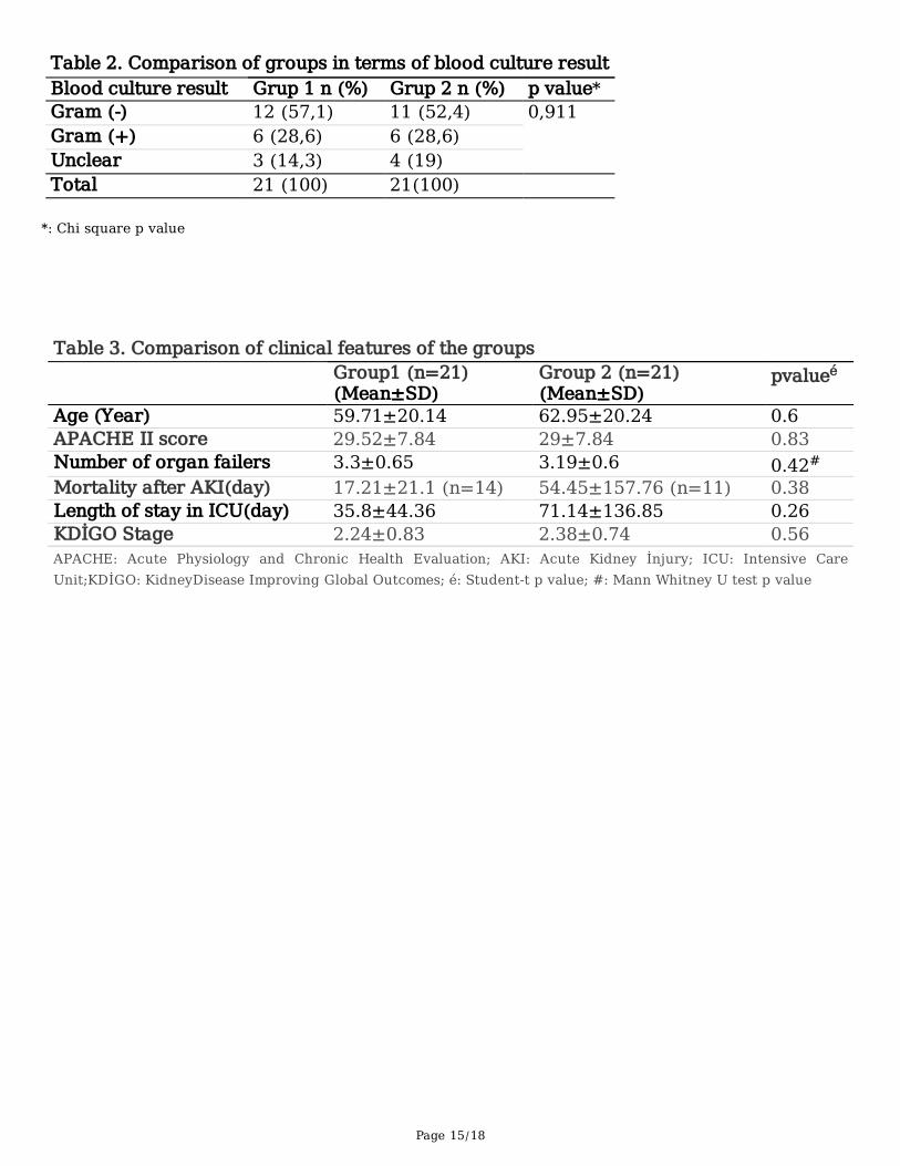
In 21 patients, CVVHDF was performed using Oxiris �lter (Group 1) and in 21 patients CVVHDF wasperformed using AN69 �lter (Group 2). When the blood cultures of the patients using Oxiris �lter wereexamined 12 patients had gram-negative, 6 patients had gram-positive bacteria growth and 3 patientshad no growth. When the blood cultures of the patients using AN69 �lter were examined 11 patients hadgram-negative, 6 patients had gram-positive bacteria growth and 4 patients had no growth. There was nostatistically signi�cant difference between the groups in terms of gram-negative and gram-positivebacteria growth in blood culture (Table 2).
The comparison of the groups in terms of clinical characteristics and laboratory values is shown inTables 3 and 4. There was no statistically signi�cant difference between the groups in terms of clinicalfeatures and laboratory values.
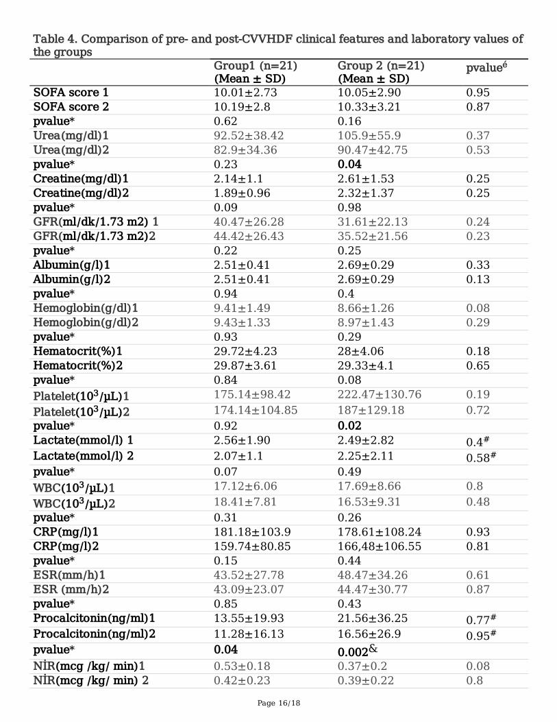
In the comparison of the pre- and post-CVVHDF values in Group 1, there was a statistically signi�cantdecrease detected in the procalcitonin (p = 0.04) and noradrenaline infusion rate (p = 0.02) levels. Interms of the other data there was no statistically signi�cant difference between pre- and post-CVVHDFvalues in Group 1. In the comparison of the pre- and post-CVVHDF values in Group 2, there was astatistically signi�cant decrease detected in the urea (p = 0.04), platelet count (p = 0.02) and procalcitonin(p = 0.002) levels. There was no statistically signi�cant difference between pre- and post-CVVHDF valuesin terms of the other data in Group 2 (Table 4).
Table 5 shows the comparison of the groups in terms of gender, mortality and anticoagulant used. Therewas no statistically signi�cant difference between the groups in terms of gender, mortality andanticoagulants used.
DiscussionIn our study, at the end of the CVVHDF for 24 hours we detected that PRC and NIR were reduced inpatients using Oxiris �lter; urea, platelet count and PRC levels were decreased signi�cantly in patientsusing AN69 �lter. There was no statistically signi�cant difference between the groups in terms ofmortality.
Page 7/18
LTA, a cell wall component speci�c to gram-positive bacteria, is the functional equivalent of LPS, themain cell wall component of gram-negative bacteria (18). LTA and LPS are also called pathogen-associated molecular patterns (PAMPs) and stimulate the natural immune response by binding to patternrecognition receptors (PRRs) such as Toll-like receptors (TLRs) expressed by monocytes, macrophages,neutrophils and other immune cell types. Gram-negative LPS mainly signals through TLR4, while Gram-positive LTA can bind to and signal through TLR2. (2). Both of these interactions stimulate the activationof Nuclear Factor kappa B (NF-κB), resulting in transcription and secretion of multiple pro-in�ammatorycytokines such as tumor necrosis factor (TNF) -α, interleukin (IL) –1, IL–6, IL–8 and IL–10 that playimportant roles in in�ammatory diseases such as sepsis (2,14). Damaged host cells express surfacedamage-associated molecular patterns (DAMPs) such as high-mobility-group-box–1 protein (HMGB1) ontheir surface. DAMPs can be released into circulation and are recognized by pattern recognition receptors(PRRs). Thus, leukocyte activation and cytokine synthesis are increased, fueling the vicious cycle ofuncontrolled immuno-in�ammatory process. Excessive release of cytokines in the blood is de�ned as a“cytokine storm” (14). As a result, sepsis or septic shock develops through vasodilatation, endothelialleakage and organ dysfunction (4). One of the organ dysfunctions in sepsis is AKI. Although thepathophysiological mechanism in septic AKI is not fully understood, it is clear that the in�ammatorycascade of sepsis contributes to AKI (19). The basic pathophysiological paradigm correlates septic AKIwith decreased global renal blood �ow, secondary tubular epithelial cell death, or acute tubular necrosis.The reason for this belief is that AKI is associated with hypoperfusion and shock, and that ischemicdamage can lead to intense cell death (eg acute tubular necrosis). However, the importance of ischemia-reperfusion is increasing (20).
Septic shock is associated with higher mortality compared to sepsis (1). Septic shock continues toaccount for 62% of general deaths and hospital mortality rates are above 40% (21). Mortality associatedwith AKI is high (40–60%) and the short- and long-term outcomes in the form of chronic or end-stagerenal disease are devastating (5). AKI develops in more than 45% of patients with septic shock (21). Themortality rate of our study patients was 59.5%. Considering the fact that our patient group had septicshock-related AKI, our mortality rate was within normal limits. There was no signi�cant differencebetween the groups in terms of mortality.
When we look at the results of four studies in the literature, which examined a large number of patientswith sepsis, the most common source of sepsis was reported to be lungs with varying rates (39–68%).Although the ranking varies, other common sources of infection are the abdomen (8–22%), unclear (17–20%), urinary tract (9–14%) and soft tissue (10%) (22–25). In our study, the most common source ofsepsis in the literature was the lungs (33%), followed by soft tissue (16%), unclear (16%), abdomen (12%),catheter-related infection (12%) and urinary tract (9%), respectively.
The Sepsis Occurrence in Acutely Ill Patients (SOAP) study reported an almost equal prevalence of gram-positive and gram-negative bacterial infections in patients with sepsis (24). Although subsequent studies(26) have suggested an increase in the incidence of gram-positive organisms, The 2012 Intensive CareOver Nations (ICON) study has shown that gram-negative bacterial infections are more common in the
Page 8/18
United States than gram-positive bacterial infections (27). In our study, gram-negative bacteria grew morein the blood cultures of the patients than gram-positive bacteria.
In a study of 13796 infected ICU patients published in 2009 S. aureus, Pseudomonas species,Enterobacteriaceae (especially E. coli) and fungi were the most common blood cultures. In this study,Acinetobacter accounted for 9% of patients with positive blood culture (25). In a review published in 2012,the most common isolated gram-negative bacteria were E. coli, P. aeruginosa and K. pneumonia; and andthe most common gram-positive bacteria were S. aureus, S. pneumonia and Enterococcus spp. inpatients with sepsis and septic shock (28). In our study, the most common gram-negative bacteria were P.aeruginosa, A. Baumannii, K. Pneumoniae and Enterobacter spp; and the most common gram-positivebacteria were S. epidermidis, S. aureus and S. hominis ssp hominis in the blood culture. Our study wasconsistent with previous studies in terms of blood culture reproduction. However, 20% of patients withpositive blood culture had acinetobacter reproduction and this rate was higher than previous studies.
Continuous renal replacement therapy (CRRT) with improvement in extracorporeal blood puri�cationtechniques and membrane materials are widely used in critical illness (10). The theoretical cut-off for anRRT membrane is about 35 kDa. Throughout this membrane, diffusion and convection can take place.Diffusion follows a concentration gradient (as in intermittent hemodialysis) and is an ideal method toremove small (<500 Da) molecules such as creatinine. Convection follows a hydrostatic pressure gradientand is the best method for elimination of medium to large (500 Da to 60 kDa, 13,750 Da beta–2microglobulin) and large molecules (60 to 100 kDa, eg 70 kDa albumin). In clinical practice, high (60 kDa)or median (50 kDa) cut-off membranes are almost never used because high cut-off membranes mayincrease the risk of albumin loss (11). Endotoxin molecules have a size of approximately 10 kDa, but canform aggregates of up to 1,000 kDa consisting of a covalently bonded lipid and polysaccharide (9). Thesmaller molecular weight of the cytokines, the more cytokines will be removed in the CRRT. The cutoffvalue of CRRT was 30–40 kDa, while IL–1β was 17 kDa, IL–1RA 15–20 kDa, IL–2 15 kDa, IL–6 26 kDa,IL–8 8 kDa, macrophage migration inhibitory factor (MIF) has a molecular weight of 12.5 kDa, IL–10 35–40 kDa and TNF-α51 kDa. Thus, only IL–10 and TNF-α are outside the threshold of CRRT; all othercytokines can be slowly removed with CRRT. High volume hemo�ltration or treatments like the ones withthe use of a high cut-off membrane can increase the clearance of in�ammatory cytokines but it is stillunknown if it could provide bene�t to patients (10).
AN69 is a copolymer of hydrophobic acrylonitrile and hydrophilic sodium metahalylsulfonate. As AN69 isnegatively charged due to sulfonate groups, the AN69 membrane adsorbs cytokines via ionic bondingbetween its sulfonate group and the amino group on the surface of a cytokine molecule (12). Oxiris is ahigh permeability polyacrylonitrile (AN69) based membrane on which a surface treatment of a positivelycharged polyethylene is added onto the hemo�lter. Thanks to this positive charge on the surface of theOxiris �lter - in addition to the bulk cytokine elimination in the mass of the membrane - endotoxin(negatively charged) adsorption takes place (13). In in-vitro studies, Oxiris �lter emerges as ahemoperfusion device capable of eliminating both endotoxin and cytokine (29). Endotoxinhemoadsorption can reduce the pathogenic activity and organ dysfunction of endotoxin. Cytokine
Page 9/18
removal by hemo�ltration or hemoadsorption can restore the status of immune homeostasis. It isthought that the use of semi-permeable membranes that can provide endotoxin and cytokine eliminationis a valuable treatment option in septic shock due to gram-negative bacterial infection (30). Endotoxinmay also be present in the circulation due to translocation from the ischemic gut in gram-positiveinfections (4). Therefore, the use of oxiris in gram-positive sepsis or septic shock may be bene�cial (14).
The main �nding of our study is that CVVHDF performed with Oxiris �lter improves hemodynamics,reduces NIR, is clinically applicable and has no side effects in patients with septic shock-related AKI. Ourresults con�rm the results of some studies in which Oxiris �lter was applied in the same patient group.Comparing Oxiris and AN69 �lters in patients with septic shock-related AKI Broman et al. found a strongdecrease in circulating endotoxin and cytokine levels as a result of CVVHDF treatment with Oxiris �lter.This reduction was associated with a favorable hemodynamic effect, such as a faster decrease in bloodlactate levels and a decrease in NIR required to maintain mean arterial pressure. There was a bluntedcytokine response in both �lter groups, but the decrease in TNF-α, IL–6, IL–8, and IFN- γ was moreoutstanding in patients treated with Oxiris than the AN69 �lter (4). In the study of Schwindenhammer etal., 31 patients were diagnosed with septic shock between 2014 and 2019 and one of the continuousvenovenous hemo�ltration (CVVH) or CVVHDF therapies with Oxiris �lter was applied. A relative decreaseof 88% was observed in NIR. Lactatemia and pH improved signi�cantly over time (13). In their study on60 septic patients published in 2019, Turani et al. found that CVVHDF with Oxiris �lter improved basiccardiorenal and respiratory parameters and decreased NIR (31).
Shum et al., in a study performed CVVH for 48 hours, found a 37% reduction in the SOFA score of sepsis-related AKI patients who underwent Oxiris �lter compared to polysulfone-based standard �lter (30). Intheir study published in 2019, Turani et al. found that the SOFA score of 60 septic patients appliedCVVHDF with Oxiris �lter decreased from 12.4 ± 2 to 9 ± 2 (31). In our study, no signi�cant decrease wasobserved in the SOFA score by CVVHDF with Oxiris �lter. This is due to the fact that our patients had verysevere diseases with high mortality (septic shock-related AKI) and we evaluated the SOFA score after 24hours of Oxiris administration. Longer CRRT could cause a signi�cant decrease in SOFA score.
In their study on 13 patients with sepsis and multiorgan failure Dahaba AA et al. found that, PRC levelsdecreased signi�cantly after 12 hours CVVH with AN69 �lter (32). Turani et al. in their studies publishedin 2019, observed a decrease in the PRC level of 60 septic patients who received CVVHDF with Oxiris �lter(31). In our study, we found that PRC levels decreased signi�cantly in both groups using Oxiris and AN69�lters after 24 hours of CVVHDF. The cut-off value of AN69 �lter is 35–40 kDa (32) and PCT molecularweight is 14.5 kDa (33). We attributed the signi�cant decrease in PCT value after CVVHDF with both�lters to the fact that the molecular weight of PCT was considerably lower than the cut-off value.
The limitation of our study is that we evaluate the blood cell counts, blood biochemistry, in�ammationindicators, clinical conditions and the mortality results without considering other intermittent conditionsafter the 24 hours of CVVHDF. We compared the changes that occurred with only one CVVHDF
Page 10/18
application. In the future, randomized, controlled, double-blind, clinical trials can be planned to comparethe changes in renal function and mortality rates of CVVHDF with longer or repeated administration.
ConclusionsCVVHDF with Oxiris �lter leads to a statistically signi�cant decrease in NIR. Therefore, we think that theuse of CVVHDF with Oxiris �lter applied for septic shock-related AKI will save us time and increase theimprovement.
Declarations1.Ethics committee: Republic of turkey, health sciences university, Gazi Yaşargil training and researchhospital Ethics committee for clinical research
Consent to participate: We obtained written informed consent from each �rst degree relative of patient.
2.Consent for publication: Not Applicable
3.Availability of data and materials: The datasets used and/or analysed during the current studyavailable from the corresponding author on reasonable request. ([email protected])
4.Competing interests: The authors declare no competing interests.
5.Funding: Not funding
6.Author contribution:
AKY, OU and CKK carried out the anterior and posterior sciatic nerve block, participated in the sequencealignment and drafted the manuscript, participated in the design of the study and performed thestatistical analysis. DK and EY conceived of the study, and participated in its design and coordinationand helped to draft the manuscript. All authors read and approved the �nal manuscript.
7.Acknowledgements: Not Applicable
References1. Singer M, Deutschman CS, Seymour C, Shankar-Hari M, Annane D, Bauer M, et al. The third
international consensus de�nitions for sepsis and septic shock (sepsis–3). JAMA - J Am Med Assoc.2016;315(8):801–10.
2. Grin PM, Dwivedi DJ, Chathely KM, Trigatti BL, Prat A, Seidah NG, et al. Low-density lipoprotein (LDL)-dependent uptake of Gram-positive lipoteichoic acid and Gram-negative lipopolysaccharide occursthrough LDL receptor. Sci Rep [Internet]. 2018;8(1):1–11. Available from:http://dx.doi.org/10.1038/s41598–018–28777–0
Page 11/18
3. Chausse JM, Malekele L, Paruk F. Improved understanding of the pathophysiology of sepsis: Settingthe scene for potential novel adjunctive therapies. South African J Crit Care. 2018;34(1):4–8.
4. Broman ME, Hansson F, Vincent JL, Bodelsson M. Endotoxin and cytokine reducing properties of theoXiris membrane in patients with septic shock: A randomized crossover double-blind study. PLoSOne. 2019;14(8).
5. Nusshag C, Weigand MA, Zeier M, Morath C, Brenner T. Issues of acute kidney injury staging andmanagement in sepsis and critical illness: A narrative review. Vol. 18, International Journal ofMolecular Sciences. MDPI AG; 2017.
�. Rhodes A, Evans LE, Alhazzani W, Levy MM, Antonelli M, Ferrer R, et al. Surviving Sepsis Campaign:International Guidelines for Management of Sepsis and Septic Shock: 2016. Intensive Care Med.2017 Mar 1;43(3):304–77.
7. Dellinger RP, Bagshaw SM, Antonelli M, Foster DM, Klein DJ, Marshall JC, et al. Effect of TargetedPolymyxin B Hemoperfusion on 28-Day Mortality in Patients with Septic Shock and ElevatedEndotoxin Level: The EUPHRATES Randomized Clinical Trial. JAMA - J Am Med Assoc. 2018 Oct9;320(14):1455–63.
�. Schädler D, Pausch C, Heise D, Meier-Hellmann A, Brederlau J, Weiler N, et al. The effect of a novelextracorporeal cytokine hemoadsorption device on IL–6 elimination in septic patients: A randomizedcontrolled trial. PLoS One. 2017 Oct 1;12(10).
9. Broman ME, Bodelsson M. Analysis of endotoxin adsorption in two swedish patients with septicshock. Blood Purif. 2019;47(Suppl3):51–3.
10. Zhang J, Tian J, Sun H, Digvijay K, Neri M, Bhargava V, et al. How does continuous renal replacementtherapy affect septic acute kidney injury? Vol. 46, Blood Puri�cation. S. Karger AG; 2018. p. 326–31.
11. Honore PM, Spapen HD. What a clinician should know about a renal replacement membrane? JTransl Intern Med. 2018 Jun 28;6(2):62–5.
12. Hattori N, Oda S. Cytokine-adsorbing hemo�lter: old but new modality for septic acute kidney injury.Ren Replace Ther [Internet]. 2016 Dec 18 [cited 2019 Dec 6];2(1):41. Available from:http://rrtjournal.biomedcentral.com/articles/10.1186/s41100–016–0051–1
13. Schwindenhammer V, Girardot T, Chaulier K, Grégoire A, Monard C, Huriaux L, et al. Oxiris® use inseptic shock: Experience of two French centres. Blood Purif. 2019 Apr 1;47(Suppl3):29–35.
14. Monard C, Rimmelé T, Ronco C. Extracorporeal Blood Puri�cation Therapies for Sepsis. Blood Purif[Internet]. 2019 [cited 2019 Dec 6];47(3):2–15. Available from: www.karger.com/bpu
15. KDIGO Clinical Practice Guideline for Acute Kidney Injury. Off J Int Soc Nephrol [Internet]. 2012 [cited2019 Dec 6];2(1). Available from: http://www.kidney-international.org
1�. Preau S, Bortolotti P, Colling D, Dewavrin F, Colas V, Voisin B, et al. Diagnostic Accuracy of the InferiorVena Cava Collapsibility to Predict Fluid Responsiveness in Spontaneously Breathing Patients WithSepsis and Acute Circulatory Failure. Crit Care Med. 2017 Mar;45(3):e290-e297.
17. Laurent Muller, Xavier Bobbia, Mehdi Toumi, Guillaume Louart, Nicolas Molinari, Benoit Ragonnet, etal. Respiratory variations of inferior vena cava diameter to predict �uid responsiveness in
Page 12/18
spontaneously breathing patients with acute circulatory failure: need for a cautious use. CritCare. 2012 Oct 8;16(5):R188.
1�. Finney SJ, Leaver SK, Evans TW, Burke-Gaffney A. Differences in lipopolysaccharide- and lipoteichoicacid-induced cytokine/chemokine expression. Intensive Care Med [Internet]. 2012 Feb 20 [cited 2019Dec 6];38(2):324–32. Available from: http://link.springer.com/10.1007/s00134–011–2444–5
19. Poston JT, Koyner JL. Sepsis associated acute kidney injury. Vol. 364, BMJ (Online). BMJ PublishingGroup; 2019.
20. Peerapornratana S, Manrique-Caballero CL, Gómez H, Kellum JA. Acute kidney injury from sepsis:current concepts, epidemiology, pathophysiology, prevention and treatment. Vol. 96, KidneyInternational. Elsevier B. V.; 2019. p. 1083–99.
21. Xu Z, Cheng B, Fu S, Liu X, Xie G, Li Z, et al. Coagulative biomarkers on admission to the ICU predictacute kidney injury and mortality in patients with septic shock caused by intra-abdominal infection.Infect Drug Resist. 2019;12:2755–64.
22. Kumar A, Zarychanski R, Light B, Parrillo J, Maki D, Simon D, et al. Early combination antibiotictherapy yields improved survival compared with monotherapy in septic shock: A propensity-matchedanalysis. Crit Care Med. 2010;38(9):1773–85.
23. Angus DC, Linde-Zwirble WT, Lidicker J, Clermont G, Carcillo J, Pinsky MR. Epidemiology of severesepsis in the United States: Analysis of incidence, outcome, and associated costs of care. Crit CareMed. 2001;29(7):1303–10.
24. Vincent JL, Sakr Y, Sprung CL, Ranieri VM, Reinhart K, Gerlach H, et al. Sepsis in European intensivecare units: Results of the SOAP study. Crit Care Med. 2006;34(2):344–53.
25. Vincent JL, Rello J, Marshall J, Silva E, Anzueto A, Martin CD, et al. International study of theprevalence and outcomes of infection in intensive care units. JAMA - J Am Med Assoc. 2009 Dec2;302(21):2323–9.
2�. Martin GS, Mannino DM, Eaton S, Moss M. The epidemiology of sepsis in the United States from1979 through 2000. N Engl J Med. 2003 Apr 17;348(16):1546–54.
27. Vincent JL, Marshall JC, Ñamendys-Silva SA, François B, Martin-Loeches I, Lipman J, et al.Assessment of the worldwide burden of critical illness: The Intensive Care Over Nations (ICON) audit.Lancet Respir Med. 2014 May 1;2(5):380–6.
2�. Artero A, Zaragoza R, Miguel J. Epidemiology of Severe Sepsis and Septic Shock. Sev Sepsis SepticShock - Underst a Serious Kill. 2012;(June):2–24.
29. Malard B, Lambert C, Kellum JA. In vitro comparison of the adsorption of in�ammatory mediators byblood puri�cation devices. Intensive Care Med Exp. 2018 Dec;6(1).
30. Shum HP, Chan KC, Kwan MC, Yan WW. Application of endotoxin and cytokine adsorptionhaemo�lter in septic acute kidney injury due to Gram-negative bacterial infection. Hong Kong Med J.2013 Dec;19(6):491–7.
31. Turani F, Barchetta R, Falco M, Busatti S, Weltert L. Continuous renal replacement therapy with theadsorbing �lter oxiris in septic patients: A case series. Blood Purif. 2019 Apr 1;47(Suppl3):54–8.
Page 13/18
32. Dahaba AA, Elawady GA, Rehak PH, List WF. Procalcitonin and proin�ammatory cytokine clearanceduring continuous venovenous haemo�ltration in septic patients. Anaesth Intensive Care [Internet].2002 Jun [cited 2019 Dec 6];30(3):269–74. Available from:http://www.ncbi.nlm.nih.gov/pubmed/12075632
33. Samsudin I, Vasikaran SD, Vasikaran S. Clinical Utility and Measurement of Procalcitonin. I ClinBiochem Rev. 2017;38(2):59–68.
TablesTable 1: Source of sepsis, blood culture growth and filter type data of the study patients
Page 14/18
Study Patient Source of sepsis Bacterial Growth in Blood Gram Filter1 Urinary tract K. pneumoniae - AN692 Lungs S. epidermidis + AN693 Soft tissue P. aeruginosa - AN694 Soft tissue S. epidermidis + Oxiris5 Urinary tract A. baumannii - AN696 Lungs S. hominis ssp hominis + Oxiris7 Catheter-related S. hominis ssp hominis + Oxiris8 Catheter-related E. faecium + AN699 Catheter-related S. aureus + AN6910 Lungs A. baumannii - Oxiris11 Abdomen Enterobacter spp. - Oxiris12 Lungs P. aeruginosa - AN6913 Lungs S. aureus + Oxiris14 Catheter-related S. haemolyticus + AN6915 Unclear Unclear Unclear AN6916 Abdomen Enterobacter spp. - AN6917 Soft tissue P. aeruginosa - AN6918 Catheter-related S. hominis ssp hominis + Oxiris19 Unclear Unclear Unclear Oxiris20 Abdomen A. baumannii - Oxiris21 Unclear Unclear Unclear AN6922 Lungs A. baumannii - AN6923 Lungs P. aeruginosa - AN6924 Lungs P. aeruginosa - AN6925 Lungs K. pneumoniae - Oxiris26 Soft tissue S. maltophilia - Oxiris27 Urinary tract A. baumannii - Oxiris28 Unclear Unclear Unclear AN6929 Unclear Unclear Unclear AN6930 Lungs A. baumannii - AN6931 Lungs A. baumannii - Oxiris32 Unclear Unclear Unclear Oxiris33 Abdomen M. R. S. aureus + AN6934 Urinary tract E. coli - Oxiris35 Abdomen Enterobacter spp. - Oxiris36 Lungs S. aureus + Oxiris37 Unclear Unclear Unclear Oxiris38 Soft tissue P. aeruginosa - Oxiris39 Lungs K. pneumoniae - AN6940 Soft tissue S. epidermidis + AN6941 Lungs K. pneumoniae - Oxiris42 Soft tissue P. aeruginosa - Oxiris
Page 15/18
Table 2. Comparison of groups in terms of blood culture resultBlood culture result Grup 1 n (%) Grup 2 n (%) p value*Gram (-) 12 (57,1) 11 (52,4) 0,911Gram (+) 6 (28,6) 6 (28,6)Unclear 3 (14,3) 4 (19)Total 21 (100) 21(100)
*: Chi square p value
Table 3. Comparison of clinical features of the groups Group1 (n=21)
(Mean±SD)Group 2 (n=21)(Mean±SD)
pvalueé
Age (Year) 59.71±20.14 62.95±20.24 0.6APACHE II score 29.52±7.84 29±7.84 0.83Number of organ failers 3.3±0.65 3.19±0.6 0.42#
Mortality after AKI(day) 17.21±21.1 (n=14) 54.45±157.76 (n=11) 0.38Length of stay in ICU(day) 35.8±44.36 71.14±136.85 0.26KDİGO Stage 2.24±0.83 2.38±0.74 0.56APACHE: Acute Physiology and Chronic Health Evaluation; AKI: Acute Kidney İnjury; ICU: Intensive CareUnit;KDİGO: KidneyDisease Improving Global Outcomes; é: Student-t p value; #: Mann Whitney U test p value
Page 16/18
Table 4. Comparison of pre- and post-CVVHDF clinical features and laboratory values ofthe groups Group1 (n=21)
(Mean ± SD)Group 2 (n=21)(Mean ± SD)
pvalueé
SOFA score 1 10.01±2.73 10.05±2.90 0.95SOFA score 2 10.19±2.8 10.33±3.21 0.87pvalue* 0.62 0.16 Urea(mg/dl)1 92.52±38.42 105.9±55.9 0.37Urea(mg/dl)2 82.9±34.36 90.47±42.75 0.53pvalue* 0.23 0.04 Creatine(mg/dl)1 2.14±1.1 2.61±1.53 0.25Creatine(mg/dl)2 1.89±0.96 2.32±1.37 0.25pvalue* 0.09 0.98 GFR(ml/dk/1.73 m2) 1 40.47±26.28 31.61±22.13 0.24GFR(ml/dk/1.73 m2)2 44.42±26.43 35.52±21.56 0.23pvalue* 0.22 0.25 Albumin(g/l)1 2.51±0.41 2.69±0.29 0.33Albumin(g/l)2 2.51±0.41 2.69±0.29 0.13pvalue* 0.94 0.4 Hemoglobin(g/dl)1 9.41±1.49 8.66±1.26 0.08Hemoglobin(g/dl)2 9.43±1.33 8.97±1.43 0.29pvalue* 0.93 0.29 Hematocrit(%)1 29.72±4.23 28±4.06 0.18Hematocrit(%)2 29.87±3.61 29.33±4.1 0.65pvalue* 0.84 0.08 Platelet(103/µL)1 175.14±98.42 222.47±130.76 0.19Platelet(103/µL)2 174.14±104.85 187±129.18 0.72pvalue* 0.92 0.02 Lactate(mmol/l) 1 2.56±1.90 2.49±2.82 0.4#
Lactate(mmol/l) 2 2.07±1.1 2.25±2.11 0.58#
pvalue* 0.07 0.49 WBC(103/µL)1 17.12±6.06 17.69±8.66 0.8WBC(103/µL)2 18.41±7.81 16.53±9.31 0.48pvalue* 0.31 0.26 CRP(mg/l)1 181.18±103.9 178.61±108.24 0.93CRP(mg/l)2 159.74±80.85 166,48±106.55 0.81pvalue* 0.15 0.44 ESR(mm/h)1 43.52±27.78 48.47±34.26 0.61ESR (mm/h)2 43.09±23.07 44.47±30.77 0.87pvalue* 0.85 0.43 Procalcitonin(ng/ml)1 13.55±19.93 21.56±36.25 0.77#
Procalcitonin(ng/ml)2 11.28±16.13 16.56±26.9 0.95#
pvalue* 0.04 0.002& NİR(mcg /kg/ min)1 0.53±0.18 0.37±0.2 0.08NİR(mcg /kg/ min) 2 0.42±0.23 0.39±0.22 0.8
Page 17/18
pvalue* 0.02 0.37 APACHE: Acute Physiology and Chronic Health Evaluation; SOFA: Sequential Organ Failure Assessment; AKI:Acute Kidney İnjury; ICU: Intensive Care Unit; KDİGO: Kidney Disease Improving Global Outcomes; GFR:Glomerular filtration rate; WBC: White blood cell; CRP: C-reactive protein; ESR: Erythrocyte sedimentation rate; NİR: Noradrenaline infusion rate; é: Student-t p value; #: Mann Whitney U test p value; *: Pairedsamples t-test pvalue, &: Wilcoxon test pvalue, 1: Pre-dialysis; 2: Post-dialysis
Table 5. Comparison of groups in terms of gender, mortality and anticoagulants Group 1 n (%) Group 2 n (%) pvalue*Gender Female 10 (47.6) 11 (52.4) 0.75 Male 11 (52.4) 10 (47.6)Mortality (-) 7 (33.3) 10 (47.6) 0.34 (+) 14 (66.7) 11 (52.4)Anticoagulant Heparin 17 (81) 17 (81) 1 Citrate 4 (19) 4 (19)Total 21 (100) 21 (100) *: Chi squarep value
Figures
Page 18/18
Figure 1
Classi�cation of patients in terms of the source of sepsis