Embed Size (px)
Citation preview
Shiga Toxin Attacks Bacterial Ribosomes as Effectively as Eucaryotic Ribosomes†
Jung-Keun Suh,‡ Carolyn J. Hovde,§ and Jon D. Robertus*,‡
Institute of Cellular and Molecular Biology, Department of Chemistry and Biochemistry, UniVersity of Texas,Austin,Texas 78712, and Deparment of Microbiology, Molecular Biology, and Biochemistry, UniVersity of Idaho,
Moscow, Idaho 83844
ReceiVed February 23, 1998; ReVised Manuscript ReceiVed May 1, 1998
ABSTRACT: Several pathogenic bacteria, includingShigella dysenteriaeand certain strains ofEscherichiacoli, produce potent class 2 ribosome inhibiting proteins (RIPs) termed Shiga toxins (Stx). The toxinsare bipartite molecules composed of a single A chain (StxA) noncovalently associated with a pentamerof receptor-binding B subunits (StxB). StxA and Stx1A fromE. coli are protoxins. Proteolysis generatesan A1 enzyme (28 kDa) and an A2 fragment (3 kDa), which remain bound, inactivating the enzyme, untila disulfide bond linking them is reduced. Efforts to express active recombinant Stx1A1 in the cytoplasmof E. coli were very difficult and led to the hypothesis that Stx1A1 is toxic toE. coli. We created thegene for a His-tagged Stx1A1 (cStx1A1) and expressed it inE. coli from a tightly controlled expressionvector. About 1-2 mg of protein can be purified in a one-step isolation from 1 L of culture. cStx1A1,RTA, and PAP exhibited similar high toxicity against theArtemiaribosomes with IC50 values near 1 nM.Surprisingly, Stx1A1 had an IC50 of 0.8 nM againstE. coli ribosomes, about the same as it had forArtemiaribosomes. This is about 250 times more active than PAP against bacterial targets, making Stx1A1the most powerful RIP toxin presently known againstE. coli ribosomes.
A variety of higher plants and some bacteria containribosome inhibiting proteins (RIPs). There are many reviewsof this copious literature (1-4). RIPs have been categorizedinto two classes based on their ability to bind to target cells.Class 1 RIPs are roughly 30 000 molecular weight N-glycosidases. The RIP enzymes catalytically remove a singleadenine from a conserved stem and loop sequence of rRNA,inactivating the ribosome (5). Examples of class 1 RIPs arepokeweed antiviral protein (PAP), gelonin, and tritin. Class2 RIPs contain an enzyme homologous to class 1 RIPs, calledA chain, that is associated with one or more B chains. TheB chain(s) facilitate toxin binding and uptake into cells. Class2 RIPs are usually lectins with an affinity for surfaceglycoproteins or glycolipids. Because class 2 RIPs bindtarget cells, they are potent cytotoxins, generally 103-104
times as cytotoxic as class 1 enzymes. Ricin is the bestunderstood of the class 2 plant RIPs and is the archetypefor the toxin family. Its X-ray structure has been solved(6), numerous site-directed mutations of active site residueshave been analyzed (7-9), and a mechanism of substratebinding and catalysis has been postulated (10).
Several bacteria produce potent class 2 RIPs termed Shigatoxins. These include Shiga toxin (Stx), produced byShigella dysenteriaetype I (11); Shiga toxin type 1 (Stx1),Shiga toxin type 2 (Stx2), and Stx2 variants (Stx2c or Stx2e),
produced by certain strains ofEscherichia coli(12, 13). Themost infamous serotype among the Stx-producingE. coli isO157:H7. This serotype or other enterohemmorhagicE. colihave been implicated in outbreaks of hemorrhagic colitis,neonatal and adult diarrhea as well as two life-threateningsequalae, the hemolytic uremic syndrome, and thromboticthrombocytopenic purpura (14, 15). Stx1 and Stx are 99%identical and immunologically cross-reactive, while Stx1 andStx2 are immunologically distinct and are about 56%identical in amino acid sequence (16). Toxins in the Stxfamily have also been called Shiga-like toxins or Vero toxins(13).
Members of the Stx family are bipartite moleculescomposed of a single A chain (StxA) noncovalently associ-ated with a pentamer of receptor-binding B subunits (StxB).The enzymatic StxA is activated by mild proteolysis generat-ing an active A1 enzyme (28 kDa) and an A2 fragment (3kDa), which remain bound until a disulfide bond linking themis reduced (17). StxA1 or the essentially identical Stx1A1(from E. coli) are sequence homologues of ricin A chain(RTA), and a hypothetical model of Stx1A1 based on theRTA structure suggests that the three-dimensional structuresare probably very similar (18). StxA1 catalyzes the samedepurination reaction as ricin (19) and the activity isdependent upon the same active site residues (19, 20). StxBchains are more closely related to those of cholera toxin thanto ricin. The X-ray structure solution of Stx suggests that itis an inactive proenzyme and confirms that the structure ofStxA1 is very similar to RTA. The holotoxin crystalstructure shows that the catalytic center in A1 is physicallyblocked by side chains of A2. Toxin activation involvesproteolysis of the chain, forming a loop at the C terminus ofStxA. Reduction of the disulfide bond between cysteines
† This work was supported by Grant GM 30048 from the NationalInstitutes of Health, by grants from the Foundation for Research andthe Welch Foundation, by Public Health Service Grant AI 33981 fromthe National Institutes of Health, and by the U.S. Department ofAgriculture Grant 92-04350.
* To whom correspondence should be addressed. Telephone: 512-471-3175. Fax: 512-471-8696. E-mail: [email protected].
‡ University of Texas.§ University of Idaho.
9394 Biochemistry1998,37, 9394-9398
S0006-2960(98)00424-3 CCC: $15.00 © 1998 American Chemical SocietyPublished on Web 06/11/1998
242 and 261 forms the A1 and A2 peptides, with A2 blockingthe active site of A1. Enzyme action results from thedissociation of the two chains (21).
The action of RIPs as specific N-glycosidases was initiallydetermined using ricin against rat liver ribosomes (22).These studies revealed that the enzyme removes a singleadenine base, A4324 in rat ribosomes, from the 28S rRNA(5). The scissile adenine is found as part of a GAGAsequence within the loop of a stem and loop structure ofrRNA that has been conserved in most species fromE. colito man. RTA can attack RNA containing a GAGA targetin naked RNA or in synthetic nucleotides (5, 23), but thehydrolysis was at least 10 000 times slower than the attackon intact eucaryotic ribosomes. Other plant RIPs havesimilar action against eucaryotic ribosomes (24) as does Stx(19). Ricin does not attack intact bacterial ribosomes,although naked bacterial rRNA can be hydrolyzed at a veryslow rate. This is consistent with the ability to expressrecombinant wild-type RTA and many site-specific RTAmutants inE. coli.
Conversely, efforts to express recombinant Mirabilisantiviral protein (MAP), a single-chain RIP, inE. coli werevery difficult and led to the discovery that MAP hasmeasurable activity against bacterial ribosomes (25). It wassubsequently discovered that several plant RIPs can attackboth bacterial and eucaryotic ribosomes, although theenzymes are generally about 100 times more efficient againsteucaryotic targets (26). In this paper, we present evidencethat Stx1A1 not only attacks bacterial ribosomes but alsodoes so with essentially the same efficiency as it attackseucaryotic ribosomes.
MATERIALS AND METHODS
Materials. Oligonucleotides for polymerase chain reaction(PCR) were purchased from Integrated DNA Technologies,Inc. (Coralville, IA). All restriction enzymes were obtainedfrom New England Biolabs (Beverly, MA). T4 DNA ligaseand E. coli S30 extract system were from Promega Corp.(Madison, WI). TA cloning kit was purchased from Invit-rogen Corp. (Carlsbad, CA).Taq DNA polymerase wasfrom Perkin-Elmer (Norwalk, CT). PAP was kindly pro-vided by Dr. J. Irvin. The Ni-NTA resin was purchasedfrom Qiagen Inc. (Santa Clarita, CA).
Bacterial Strains and Plasmids. E. colistrains INVRF′(Invitrogen Corp.) [F′ endA1 recA1 hsdR17(rk-, mk-)supE44 thi-1 gyrA96 relA1 φ80lacZ15D(lacZYA-argF)-u169] and BL21(DE3)pLysS (27) (Novagen Inc., Madison,WI) [F- ompT hadSB (rb-, mb-)gal dcm(DE3)pLysS] wereused for cloning and expression experiments. The expressionplasmid, pAII17 (28), was obtained from New EnglandBiolabs (Beverly, MA), and pREP4groESL (29) was fromDr. M. Stieger (Hoffman-La Roche, Basel, Switzerland).
Construction of an Expression Vector for CytoplasmicStx1A1 (pcSTA1).For cytoplasmic expression, PCR wasused to amplify the Stx1A1 gene minus the signal sequenceand with three additional histidine residues at the C-terminus.pRD500 (20), an expression vector encoding the signalpeptide and the first 255 amino acid of Stx1A1, was used asthe template for amplification. Briefly, two primers wereused: a sense strand primer (5′-GCCATATGGAATTTAC-CTTAGACTTC-3′) containing the N-terminal first amino
acid codon after the signal sequence and an antisense primer(5′-CCACTAGTGGTGGTGATGATGATGACAATTCA-GTAT-3′) having three extra histidine codons and the stopcodon at the C-terminal end of the protein. Twenty-fivecycles of amplification were carried out usingTaq DNApolymerase (Perkin-Elmer), and the resultant DNA productwas cloned into the TA cloning vector pCRII (InvitrogenCorp.). The recombinant plasmid pTASTA1 having thecoding sequences for Stx1A1, minus the signal sequence,and three C-terminal His residues was digested with ap-propriate restriction enzymes. The resulting DNA fragmentwas ligated into the expression vectors, and the ligationmixtures were transformed. The transformants having theStx1A1 coding sequences were selected and confirmed byrestriction analysis and sequencing. The resultant recom-binant plasmid, pcSTA1, was transformed into the expressionE. coli host BL21(DE3)pLysS. To co-express bacterialchaperones (GroEL and GroES) pREP4groESL was co-transformed with the pcSTA1. This recombinant cytoplas-mic form of Stx1A1 is referred to as cStx1A1 throughout.
Expression of Recombinant cStx1A1 and RTA from E. coli.A single colony expressing cStx1A1 or RTA (8) wasinoculated into 10 mL of 2xYT medium. This culture wasgrown overnight at 37°C and added to 1 L of 2xYTsupplemented with appropriate antibiotics. The culture wasgrown at 37°C for 4-5 h with shaking. IPTG was addedto a final concentration of 0.5-1 mM for Stx1A1 and 0.1mM for RTA to induce expression. To co-express GroELand GroES with cStx1A1, ATP was added to a finalconcentration of 1 mM. The culture was grown at 30°Cfor 3 more hours, and the cells were harvested. The cellpellet for cStx1A1 was resuspended in 1/20 volume of lysisbuffer (20 mM Tris-Cl, pH 8.0, and 0.3 M NaCl), and thecell pellet for RTA was resuspended in 1/20 volume of 5mM sodium phosphate (pH 6.5). The resuspended cells werebroken in a precooled French Press Cell (SLM Aminco,Urbana, IL) at 20 000 psi twice. The lysed cells werecentrifuged at 3000g for 20 min. The supernatant was savedand subjected to ultracentrifugation in a Ti 60 rotor (Beck-man) at 4°C for 1 h at100000g. The supernatants (S100)were saved for the further purification.
Isolation of the Recombinant cStx1A1 and RTA from E.coli. To purify cStx1A1, the S100 fraction was applied toa 5-mL Ni-NTA column that had been previously equili-brated in lysis buffer with 5 mM imidazole. After applicationof toxin, the column was washed with the same buffer andwith wash buffer (20 mM Tris-Cl, pH 8.0, 0.3 M NaCl, and60 mM imidazole) until the A280 of the effluent was lessthan 0.05. The bound proteins were eluted with 0.5 Mimidazole in wash buffer and collected in 2-mL fractions.Fractions containing cStx1A1 were pooled and dialyzed intostorage buffer (20 mM Tris-Cl, pH 8.0, and 10% glycerol).To purify RTA, the S100 fraction was applied to a car-boxymethyl-Sepharose (Pharmacia) column equilibrated inthe same buffer. Unbound proteins were washed with thesame buffer and the buffer containing 0.1 M NaCl. RTAwas eluted with a linear gradient of 0.1-0.3 M NaCl. RTA-containing fractions were pooled and stored at 4°C.
Growth Inhibition of E. coli. E. colicells expressingcStx1A1 or control cells containing the pAII17 vector wereinoculated into 2xYT and grown overnight. These overnightcultures were used to initiate cultures in 2xYT containing 1
Shiga Toxin Inactivates Bacterial Ribosomes Biochemistry, Vol. 37, No. 26, 19989395
mM IPTG. The OD600 of the cultures was measured every30 min.
Protein Synthesis Inhibition Assay.The inhibition of invitro protein synthesis by toxins was assessed using eucary-otic Artemia salinasribosomes. Polyuridylate mRNA wasused to direct synthesis of radiolabeled polyphenylalanine(30) or prokaryoticE. coli ribosomes obtained in theE. coliS30 extract system (Promega). Briefly, toxins were incu-bated for 5 min at 25°C with ribosomes, the reactions werestopped by the addition of antibodies, and the residualribosomes activities were measured as previously described(31).
RESULTS
Expression and Purification of Soluble cStx1A1 in E. coli.When the coding sequence for cStx1A1 (StxA1 modifiedwith three C-terminal His residues) was inserted into thenonexpressing TA system, transformation was efficient.However, when the construct was moved to leaky high-levelexpression vectors such as pGEX4T3 (Pharmacia) or pCYB1(New England Biolabs.), no transformants were obtained.
It had been shown that the T7 promoter of pAII17 giveshigh and tightly controlled expression after IPTG induction(28). Using pAII17 together withE. coli strain BL21(DE3)-pLysS gives even more stringent conditions for expressionby preventing leaky expression of T7 RNA polymerases (27).Moving the cStx1A1 construct into this system allowedefficient transformation. Subsequent induction of the pro-moter system gave modest, but useful, expression levels ofthe active enzyme. These results suggest that cStx1A1 maybe toxic forE. coli.
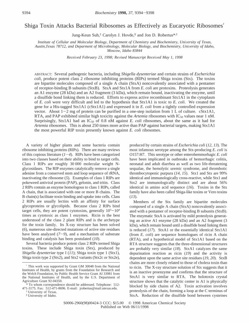
To test this hypothesis, the influence of protein expressionon E. coli growth was observed. The 20-mL cultures wereinitiated from overnight cultures, and the growth wasmeasured (Figure 1). As shown by circles, induction of theparent pAII17 vector system with 1 mM IPTG retarded cellgrowth slightly. Cells with the inducer showed the same 2h lag as the uninduced cells, but the subsequent growth rateand total growth density was about 60% of the control.Presumably, this is due to the added expression load ofnontoxic proteins from the vector, including the T7 poly-merase, GroEL, and GroES. Cells with the vector carrying
the toxin increased the lag time from 2 to about 6 h buteventually reached a normal growth density. The reason forthe increased lag time is not clear. However, analysis ofstationary cells showed that active toxin could be inducedby the addition of IPTG, suggesting that no mutations hadoccurred in the toxin gene or in the induction system duringthe extended lag period. As shown in Figure 1, cells carryingthe gene for cStx1A1 could not grow in the presence ofinducer (1 mM IPTG). Since toxin induction prevented anymeasurable bacterial growth, it seemed reasonable that theenzyme was lethal to the bacterial host cell.
The engineered cStx1A1 has 248 amino acids, includingthree extra histidine residues and a stop codon added afterhistidine 245 of the wild-type Stx1A1 enzyme. The con-struction therefore has six histidine residues at the C-terminus, forming a polyhistidine tag to facilitate purification.
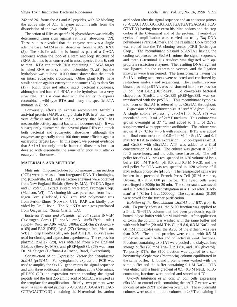
After disrupting the cells, the extract was applied to a Ni-NTA column and eluted with 0-0.5 M gradient of imidazole;the recombinant cStx1A1 was eluted at 0.12 M imidazole.Figure 2 shows the purity of the isolated cStx1A1 with anestimated molecular weight of 27 400, based on polyacry-lamide gels. Applying this one-step purification protocol,1-2 mg of highly purified cStx1A1 was obtained from a1-L culture.
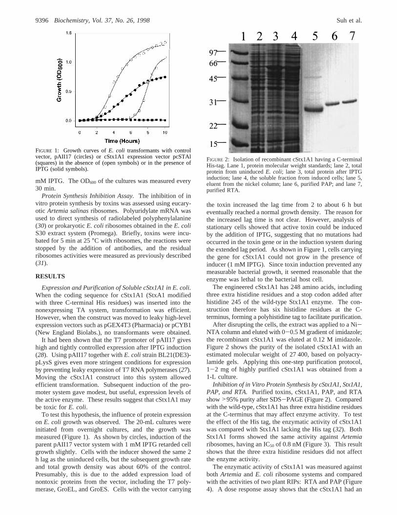
Inhibition of in Vitro Protein Synthesis by cStx1A1, Stx1A1,PAP, and RTA.Purified toxins, cStx1A1, PAP, and RTAshow>95% purity after SDS-PAGE (Figure 2). Comparedwith the wild-type, cStx1A1 has three extra histidine residuesat the C-terminus that may affect enzyme activity. To testthe effect of the His tag, the enzymatic activity of cStx1A1was compared with Stx1A1 lacking the His tag (32). BothStx1A1 forms showed the same activity againstArtemiaribosomes, having an IC50 of 0.8 nM (Figure 3). This resultshows that the three extra histidine residues did not affectthe enzyme activity.
The enzymatic activity of cStx1A1 was measured againstboth Artemia and E. coli ribosome systems and comparedwith the activities of two plant RIPs: RTA and PAP (Figure4). A dose response assay shows that the cStx1A1 had an
FIGURE 1: Growth curves ofE. coli transformants with controlvector, pAII17 (circles) or cStx1A1 expression vector pcSTAl(squares) in the absence of (open symbols) or in the presence ofIPTG (solid symbols).
FIGURE 2: Isolation of recombinant cStx1A1 having a C-terminalHis-tag. Lane 1, protein molecular weight standards; lane 2, totalprotein from uninducedE. coli; lane 3, total protein after IPTGinduction; lane 4, the soluble fraction from induced cells; lane 5,eluent from the nickel column; lane 6, purified PAP; and lane 7,purified RTA.
9396 Biochemistry, Vol. 37, No. 26, 1998 Suh et al.
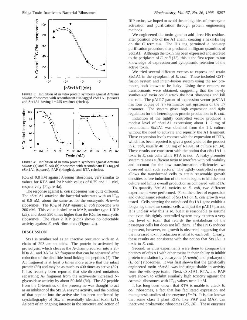
IC50 of 0.8 nM againstArtemia ribosomes, very similar tovalues for RTA and PAP with values of 0.75 and 1.1 nM,respectively (Figure 4a).
The response againstE. coli ribosomes was quite different.The cStx1A1 attacked the bacterial substrates with an IC50
of 0.8 nM, about the same as for the eucaryoticArtemiaribosomes. The IC50 of PAP againstE. coli ribosome was200 nM. This value is similar to MAP, another type 1 RIP(25), and about 250 times higher than the IC50 for eucaryoticribosomes. The class 2 RIP (ricin) shows no detectableactivity againstE. coli ribosomes (Figure 4b).
DISCUSSION
Stx1 is synthesized as an inactive precursor with an Achain of 293 amino acids. The protein is activated byproteolysis, which cleaves the A-chain precursor into a 28-kDa A1 and 3-kDa A2 fragment that can be separated afterreduction of the disulfide bond linking the peptides (1). TheA1 fragment is at least 6 times more active that the intactprotein (33) and may be as much as 400 times as active (32).It has recently been reported that site-directed mutationsseparating A2 fragment from the active-site increased N-glycosidase activity by about 50-fold (34). The A2 peptidefrom the C-terminus of the proenzyme was thought to actas an inhibitor of the Stx1A enzyme activity, and the bindingof that peptide into the active site has been confirmed bycrystallography of Stx, an essentially identical toxin (21).As part of an ongoing interest in the structure and action of
RIP toxins, we hoped to avoid the ambiguities of proenzymeactivation and purification through protein engineeringmethods.
We engineered the toxin gene to add three His residuesafter position 245 of the A1 chain, creating a hexaHis tagon the C terminus. The His tag permitted a one-steppurification procedure that produced milligram quantities ofStx1A1. Although the toxin has been expressed and secretedto the periplasm ofE. coli (32), this is the first report to ourknowledge of expression and cytoplasmic retention of theactive toxin.
We tried several different vectors to express and retainStx1A1 in the cytoplasm ofE. coli. These included GST-fusion system and intein-fusion system using thetac pro-moter, both known to be leaky. Using these vectors, notransformants were obtained, suggesting that the newlysynthesized toxin could attack the host ribosomes and killthe cell. The pAII17 parent of expression vector pcSTA1has four copies ofrrn terminator just upstream of the T7promoter. The system gives high expression and tightregulation for the heterologous protein production inE. coli.
Induction of the tightly controlled vector produced amodest level of cStx1A1 expression; about 1-2 mg ofrecombinant Stx1A1 was obtained from the 1-L culturewithout the need to activate and repurify the A1 fragment.These expression levels contrast with the expression of RTA,which has been reported to give a good yield of the productin E. coli, usually 40-50 mg of RTA/L of culture (8, 34).These results are consistent with the notion that cStx1A1 istoxic to E. coli cells while RTA is not. A leaky promotersystem releases sufficient toxin to interfere with cell viabilityand account for the low transformation efficiencies weobserved with such vectors. The tightly controlled systemallows the transformed cells to attain reasonable growthdensities before induction of the toxin begins to kill the hostculture and limits overall production as compared with RTA.
To quantify Stx1A1 toxicity toE. coli, two differentexperiments were performed. First, the effect of expressionand cytoplasmic retention of Stx1A1 onE. coli growth wastested. Cells carrying the uninduced Stx1A1 gene exhibit alonger lag time than control cells with just the pAII17 parent.It is unclear why this is so, but it is reasonable to assumethat even this tightly controlled system may express a verylow level of toxin that retards the metabolism of thepassenger cells but does not kill them. If the IPTG induceris present, however, no growth is observed, suggesting thatthe increased toxin production is lethal to each cell. Clearly,these results are consistent with the notion that Stx1A1 istoxic to E. coli.
Second, in vitro experiments were done to compare thepotency of cStxA1 with other toxins in their ability to inhibitprotein translation by eucaryotic (Artemia) and prokaryotic(E. coli) ribosomes. It was first shown that the geneticallyengineered toxin cStxA1 was indistinguishable in activityfrom the wild-type toxin. Next, cStx1A1, RTA, and PAPwere shown to exhibit similarly high toxicity against theArtemia ribosomes with IC50 values near 1 nM.
It has long been known that RTA is unable to attackE.coli ribosomes, a fact that has facilitated expression andmutagenesis studies of the enzyme (7-9). It is also knownthat some class 1 plant RIPs, like PAP and MAP, caninactivate prokaryotic ribosomes (25, 26). These enzymes
FIGURE 3: Inhibition of in vitro protein synthesis againstArtemiasalinasribosomes with recombinant His-tagged cStx1A1 (square)and Stx1A1 having 1-255 residues (circles).
FIGURE 4: Inhibition of in vitro protein synthesis againstArtemiasalinas(a) andE. coli (b) ribosomes with recombinant His-taggedcStx1A1 (squares), PAP (triangles), and RTA (circles).
Shiga Toxin Inactivates Bacterial Ribosomes Biochemistry, Vol. 37, No. 26, 19989397
have IC50 values around 200 nM againstE. coli ribosomes,generally around 200 times higher than for their actionagainst eucaryotic ribosomes.
Surprisingly, Stx1A1 had an IC50 of 0.8 nM againstE.coli ribosomes, about the same as it had forArtemiaribosomes. Therefore it was about 250 times more activethan PAP against bacterial targets. This result makes Stx1A1the most powerful RIP toxin presently known againstE. coliribosomes. This rationalizes the observation that the toxinis synthesized as an inactive precursor and is activated onlywhen it is transferred away from the host ribosomes.
Although the key catalytic residues in the active site ofthe three RIPs used in this study are conserved and themechanism of depurination is the same, substrate specificityis clearly different in the sense that the three have verydifferent abilities to attack bacterial ribosomes. It has beenproposed that this difference in specificity may result frommodest structural differences at noncatalytic positions on theRIP protein (35). There is also reason to believe that thedifferences may result from differing interaction between theRIP and the ribosomal proteins. For example, intactE. coliribosomes are insensitive to ricin, but the deproteinizedE.coli 23S rRNA is depurinated by RTA. The rate is at least1000 times slower than for intact ribosomes but is roughlyequivalent to the rates that other toxins attack naked RNA(22). This suggests that, in the absence of ribosomal proteins,prokaryotic rRNA has a structure suitable for depurination(36). Comparison of the crystal structure of RTA and PAPsuggested that several regions show enough difference towarrant investigation as the possible cause for the specificitydifferences (35). Peptide swap experiments between RTAand PAP show that some structural differences in the amino-terminal half of the proteins do affect ribosome specificity,but to date no detailed information about the nature of thisinteraction is available (36).
ACKNOWLEDGMENT
We thank Paula R. Austin for technical expertise.
REFERENCES
1. Olsnes, S., and Pihl, A. (1982) Toxic Lectins and RelatedProteins, inThe Molecular Action of Toxins and Viruses(Cohen, P., and Van Heynigen, S., Eds.) pp 52-105, ElsevierBiomedical Press, New York.
2. Stirpe , F., and Barbieri, L. (1985)FEBS Lett. 195, 1-8.3. Lord, J. M., Roberts, L. M., and Robertus, J. D. (1994)FASEB
J. 8, 201-208.4. Robertus, J. D., and Monzingo, A. F. (1996) inProtein toxin
structure, ed. (Parker, M., Ed.) pp 253-270, R. G. Landes,Austin, TX.
5. Endo, Y., and Tsurugi, K. (1988)J. Biol. Chem. 263, 8735-8739.
6. Rutenber, E., Katzin, B. J., Collins, E. J., Mlsna, D., Ernst, S.E., Ready, M. P., and Robertus, J. D. (1991)Proteins 10, 240-250.
7. Frankel, A., Welsh, P., Richardson, J., and Robertus, J. D.(1990)Mol. Cell. Biol. 10, 6257-6263.
8. Ready, M. P., Kim, Y., and Robertus, J. D. (1991)ProteinsStruct. Funct. Genet. 10, 270-278.
9. Kim, Y. S., and Robertus, J. D. (1992)Protein Eng. 5, 775-779.
10. Monzingo, A. F., and Robertus, J. D. (1992)J. Mol. Biol. 227,1136-1145.
11. Strockbine, N. A., Jackson, M. P., Sung, L. M., Holmes, R.K., and O’Brien, A. D. (1988)J. Bacteriol. 170, 1116-1122.
12. Calderwood, S. B., Auclair, F., Donohue-Rolfe, A., Keusch,G. T., and Mekalanos, J. J. (1987)Proc. Natl. Acad. Sci. U.S.A.84, 4364-4368.
13. Calderwood, S. B., Acheson, D. W. K., Keusch, G. T., Barrett,T. J., Griffin, P. M., Strockbine, N. A., Swaminathan, B.,Kaper, J. B., Levine, M. M., Kaplan, B. S., Karch, H., O’Brien,A. D., Obrig, T. G., Takeda, Y., Tarr, P. L., and Wachsmuth,I. K. (1996).ASM News 62, 118-119.
14. Service, R. F. (1994)Science 265, 475.15. Tarr, P. I., Fouser, L. S., Stapleton, A. E., Wilson, R. A., Kim,
H. H., Vary, J. C., and Clausen, C. R. (1996)N. Engl. J. Med.335, 635-638.
16. O’Brien, A. D., and Holmes, R. K. (1987)Microbiol. ReV.51, 206-220.
17. Olsnes, S., Reisbig, R., and Eiklid, K. (1981)J. Biol. Chem.256, 8732-8738.
18. Deresiewicz, R. L., Calderwood, S. B., Robertus, J. D., andCollier, R. J. (1992)Biochemistry 31, 3272-3280.
19. (a) Endo, Y., Tsurugi, K., Yutsudo, T., Takeda, Y., Ogasawara,T., and Igarashi, K. (1988)Eur. J. Biochem. 171, 45-50. (b)Hovde, C. J., Calderwood, S. B., Mekalanos, J. J., and Collier,R. J. (1988)Proc. Natl. Acad. Sci. U.S.A. 85, 2568-2572.
20. Deresiewicz, R. L., Austin, P. R., and Hovde, C. J. (1993).Mol. Gen. Genet. 241, 467-473.
21. Fraser, M. E., Chernaia, M. M., Kozlov, Y. V., and James,M. N. (1994)Nat. Struct. Biol. 1, 59-64.
22. Endo, Y., and Tsurugi, K. (1987)J. Biol. Chem. 262, 8128-8130.
23. Endo., Y., Gluck, A., and Wool, I. G. (1991)J. Mol. Biol.221, 193-207.
24. Endo, Y., Tsurugi, K., and Lambert, J. M. (1988)Biochem.Biophys. Res. Commun. 150, 1032-1036.
25. Habuka, N., Akiyama, K., Tsuge, H., Miyano, M., Matsumoto,T., and Noma, M. (1990)J. Biol. Chem. 265, 10988-10992.
26. Hartley, M. R., Legname, G., Osborn, R., Chen, Z., and Lord,J. M. (1991)FEBS Lett. 290, 65-68.
27. Studier, F. W., Rosenberg, A. H., Dunn, J. J., and Dubendorff,J. W. (1990)Methods Enzymol. 185, 60-89.
28. Kong, H., Kucera, R. B., and Jack, W. E. (1993)J. Biol. Chem.268, 1965-1975.
29. Dale, G. E., Schonfeld, H. S., Langen, H., and Stieger, M.(1994)Protein Eng. 7, 925-931.
30. Kramer, G. A., Pinphanichararn, P., Konecki, D., and Hardesty,B. A. (1975)Eur. J. Biochem. 53, 471-480.
31. Ready, M. P., Bird, S., Rothe, G., and Robertus, J. D. (1983)Biochim. Biophys. Acta 740, 19-28.
32. Zollman, T. M., Austin, P. P., Jablonski, P. E., and Hovde, C.J. (1994)Protein Expression Purif. 5, 291-295.
33. Reisbig, R., Olsnes, S., and Eiklid, K. (1981)J. Biol. Chem.256, 8739-8744.
34. (a) Day, P. J., Ernst, S. R., Frankel, A. E., Monzingo, A. F.,Pascal, J. M., Molina-Svinth, M. C., and Robertus, J. D. (1996).Biochemistry 35, 11098-11103. (b) Polesskaya, A. N., Garred,O., Olsnes, S., and Kozlov, Y. V. (1997)Mol. Biol. 31, 528-535.
35. Monzingo, A. F., Collins, E. J., Ernst, S., R., Irvin, J. D., andRobertus, J. D. (1993)J. Mol. Biol. 233, 705-715.
36. Chaddock., J. A., Monzingo., A. F., Robertus, J. D., Lord, J.M., and Roberts, L. M. (1996)Eur. J. Biochem. 235, 159-166.
BI980424U
9398 Biochemistry, Vol. 37, No. 26, 1998 Suh et al.