Embed Size (px)
Citation preview

Pyranopterin Coordination Controls Molybdenum Electrochemistry in Escherichia coli Nitrate
Reductase
by
Sheng Yi Wu
A thesis submitted in partial fulfillment of the requirements for the degree of
Master of Science
Department of Biochemistry
University of Alberta
© Sheng Yi Wu, 2015

ii
Abstract
Molybdenum is an essential trace element for most species on the earth. Enzymes that contain it
are called molybdoenzymes. Mononuclear molybdoenzymes play diverse roles in global
geochemical cycles, bacterial metabolism, and human health. The catalytic molybdenum atom is
usually incorporated into a molybdenum cofactor through a complicated biosynthetic pathway
before being inserted in the enzyme. The molybdenum cofactors contain either one or two
heterocyclic moieties known as pyranopterins that coordinate a single molybdenum atom with
dithiolene linkages. The conventional view of the active site is that it is the immediate
coordination environment of the molybdenum atom that defines catalysis. Using the Escherichia
coli respiratory nitrate reductase (NarGHI) as a model system, I tested the hypothesis that protein
coordination of the pyranopterin ring system also plays a role in defining redox chemistry and
reactivity of the active site molybdenum atom. The molybdenum cofactor in the catalytic subunit
NarG contains two pyranopterins coordinated by conserved residues that include a charge
transfer relay (NarG-Ser719, NarG-His1163, NarG-His118, and three H2O molecules) and two
critical histidines defining the piperazine ring of pyranopterins (NarG-His1092 and NarG-
His1098). NarG-Ser719 and NarG-His1163 coordinate O1 of the open pyran ring of the distal
pyranopterin through H-bonds. NarG-His1184 hydrogen bonds with NarG-His1163 and three
conserved H2O molecules. NarG-His1092 links two pyranopterins via hydrogen bonding with
their piperazine N-5 nitrogen atoms. The proximal piperazine N-5 nitrogen is also coordinated by
NarG-His1098. The results from the charge transfer relay suggest the role of the distal
pyranopterin in modulating the Mo potential and reactivity. His1092 and His1098 variants reveal
the critical roles of the bridging His1092 residue and the proximal-pyranopterin-coordinating
His1098 residue in controlling substrate reactivity at the Mo atom. These results support an
emerging paradigm of the importance of pyranopterin coordination in defining molybdoenzyme
catalysis.

iii
Preface
Chapter 2 of the thesis has been submitted with the title Pyranopterin Coordination Controls
Molybdenum Electrochemistry in Escherichia coli Nitrate Reductase. The framework was
mostly brainstormed by Dr. Richard Rothery as a follow-up of his previous work on the
pyranopterin conformational analyses in Rothery et al. (2012). I was responsible for the
experimental planning, data collection and analyses under his direct supervision. Dr. Richard
Rothery contributed significantly to the redox titration experiments and we both collected data
conducting the titration experiments independently. He also provided suggestions on
experimental result interpretation and the visual presentation of data analyses. I wrote the draft of
the manuscript of this work. Dr. Rothery contributed to the extensive edits.

iv
Acknowledgments
It is a great pleasure to honour the ones who have been supportive during my program years. I
owe the biggest thank-you to my family. There are moments of doubts and hesitation in my life
while I was in the program. I might have not been here, if there was an absence of their
reassurance and encouragement.
I thank my supervisor Dr. Joel H. Weiner for taking me in as his student. I started as a summer
student in 2011. I really enjoyed my internship and decided to stay in the lab for my
undergraduate research project. Eventually, I became a graduate student in the lab. Joel is very
open-minded and provides great freedom for me to plan and carry out my experiments
independently. He is also a great role-model scientist who is always eager to learn new things.
Another big thank-you goes to our research associate, Dr. Richard Rothery. He was very
knowledgeable in the field and provided detailed guidance for my thesis project. I improved my
technical writing and figure making by learning from him, though I have to admit that I am still
quite far away from his high standard. He and I also initialized goEdit Alberta to provide
translation and editing service in technical writing. Even though it was small and we are still
trying to increase our customer base, I had the opportunity to get the first-hand experience with
website and business card design, and preliminary editing which is probably a rare opportunity
for people at my age.
I would also like to thank my graduate student colleagues, double-Dr. Yanfei Zhang and Dr.-to-
be Justin Fedor. Both of them are more senior than me and willing to share their stories and
perspectives. They both offered suggestions during discussions of the project. Yanfei, in
particular, shared many helpful tips with his multi-disciplinary academic background. It is also
worth mentioning that both Yanfei and Justin are keeners in culinary art, and they are kind
enough to share their food products. Yanfei’s beef noodle soup is so far the best homemade
Chinese dish I had in my seven-year Edmontonian life and Justin’s liquid nitrogen ice cream is
absolutely a Weiner lab specialty. Their contributions made my gut microbiome happy and
therefore made me happy.

v
There are many more people who have been supportive to me during my program. Those people
include several current and past lab members: Shannon Murphy, Michelina Kierzek, Francesca
Sabastian, Glen Zhang, and Victor Cheng. I would also like to highlight some members of
Department of Biochemistry who made my time in the program better: Dr. Bernard Lemire, Dr.
Howard Young, and Dr. Richard Falhman who have served on my 670/671 evaluation
committee and/or supervisory committee; Lucy Sun who invited to the Let’s Talk Science
workshops and volunteered with me for almost three years; Dr. Adrienne Wright, Dr. Jonathan
Parrish, and Dr. Rachel Milner, who have been great mentors during TA sessions; and many
other good friends for the company during the program. In addition, I would like to thank Dr.
Xing-Zhen Chen from Department of Physiology for taking time to be my internal external
committee member.
I would also like to acknowledge the International Research Training Group (IRTG) for
generously providing the funding during my program and the opportunity of German exchange. I
thank the Keller lab, especially Dr. Sandro Keller and PhD student Johannes Klingler, for
making my stay comfortable and educational.
With all that, I would like to conclude my acknowledge section. The three year MSc program in
the Department of Biochemistry at University of Alberta is an experience that I will never regret.
While I will move onto the next stage of my life, I wish all the best to all my colleagues and
peers.

vi
Table of Contents
CHAPTER 1. Introduction.............................................................................................................. 1
1.1 Introduction ............................................................................................................................... 2
1.2 Bioenergetics and respiration .................................................................................................... 2
1.3 E. coli respiratory regulation..................................................................................................... 5
1.4 Molybdoenzymes and tungstoenzymes .................................................................................... 7
1.4.1 Molybdoenzyme classification ........................................................................................... 8
1.4.2 Molybdoenzymes in human health .................................................................................... 8
1.4.3 Molybdoenzymes in geochemical cycles ........................................................................... 9
1.4.3.1 Nitrogen fixation by nitrogenase ............................................................................... 10
1.4.3.2 Nitrogen assimilation by plant nitrate reductase ....................................................... 10
1.4.3.3 Denitrification by bacterial nitrate reductase ............................................................. 11
1.5 Molybdenum cofactors and tungsten cofactor ........................................................................ 12
1.5.1 Categories of Mononuclear Mo-/W- cofactors ................................................................ 12
1.5.1.1 Simplest molybdo-pyranopterin cofactor (Mo-PPT) ................................................. 13
1.5.1.2 Molybdo-pyranopterin cytosine dinucleotide (Mo-PCD) ......................................... 14
1.5.1.3 Tungsto-bispyranopterin (W-bisPPT) ....................................................................... 14
1.5.1.4 Molybdo-bis (pyranopterin guanine dinucleotide) (Mo-bisPGD) ............................. 15
1.5.2 Pyranopterin (PPT) ........................................................................................................... 15
1.5.2.1 Pyranopterin conformational analysis ....................................................................... 16
1.5.3 Biosynthesis of Mo-/W- cofactors ................................................................................... 17

vii
1.5.3.1 Circularization of GTP to form cPMP ....................................................................... 17
1.5.3.2 Formation of dithiolene group to form MPT ............................................................. 18
1.5.3.3 Formation of MPT-AMP ........................................................................................... 20
1.5.3.4 Mo insertion ............................................................................................................... 20
1.6 NarGHI ................................................................................................................................... 21
1.6.1 Overview .......................................................................................................................... 21
1.6.2 The nar operon ................................................................................................................. 22
1.6.3 NarG ................................................................................................................................. 22
1.6.4 NarH ................................................................................................................................. 23
1.6.5 NarI................................................................................................................................... 25
1.6.6 NarGHI Molybdo-bis (pyranopterin guanine dinucleotide) (Mo-bisPGD) ..................... 26
1.7 Thesis objectives ..................................................................................................................... 27
1.8 Figures..................................................................................................................................... 30
CHAPTER 2. Pyranopterin Coordination Controls Molybdenum Electrochemistry in Escherichia
coli Nitrate Reductase ................................................................................................................... 44
2.1 Introduction ............................................................................................................................. 45
2.2 Experimental procedures ........................................................................................................ 48
2.2.1 Bacterial strains and plasmids .......................................................................................... 48
2.2.2 Site-directed mutagenesis ................................................................................................. 49
2.2.2.1 Generation of a NarG-Ser719Ala variant .................................................................. 49
2.2.2.2 Generation of NarG-His1163Ala and NarG His1184Ala variants ............................ 49

viii
2.2.2.3 Generation of NarG-His1092Ala, NarG-His1092Arg, and NarG-His1098Ala variants
............................................................................................................................................... 49
2.2.3 Growth of cells ................................................................................................................. 50
2.2.4 Bacterial growth on glycerol-nitrate minimal medium .................................................... 50
2.2.5 Redox potentiometry and EPR spectroscopy ................................................................... 51
2.2.6 Protein assays ................................................................................................................... 52
2.2.7 Enzyme Assays ................................................................................................................ 52
2.3 Results and discussion ............................................................................................................ 52
2.3.1 Residues targeted for site-directed mutagenesis .............................................................. 52
2.3.2 Impact of the variants on the NarG Mo(V) EPR spectrum .............................................. 53
2.3.3 Influence of the NarGHI variants on Mo electrochemistry.............................................. 54
2.3.4 Correlation between enzyme activity, cell growth and Mo electrochemistry .................. 56
2.3.5 Role of the His1163/His1184 charge-transfer relay in NarGHI maturation and in
modulation of Mo electrochemistry .......................................................................................... 58
2.4 Conclusion .............................................................................................................................. 59
2.5. Table ...................................................................................................................................... 60
2.6. Figures.................................................................................................................................... 61
CHAPTER 3. Conclusions............................................................................................................ 68
References ..................................................................................................................................... 70

ix
Table of Figures
Figure 1.1. Dehydrogenases and reductases ................................................................................. 30
Figure 1.2. Global nitrate homeostasis is maintained by the nitrogen cycle. ............................... 31
Figure 1.3. Molybdenum cofactor classification .......................................................................... 32
Figure 1.4. SUOX family functional distribution ......................................................................... 33
Figure 1.5. XDH family functional distribution ........................................................................... 34
Figure 1.6. AOR family functional distribution............................................................................ 35
Figure 1.7. DMSOR family functional distribution ...................................................................... 36
Figure 1.8. Molybdenum cofactor conformational alignment ...................................................... 37
Figure 1.9. Molybdenum cofactor biosynthetic pathway ............................................................. 38
Figure 1.10. Redox loops formed by nitrate reductase and formate dehydrogenase .................... 39
Figure 1.11. Crystal structure of NarGHI (1Q16) ......................................................................... 40
Figure 1.12. NarG structural information ..................................................................................... 41
Figure 1.13. NarI structural information ....................................................................................... 42
Figure 1.14. The catalytic mechanism of nitrate reduction .......................................................... 43
Figure 2.1. Charge transfer relay .................................................................................................. 61
Figure 2.2. Residues defining pyranopterin piperazine ring coordination.................................... 62
Figure 2.3. Mo(V) EPR spectra of redox-poised NarGHI variants of residues involved in
pyranopterin coordination. ............................................................................................................ 63
Figure 2.4. Potentiometric titrations of membranes containing variants of residues coordinating
the distal pyranopterin of NarGHI ................................................................................................ 64
Figure 2.5. Potentiometric titrations of membranes containing variants of His1092 and His1098
....................................................................................................................................................... 65
Figure 2.6. Correlation between enzyme activity and overall Mo reduction potential. ................ 66
Figure 2.7. Proposed mechanism of pyran ring opening of the distal pyranopterin of NarGHI. . 67

x
List of the table
Table 2.1. Effects of variants of pyranopterin-coordinating residues on Mo reduction potentials
and enzyme activity ...................................................................................................................... 60

xi
List of Abbreviations
AOR aldehyde oxidoreductase
ATP adenosine triphosphate
BV/BVH• benzyl viologen/ reduced benzyl viologen
Cnx cofactors for nitrate reductase and xanthine dehydrogenase
cPMP cyclic pyranopterin monophosphate
DH dehydrogenase
DmsABC E. coli dimethyl sulfoxide reductase
DMSO dimethyl sulfoxide (Me2SO)
DMSOR dimethyl sulfoxide reductase
EbdABC Aromatoleum aromaticum ethylbenzene dehydrogenase
E. coli Escherichia coli
Em midpoint potential
EPR electron paramagnetic resonance
ETC electron transport chain
FdnGHI E. coli formate dehydrogenase-N
FeS iron sulfur cluster

xii
GTP guanosine triphosphate
IPTG isopropyl-1-thio--D-galactopyranoside
MCOS molybdenum cofactor synthesis
MPT molybdopterin
Mo-bisPGD molybdo-bis(pyranopterin guanine dinucleotide)
Moco molybdenum cofactor
Mo-enzymes molybdoenzymes
Mo-PCD molybdo-pyranopterin cytosine dinucleotide
Mo-PPT molybdo-pyranopterin
MQ/MQH2 menaquinone/menaquinol
NapA periplasmic nitrate reductase
NarGHI respiratory nitrate reductase
PGD pyranopterin guanine dinucleotide
pmf proton motive force
PMSF phenylmethylsulfonyl fluoride
PPT pyranopterin
Q-site quinone binding site
xiii
SDS-PAGE sodium docdecyl sulfate polyacrylamide gel electrophoresis
SUOX sulfite oxidoreductase
W-enzymes tungstoenzymes
W-bisPPT tungsto- bispyranopterin
WT wild-type
XDH xanthine dehydrogenase

1
CHAPTER 1. Introduction

2
1.1 Introduction
Energy is essential for the survival of species. Many amazing adaptations in nature were solely
developed to increase efficiency in obtaining food and absorbing nutrients from food. Some
examples are sharp claws and teeth of carnivores for capturing and tearing preys; complex and
strong digestive system of herbivores for heavy-duty digestion; and various shapes of bird beaks
for catching insects and worms at different locations (1–3). This ongoing conquest of energy
acquirement began at the very beginning of evolution billions of years ago at microscopic scales.
As one group of the earliest biological organisms, bacteria are an extremely diverse group of
organisms living under many different conditions, some of which are considered uninhabitable to
any other living thing (4, 5). Their brilliant adaptations are largely dependent on their ability to
metabolize the finite amount and limited selection of substrates present in the environment. This
provides an opportunity to explore bacterial bioenergetics and its role in metabolic diversity.
1.2 Bioenergetics and respiration
Bioenergetics is a biochemical field focusing on energy transformation to the cellular ―currency‖,
adenosine triphosphate (ATP), in living systems. The source of the energy can be chemical
compounds, photons from the sunlight, or sometimes thermal energy. There are two common
methods for cells of prokaryotes and eukaryotes to generate ATP: fermentation and respiration
(6). Fermentation requires the presence of neither external electron acceptors nor the electron
transport chain (ETC). It generates energy through glycolysis, in which one glucose molecule is
broken down into two pyruvates with net yield of two ATP molecules and two NADH molecules.
To ensure continuing glycolysis, NAD+ is regenerated from NADH. Under anaerobic condition,

3
the regeneration is typically carried out by enzymes such as lactate dehydrogenase and alcohol
dehydrogenase, and no ATP is produced in the process. Respiration on the other hand, requires
the presence of both external electron acceptors and the ETC. The process of respiration also
utilizes the glycolysis pathway; however, there are many more ATPs produced in regeneration of
the electron carriers NAD+ and FAD. Reduced NADH and FADH2 are generated in process of
glycolysis, Acetyl-CoA synthesis, and the Kreb cycles under aerobic conditions. In
mitochondrial respiration, up to 32 ATPs are generated with initial input of one glucose through
the ETC composed of Complex I (NADH:ubiquinone oxidoreductase), II (succinate:ubiquinone
oxidoreductase), III (cytochrome bc1 complex), IV (cytochome c oxidase), and V (ATP synthase)
(7, 8). Complex I regenerates NAD+ while reduces ubiquinone in the membrane to ubiquinol.
Complex II oxidazes succinate (an intermediate in the Kreb cycles) to fumarate. The electrons
from succinate are used to reduce FAD to FADH2, from which the electrons are used to reduce
ubiquinone to ubiquinol. The ubiquinol generated by Complex I and II is used to reduce
cytochrome c (Fe3+
) to cytochrome c (Fe2+
) by Complex III. Complex IV reduces O2 to H2O with
electrons from cytochrome c (Fe2+
). Complex I, III, and IV are capable of proton translocation by
which a proton gradient is generated for ATP synthesis. The ETC allows mitochondrial
respiration to be a more effective in energy production. The most common electron acceptor in
biology is oxygen as in mitochondrial respiration, and the respiration is referred to as aerobic
respiration. The less common electron acceptors include nitrogen, carbon, and sulfur compounds.
They are more likely found in prokaryotes and allow for anaerobic respiration. Anaerobic
respiration usually generates less energy than aerobic respiration, but is more efficient than
fermentation.

4
In respiration, the pathways connecting the substrates and final electron acceptors typically
involve many intermediate compounds generated in a series of chemical reactions catalyzed by
multiple enzymes. The production of ATP during those reactions was explained by the
chemiosmotic model proposed by Peter Mitchell in 1961(9). In his model, the electrochemical
gradient of electrolytes generated by the series of chemical reactions is key to ATP synthesis.
The most common electrolytes are protons and its gradient is also called the proton motive force
(pmf). Proton translocation against the concentration gradient is usually coupled with the
production of high-energy electrons in oxidoreduction reactions. The process requires a
membrane that is impermeable to those protons to maintain the proton gradient which has a
higher concentration of protons on one side and lower concentration on the other side. This
accumulated potential energy from the proton gradient is the driving force for the endothermic
ATP synthesis. The enzyme responsible for this process is called ATP synthase. It is a
transmembrane protein that allows protons to pass through along the concentration gradient,
converting the potential energy from the gradient to chemical energy of phosphodiester bonds in
ATP molecules. A redox loop, also proposed by Peter Mitchell, is an example of how the proton
gradient is used to create ATPs. Two coupled components, usually a dehydrogenase and a
reductase, and an electron carrier are required in a loop. A dehydrogenase oxidizes its substrate
and provides electron(s) for the electron carrier; a reductase uses electron(s) from the electron
carrier to reduce its substrate. Protons are translocated, consumed, or generated by the
dehydrogenase and the reductase during the catalysis. This normally creates a net proton
difference across the membrane. A common example of redox loops is a Q cycle, which uses
quinone molecules as electron carriers (10). Q stands for quinone/quinol (Q/QH2) species,
including menaquinone/menaquinol (MQ/MQH2) and ubiquinone/ubiquinol (UQ/UQH2). They

5
are able to support two-electron redox reactions with various combinations of dehydrogenases
and reductases, including 15 primary dehydrogenases and 10 terminal reductases identified so far
(Figure 1.1). Not all of listed proteins above are present in a single cell. A detailed example of
Escherichia coli (E. coli) nitrate reductase A and formate dehydrogenase is illustrated in the
section 1.6.1.
1.3 E. coli respiratory regulation
E. coli is an excellent model for studying the bacterial respiratory system and its regulation. It is
a facultative anaerobic diderm bacterium that can switch its dominant metabolic pathway to
adjust to the environmental oxygen level and substrate availability (10, 11). The regulation in
respiration is under a strict hierarchy (12). Under aerobic condition with a sufficient amount of
carbon source, the aerobic pathways are induced to use oxygen as the terminal electron acceptor
for ATP synthesis, while the anaerobic pathways are repressed. When there is a lack of oxygen,
the anaerobic pathways are induced. Nitrate is the preferential electron acceptor under such
condition. When nitrate is not available, fumarate or dimethyl sulfoxide (DMSO) will be
accepted as the next best substrate.
Such strict electron acceptor hierarchy is achieved through substantial regulation of E. coli gene
expression. Some key regulators are the ArcB/A (aerobic respiratory control) system, FNR
(fumarate nitrate reduction) protein, and Nar system. All three regulators are capable of
switching the metabolism towards the anaerobic pathways under anaerobic conditions. ArcB/A
and FNR are also responsible for favouring aerobic pathways over anaerobic ones through global
regulations when oxygen is available (13).

6
ArcB/A is a two-component system with ArcB being the membrane oxygen sensor
kinase/phosphatase and ArcA being the associated transcription factor in the cytosol (14–19).
ArcB indirectly senses the oxygen level through the oxidation state of the quinone pool in the
membrane (20, 21). ArcB is structurally composed of a 16-amino-acid transmembrane domain, a
Per-Arnt-Sim (PAS) domain, and a catalytic domain. The PAS domain is a conserved feature in
signal sensors that can detect changes such as light, redox states, and energy level in the cell. The
PAS domain of ArcB contains key cysteine residues, Cys180 and Cys241, which can alter the
function of the catalytic domain according to the quinone oxidation state. Under aerobic
condition, an intermolecular disulfide bond is formed between two ArcB monomers to inhibit
kinase activity, and the catalytic domain functions as a phosphatase specifically targeting
phosphorylated ArcA. ArcA is not capable of interacting with DNA when dephosphorylated.
Under anaerobic conditions, the two cysteines are reduced, and the catalytic domain functions as
a kinase that auto-phosphorylates ArcB at conserved His292 and this phosphate group is
sequentially transferred onto conserved Asp576 and His717 of ArcB (17, 18). The phosphate
group on His717 is eventually transferred onto Asp54 of ArcA, and phosphorylated ArcA is
capable of interacting with many E. coli operons globally with its helix-turn-helix motif (13, 22,
23).
Like the ArcB/A system, FNR protein is also capable of switching metabolic pathways between
anaerobic and aerobic respirations (24). It is a transcription factor with an N terminus as a sensor
domain and a C terminus capable of binding DNA (25). The protein binds an oxygen labile [4Fe-
4S] cluster in the sensor domain under anaerobic conditions, which results in dimerization of
FNR though a disulfide bond (26). The FNR dimer has enhanced ability to bind to DNA at the
promoters for genes associated with anaerobic respiration (27–29). When oxygen is available,

7
the [4Fe-4S] cluster is converted to a [2Fe-2S] cluster through a [3Fe-4S] intermediate (26, 30–
33). The [2Fe-2S]-bound FNR can no longer maintain its dimerization. As a result, the specific
DNA binding is inhibited.
Unlike the previous two regulating systems, the Nar system is more specific for enhancing
anaerobic respiration with the substrate nitrate. There are two homologues of the two-component
Nar systems: NarX/L and NarQ/P, both of which are responsible for anaerobic respiration
regulation (34). Similar to the ArcB/A system, NarX and NarQ are sensor kinases/phosphatases,
and NarL and NarP are response transcription factors. NarX or NarQ senses the concentration of
nitrate and nitrite (35). NarQ can bind to both substrates equally well, while NarX has a
preference for nitrate. When the kinase activity of NarX is turned on by available nitrate (36),
NarX autophosphorylates and transphosphorylates NarL. Phospho-NarL is capable of enhancing
or repressing transcription of genes by binding to DNA. The expression of nitrate reductase is
enhanced and that of fumarate reductase and DMSO reductase are repressed to ensure the nitrate
metabolism is prioritized (37, 38). When there is an absence of nitrate, NarX becomes a
phosphatase to dephosphorylate NarL, and the dephosphorylated NarL is no longer capable of
binding to DNA. The NarQ/P system works in a similar fashion. The two parallel
two-component systems are alternative to each other and ensure the nitrate/nitrite metabolism
has the priority under anaerobic condition.
1.4 Molybdoenzymes and tungstoenzymes
Molybdoenzymes are an important group of enzymes catalyzing oxidoreductive reactions in
respiration (39, 40). They are metalloenzymes characterized by incorporation of molybdenum,
which is a rare transition metal present in the environment. They play roles in bacterial metabolic

8
diversity, human health, and global geochemical cycles, such as carbon, sulfur, and nitrogen
cycles (41). Their presence is found in all animals and plants, and most prokaryotes.
Tungstoenzymes are analogous to molybdoenzymes (42). Current knowledge shows their
presence in archaea and bacteria. Like molybdoenzymes, tungstoenzymes are also important for
the metabolic diversity and global geochemical cycles (43).
1.4.1 Molybdoenzyme classification
Molybdoenzymes are classified into four families based on their cofactor coordination protein
fold structure (44). They are the sulfite oxidoreductase (SUOX-fold) family, the xanthine
dehydrogenase (XDH-fold) family, the aldehyde oxidoreductase (AOR-fold) family, and the
dimethylsulfoxide reductase (DMSOR-fold) family. Their molybdenum cofactors and their
associated functions are discussed in detail in Section 1.5.1. Most of molybdoenzymes are found
in prokaryotes. The DMSOR family is the biggest family of molybdoenzymes and the most
functionally diverse one. Most AOR family members are found in archaea and none from
eukaryotes.
1.4.2 Molybdoenzymes in human health
Some known molybdoenzymes have a particularly important role in human health, which make
the group of enzymes a research target to understand the cause of diseases and to facilitate drug
discoveries (45).
Human xanthine dehydrogenase participates in purine metabolism. Deficiency or mutation can
cause serious health consequences. Xanthinuria, for example, is a medical condition when purine
cannot be metabolized to uric acid either due to lack of xanthine dehydrogenase (type I) or lack

9
of molybdenum cofactor (type II) (46). It is a disease occurring at an approximate rate of
1/69000 (47). The consequences include urinary tract calculi, acute renal failure and myositis
(tissue deposition of xanthine) (48).
Human aldehyde oxidase belongs to the AOR family. The enzyme can be found in metabolic
pathways of heterocycles, aldehyde, purine, and pteridine (49). It was suggested that aldehyde
oxidase plays an endogenous role in the synthesis and deposition of retinoids in Hardarian glands
and skin, and a role on metabolism of neurotransmitters, certain amino acid (valine, isoleucine,
and leucine), and vitamins (50). It is a research target for metabolism of drugs and xenobiotics,
as it is found at a high concentration in liver (51). The drugs that have been tested against
aldehyde oxidase include antitumor agents (methotrexate and 6-mercaptopurine) and
antidepressant (citalopram) (51–54).
Sulfite oxidase is a critical enzyme in cysteine catabolism. It converts sulfite into sulfate in the
catabolic pathway. The deficiency in or mutation of sulfite oxidase can cause sulfite
accumulation and toxicity (55). Molybdenum cofactor deficiency can also result in dysfunctional
sulfite oxidase and the same clinical outcome. The result is severe neurodegeneration: intractable
seizures, hyper- and hypotonus, mental retardation, developmental delay, and even lethality in
infancy (56).
1.4.3 Molybdoenzymes in geochemical cycles
Molybdoenzymes are critical for global carbon, nitrogen, and sulfur cycles. In the nitrogen cycle,
for example, molybdoenzymes participate in reactions producing different nitrogen compounds
that allow nitrogen absorption and utilization by different organisms and maintain global
nitrogen homeostasis between four spheres: biosphere, atmosphere, lithosphere, and hydrosphere.

10
Some processes involving molybdoenzymes, for instance, are nitrogen fixation, nitrogen
assimilation, and nitrogen denitrification (Figure 1.2).
1.4.3.1 Nitrogen fixation by nitrogenase
Nitrogen is an essential element for all life found on our planet. It is an abundant element in the
environment: 79% of the atmosphere volume is nitrogen. However, most organisms cannot
utilize atmospheric nitrogen directly. To fulfill the demand of absorbable nitrogen, the
atmospheric nitrogen is converted to nitrogen compounds and this chemical conversion is called
nitrogen fixation (Figure 1.2). The organisms who can fix atmospheric nitrogen are called
diazotrophs, and they do so through a molybdoenzyme called nitrogenase (57). It converts
atmospheric nitrogen into ammonium, which is the one of nitrogen compounds absorbed by
plants. Some diazotrophic bacteria and their host plants build symbiotic relationships in which
the diazotrophs provide ammonium to the plants and the plants provide other nutrients to the
bacteria in return. As nitrogen is typically 2% of plant weight, studies on nitrogenase became a
particular interest in the field of agriculture (57). Other diazotrophic organisms are non-
symbiotic and directly release ammonium into their environments and maintain the lithospheric
and hydrospheric ammonium concentration for non-diazotrophic organisms.
1.4.3.2 Nitrogen assimilation by plant nitrate reductase
After inorganic nitrogen compounds are absorbed by plants, they need to be further modified for
biosynthesis of biological nitrogen compounds such as amino acids and nucleotides. This is
fundamental to not only plants, but also to other higher eukaryotes such as animals, as they
obtain some of these nutrients directly or indirectly from plants through the food chain. This

11
process of converting inorganic nitrogen compounds to organic nitrogen compounds is called
nitrogen assimilation.
Nitrate reduction is the first reaction in nitrogen assimilation by converting nitrate to ammonium
in two steps (Figure 1.2). Even though both ammonium and nitrate can be absorbed by plants,
ammonium can be directly used for amino acid synthesis while nitrate needs to be converted to
ammonium first. The two steps are nitrate-to-nitrite conversion by nitrate reductase and nitrite-
to-ammonium by nitrite reductase. Nitrate reductase is a molybdoenzyme, and it has been
reported that the deficiency in molybdenum can cause deficiency in several amino acids in plants
(58).
1.4.3.3 Denitrification by bacterial nitrate reductase
Similar to plant nitrate reductase, bacterial nitrate reductase also catalyzes the reaction of nitrate
to nitrite. Unlike in plants, the product nitrite from bacterial nitrate reductase is further modified
by a series of enzymes to molecular nitrogen (N2), which is released to the atmosphere. The
process of producing atmospheric nitrogen is called denitrification and organisms containing
enzymes in the process are called denitrifiers.
Denitrifiers and diazotrophs work in the opposite direction to maintain the homeostasis between
atmospheric nitrogen and nitrogen in other spheres (59). This homeostasis is interrupted in the
modern history by human pollutions such as urban and agricultural runoff containing nitrate
fertilizers. The excess nitrate in the water system triggers a series of natural responses called
eutrophication which includes oxygen-depleted water environment caused by overgrowing algae
supported by the excess nutrients, and decreased biodiversity as many aerobic species fail to
compete with algae for oxygen. Because of this current crisis, denitrification became important

12
for wastewater treatment. Bacteria containing nitrate reductase, as denitrifiers, have been widely
used in many water treatment facilities (60).
1.5 Molybdenum cofactors and tungsten cofactor
The molybdenum (tungsten) cofactor is considered one of the most important functional groups
in molybdoenzymes (tungstoenzymes), if not the most important one. Studies of model
molybdenum cofactors have been and still are providing information for understanding of
molybdenum/tungsten cofactors (Mo-/W- cofactor). These cofactors are the signature feature of
molybdo-/tungsto- enzymes (Mo-/W- enzymes) and are required for Mo/W insertion (39, 40, 61,
62). They are where redox reactions occur. The catalytic metals, molybdenum (42Mo, period 5)
and tungsten (74W, period 6), are both d block transition metals with similar physical and
chemical properties. They both have oxidation states of +6, +5, +4, +3, +2, +1, -1, and -2 and
atomic radii of 39 pm. It is found that they are interchangable in some enzymes for structural
assembly. Mo-/W- cofactors are energetically expensive structures, as a single molybdenum
cofactor in E. coli requires at least five operons in its biosynthesis (63, 64). The core structure of
a Mo-/W- cofactor involves a Mo or W atom coordinated by one or two pyranopterin structures.
More details of their categories, structures, and functions are discussed below.
1.5.1 Categories of Mononuclear Mo-/W- cofactors
The structures of Mo-/W- cofactors vary from one Mo-/W- enzyme to another. The variation
allows the cofactor to catalyze different reactions. Most Mo-/W- cofactors are mononuclear with
only one metal center. A few exceptions are multinuclear cofactors, such as the iron-
molybdenum cofactor ([MoFe7S9] cluster) from nitrogenase (65, 66). Only mononuclear

13
Mo-/W- cofactors will be discussed herein. A Mo-/W- cofactor is so important for the enzyme
that the classification of Mo-/W- enzymes is based on the type of protein fold coordinating the
Mo-/W- cofactors. The complexity of the cofactor is positively correlated to the level of
substrate variation (44, 63, 67). The four categories of Mo-/W- enzymes in the order of
increasing cofactor complexity are sulfite oxidoreductase (SUOX-fold) family, xanthine
dehydrogenase (XDH-fold) family, aldehyde oxidoreductase (AOR-fold) family, and
dimethylsulfoxide reductase (DMSOR-fold) family. The four general types of Mo-/W- cofactors
and their corresponding Mo-/W-enzymes are illustrated in Figure 1.3 and discussed in details
below.
1.5.1.1 Simplest molybdo-pyranopterin cofactor (Mo-PPT)
The simplest form of a Mo-/W- cofactor is the molybdo-pyranopterin structure (Mo-PPT) with
mononuclear molybdenum coordinated by a single pyranopterin. With current structure
information, this type of cofactor is present in all eukaryotic molybdoenzymes, all SUOX family
members, and some bacterial XDH family members. SUOX family enzymes are present in
bacteria, plants, and animals. Many of them are important for sulfur metabolism in living
systems and deficiency or mutations can sometimes lead to severe consequences including
lethality. Examples are sulfite oxidase from Gallus gallus and Arabidopsis thaliana and sulfite
dehydrogenase from Starkeya novella. Assimilatory nitrate reductase also belongs to this family.
In addition, there are another two enzymes in the SUOX family with unknown functions: YedY
and YuiH (68). They are under current investigation to determine their roles. The SUOX family
functional distribution is concluded in Figure 1.4. The simple molybdo-pyranopterin cofactors
are also found in some bacterial XDH proteins such as Rhodobacter capsulatus xanthine
dehydrogenase.

14
1.5.1.2 Molybdo-pyranopterin cytosine dinucleotide (Mo-PCD)
The molybdo-pyranopterin cytosine dinucleotide has a structural addition to the simplest
molybdo-pyranopterin cofactors — cytosine dinucleotide. The cytosine dinucleotide is
covalently linked to the pyranopterin through a phosphoester bond. This type of cofactor is
incorporated in most bacterial members of the XDH family. Some examples are Desulfovibrio
gigas aldehyde dehydrogenase, Pseudomonas putida isoquinoline oxidoreductase, and Thauera
aromatica 4-hydroxybenzoyl-CoA reductase (69). About half of the XDH family catalyzes
purine metabolism, and the rest catalyzes reactions on isoquinoline, niacin (B3γ 4-
hydroxybenzoyl-CoA), aldehyde, carbon monoxide, and other substrates (Figure 1.5). A
modified Mo-PCD has been found in carbon monoxide oxidoreductase from Oligotropha
carboxidovorans (70). This unique dinuclear heterometal cofactor contains a Cu atom in addition
to the Mo atom and is denoted as (CuSMo(O=)OH) (71).
1.5.1.3 Tungsto-bispyranopterin (W-bisPPT)
The third complex level of Mo-/W- cofactor is featured by two pyranopterins forming two
dithiolene linkages instead of one pyranopterin in the previous two levels. It is found in AOR
family members, many of whom are in archaeal proteins. Examples are aldehyde:ferrodoxin
oxidoreductase and formaldehyde oxidoreductase in thermophilic pyrococcus furiosus (72–74).
The cofactor is quite weak on functional diversity despite its complexity: the functions of the
AOR family members are limited to aldehyde and formaldehyde catalysis as shown in Figure
1.6.

15
1.5.1.4 Molybdo-bis (pyranopterin guanine dinucleotide) (Mo-bisPGD)
This is the most structurally complex and functionally diverse category of Mo-/W- cofactors. In
addition to the structure of the central metal coordinated by two pyranopterins as in W-bisPPT,
two guanine nucleotides are covalently linked to the pyranopterins. This type of cofactor is found
in the DMSOR family of Mo-/W- enzymes and utilizes diverse substrates as shown in Figure 1.7.
The cofactor is present in the following enzymes: formate dehydrogenases, S- and O-
oxidoreductase, nitrate/selenate/percholorate reductases, sulfur anion reductases,
formylmethanofuran dehydrogenases, arsenite oxidases, and many other enzymes with known or
unknown functions (44). The metabolic diversity of DMSOR family is suggested as a result of
Mo electron potential variation, such as Em,pH8 = -420 mV for E. coli formate dehydrogenase, and
Em,pH8 = +420 mV for E. coli nitrate reductase. The Mo electron potential is determined by its
coordinating environment. The impact of its surrounding dithiolene linkage, protein ligand(s),
and general protein environment has been investigated for decades.
1.5.2 Pyranopterin (PPT)
A pyranopterin refers to a cofactor component coordinating a metal ion through sulfur atoms. It
is a conserved feature of molybdenum or tungsten cofactor. Most pyranopterins are in tricyclic
form consisted of a pyran, a pyrimidine, and a piperazine ring. In bis-pyranopterin cofactors, the
two pyranopterins are named according to their distance to the juxtapositional iron sulfur cluster
present in almost all DMSOR family enzymes: the pyranopterin positioned closer to the iron
sulfur cluster is named the proximal pyranopterin, and the farther one is named the distal
pyranopterin (75, 76). A bicyclic pyranopterin with open pyran ring has been observed in E. coli
NarGHI and ethylbenzene dehydrogenase (77, 78). The number of pyranopterins in a cofactor is

16
important for cofactor classification: the simplest Mo-PPT and Mo-PCT contain one
pyranopterin, and more complex W-bisPPT and Mo-bisPGD contain two pyranopterins.
1.5.2.1 Pyranopterin conformational analysis
The three dimensional conformation of a pyranopterin is influenced by redox states of the ring
structure and the interaction of the pyranopterin with the surrounding protein environment.
Pyranopterins are highly enriched with double bonds, which is common in electron-carrying
prosthetic groups. The carbon atoms in a double bond can adopt sp2 hybridization which allows
electron delocalisation and a more planar pyranopterin conformation. A change in the
pyranopterin redox states alters the electron distribution in the pyranopterin ring structures, and
therefore, the conformation of the pyranopterin. Interactions such as hydrogen bonding,
electrostatic interaction, and hydrophobic interaction also influence the conformation of a
pyranopterin.
In Rothery et al. 2012, it was determined that the conformations of distal and proximal
pyranopterins are different: the proximal pyranopterin adopts a more distorted structure in
comparison to the distal pyranopterin (76). The distortion is most significant at pyrazine and
pyran ring positions. The analyses indicated the oxidation state varies between the two
pyranopterins. The more distorted proximal pyranopterin is in the more reduced tetrahydro form
and the distal pyranopterin is in the more oxidized 10-10a dihydro form. The structural
assignment suggests this pattern is systemic in all bis-pyranopterin cofactors of DMSOR family
members. The pyranopterin of XDH family members adopts the conformation of DMSOR
proximal pyranopterin, and the pyranopterin of SUOX family members adopts the conformation
of DMSOR distal pyranopterin as shown in Figure 1.8. Both XDH pyranopterins and proximal
DMSO pyranopterins are located between the Mo and the nearest FeS cluster; both SUOX

17
pyranopterins and distal DMSO pyranopterins are located on the other side of the Mo, away from
the closest FeS cluster in the electron transfer system (76). This suggests the tetrahydro form of
the pyranopterin may serve a role in electron flow between FeS cluster and Mo and the dihydro
form may serve a different role. This is the first study that suggests a functional division between
the two pyranopterin in bis-pyranopterin cofactors.
1.5.3 Biosynthesis of Mo-/W- cofactors
Mo-/W- cofactors in general are complex structures requiring many proteins to be properly
synthesized and incorporated into an apoenzyme. The biosynthesis of a molybdenum cofactors is
divided into four steps based on intermediates precursor Z, molybdo-pterin (MPT), and
adenylated MPT; and the product Moco (62, 63, 67). The synthetic pathways are well conserved
for molybdenum cofactors. There are six proteins identified for such purpose in fungi, plants,
and humans (79–83). They are homologues of their prokaryotic counterparts. The prokaryotic
proteins can functionally substitute for the eukaryotic proteins. Genes and proteins for Moco
synthesis use the nomenclature cnx (cofactor for nitrate reductase and xanthine dehydrogenase)
in plants and MCOS (molybdenum cofactor synthesis) in humans. The biosynthetic process is
summarized in Figure 1.9 and discussed below.
1.5.3.1 Circularization of GTP to form cPMP
The first step is to convert 5’- GTP (5’- Guanosine triphosphate) to cPMP (cyclic pyranopterin
monophosphate) (84). The intermediate cPMP is the most stable species in the pathway of Moco
biosynthesis (85). Its structure was solved by H-NMR, showing a fully reduced tricyclic
tetrahydro structure (86, 87). The cPMP is formed by radical rearrangement of GTP, supported
by the observation in the H-NMR experiments that all the C atoms from labelled GTP become

18
part of cPMP (88). The intermediate cPMP is still sulfur-free. There are two proteins responsible
for the conversion of GTP to cPMP:
(1) Radical S-adenosylmethionine (SAM)-dependent enzyme
The name for the enzyme is Cnx2 in plants, MOCS1A in humans, and MoaA in bacteria
E. coli and S. aureus (89). The enzyme catalyzes the rearrangement of the GTP in which
the one-ring deoxyribose group is fused with the two-ring guanine nitrogen base to form
a tricyclic structure (90). This free-radical reaction is catalyzed by the radical SAM. FeS
clusters found in radical SAM-dependent enzyme are important in generating the radical
SAM (89). The entire reaction mechanism is well defined in MoaA (84, 91). The
mechanism for Cnx2 and MOCS1A is speculated to be similar, as they can be used to
substitute MoaA (82, 92). The N-terminal [4Fe-4S] cluster of MoaA binds SAM, which
leads to subsequent reductive cleavage of SAM, resulting in a 5’-deoxyadenosyl radical.
The radical can initiate further reaction to transform the GTP into a tricyclic structure.
(2) Functionally unknown protein
The identified members of the protein are Cnx3 in plants, MOCS1B in humans, and
MoaC in bacteria. The function of the protein is not identified yet. MoaC, for instance,
has no sequence or structural similarity to any functionally identified protein group (93).
One speculation is that the protein is involved in pyrophosphate release during molecular
rearrangement (63). There are other proposals for its involvement in the catalysis, such as
anionic charge stabilization (93).
1.5.3.2 Formation of dithiolene group to form MPT
The second step involves MPT (molybdopterin) synthesis, where sulfur is added to the cPMP.
The enzyme that catalyzes this step is called MPT synthase. It is a heterotetrameric protein
19
complex consisting of two small subunits (plant Cnx7, E. coli MoaD, or human MOCS2B) and
two large subunits (plant Cnx6, E. coli MoaE, or human MOCS2A). The sulfur atom for transfer
is bound to the C-terminus of a small subunit as a thiocarboxylate group. The C-terminal region
is well conserved, with a conserved double glycine feature where thiocarboxylation occurs (94,
95).
The mechanism of MPT synthase is characterized in E. coli MPT synthase (67). E. coli MPT
synthase is an elongated complex containing two active sites, each formed by the C-terminus of a
small subunit deeply inserted into a large subunit (96). The two separate active sites also suggest
that the two sulfur groups are not simultaneously transferred, as verified by the observation of a
monosulfurated intermediate (97). The sulfuration site is on C2’ of the cPMP molecule. There is
one question remaining: whether the intermediate, the cPMP molecule with a single thiolene, is
transferred to the other active site through the complex or dissociates from the complex first.
There is little direct knowledge on the catalytic mechanism of MPT synthase from other
organisms; however, it has been noticed that the exchange of large subunits between species
allow the complex to function (94). It is reasonable to conclude that the catalytic mechanism is
the same in MPT synthases from different species.
Another enzyme worth mentioning is the MPT-synthase sulfurase (plant Cnx5, E. coli MoaB, or
Human MOCS3). This enzyme resulfurates the small subunit C terminal double-glycine after a
cycle of sulfur transfer catalysis. The protein contains two domains. The N-terminal domain is an
adenylating domain and the C-terminal domain is a rhodanese-like domain (RLD) where sulfur
bound to a conserved Cys forms persulfide (98, 99). The two domains allow the protein to be
multifunctional: after sulfur transfer by the RLD domain, the resulfurated MPT synthase small

20
subunit is activated through adenylation by the adenylating domain of MPT-synthase resulfurase
(100).
1.5.3.3 Formation of MPT-AMP
The formation of intermediate MPT-AMP is characterized in plant Cnx1 and verified in E. coli
MogA (67). Cnx 1 contains a G-domain for adenylation and an E-domain for Mo-insertion. The
two reactions are catalyzed by two separate enzymes in E.coli: MogA for adenylation and MoeA
for Mo-insertion. The adenylation reaction catalyzed by the Cnx1 G-domain is Mg2+
and ATP
dependent. The high-energy MPT-AMP intermediate energetically favours the subsequent Mo
insertion, as the transfer of molybdate from the molybdate uptake system to MPT pathway is a
non-spontaneous process.
1.5.3.4 Mo insertion
The enzyme responsible for Mo ligation to MPT is Mo-insertase (plant Cnx1, E. coli
MogA/MoeA, and human Gephyrin). It has been shown that a defective mutation in the Mo-
insertase Cnx1 can be rescued by a high level of molybdate (1-10mM) which does not occur
physiologically (101). Mo-insertion is carried out by Cnx1 E-domain in plants. The hydrolysis of
MPT-AMP is a Mg2+
and molybdate dependant reaction (102, 103). The end product is a MPT in
which the Mo bonds with two oxo ligands and one deprotonated hydroxyl group (102).
The presence of Cu was observed in a Cnx1G crystal structure (40, 86). The metal binds to MPT
dithiolate sulfur in a tetragonal coordination. This is an indication of metal replacement event
where Cu acts as the leaving group and Mo comes for replacement. It is speculated that Cu
protects MPT dithiolate from oxidation.

21
1.6 NarGHI
1.6.1 Overview
E. coli nitrate reductase A (NarGHI) is a molybdoenzyme with a Mo-bisPGD cofactor. As an
important member of E. coli respiratory proteins, it can couple with dehydrogenases to complete
a redox loop for anaerobic respiration (104). One example involves the redox loop formed by E.
coli nitrate reductase and formate dehydrogenase (FdnGHI) as illustrate in Figure 1.10. The
periplasm-facing FdnGHI reduces menaquinone to menaquinol in the membrane pool, and the
cytoplasm-facing NarGHI oxidizes menaquinol and reduces nitrate to nitrite (105). The cycles of
generation and consumption of menaquinol allows the loop to continue. The two proteins are
oriented in opposite directions to allow the two scalar reactions to create a vectorial reaction
where a unidirectional proton gradient is generated. The detailed proton translocation is
described in Figure 1.10 and chemical reactions below. For each cycle, seven-proton difference
across membrane is created: two protons are consumed in cytoplasmic nitrate reduction by
NarGHI; one proton is released into the periplasm by FdnGHI; two protons are picked up by
menaquinone from the cytoplasmic side and released into the periplasmic side after menaquinol
oxidation by NarGHI (78). The reactions are illustrated below:
NarGHI: NO3- + MQH2 + 2H
+ (cytoplasm) → NO2
- + H2O + MQ + 2H
+ (periplasm)
ΔH+=4
FdnGHI: HCOO- +MQ +2H
+ (cytoplasm) → CO2 +MQH2 +H
+ (periplasm)
ΔH+=3

22
Net reaction: NO3- + HCOO
- +4H
+ (cytoplasm) → NO2
- + CO2 + H2O + 3H
+ (periplasm)
ΔH+=7
To further explore the mechanism of NarGHI, the structure of this important enzyme was
determined as described in Bertero et.al. Nature Structural & Molecular Biology (2003) (78).
NarGHI is a cytoplasmic membrane-bound heterotrimeric protein facing the cytoplasm and is
found in a functional unit of homodimers. The three subunits are named NarG (catalytic subunit),
NarH (electron transfer subunit), and NarI (membrane anchor subunit). The complex has a
dimension of 90 X 128 X 70 Å and a size of 223,921 Da. It contains 1983 amino acid residues
and 8 prosthetic groups.
1.6.2 The nar operon
The typical nar operon is in a cluster of narXL-narK-narGHJI. narXL encodes for NarX/L two-
component regulatory complex, which has positive impact on expression of Nar proteins when
there is a lack of oxygen and presence of nitrate (106). narK encodes for nitrate and/or nitrite
transporter and usually presents in one or two copies of the genes (107). narGHJI encodes for the
membrane-bound nitrate reductase NarGHI. NarJ is not a part of an active NarGHI, but a very
critical chaperone for NarGHI (108). During the enzyme assembly process, NarJ binds NarG of
the apoenzyme NarGH to keep it at a conformation for Moco insertion (109). After Moco
incorporation, NarJ dissociates from the complex before NarGH attaches to NarI on the
membrane.
1.6.3 NarG
NarG is the catalytic subunit which contains 1246 amino acid residues and two prothetic groups:
the molybdenum cofactor and [4Fe-4S] cluster FS0 (78). The protein component of NarG

23
structurally consists of four conserved α-β domains denoted as Domains I to IV. The domains are
illustrated in Fig. 1.11 and some of their functions are highlighted in detail below.
Domain I consists of 2 mixed β sheets, 3 α helices and a structured N-terminal tail (78). One of
its roles is to coordinate FS0 though His50, Cys54, Cys58, and Cys93. The combination of one
histidine and three cysteine as FeS ligands is relatively uncommon and has only been previously
observed in the [Ni-Fe] hydrogenase from Desulfovibrio gigas and the Fe-only hydrogenase
from Clostridium pasteurianum (110). Based on the calculation of edge-to-edge distance, FS0 is
7 Å and 11 Å away from Mo-bisPGD and FS1, respectively. The protein environment
surrounding the FS0 contains many aromatic residues to stabilize its redox potential. The
N-terminal tail extended from Domain I serves as a regulatory/accessory protein binding site and
is important for enzyme assembly. It is where NarH interacts with NarG to form the NarGH
complex. In addition, the nitrate reductase chaperone NarJ binds at the N terminus to ensure
proper incorporation of Moco and delivery of complex NarGH to the membrane domain NarI
(108).
Domain II and III are formed by mixed α-β structure and important in coordinating Mo-bisPGD.
Domain II interacts with the proximal pyranopterin of the Mo-bisPGD, and Domain III links to
the distal pyranopterin (78). The two pyranopterins and their interacting partners will be
explained in detail in Section 1.6.6. It is also evident that the residues at the Mo active site may
play a role in enzyme catalytic mechanism and details are also included in Section 1.6.6.
1.6.4 NarH
The electron transfer subunit NarH contains 512 residues and four iron-sulfur clusters that each
is capable of holding one electron at a time. The subunit belongs to the superfamily of bacterial

24
oxidoreductase electron transfer subunits. This subunit is important in transferring the electron
from NarI to NarG. There are two domains in NarH, and each of them holds one high-potential
FeS cluster and one low-potential FS cluster (78). The two high-potential FeS clusters are FS1
and FS4 with midpoint potentials of 130 mV and 180 mV, respectively; the two low-potential
FeS clusters are FS2 and FS3 with midpoint potentials of -420 mV and -55 mV, respectively.
The underlying cause for the variation in the midpoint potential of iron sulfur clusters is not
clearly identified yet. Some speculated causes are the degree of hydrophobic interactions, protein
folds, surrounding residues, or solvent interactions. With electrons flowing from FS1 to FS4 in
sequence as illustrated in Figure 1.11, it is important to recognize that the midpoint potentials of
FeS clusters are not exactly in an increasing order. However, the overall potential difference
between FS1 and FS4 (50 mV) is significant enough to drive the electron flow.
The FeS clusters are ubiquitous structures found in electron transfer enzymes and studies of their
function, structure, and biosynthesis have been conducted to obtain a comprehensive
understanding of them (111). To obtain a more detailed understanding of the FeS clusters
involved in NarGHI, we need to examine their structures. With the exception of FS4 being a
[3Fe-4S] cluster, all other FeS clusters are [4Fe-4S]. The [4Fe-4S] clusters have alternating Fe
and S in a cubane structure in which each iron coordinates with three sulfur atoms (112). The
[3Fe-4S] cluster has one fewer iron, with three sulfur atoms each coordinating two irons and the
fourth sulfur coordinating three irons. The structure of the cluster is very different from that of
the [4Fe-4S] cluster (113). In both [4Fe-4S] and [3Fe-4S] clusters, each iron is coordinated by
Cys residues of NarH. The mutation of Cys can cause detrimental effects on electron transfer as
previously indicated in other iron-sulfur enzymes such as fumarate reductase and DMSO
reductase (114, 115).

25
1.6.5 NarI
The subunit NarI contains 225 amino acids and two low-spin heme groups, the proximal heme
(Heme bP) and the distal heme (Heme bD). The proximal heme is closer to the NarGH than the
distal heme. The detailed structure is shown in Figure 1.13. The protein component of NarI
contains five transmembrane helices, denoted as TM I to V, and two horizontal helices between
TM IV and TM V, denoted as helices 2 and 310 (78). The transmembrane helices are 30° tilted
from the normal of the membrane bilayer. The C terminus of the subunit interacts with NarGH
complex to assemble and stabilize the heterotrimeric structure, NarGHI.
The heme groups in NarI are important redox cofactors and have been studied to a great extent.
Heme bD is where the menaquinol oxidation occurs, which is the rate-limiting step of the entire
NarGHI catalysis. The two electrons from the quinol are passed sequentially through hemes
towards the Moco. The equations of menaquinol oxidation/heme reduction are below.
Q site: MQH2 → MQ + 2H+ + 2e
-
Heme bD: oxidized Heme bD + e- → reduced Heme bD
Overall: 2 oxidized Heme bD + MQH2 → 2 reduced Heme bD + MQ + 2H+
(Note: There is only one Heme bD in the structure. The reduced Heme bD passes its electron to
Heme bP before accepting the second electron from menaquinol. Thus, Heme bD is reduced twice
by the electrons from one menaquinol molecule)
The reaction described is greatly facilitated by the protein environment in NarI. Several
conserved residues at the Q site have been found to be critical for menaquinol binding and
oxidation. Lys 86, in particular, shows great flexibility between different side chain orientations
26
in different crystal structures, and is speculated as a swing arm to modulate the protonation state
of heme bD for the reaction (116).
1.6.6 NarGHI Molybdo-bis (pyranopterin guanine dinucleotide)
(Mo-bisPGD)
With NarGHI belonging to the DMSOR family, its cofactor is in the group of Mo-bisPGD
cofactors. This is where the nitrate reduction occurs. The Mo atom of this cofactor can reach all
the biological accessible oxidation states: +4, +5, and +6. Because all the hemes and FeS clusters
can only hold one electron at a time, the two electrons from menaquinol are passed through the
prosthetic groups one by one. The Mo-bisPGD can hold the first electron and wait for the other
one. When both electrons are received, the cofactor carries out two-electron reduction to convert
nitrate to nitrite. The net reaction is described in the following equation:
NO3- + 2e
- + 2H
+ → NO2
- + H2O
Molybdenum coordination plays an essential role in the nitrate reduction catalytic mechanism.
The Mo atom can have six coordination partners, of which four are dithiolene sulfurs from the
MPTs. The last two coordinations are filled with a bidentate coordination of carboxyl oxygens of
residue NarG-Asp222, or an oxo group with or without a monodentate coordination of Asp 222
carboxyl group (Figure 1.14) (78). An Asp or Glu residue at the position of molybdenum active
site is highly conserved and the carboxylate group from either residue appears to be critical (117).
Asp222 adds additional chelation through monodentate coordination from one oxo group of the
Asp side chain or bidentate coordination from both oxo groups. The alteration between the two
forms is speculated to have a role in change of Mo oxidation state between +4 and +6. The
monodentate and bidentate coordinations were seen in two crystal structures solved

27
independently (1Q16 and 1R27). The oxidation state of Mo in the sample prepared for X-ray
crystallography was presumed at +6 due to the presence of atmospheric oxygen during the
experiment; however, it is possible the Mo atom was reduced by the high-energy X-ray laser.
The alteration between Asp222 monodentate and bidentate coordinations in structures may
represent different stages of catalysis.
The Mo-bisPGD is coordinated by the conserved residues from NarG which includes His1098,
His1163, His1092, Val578, Asp222, and Tyr220. Among those residues, some residues such as
Asp222, His546, and Asn1217 may be involved in Mo catalysis. The Asp222, as mentioned
earlier, can modulate the Mo oxidation states. It can be coordinated by His546 and His546 is
coordinated by Asn1217 (78, 118). A mechanism was proposed in our lab in which Asp222
maintains a bidentate coordination to the Mo(IV) until the nucleophilic attack from NO3- oxygen
(Figure 1.14). Then, the Asp switches to a monodentate coordination and the other oxygen of the
carboxyl group is stabilized by the His546. At this moment, Mo(IV) is coordinated by one
oxygen of NO3- and one oxygen from Asp222. Further rearrangement of the molecular complex
occurs when two electrons from the molybdenum leave for nitrate reduction. A nitrite is formed
and leaves behind an oxo group coordinating Mo(VI). Upon receiving another two electrons
from the membrane menaquinol, the oxo group is released as a H2O molecule, and Asp222 re-
establish its bidentate coordination to the Mo(IV).
1.7 Thesis objectives
The role of pyranopterins has been perceived as the structural anchor for the Mo. In other words,
pyranopterins are chemically innocent. The overall reduction potential of Mo is controlled by its

28
dithiolene bonds with Mo, protein ligand(s) such as Ser and Asp, and overall protein
environment. The rest of the cofactor is entirely for structural support.
Based on research in the lab and studies with model compounds, we proposed that the
pyranopterins are more than a structural support (76, 119–121). First, the complexity and energy
consumption of pyranopterin synthesis suggested likelihood that pyranopterins are more
important than an anchor, since the structural support can be provided by a much simpler
structure such as Cys residues. Second, conformational analyses indicated that two pyranopterins
in bispyranopterin cofactors systematically adopt different conformations. The conformational
difference suggests potentially different roles for the two pyranopterins in addition to their
supporting role.
Based on available structural information and previous conservation analysis, we propose that
pyranopterins are involved in modulation of Mo electron chemistry. To be more specific, the
hypothesis involves the following.
(1) The proximal pyranopterin is acting as an electron conduit to conduct electrons from FS0
towards Mo;
(2) The distal pyranopterin plays a role in fine-tuning electron potential of Mo through
conserved charge transfer relay (NarG-His1163, His1184, and three water molecules).
We conducted our experiments by characterizing the electron potential and activity of variants of
important conserved residues involved in coordinating the pyranopterins. They are divided into
two groups:
(1) His1163, His1184 and Ser719 coordinating the distal pyranopterin;

29
(2) His1092 bridging the proximal and distal pyranopterins, and His1098 coordinating the
proximal pyranopterin.

30
1.8 Figures
Figure 1.1. Dehydrogenases and reductases
The figure illustrates metabolic dehydrogenases on the left side and terminal reductases on the
right side. All the above enzymes are aerobic or anaerobic respiratory enzymes in E. coli. The
combination of a dehydrogenase and a reductase forms a redox loop via an electron carrier
quinol molecule from the membrane Q-pool. The redox loop is usually coupled with proton
gradient generation. The dehydrogenases are NADH dehydrogenase I (NuoA-N), succinate
dehydrogenase (SdhCDAB), glycerol-3-phosphate dehydrogenase/DHO (GlpABC), glycerol-3-
phophate dehydrogenase/DHN (GlpD), NADH dehydrogenase II (Ndh), D-lactate dehydrogenase
(Dld), L-lactate dehydrogenase (LetD), D-amino acid dehydrogenase (DadA), pyruvae oxidase
(PoxB), formate dehydrogenase/DHO (FdoGHI) formate dehydrogenase/DHN (FdnGHI),
hydrogenase 1 (HyaABC), and hydrogenase 2 (HybABC). The terminal reductases are quinol
oxidase bo3 (CyoABCD), quinol oxidase bd (CydAB), nitrate reductase A (NarGHI), fumarate
reductase (FrdABCD), nitrite reductase (NrfABCD), and TMAO reductase (TorCAD). This
figure is adapted from Unden,G., and Bongaerts, J. (1997) (10).

31
Figure 1.2. Global nitrogen homeostasis is maintained by the nitrogen cycle.
The nitrogen cycle is completed by many processes including nitrification, nitrogen assimilation,
nitrogen fixation, and denitrification. Many molybdoenzymes participate in reactions above and
some are highlighted: nitrogenase in green, plant nitrate reductase in light blue, and bacterial
nitrate reductase in dark blue. The figure is modified from Sparacino-Watkins et al. (2014) (122).
nitrogenase
Plant nitrate reductase
bacterial nitrate
reductase

32
Figure 1.3. Molybdenum cofactor classification
The molybdenum/tungsten cofactor is categorized into four levels based on their component and
complexity. (a) Mo-PPT contains one pyranopterin coordinating the molybdenum; (b) Mo-PCD
contains the basic structure of a Mo-PPT with an additional nucleotide group (cytosine); the inset
illustrates a special structure from carbon monoxide oxidoreductase; (c) W-bisPPT contains two
pyranopterins coordinating a tungsten atom; (d) Mo-bisPGD contains two pyranopterins each
linked to a GDP group. The figure is adapted from Rothery et al. (2015) (44).

33
Figure 1.4. SUOX family functional distribution
The distribution of SUOX family molybdoenzymes are illustrated in the diagram. The blue
portion represents a significant proportion of SUOX family members that catalyzes sulfate
reduction to sulfite. The orange part represents the portion of members catalyzing nitrite
oxidation to nitrate. The grey portions are YuiH and YedY with unknown function(s). The figure
is adapted from Rothery et al. (2015) (44).

34
Figure 1.5. XDH family functional distribution
The figure illustrates the functional distribution of the XDH family. The blue part represents
about half of the XDH family members that catalyzing purine metabolism. The dark red part is a
small percentage of XDH family members catalyzing carbon monoxide oxidation to carbon
dioxide. The green part, yellow part, and orange part represent the XDH family members
catalyzing aldehyde oxidation, niacin oxidation, and isoquinoline oxidation, respectively. The
figure is adapted from Rothery et al. (2015) (44).

35
Figure 1.6. AOR family functional distribution
The figure illustrates the functional distribution of AOR family members of molybdoenzymes.
Most AOR family members are aldehyde oxidizing represented by blue. The grey, yellow and
orange represent YdhV with unknown function, Glyceraldehyde-3-phosphate oxidizing enzymes,
and formaldehyde oxidizing enzymes, respectively. The figure is adapted from Rothery et al.
(2014) (44).
.

36
Figure 1.7. DMSOR family functional distribution
The functional distribution of DMSOR family is most diverse among the four groups of
molybdoenzymes. A significant portion of DMSOR family members have identified functions.
The ones with most representations are formate oxidation, nitrate/selenate/perchlorate reduction,
S-/N- oxide oxidoreduction, and sulfur compound oxidoreduction illustrated in blue, orange,
yellow, and green, respectively. There are many other functions with smaller representation in
the family. Grey area represents DMSOR family with unknown functions. The figure is adapted
from Rothery et al. (2015) (44).
37
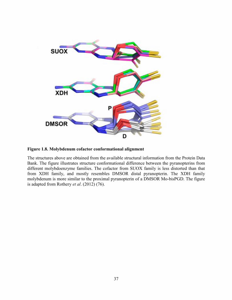
Figure 1.8. Molybdenum cofactor conformational alignment
The structures above are obtained from the available structural information from the Protein Data
Bank. The figure illustrates structure conformational difference between the pyranopterins from
different molybdoenzyme families. The cofactor from SUOX family is less distorted than that
from XDH family, and mostly resembles DMSOR distal pyranopterin. The XDH family
molybdenum is more similar to the proximal pyranopterin of a DMSOR Mo-bisPGD. The figure
is adapted from Rothery et al. (2012) (76).

38
Figure 1.9. Molybdenum cofactor biosynthetic pathway
The figure illustrates four basic steps in the Moco biosynthesis pathway: circularization of GTP,
formation of dithiolene on cPMP, adenylation of MPT, and Mo insertion. The enzymes
catalyzing those steps are shown on the left side with red represents human enzymes, green
represents plant enzymes, and black represents bacterial enzymes. The ATP equivalent
molecules are squared in red. There are minimum five ATP equivalent molecules consumed for
synthesis of each pyranopterin. The figure is adapted from Mendel. (2013) (63).
X2

39
Figure 1.10. Redox loop formed by nitrate reductase and formate dehydrogenase
The figure illustrates the redox loop formed by formate dehydrogenase and nitrate reductase A.
FdnGHI converts formate to carbon dioxide and reduces MQ to MQH2, while NarGHI oxidizes
MQH2 to MQ and uses the two electrons to convert nitrate to nitrite. There is a net seven-proton
difference produced in each cycle of the redox loop. The loop is one of the mechanisms allowing
E. coli to survive anaerobically. The figure is adapted from Bertero et al. (2003) (78).

40
Figure 1.11. Crystal structure of NarGHI (1Q16)
(a) The overall crystal structure of a NarGHI protein. The protein is membrane-bound and faces
the cytoplasm. The green represents NarG, the catalytic subunit; the red represents NarH, the
electron transfer subunit; and the blue represents NarI, the membrane anchor subunits. There are
eight prosthetic groups shown in sticks: Mo-bisPGD and FS0 from NarG, FS1 to FS4 from NarH,
Heme bP and Heme bD from NarI. Mo, Fe, and S are labelled as cyan, orange, and yellow spheres,
respectively. (b) The prosthetic groups are shown in the same position and orientation as in (a).
The center-to-center and edge-to-edge (in parenthesis) distances between adjacent prosthetic
groups are labelled on the right hand side. (c) NarGHI is in the form of dimers in crystal
structures. The dimension of a dimer is 143 Å by 128 Å. The monomers in the diagram are ~90°
rotation from (a). The figure is adapted from Bertero et al. (2003) (78).

41
Figure 1.12. NarG structural information
(a) The four domains of NarG are Domain I in green, Domain II in purple, Domain III in blue,
and Domain IV in orange. Other structural components are labelled in lighter colors. The
molybdenum cofactor is coordinated by domain II and III. Domain I coordinates FSO and
contains the extended N terminus that is capable of interacting with other subunits. (b) Detail
illustration of the active site Moco with its interacting partners. The molybdenum in a cyan
sphere and the cofactor is in red sticks. The residues are yellow sticks with N represented by blue
colour. All the residues highlighted are well conserved. The figure is adapted from Bertero et al.
(2003) (78).
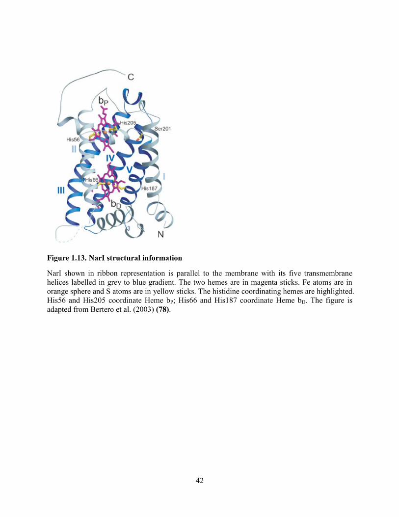
42
Figure 1.13. NarI structural information
NarI shown in ribbon representation is parallel to the membrane with its five transmembrane
helices labelled in grey to blue gradient. The two hemes are in magenta sticks. Fe atoms are in
orange sphere and S atoms are in yellow sticks. The histidine coordinating hemes are highlighted.
His56 and His205 coordinate Heme bP; His66 and His187 coordinate Heme bD. The figure is
adapted from Bertero et al. (2003) (78).

43
Figure 1.14. The catalytic mechanism of nitrate reduction
The catalytic mechanism proposed by our lab involves Mo atom, NarG-Asp222, and NarG-
His546. The Mo(IV) is coordinated by four dithiolene sulfurs and two oxygens from Asp222.
The substrate NO3- carries out nucleophilic attach onto Mo(IV) and Asp222 switches form the
bidentate coordination to a monodentate coordination. The His546 coordinates the Asp carboxyl
oxygen that has left the Mo coordination. His546 is further coordinated by Asn1217 which is not
illustrated in the figure. Nitrate reduction occurs and Mo is oxidized to +6. Nitrite leaves the Mo
center, leaving an oxo group coordinating the Mo(VI). The oxo group leaves as water when Mo
is reduced to +4 again and Asp222 re-establish the bidentate coordination. The Figure is created
by Dr. Richard Rothery.

44
CHAPTER 2. Pyranopterin Coordination
Controls Molybdenum Electrochemistry in
Escherichia coli Nitrate Reductase
This work is accepted and published online in August 2015 by Journal of Biological Chemistry
Author contributions
Sheng Yi Wu designed, performed and analyzed the experiments presented in the manuscript. Dr.
Richard A. Rothery wrote the paper and contributed to the data analyses. Dr. Joel H. Weiner
conceived and coordinated the study.
Acknowledgments: This work was funded by the Canadian Institutes of Health Research (Grant
MOP15292), the Canada Foundation for Innovation, the Alberta Heritage Foundation for
Medical Research, and Natural Science and Engineering Research Council - Collaborative
Research and Training Experience Program. We thank Justin Fedor for critical reading of the
manuscript, and Shannon Murphy for providing technical support.

45
2.1 Introduction
Enzymes containing the mononuclear molybdenum cofactor (Mo-enzymes) support an
extraordinary diversity of redox transitions spanning a reduction potential range of
approximately one volt (10, 123). Their active sites comprise a Mo atom coordinated by one or
two pyranopterin dithiolene chelates, with additional metal coordination being provided by
oxo/sulfido groups and/or oxygen/sulfur atoms derived from amino acid side chains (124, 125).
Mo-enzymes fall into three major families, each of which has a distinct protein fold: the sulfite
oxidases and plant nitrate reductases [the SUOX family (68)]; the xanthine dehydrogenases and
carbon-monoxide dehydrogenases [the XDH family (126)]; and enzymes related to the bacterial
DMSO reductases [the DMSOR family (75, 126)]. The metal atom of the SUOX and XDH
families is coordinated by a single pyranopterin dithiolene chelate, whereas in the DMSOR
family it is coordinated by two such moieties (44, 75, 126). The situation is further complicated
by the presence of a cytosine nucleotide attached via a phosphodiester linkage to the methyl
group of the pyranopterin in some members of the XDH family (69), and by the presence of a
guanine nucleotide similarly attached to each pyranopterin in members of the DMSOR family
(44, 75, 126). The precise function of the pyranopterin component of the molybdenum cofactor
remains unclear: it clearly functions as part of the molecular scaffold holding the Mo atom in an
optimum position for catalysis (127, 128); and it has recently been proposed that it may also
modulate Mo reduction potentials and hence reactivity via alternate oxidation states (e.g.
dihydro- versus tetrahydro- oxidation states) (44, 76, 119, 120, 129, 130).
Evolutionary analyses have established the presence of the DMSOR protein fold in the ―last
universal common ancestor‖ from which all extant species evolved (131, 132). An important
consequence of this is that the fold has evolved to support catalytic functions encompassing

46
those necessary prior to and after the evolution of oxygenic photosynthetic water oxidation (133).
This process likely encompassed substitutions of the protein-metal ligands, modulation of the
metal coordination sphere via the bis-dithiolene chelate, and modification of the pyranopterin
coordination environment. The DMSOR family is able to support a broad range of redox
transitions including the following: oxidation of formate, arsenite, ethylbenzene, and
dimethylsulfide; and the reduction of polysulfide, nitrate, chlorate, trimethylamine-N-oxide and
dimethylsulfoxide (44, 75, 123, 126). A plausible hypothesis explaining the presence of the
second pyranopterin in the DMSOR family is that it adds additional facets of fine tuning to
facilitate reactivity towards the observed broad range of substrates. The pyranopterin
coordination environments may play a critical role in modulating dithiolene chelate electrostatics
via differences in hydrogen bonding environment, conformation, and oxidation state (44, 76,
119–121, 130, 134). While much attention has been paid to the role of the immediate metal
environment in defining catalysis (66, 128, 135, 136), little is known about the role of the
pyranopterin coordination environment.
The molybdenum cofactor found in the DMSOR family is a molybdo-bis(pyranopterin guanine
dinucleotide) (Mo-bisPGD) (44, 75, 123, 126). The pyranopterins are referred to as proximal and
distal based on their positions relative to a conserved [4Fe-4S] cluster, known as FS0, that is
present in almost every member of the DMSOR enzyme family. In enzymes for which high-
resolution structures are available, the proximal pyranopterin has a more distorted conformation
than that of the distal pyranopterin, consistent with the former being in a fully-reduced
tetrahydro-form; and with the latter having accessibility to the partially-oxidized 10,10a dihydro
form (76). In two members of the family, Escherichia coli respiratory nitrate reductase (NarGHI)
(78) and Aromatoleum aromaticum ethylbenzene dehydrogenase (EbdABC) (77), high-

47
resolution protein structures reveal that the distal pyranopterin has a bicyclic structure with an
open pyran ring1. These observations raise the question of the role of the alternate open pyran
ring structure in defining active site redox chemistry and catalysis. In the case of NarGHI,
protein crystallography reveals that the open pyran ring oxygen participates in hydrogen bonding
interactions with two conserved residues in NarG: NarG-His1163 and NarG-Ser719 [((125)
Figure 2.1 A)]. The distances between the Ser719 OG proton and the pyran oxygen and between
the His1163 NE2 proton and the pyran oxygen are both approximately 2 Å. Closer inspection
reveals the presence of a second conserved His residue (NarG His1184) that completes a charge-
transfer relay connecting the pyran oxygen with three structurally-conserved water molecules at
its distal end (Figure 2.1). It would therefore be of interest to generate variants of NarG-Ser719,
His1163, and His1184 and determine their effects on Mo electrochemistry and catalysis.
We recently examined the coordination environment of the pyranopterin piperazine nitrogens in
members of the DMSOR family for which high-resolution structures are available (44, 76),
revealing that all members of the family contain a His or Arg residue that bridges the two
piperazine N-5 atoms. With the exception of the Thermus thermophilus polysulfide reductase
(137), all DMSOR fold enzyme structures obtained to date contain an additional residue (a
His/Ser/Gln) that stabilizes the N-5 of the proximal pyranopterin in an sp3 hybridized state.
Figure 2.2 A illustrates piperazine nitrogen coordination of the NarGHI pyranopterins. The N-10
atoms of both piperazines are hydrogen-bond donors to backbone amide oxygens (of NarG
Thr259 and NarG Ser720). The N-5 atoms are bridged by the imidazole of NarG His1092, with
the N-5 atom of the proximal pyranopterin participating in an additional H-bond with the NE2
nitrogen of NarG His1098. These two residues are referred to as the ―bridging‖ and ―stabilizing‖
1 The bicyclic form is therefore formally a pterin rather than a pyranopterin. However, for the sake of brevity, it is
referred to as a pyranopterin herein.

48
histidines, respectively, and their H bonding contacts are summarized in Figure 2.2 B. That
almost all of the proximal N-5 atoms in the DMSOR family have two hydrogen bonding contacts
favors a tetrahydro oxidation state for the proximal pyranopterin. The single H-bonding contact
observed for the N-5 atoms of all the distal pyranopterins provides further support for the
suggestion that these have accessibility to the 10,10a-dihydro oxidation state (44, 76). In some
members of the DMSOR family, [e.g. E. coli formate dehydrogenase N (FdnGHI (138)) and the
periplasmic nitrate reductase from Cupriavidus necator (NapAB (139))], the bridging ligand is
an Arg residue. It would therefore be of interest to generate variants of His1092 and His1098 and
determine their effects on Mo electrochemistry and catalysis.
In this paper, we tested the hypothesis that the pyranopterins of the DMSOR family of
molybdoenzymes have a non-innocent role in defining Mo electrochemistry and catalysis. If the
role of the pyranopterin dithiolene chelates is merely to contribute to a Mo-binding scaffold at
the active site, then variants of the target residues studied herein should have little or no effect on
enzyme function. If, however, the non-innocence reported for model compounds extends beyond
the dithiolene chelate and into the heterocyclic ring system of the pyanopterin (76, 119–121),
variants of pyranopterin-coordinating residues should have a dramatic effect on molybdenum
electrochemistry and catalysis.
2.2 Experimental procedures
2.2.1 Bacterial strains and plasmids
E. coli LCB79 (araD139 Δ(lacIPOZYA-argF) rpsL, thi Ø79(nar-lac)) (140) was used as the host
for all the experiments described herein. NarGHI was expressed from the plasmid pVA700 (141).

49
2.2.2 Site-directed mutagenesis
Mutant plasmids were generated using the QuikChange site-directed mutagenesis kit from
Stratagene. DpnI was purchased from Invitrogen, and DNA purification kits were purchased
from Qiagen. Mutants were verified by DNA sequencing (DNA Core Facility, Department of
Biochemistry, University of Alberta). Preparation of competent cells and their transformations
with plasmids were carried out as previously described (142).
2.2.2.1 Generation of a NarG-Ser719Ala variant
A 4.5 kbp SacI-AvaI fragment of pVA700 was subcloned into pTZ18R and the resultant plasmid
was used for site-directed mutagenesis. A 3.3 kbp SacI-NcoI fragment was cloned back into the
pVA700 expression vector, creating pVA700/Ser719Ala.
2.2.2.2 Generation of NarG-His1163Ala and NarG His1184Ala variants
A 4.6 kbp EcoRI-SacII fragment of pVA700 was subcloned into pBlueScript to generate a
template for site-directed mutagenesis. A 2.1 kb NcoI-SacII fragment was cloned back into the
pVA700, creating pVA700/His1163Ala or pVA700/His1184Ala.
2.2.2.3 Generation of NarG-His1092Ala, NarG-His1092Arg, and NarG-
His1098Ala variants
The EcoRI-SacII fragment cloned into pBluescript was used as a site-directed mutagenesis
template, after which a 1.2 kbp NcoI/BstBI fragment was cloned back into the pVA700, creating
pVA700/His1092Ala, pVA700/His1092Arg, or pVA700/His1098Ala.
50
2.2.3 Growth of cells
Cells were grown microaerobically in 2 L batch cultures of Terrific Broth (142) at 30 °C in the
presence of 100 μg mL-1
streptomycin and ampicillin. A 10 % innoculum of a stationary phase
culture was used and NarGHI overexpression was induced by addition of 0.2 mM isopropyl-1-
thio-β-D-galactopyranoside (IPTG). Following addition of the innoculum and IPTG, cells were
grown overnight with gentle shaking, and were harvested by centrifugation, and subsequently
washed in a buffer containing 100 mM MOPS and 5 mM EDTA (pH 7.0). Cells were
resuspended in buffer, and phenylmethylsulfonyl fluoride was added to a final concentration of
0.2 mM. Cell lysis was achieved by three passages through an Emulsiflex C3 microfluidizer
(Avestin) at a pressure of 17,000 p.s.i. Crude membranes were prepared by differential
centrifugation as previously described (143). These were resuspended in buffer and layered on
top of a 55% (w/v) sucrose step (made up in buffer). Following ultracentrifugation at 40,000
r.p.m. for 1.5 hours, the floating band enriched in cytoplasmic membrane vesicles was collected.
Excess sucrose was removed by two dilution and ultracentrifugation steps. The final membrane
pellet was resuspended in buffer to a concentration of approximately 30 mg mL-1
, and were flash
frozen in liquid nitrogen prior to being stored at -70 °C until use. Where appropriate, buffer
exchange prior to EPR analysis was achieved by dilution and re-centrifugation. Overexpression
of NarGHI was evaluated by polyacrylamide gel electrophoresis (144).
2.2.4 Bacterial growth on glycerol-nitrate minimal medium
Anaerobic growth of E. coli harboring LCB79/pVA700 and mutant derivatives was assessed in a
glycerol-nitrate (GN) minimal medium essentially as previously described (145, 146). Nitrate
was added (as KNO3) to a final concentration of 40 mM. Growth was evaluated at 37 °C, and
51
culture turbidity was measured using a Klett-Summerson spectrophotometer equipped with a No.
66 filter. Maximal growth rates were calculated as described by Zwietering et al. (147), and were
expressed as the parameter μm in units of (Klett units) hour-1
.
Form A fluorescence of the pyranopterin cofactor. Pyranopterin was assayed by generation of its
Form A fluorescent derivative following protein denaturation (148, 149), essentially as
previously described (146). As starting material, 10 mg of membrane protein were used.
2.2.5 Redox potentiometry and EPR spectroscopy
Redox titrations were carried out under argon at 25 °C as previously described (150, 151) in 100
mM Tricine and 5 mM EDTA (pH 8.0). The protein concentration used was approximately 30
mg mL -1
. The following redox mediators were used at a concentration of 25 μM: quinhydrone,
2,6-dichloroindophenol, 1,2 naphthoquinone, toluylene blue, phenazine methosulfate, thionine,
methylene blue, resorufin, indigotrisulfonate, indigocarmine, anthraquinone-2-sulfonic acid,
phenosafranine, and neutral red. All samples were prepared in quartz EPR tubes with a 3 mm
internal diameter, and were rapidly frozen in liquid nitrogen-chilled ethanol and stored at 77 K
until use. EPR spectra were recorded at 150 K using a Bruker Elexsys E500 spectrometer
equipped with a Bruker SHQE microwave cavity and a Bruker ER4131 Variable Temperature
Unit using liquid nitrogen as a cryogen. Potentiometric titration data were analyzed by plotting
the intensity of the molybdenum peak-trough versus Eh and fitting the data to two Em values (E1
and E2) (152). Mo(V) stability constants (Kstab) were calculated as described by Ohnishi et al.
(153). Fitting was performed using the Solver for Nonlinear Programming extension of Libre
Office Calc. Where appropriate the statistical significance of differences in Em values was
evaluated using a t-test.

52
2.2.6 Protein assays
Protein concentrations were assayed as described by Lowry et al. (154) modified by the inclusion
of 1% (w/v) sodium dodecyl sulfate in the incubation mixture to solubilize membrane proteins
(155).
2.2.7 Enzyme Assays
Nitrate reductase activities were measured using the non-specific electron donor benzyl viologen
(33). Assays were carried out at pH 8.0 in a buffer containing 100 mM Tricine, 0.23 mM benzyl
viologen, 5 mM EDTA and 4 mM KNO3. Benzyl viologen was reduced by adding an excess of
sodium dithionite (approximately 0.4 mM), and the reaction was initiated by addition of a
catalytic amount of enzyme.
2.3 Results and discussion
2.3.1 Residues targeted for site-directed mutagenesis
During nitrate reduction, the Mo atom of the Mo-bisPGD cycles through the IV, V, and VI
oxidation states to catalyze the two electron reduction of nitrate to nitrite (156). To investigate
the effect of pyranopterin coordination on Mo electrochemistry, we generated variants of two
sets of pyranopterin-coordinating residues in NarG: (i) those interacting directly or indirectly
with the oxygen of the open pyran ring of the distal pyranopterin (NarG-Ser719, NarG-His1163
and NarG-His1184; Figure 2.1); and (ii) those involved in bridging the proximal and distal
pyranopterins and stabilizing the oxidation state of the proximal pyranopterin (NarG-His1092
and NarG His1098; Figure 2.2). As judged by SDS-PAGE and Form A pyranopterin assays,

53
each variant was expressed in a Mo bisPGD-containing form at levels comparable to that of the
wild-type. We used these variants to test the hypothesis that residues coordinating the
pyranopterin moiety of the Mo bisPGD cofactor play a critical role in defining molybdenum
electrochemistry and substrate reactivity.
2.3.2 Impact of the variants on the NarG Mo(V) EPR spectrum
Of the three accessible Mo oxidation states, only the intermediate Mo(V) state is paramagnetic
and EPR-visible. It can be observed in spectra recorded of samples poised at appropriate
reduction potentials. Because the NarGHI Mo(V) signal is sensitive to pH in an anion-dependent
way (157, 158), we recorded spectra of samples poised at pH8.0, wherein the so-called ―high pH‖
form is dominant. This form exhibits a simple rhombic EPR spectrum with an absence of
resolved hyperfine (1H) couplings (157) that simplifies subsequent analyses of potentiometric
data (see below).
Figure 2.3 A shows spectra of redox-poised samples containing variants of residues implicated
in coordinating the open pyran oxygen of the distal pyranopterin (Ser719Ala, His1163Ala and
His1184Ala). In general, these spectra are similar to those previously observed for the Mo(V)
species of NarGHI (150, 157, 158), wherein a spectrum of a rhombic Mo(V) species is observed
with g1,2,3 values of approximately 1.988, 1.982, and 1.962. As was the case in previous studies
(157–159), no hyperfine coupling was resolved with the isotopes of Mo having a nuclear spin of
5/2 (95Mo and 97Mo, with natural abundances of ~16 % and 10 %, respectively). These data
indicate that the Ser719Ala, His1163Ala and His1184Ala variants do not significantly impact the
Mo coordination sphere.

54
Figure 2.3 B shows spectra of redox-poised samples of variants of residues implicated in
bridging the two pyranopterins (His1092Ala and His1092Arg), or stabilizing the proximal
pyranopterin in its tetrahydro form (His1098Ala). Of these, the His1092Ala variant has the most
dramatic effect on the Mo(V) EPR spectrum, shifting g2 upfield from approximately g = 1.982
(wild-type) to approximately g = 1.962 (His1092Ala). The His1092Arg variant exhibits an EPR
spectrum wherein heterogeneity is observed, especially in the g3 feature. Finally, the His1098Ala
variant exhibits a spectrum with clearly defined g1,2,3 values, with greater rhombicity than that of
the wild-type. Overall, Mo(V) species are clearly visible in all of the NarGHI variants studied
herein, with the His1092Ala variant having the greatest effect on the Mo(V) EPR spectrum (see
below).
2.3.3 Influence of the NarGHI variants on Mo electrochemistry
Molybdenum redox cycling is defined by two reduction potentials, E1 and E2, corresponding to
the Mo(VI/V) and Mo(V/IV) reduction potentials, with the average of these being defined as the
overall reduction potential (Em). The difference between E1 and E2 is a reflection of the stability
of the intermediate Mo(V) species, and can be used to calculate its stability constant (Kstab) (153).
We performed potentiometric titrations on all the variants of NarGHI studied herein, and in each
case titrations were carried out on samples generated from 3-4 independent biological replicates.
Figure 2.4-A shows representative titrations for the variants of residues interacting with the O-1
pyran oxygen (Figure 2.1). Figure 2.4 B shows plots of the residuals between the experimental
data and the fits. For the wild-type enzyme, we estimate values of Em and Kstab of 142 mV and 28,
respectively (summarized in Figure 2.4 C). These values are in reasonable agreement with those
previously obtained (150, 160–162). The Ser719Ala variant has a statistically-insignificant effect
on the Em (ΔEm = 4 mV, P = 0.57) and decreases the Kstab from 28 to 8, while the His1163Ala

55
variant elicits a large decrease in the Em (ΔEm = -88 mV, Table 2.1) and a concomitant decrease
in the Kstab from 28 to 1. The His1184Ala variant has an intermediate effect, eliciting a ΔEm of -
36 mV, and decreases the Kstab, again from 28 to 1. Thus, the charge-transfer relay depicted in
Figure 2.1 B appears to play two critical roles: (i) modulating the Mo reduction potentials (see
below for further discussion of this); and (ii) stabilizing the Mo(V) intermediate.
Figure 2.5 A shows representative potentiometric titrations of the His1092Ala, His1092Arg, and
His1098Ala variants. The His1092Arg variant replaces the bridging His residue with an Arg, and
mimics the pyranopterin-coordination environment observed in the formate dehydrogenases and
periplasmic nitrate reductases [e.g. FdnGHI and NapAB, respectively (44)]. The His1092Arg
variant elicits a modest increase of marginal statistical significance in the overall Mo Em (ΔEm =
18 mV, P = 0.07; Table 2.1), consistent with there being no change in overall charge of the
bridging residue when it is changed from a His to an Arg (the pKa of the Arg guanidinium group
is approximately 12.5). This suggests that the imidazole pKa is significantly increased from
approximately 6.0 in aqueous solution to > 8.0 in the bridging environment between the two
pyranopterins. The His1092Ala variant elicits a ΔEm on the Mo center of approximately -143
mV along with a modest decrease in its Kstab (from 28 to 19, Table 2.1). A possible explanation
for the large negative ΔEm is that the void between the two pyranopterins contains waters in the
His1092Ala variant, and that these waters lack an overall positive charge, thus decreasing the
overall Mo reduction potential. Alternatively, given that the His1092Ala variant has the most
severe effect on the Mo(V) EPR spectrum, loss of ―scaffolding‖ by the bridging imidazole
moiety may result in a significant alteration of the geometry of dithiolene coordination that may
cause the large observed ΔEm.

56
The His1098Ala variant elicits a ΔEm of 101 mV, along with a significant increase in Mo(V)
stability, with the Kstab increasing from 28 to 1822 (Figure 2.5 A and 2.5 C, Table 2.1). Figure
2.2 B shows a model for how His1092 and His1098 define the H bonding environment of the
proximal pyranopterin piperazine N-5 atom. As suggested above, formal protonation of the
imidazole of His1092 is supported by the relative lack of effect of the His1092Arg variant on the
overall Mo Em. One of the H-bonds is eliminated in the His1098Ala variant, which may result in
transfer of the proton from the ND1 atom to the piperazine N-5, resulting in loss of a positive
charge and the observed decrease in the overall Mo Em.
We proposed that in the DMSOR family of enzymes, the role of the stabilizing residue (His1098
in NarG) is to maintain the N-5 atom of the proximal pyranopterin in an sp3-hybridized form,
thus stabilizing the tetrahydro oxidation state (44, 76). This suggests a possible explanation for
the observed increase in Mo(V) stability in the His1098Ala variant. Removal of the hydrogen-
bonding contact provided by the His1098 imidazole may result in the 10,10a dihydro oxidation
state becoming available to the proximal pyranopterin. If the 10,10a dihydro to tetrahydro
reduction potential occurs within the range in which Mo(V) is extant, then E1 (the Mo(VI/V)
reduction potential) could be shifted to the observed value of 139 mV (Table 2.1; see reference
(76)).
2.3.4 Correlation between enzyme activity, cell growth and Mo
electrochemistry
How do changes in Mo electrochemistry impact enzyme activity and anaerobic respiratory
growth on nitrate? Specific activities in enriched membranes for the variants studied herein range
from 1 to 54 μmoles min-1
mg-1
for the His1092Ala and Ser719Ala variants, respectively (Table

57
2.1). With the exception of the His1092Arg variant, a strong correlation exists between overall
Mo reduction potential and enzyme activity (R = 0.99, Figure 2.6). A correlation also exists
between respiratory growth rate and Mo reduction potential across all variants (R = 0.95, inset to
Figure 2.6). Thus, catalysis and growth are convincingly correlated with increased Mo Em. A
possible explanation for this is that catalytic turnover depends on the energetics of electron
transfer from FS0 (Em = -55 mV (161)) to the Mo.
It is notable that NarGHI catalytic activity appears to be dependent on the overall Mo Em, but not
on the Mo(V) Kstab (Table 2.1). Protein film voltammetry studies of NarGHI (156) and two other
members of the DMSOR family (the periplasmic nitrate reductase from Rhodobacter
sphaeroides, NapAB (163), and the Me2SO reductase from E. coli (164) demonstrated that
enzyme turnover decreases below a reduction potential referred to as Eswitch. It was therefore
proposed that observation of an Eswitch arises because substrate binds preferentially to the Mo(V)
form of the cofactor (156, 163, 164). In the case of NarGHI, this is corroborated by the
observation of a stable nitrate adduct to the Mo(V) (158). If the mechanism of nitrate reduction
proceeds via substrate binding to a Mo(V) species, we would expect a variant that increases the
Mo(V) Kstab to retain significant catalytic activity. The His1098Ala variant exhibits a very large
Mo(V) Kstab (~1822; Table 2.1), but has low specific activity and growth rate compared to the
wild-type (Figure 2.6).
As previously noted (76), an alternative explanation for the observed Eswitch is that it controls a
reduction of the distal pyranopterin from an oxidation state equivalent to the 10,10a-dihydro
form to one equivalent to the tetrahydro form. This provides a possible explanation for why the
His1092Arg variant supports respiratory growth on nitrate, but has decreased enzyme activity in
our in vitro assay (Figure 2.6). It is possible that a significant positive ΔEm is elicited on the

58
Eswitch, resulting in lower activity measured using benzyl viologen as electron donor (Em = -374
mV (165)). NarGHI is able to function in vivo with either ubiquinol or menaquinol as electron
donor (166), and these have reduction potentials of +110 and -80 mV, respectively (10),
rendering them both less likely to reduce the distal pyranopterin than the artificial electron donor
benzyl viologen.
2.3.5 Role of the His1163/His1184 charge-transfer relay in NarGHI
maturation and in modulation of Mo electrochemistry
NarGHI maturation is a carefully orchestrated process involving the NarJ system-specific
chaperone (167) and the twin arginine translocase (168). In general, the process ensures
assembly of correctly folded enzyme with a complete complement of cofactors (167, 169).
Insertion of the Mo-bisPGD cofactor depends on the presence of the FS0 [4Fe-4S] cluster (167,
169). With the exception of EbdABC (77), NarGHI is the only enzyme examined to date that has
a bicyclic distal pyranopterin (78). Closer examination of the charge-transfer relay comprising
His1163 and His1184 (Figure 2.1) suggests a possible mechanism for pyran ring opening
(Figure 2.7), which proceeds as follows. The His1163/His1184 charge-transfer relay catalyzes
elimination of the C-4a proton (Figure 2.7 A), generating an open ring-form with a piperazine
ring in a form equivalent to the 5,10-dihydro form (Figure 2.7 B) in a mechanism essentially
identical to that previously described (170). This form rearranges to the lowest energy tautomer
(equivalent to the 10,10a-dihydro form (76, 170)) shown in Figure 2.7 C. A mechanism
essentially identical to that presented in Figure 2.7 A-C can be proposed for ring-opening of the
distal pyranopterin in EbdABC, with the distinction that ring opening would be catalyzed by a
guanidinium side chain rather than by an imidazole. The equilibrium between the structures

59
shown in Figure 2.7 C and 2.7 D illustrates how the formal change on the pyran oxygen (and the
overall Mo reduction potential) can be modulated by the His1163/His1184 charge-transfer relay.
Stabilization of the pyran oxygen in its alkoxide form would decrease the overall Mo reduction
potential, whereas formal protonation to the hydroxyl form would increase the overall Mo
reduction potential.
In the context of the model presented in Figure 2.7 C and 2.7 D for charge-transfer induced Mo
reduction potential modulation, it is notable that the His1163Ala variant elicits a large ΔEm that
is approximately double that elicited by the His1184Ala variant. This is consistent with the entire
charge transfer relay being involved in Mo reduction potential modulation. It is possible to
speculate that the His1163Ala variant may impact an equilibrium between the open and closed
pyran ring forms of the distal pyranopterin, and we are currently addressing this issue using
protein crystallography.
2.4 Conclusion
We have demonstrated the importance of the pyranopterin coordination environment in defining
Mo electrochemistry and substrate reactivity, with enzyme activity correlating with increasing
overall Mo reduction potential. Our studies demonstrate the importance of factors beyond the
immediate Mo coordination sphere in defining enzyme activity.

60
2.5. Table
Table 2.1. Effects of variants of pyranopterin-coordinating residues on Mo reduction
potentials and enzyme activity
Variant Em
(mV)a
ΔEm
(mV)b
E1
(mV)c
E2
(mV)d
Kstab Maximal growth
rate, μm ((Klett
units) hr-1
)
BV·-:NO3
- activity
(μmoles min-1
mg-1
)
Wild-type 142 ± 12
(n = 4)
0 184 ± 10 99 ± 16 28 15.4 ± 0.7
(n = 3)
48 ±14
(n = 3)
Ser719Ala 146 ± 7
(n = 3)
4 170 ± 19 122 ± 7 8 15.5 ± 0.9
(n = 3)
54 ±11
(n = 3)
His1163Ala 54 ± 3
(n = 4)
-88 53 ± 5 54 ± 9 1 6.1 ± 0.2
(n = 3)
23 ± 4
(n = 3)
His1184Ala 106 ± 5
(n = 3)
-36 106 ± 14 106 ± 5 1 11.4 ± 0.6
(n = 3)
36 ± 1
(n = 3)
His1092Ala -1 ± 3
(n = 3)
-143 35 ± 9 -38 ± 11 19 4.4 ± 0.5
(n = 2)
0.9 ± 0.1
(n = 3)
His1092Arg 160 ± 7
(n = 3)
18 225 ± 15 95 ± 2 171 12.8 ± 0.4
(n = 2)
8.6 ± 0.6
(n = 3)
His1098Ala 41 ± 4
(n = 4)
-101 139 ± 7 -57 ± 2 1822 4.8 ± 0.5
(n = 2)
16 ± 2
(n = 3)
a – Overall Mo(VI/V/IV) reduction potential at pH 8.0.
b – Em (variant) minus Em (wild-type). Where appropriate, P values indicating the statistical
significance of each ΔEm are given in the text.
c – Mo(VI/V) reduction potential.
d – Mo(V/IV) reduction potential.

61
2.6. Figures
Figure 2.1. Charge transfer relay
(A) Charge-transfer relay in NarG. Three water molecules are conserved across a range of
NarGHI structures, including those described by PDB codes 1Q16, 1R27, 1YZ4 and 1Y5I
(Bertero et al. 2003; Jormakka et al. 2004; Bertero et al. 2005). These waters are linked to the
pyran oxygen of the bicyclic distal pyranopterin by NarG-His1184 and NarG-H1163. The OG
oxygen of NarG-Ser719 is also within H-bonding distance of the pyran oxygen. The image was
generated using the Pymol molecular visualization package (version 1.4.1, Schrödinger LLC). (B)
Mechanism of charge-transfer between the pyran oxygen and the conserved water molecules.
The indicated charge-transfer results in protonation of the alkoxide anion form of the pyran
oxygen. The image was generated using the MarvinSketch software package
(www.chemaxon.com). In both panels, some predicted hydrogens have been added for clarity.

62
Figure 2.2. Residues defining pyranopterin piperazine ring coordination
(A) Residues defining pyranopterin piperazine ring coordination. The image was generated
using the NarGHI structure described by PDB code 1Q16 (Bertero et al. 2003). His1092
functions to bridge the two pyranopterins, with its ND1 nitrogen functioning as an H-bond donor
to the proximal piperazine N-5 atom, and its NE2 nitrogen functioning as an H-bond donor to the
distal piperazine N-5 atom. Note that His1092 is shown in its protonated form to facilitate H-
bond donation to both piperazine N-5 atoms. For further details, see the text. (B) Proposed H-
bonding network around the piperazine rings of the two pyranopterins. The proximal
pyranopterin (labeled P) is shown in its tetrahydro form, with this redox state stabilized by both
the bridging His1092 and the stabilizing His1098, while the piperazine of the distal pyranopterin
(labeled D) is shown in a form equivalent to the 10,10a-dihydro pyranopterin. In both panels,
some predicted hydrogens have been added for clarity. Images were generated as described in
the legend to Figure 2.1.

63
Figure 2.3. Mo(V) EPR spectra of redox-poised NarGHI variants of residues involved in
pyranopterin coordination.
(A) Ala variants of NarG-Ser719, NarG-His1163 and NarG-H1184; and (B) Ala variants of
His1092 and His1098, and an Arg variant of His1092. Spectra were recorded at 150K using a
microwave power of 20 mW (at a frequency of 9.332 GHz), a modulation amplitude of 4 Gpp at a
frequency of 100 kHz. Approximate g1,2,3 values and potentials at which the samples are poised
are: 1.988, 1.982, 1.962 (wild-type, Eh = 150 mV); 1.988, 1.982, 1.962 (Ser719Ala, Eh =
152 mV); 1.992, 1.985, 1.964 (His1163Ala, Eh = 48 mV); 1.986, 1.980, 1.961 (His1184Ala, Eh =
103 mV); 1.978, 1.962, 1.945 (His1092Ala, Eh = -5 mV), 1.989, 1.984, 1.965 (His1092Arg, Eh =
146 mV); 1.992, 1.985, 1.951 (His1098Ala, Eh = 28 mV).
1.941.961.982.002.02 1.921.941.961.982.002.02
1 2
3
1 2
3
1 2
3
1 2
3
1 2
3
1 2
3
2
3
1
1 2
3
(A) (B)
g value g value
WT WT
S719A
H1163A
H1184A
H1092A
H1092R
H1098A

64
Figure 2.4. Potentiometric titrations of membranes containing variants of residues
coordinating the distal pyranopterin of NarGHI
(A) Comparison of representative titrations of wild-type (■), Ser719Ala (▼), His1163Ala (►),
and His1184Ala (●) enzymes. Data were fit to the following parameters: wild-type, Em =
140 mV (E1 = 183 mV, E2 = 97 mV); Ser719Ala, Em = 150 mV (E1 = 176 mV, E2 = 124 mV);
His1163Ala, Em = 58 mV (E1 = 49 mV, E2 = 68 mV); His1184Ala, Em = 103 mV (E1 = 96 mV,
E2 = 111 mV). (B) Plot of residuals. (C) Summary of Em, E1 and E2 values for the variants based
on the data presented in Table 1. Colored squares indicate the mean Em values and the horizontal
lines indicate the E1 and E2 values in the cases where E1 – E2 > 0 (Kstab ≥ 1).
-100 -50 0 50 100 150 200 250 3000
50
100
150
200
250
Eh (mV)
Sig
na
l h
eig
ht
-100 -50 0 50 100 150 200 250 300-30
0
30
Re
sid
ua
l
WT
S719A
H1163A
H1184A
-100 -50 0 50 100 150 200 250 300
(A)
(B)
(C)

65
Figure 2.5. Potentiometric titrations of membranes containing variants of His1092 and
His1098
(A) Comparison of representative titrations of wild-type (■), His1092Arg (▼), His1098Ala (►),
and His1092Ala (●) enzymes. Data were fit to the following parameters: wild-type, Em =
140 mV (E1 = 183 mV, E2 = 97 mV); His1092Arg, Em = 155 mV (E1 = 213 mV, E2 = 97 mV);
His1098Ala, Em = 35 mV (E1 = 129 mV, E2 = -60 mV); His1092Ala, Em = -2 mV (E1 = 24 mV,
E2 = -29 mV). (B) Plot of residuals. (C) Summary of Em, E1 and E2 values for the variants based
on the data presented in Table 1. Colored squares indicate the mean Em values and the horizontal
lines indicate the E1 and E2 values.
WT
H1092A
H1092R
H1098A
Eh (mV)
-150 -100 -50 0 50 100 150 200 250 300
-150 -100 -50 0 50 100 150 200 250 3000
50
100
150
200
250
300S
ign
al h
eig
ht
-150 -100 -50 0 50 100 150 200 250 300-30
0
30
Resid
ual
(A)
(B)
(C)
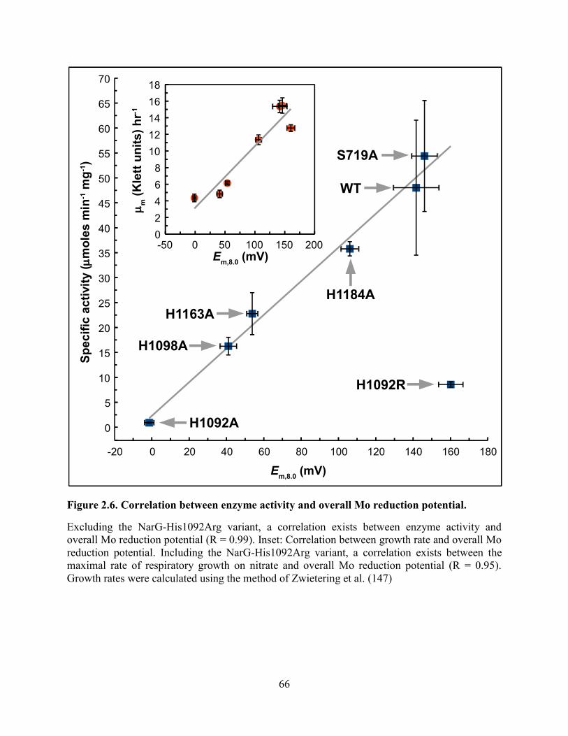
66
Figure 2.6. Correlation between enzyme activity and overall Mo reduction potential.
Excluding the NarG-His1092Arg variant, a correlation exists between enzyme activity and
overall Mo reduction potential (R = 0.99). Inset: Correlation between growth rate and overall Mo
reduction potential. Including the NarG-His1092Arg variant, a correlation exists between the
maximal rate of respiratory growth on nitrate and overall Mo reduction potential (R = 0.95).
Growth rates were calculated using the method of Zwietering et al. (147)
-20 0 20 40 60 80 100 120 140 160 180
0
5
10
15
20
25
30
35
40
45
50
55
60
65
70
-50 0 50 100 150 2000
2
4
6
8
10
12
14
16
18
WT
S719A
H1092R
H1184A
H1163A
H1098A
H1092A
Em,8.0
(mV)
Sp
ecif
ic a
cti
vit
y (m
mo
les m
in-1 m
g-1)
Em,8.0
(mV)
mm (
Kle
tt u
nit
s)
hr-1

67
Figure 2.7. Proposed mechanism of pyran ring opening of the distal pyranopterin of
NarGHI.
(A) The electron transfer relay comprising NarG-His1163 and NarG-His1184 catalyzes proton
abstraction from the C-4a atom and pyran ring-opening. The product of this reaction has a
piperazine oxidation state and structure equivalent to the 5,10-dihydro pyranopterin (B). A
tautomerization reaction results in a form equivalent to the 10,10a-dihydro pyranopterin with a
protonated oxygen equivalent to O-1. The pterin core of this form is equivalent to the lowest
energy dihydropterin tautomer (76, 170). (C). The NarG-His1163/His1184 charge-transfer relay
functions to modulate an equilibrium between the protonated (hydroxyl) and deprotonated
(alkoxide) forms of the bicyclic distal pyranopterin, with the protonated form having a higher
overall predicted Mo(VI/VIV) reduction potential than the deprotonated form (D).
HN
N
HN
NH
O
O
S
S
Mo
R1
R
N
N
H
R
NN
H
O+H
H
H
H2N
H
HN
N
N
NH
O
O
S
S
Mo
R1
R
N
N
H
R
NN
H
OH
H
H
H2N
H
H+
HN
N
N
NH
O
O
S
S
Mo
R1
R
N
N
H
R
NN
H
OH
H
H
H2N
HN
N
N
NH
O
–O
S
S
Mo
R1
R
N
N
H
R
NN
H
O+H
H
H
H2N
(A) (B)
(C)(D)
LowerMo potentials
HigherMo potentials
12
34
4a55a6
7
8
9
9a 1010a

68
CHAPTER 3. Conclusions

69
In Chapter 2, we described how pyranopterin coordination can play a role in guiding electrons
and modulating the potential of the Mo. The pyranopterins of the Moco have been undervalued
for decades and this work sheds light on them by providing direct experimental evidence
showing their functional importance. EPR experiments coupled with redox titrations, along with
functional assays, allowed us to accurately assess the changes in Mo electrochemistry and
reactivity caused by variants of pyranopterin coordinating residues.
As a part of on-going research of Moco, the Mo active site has always been of focused interest.
As mentioned in Section 1.6.6 and illustrated in Figure 1.14, we have a proposal of the reaction
mechanism at the active site and the immediate Mo coordination is under investigation, in
particular the conserved residues Asn52, His546, and Asn1217. Asn52 is within hydrogen-
bonding distance to one of the dithiolene sulfurs and is speculated to have a role in influencing
the Mo coordination environment. His546 is within hydrogen-bonding distance to Asp222, the
residue that is directly involved in the catalytic mechanism. Asn1217 is within hydrogen-
bonding distance to the His546.
The pyranopterin study and the on-going active site study provide an understanding of the Moco.
In addition, there are tremendous opportunities to further explore this important cofactor in
biology, which is key to understanding the biological diversity of metabolism and evolution of
Moco-containing organisms. The current structural and functional information about Moco is
only the tip of an iceberg of the knowledge in the field. More effort is required to pursue this
curiosity-driven research.

70
References
1. Hamrick MW (1998) Functional and adaptive significance of primate pads and claws:
evidence from new world anthropoids. Am J Phys Anthropol 106(2):113–127.
2. Müller DWH, et al. (2013) Assessing the jarman–bell principle: scaling of intake,
digestibility, retention time and gut fill with body mass in mammalian herbivores. Comp
Biochem Physiol A Mol Integr Physiol 164(1):129–140.
3. Price TD (2011) Adaptive radiations: there’s something about finches. Curr Biol
21(23):R953–R955.
4. Braakman R, Smith E (2014) Metabolic evolution of a deep-branching hyperthermophilic
chemoautotrophic bacterium. PLoS ONE 9(2). doi:10.1371/journal.pone.0087950.
5. Bryant WA, Faruqi AA, Pinney JW (2013) Analysis of metabolic evolution in bacteria
using whole-genome metabolic models. J Comput Biol 20(10):755–764.
6. Campbell N, et al. (2008) Biology (Pearson, San Francisco). 8th Ed.
7. Mitchell P (1979) Keilin’s respiratory chain concept and its chemiosmotic consequences.
Science 206(4423):1148–1159.
8. Rich P (2003) The molecular machinery of Keilin’s respiratory chain. Available at:
http://www.biochemsoctrans.org/bst/031/1095/bst0311095.htm [Accessed March 23,
2015].
9. Mitchell P (1967) Proton-translocation phosphorylation in mitochondria, chloroplasts and
bacteria: natural fuel cells and solar cells. Fed Proc 26:1370–1379.
10. Unden G, Bongaerts J (1997) Alternative respiratory pathways of Escherichia coli:
energetics and transcriptional regulation in response to electron acceptors. Biochim
Biophys Acta BBA - Bioenerg 1320(3):217–234.
11. Neidhardt FC, Curtiss R (1996) Escherichia coli and Salmonella: cellular and molecular
biology (ASM Press).
12. Rolfe MD, et al. (2012) Systems analysis of transcription factor activities in environments
with stable and dynamic oxygen concentrations. Open Biol 2(7). doi:10.1098/rsob.120091.
13. Gunsalus RP, Park SJ (1994) Aerobic-anaerobic gene regulation in Escherichia coli:
control by the ArcAB and Fnr regulons. Res Microbiol 145(5-6):437–450.
14. Iuchi S, Lin EC (1988) arcA (dye), a global regulatory gene in Escherichia coli mediating
repression of enzymes in aerobic pathways. Proc Natl Acad Sci U S A 85(6):1888–1892.

71
15. Iuchi S, Cameron DC, Lin EC (1989) A second global regulator gene (arcB) mediating
repression of enzymes in aerobic pathways of Escherichia coli. J Bacteriol 171(2):868–
873.
16. Iuchi S, Matsuda Z, Fujiwara T, Lin EC (1990) The arcB gene of Escherichia coli encodes
a sensor-regulator protein for anaerobic repression of the arc modulon. Mol Microbiol
4(5):715–727.
17. Iuchi S (1993) Phosphorylation/dephosphorylation of the receiver module at the conserved
aspartate residue controls transphosphorylation activity of histidine kinase in sensor
protein ArcB of Escherichia coli. J Biol Chem 268(32):23972–23980.
18. Georgellis D, Lynch AS, Lin EC (1997) In vitro phosphorylation study of the Arc two-
component signal transduction system of Escherichia coli. J Bacteriol 179(17):5429–5435.
19. Kwon O, Georgellis D, Lin EC (2000) Phosphorelay as the sole physiological route of
signal transmission by the arc two-component system of Escherichia coli. J Bacteriol
182(13):3858–3862.
20. Georgellis D, Kwon O, Lin EC (2001) Quinones as the redox signal for the arc two-
component system of bacteria. Science 292(5525):2314–2316.
21. Malpica R, Franco B, Rodriguez C, Kwon O, Georgellis D (2004) Identification of a
quinone-sensitive redox switch in the ArcB sensor kinase. Proc Natl Acad Sci U S A
101(36):13318–13323.
22. Liu X, De Wulf P (2004) Probing the ArcA-P modulon of Escherichia coli by whole
genome transcriptional analysis and sequence recognition profiling. J Biol Chem
279(13):12588–12597.
23. Lynch AS, Lin EC (1996) Transcriptional control mediated by the ArcA two-component
response regulator protein of Escherichia coli: characterization of DNA binding at target
promoters. J Bacteriol 178(21):6238–6249.
24. Kang Y, Weber KD, Qiu Y, Kiley PJ, Blattner FR (2005) Genome-wide expression
analysis indicates that FNR of Escherichia coli K-12 regulates a large number of genes of
unknown function. J Bacteriol 187(3):1135–1160.
25. Lazazzera BA, Beinert H, Khoroshilova N, Kennedy MC, Kiley PJ (1996) DNA binding
and dimerization of the Fe-S-containing FNR protein from Escherichia coli are regulated
by oxygen. J Biol Chem 271(5):2762–2768.
26. Achebach S, Selmer T, Unden G (2005) Properties and significance of apoFNR as a
second form of air-inactivated [4Fe-4S]·FNR of Escherichia coli. FEBS J 272(16):4260–
4269.
27. Kiley PJ, Beinert H (1998) Oxygen sensing by the global regulator, FNR: the role of the
iron-sulfur cluster. FEMS Microbiol Rev 22(5):341–352.

72
28. Crack J, Green J, Thomson AJ (2004) Mechanism of oxygen sensing by the bacterial
transcription factor fumarate-nitrate reduction (FNR). J Biol Chem 279(10):9278–9286.
29. Moore LJ, Kiley PJ (2001) Characterization of the dimerization domain in the FNR
transcription factor. J Biol Chem 276(49):45744–45750.
30. Reinhart F, Achebach S, Koch T, Unden G (2008) Reduced apo-fumarate nitrate reductase
regulator (apoFNR) as the major form of FNR in aerobically growing Escherichia coli. J
Bacteriol 190(3):879–886.
31. Khoroshilova N, Popescu C, Münck E, Beinert H, Kiley PJ (1997) Iron-sulfur cluster
disassembly in the FNR protein of Escherichia coli by O2: [4Fe-4S] to [2Fe-2S]
conversion with loss of biological activity. Proc Natl Acad Sci U S A 94(12):6087–6092.
32. Crack JC, et al. (2008) Influence of the environment on the [4Fe-4S]2+
to [2Fe-2S]2+
cluster switch in the transcriptional regulator FNR. J Am Chem Soc 130(5):1749–1758.
33. Morpeth FF, Boxer DH (1985) Kinetic analysis of respiratory nitrate reductase from
Escherichia coli K12. Biochemistry (Mosc) 24(1):40–46.
34. Rabin RS, Stewart V (1993) Dual response regulators (NarL and NarP) interact with dual
sensors (NarX and NarQ) to control nitrate- and nitrite-regulated gene expression in
Escherichia coli K-12. J Bacteriol 175(11):3259–3268.
35. Cavicchioli R, Chiang RC, Kalman LV, Gunsalus RP (1996) Role of the periplasmic
domain of the Escherichia coli NarX sensor-transmitter protein in nitrate-dependent signal
transduction and gene regulation. Mol Microbiol 21(5):901–911.
36. Lee AI, Delgado A, Gunsalus RP (1999) Signal-dependent phosphorylation of the
membrane-bound NarX two-component sensor-transmitter protein of Escherichia coli:
nitrate elicits a superior anion ligand response compared to nitrite. J Bacteriol
181(17):5309–5316.
37. Stewart V (1982) Requirement of Fnr and NarL functions for nitrate reductase expression
in Escherichia coli K-12. J Bacteriol 151(3):1320–1325.
38. Iuchi S, Lin EC (1987) The narL gene product activates the nitrate reductase operon and
represses the fumarate reductase and trimethylamine N-oxide reductase operons in
Escherichia coli. Proc Natl Acad Sci U S A 84(11):3901–3905.
39. Hille R (1996) The Mononuclear Molybdenum Enzymes†. Chem Rev 96(7):2757–2816.
40. Schwarz G, Mendel RR, Ribbe MW (2009) Molybdenum cofactors, enzymes and
pathways. Nature 460(7257):839–847.
41. Romão MJ (2009) Molybdenum and tungsten enzymes: a crystallographic and
mechanistic overview. Dalton Trans (21):4053–4068.

73
42. Hille R (2002) Molybdenum and tungsten in biology. Trends Biochem Sci 27(7):360–367.
43. Kletzin A, Adams MW (1996) Tungsten in biological systems. FEMS Microbiol Rev
18(1):5–63.
44. Rothery RA, Weiner JH (2014) Shifting the metallocentric molybdoenzyme paradigm: the
importance of pyranopterin coordination. J Biol Inorg Chem JBIC Publ Soc Biol Inorg
Chem. doi:10.1007/s00775-014-1194-6.
45. Sigel A, Sigel H, Sigel RKO eds. (2013) Interrelations between essential metal ions and
human diseases (Springer Netherlands, Dordrecht) Available at:
http://link.springer.com/10.1007/978-94-007-7500-8 [Accessed March 31, 2015].
46. Dickinson CJ, Smellie JM (1959) Xanthinuria. Br Med J 2(5161):1217–1221.
47. Harkness RA, McCreanor GM, Simpson D, MacFadyen IR (1986) Pregnancy in and
incidence of xanthine oxidase deficiency. J Inherit Metab Dis 9(4):407–408.
48. Ichida K, et al. (1997) Identification of two mutations in human xanthine dehydrogenase
gene responsible for classical type I xanthinuria. J Clin Invest 99(10):2391–2397.
49. Garattini E, Mendel R, Romão MJ, Wright R, Terao M (2003) Mammalian molybdo-
flavoenzymes, an expanding family of proteins: structure, genetics, regulation, function
and pathophysiology. Biochem J 372(Pt 1):15–32.
50. Terao M, et al. (2009) Role of the molybdoflavoenzyme aldehyde oxidase homolog 2 in
the biosynthesis of retinoic acid: generation and characterization of a knockout mouse.
Mol Cell Biol 29(2):357–377.
51. Garattini E, Terao M (2013) Aldehyde oxidase and its importance in novel drug discovery:
present and future challenges. Expert Opin Drug Discov 8(6):641–654.
52. Jordan CG, et al. (1999) Aldehyde oxidase-catalysed oxidation of methotrexate in the liver
of guinea-pig, rabbit and man. J Pharm Pharmacol 51(4):411–418.
53. Rashidi M-R, Beedham C, Smith JS, Davaran S (2007) In vitro study of 6-mercaptopurine
oxidation catalysed by aldehyde oxidase and xanthine oxidase. Drug Metab
Pharmacokinet 22(4):299–306.
54. Rochat B, et al. (1998) Stereoselective biotransformation of the selective serotonin
reuptake inhibitor citalopram and its demethylated metabolites by monoamine oxidases in
human liver. Biochem Pharmacol 56(1):15–23.
55. Schwarz G, Belaidi AA (2013) Molybdenum in human health and disease. Met Ions Life
Sci 13:415–450.
56. Tan W-H, et al. (2005) Isolated sulfite oxidase deficiency: a case report with a novel
mutation and review of the literature. Pediatrics 116(3):757–766.

74
57. Santi C, Bogusz D, Franche C (2013) Biological nitrogen fixation in non-legume plants.
Ann Bot 111(5):743–767.
58. Mulder EG, Bakema K, Veen WLV (1959) Molybdenum in symbiotic nitrogen fixation
and in nitrate assimilation. Plant Soil 10(4):319–334.
59. Fan H, Bolhuis H, Stal LJ (2015) Drivers of the dynamics of diazotrophs and denitrifiers
in North Sea bottom waters and sediments. Front Microbiol 6:738.
60. Bedzyk L, Wang T, Ye RW (1999) The Periplasmic Nitrate Reductase in Pseudomonas sp.
Strain G-179 Catalyzes the First Step of Denitrification. J Bacteriol 181(9):2802–2806.
61. Rajagopalan KV (1991) Novel aspects of the biochemistry of the molybdenum cofactor.
Adv Enzymol Relat Areas Mol Biol 64:215–290.
62. Rajagopalan KV, Johnson JL (1992) The pterin molybdenum cofactors. J Biol Chem
267(15):10199–10202.
63. Mendel RR (2013) The molybdenum cofactor. J Biol Chem 288(19):13165–13172.
64. Schwarz G, Mendel RR (2006) Molybdenum cofactor biosynthesis and molybdenum
enzymes. Annu Rev Plant Biol 57(1):623–647.
65. McWilliams SF, Holland PL (2015) Dinitrogen binding and cleavage by multinuclear iron
complexes. Acc Chem Res 48(7):2059–2065.
66. Hille R, Hall J, Basu P (2014) The mononuclear molybdenum enzymes. Chem Rev
114(7):3963–4038.
67. Leimkühler S, Wuebbens MM, Rajagopalan KV (2011) The history of the discovery of
the molybdenum cofactor and novel aspects of its biosynthesis in bacteria. Coord Chem
Rev 255(9-10):1129–1144.
68. Workun GJ, Moquin K, Rothery RA, Weiner JH (2008) Evolutionary persistence of the
molybdopyranopterin-containing sulfite oxidase protein fold. Microbiol Mol Biol Rev
MMBR 72(2):228–248, table of contents.
69. Rebelo JM, Dias JM, Huber R, Moura JJ, Romão MJ (2001) Structure refinement of the
aldehyde oxidoreductase from Desulfovibrio gigas (MOP) at 1.28 A. J Biol Inorg Chem
JBIC Publ Soc Biol Inorg Chem 6(8):791–800.
70. Meyer O, et al. (2000) The role of Se, Mo and Fe in the structure and function of carbon
monoxide dehydrogenase. Biol Chem 381(9-10):865–876.
71. Dobbek H, Gremer L, Kiefersauer R, Huber R, Meyer O (2002) Catalysis at a dinuclear
[CuSMo(==O)OH] cluster in a CO dehydrogenase resolved at 1.1-A resolution. Proc Natl
Acad Sci U S A 99(25):15971–15976.

75
72. Mukund S, Adams MW (1990) Characterization of a tungsten-iron-sulfur protein
exhibiting novel spectroscopic and redox properties from the hyperthermophilic
archaebacterium Pyrococcus furiosus. J Biol Chem 265(20):11508–11516.
73. Hu Y, Faham S, Roy R, Adams MW, Rees DC (1999) Formaldehyde ferredoxin
oxidoreductase from Pyrococcus furiosus: the 1.85 A resolution crystal structure and its
mechanistic implications. J Mol Biol 286(3):899–914.
74. Chan MK, Mukund S, Kletzin A, Adams MW, Rees DC (1995) Structure of a
hyperthermophilic tungstopterin enzyme, aldehyde ferredoxin oxidoreductase. Science
267(5203):1463–1469.
75. Rothery RA, Workun GJ, Weiner JH (2008) The prokaryotic complex iron–sulfur
molybdoenzyme family. Biochim Biophys Acta BBA - Biomembr 1778(9):1897–1929.
76. Rothery RA, Stein B, Solomonson M, Kirk ML, Weiner JH (2012) Pyranopterin
conformation defines the function of molybdenum and tungsten enzymes. Proc Natl Acad
Sci U S A 109(37):14773–14778.
77. Kloer DP, Hagel C, Heider J, Schulz GE (2006) Crystal structure of ethylbenzene
dehydrogenase from Aromatoleum aromaticum. Structure 14(9):1377–1388.
78. Bertero MG, et al. (2003) Insights into the respiratory electron transfer pathway from the
structure of nitrate reductase A. Nat Struct Biol 10(9):681–687.
79. Mendel RR, Hänsch R (2002) Molybdoenzymes and molybdenum cofactor in plants. J
Exp Bot 53(375):1689–1698.
80. Millar LJ, et al. (2001) Deletion of the cnxE gene encoding the gephyrin-like protein
involved in the final stages of molybdenum cofactor biosynthesis in Aspergillus nidulans.
Mol Genet Genomics MGG 266(3):445–453.
81. Reiss J, et al. (1998) Mutations in a polycistronic nuclear gene associated with
molybdenum cofactor deficiency. Nat Genet 20(1):51–53.
82. Stallmeyer B, Drugeon G, Reiss J, Haenni AL, Mendel RR (1999) Human molybdopterin
synthase gene: identification of a bicistronic transcript with overlapping reading frames.
Am J Hum Genet 64(3):698–705.
83. Stallmeyer B, et al. (1999) The neurotransmitter receptor-anchoring protein gephyrin
reconstitutes molybdenum cofactor biosynthesis in bacteria, plants, and mammalian cells.
Proc Natl Acad Sci U S A 96(4):1333–1338.
84. Hänzelmann P, Schindelin H (2006) Binding of 5’-GTP to the C-terminal FeS cluster of
the radical S-adenosylmethionine enzyme MoaA provides insights into its mechanism.
Proc Natl Acad Sci U S A 103(18):6829–6834.

76
85. Wuebbens MM, Rajagopalan KV (1993) Structural characterization of a molybdopterin
precursor. J Biol Chem 268(18):13493–13498.
86. Kuper J, Llamas A, Hecht H-J, Mendel RR, Schwarz G (2004) Structure of the
molybdopterin-bound Cnx1G domain links molybdenum and copper metabolism. Nature
430(7001):803–806.
87. Santamaria-Araujo JA, Wray V, Schwarz G (2012) Structure and stability of the
molybdenum cofactor intermediate cyclic pyranopterin monophosphate. J Biol Inorg
Chem JBIC Publ Soc Biol Inorg Chem 17(1):113–122.
88. Rieder C, et al. (1998) Rearrangement reactions in the biosynthesis of molybdopterin--an
NMR study with multiply 13C/15N labelled precursors. Eur J Biochem FEBS 255(1):24–
36.
89. Hänzelmann P, et al. (2004) Characterization of MOCS1A, an oxygen-sensitive iron-
sulfur protein involved in human molybdenum cofactor biosynthesis. J Biol Chem
279(33):34721–34732.
90. Mehta AP, Abdelwahed SH, Xu H, Begley TP (2014) Molybdopterin biosynthesis:
trapping of intermediates for the MoaA-catalyzed reaction using 2′-deoxyGTP and 2′-
chloroGTP as substrate analogues. J Am Chem Soc 136(30):10609–10614.
91. Mehta AP, et al. (2013) Catalysis of a new ribose carbon-insertion reaction by the
molybdenum cofactor biosynthetic enzyme MoaA. Biochemistry (Mosc) 52(7):1134–1136.
92. Hoff T, Schnorr KM, Meyer C, Caboche M (1995) Isolation of two Arabidopsis cDNAs
involved in early steps of molybdenum cofactor biosynthesis by functional
complementation of Escherichia coli mutants. J Biol Chem 270(11):6100–6107.
93. Hover BM, Tonthat NK, Schumacher MA, Yokoyama K (2015) Mechanism of
pyranopterin ring formation in molybdenum cofactor biosynthesis. Proc Natl Acad Sci
112(20):6347–6352.
94. Leimkühler S, Freuer A, Araujo JAS, Rajagopalan KV, Mendel RR (2003) Mechanistic
studies of human molybdopterin synthase reaction and characterization of mutants
identified in group B patients of molybdenum cofactor deficiency. J Biol Chem
278(28):26127–26134.
95. Gutzke G, Fischer B, Mendel RR, Schwarz G (2001) Thiocarboxylation of molybdopterin
synthase provides evidence for the mechanism of dithiolene formation in metal-binding
pterins. J Biol Chem 276(39):36268–36274.
96. Rudolph MJ, Wuebbens MM, Rajagopalan KV, Schindelin H (2001) Crystal structure of
molybdopterin synthase and its evolutionary relationship to ubiquitin activation. Nat
Struct Biol 8(1):42–46.

77
97. Wuebbens MM, Rajagopalan KV (2003) Mechanistic and mutational studies of
Escherichia coli molybdopterin synthase clarify the final step of molybdopterin
biosynthesis. J Biol Chem 278(16):14523–14532.
98. Matthies A, Rajagopalan KV, Mendel RR, Leimkühler S (2004) Evidence for the
physiological role of a rhodanese-like protein for the biosynthesis of the molybdenum
cofactor in humans. Proc Natl Acad Sci U S A 101(16):5946–5951.
99. Matthies A, Nimtz M, Leimkühler S (2005) Molybdenum cofactor biosynthesis in humans:
identification of a persulfide group in the rhodanese-like domain of MOCS3 by mass
spectrometry. Biochemistry (Mosc) 44(21):7912–7920.
100. Mendel RR, Schwarz G (2011) Molybdenum cofactor biosynthesis in plants and humans.
Coord Chem Rev 255(9–10):1145–1158.
101. Schwarz G, et al. (2000) The molybdenum cofactor biosynthetic protein Cnx1
complements molybdate-repairable mutants, transfers molybdenum to the metal binding
pterin, and is associated with the cytoskeleton. Plant Cell Online 12(12):2455–2471.
102. Llamas A, Otte T, Multhaup G, Mendel RR, Schwarz G (2006) The mechanism of
nucleotide-assisted molybdenum insertion into molybdopterin a novel route toward metal
cofactor assembly. J Biol Chem 281(27):18343–18350.
103. Llamas A, Mendel RR, Schwarz G (2004) Synthesis of adenylated molybdopterin an
essential step for molybdenum insertion. J Biol Chem 279(53):55241–55246.
104. Blasco F, et al. (2001) The coordination and function of the redox centres of the
membrane-bound nitrate reductases. Cell Mol Life Sci CMLS 58(2):179–193.
105. Rothery RA, Blasco F, Magalon A, Weiner JH (2001) The diheme cytochrome b subunit
(Narl) of Escherichia coli nitrate reductase A (NarGHI): structure, function, and
interaction with quinols. J Mol Microbiol Biotechnol 3(2):273–283.
106. Kalman LV, Gunsalus RP (1989) Identification of a second gene involved in global
regulation of fumarate reductase and other nitrate-controlled genes for anaerobic
respiration in Escherichia coli. J Bacteriol 171(7):3810–3816.
107. Goh E-B, et al. (2005) Hierarchical control of anaerobic gene expression in Escherichia
coli K-12: the nitrate-responsive NarX-NarL regulatory system represses synthesis of the
fumarate-responsive DcuS-DcuR regulatory system. J Bacteriol 187(14):4890–4899.
108. Zakian S, et al. (2010) Basis of recognition between the NarJ chaperone and the N-
terminus of the NarG subunit from Escherichia coli nitrate reductase. FEBS J
277(8):1886–1895.
109. Blasco F, et al. (1998) NarJ is a specific chaperone required for molybdenum cofactor
assembly in nitrate reductase A of Escherichia coli. Mol Microbiol 28(3):435–447.

78
110. Volbeda A, et al. (1995) Crystal structure of the nickel-iron hydrogenase from
Desulfovibrio gigas. Nature 373(6515):580–587.
111. Johnson DC, Dean DR, Smith AD, Johnson MK (2005) Structure, function, and formation
of biological iron-sulfur clusters. Annu Rev Biochem 74(1):247–281.
112. Fritsch J, et al. (2011) The crystal structure of an oxygen-tolerant hydrogenase uncovers a
novel iron-sulphur centre. Nature 479(7372):249–252.
113. Shomura Y, Yoon K-S, Nishihara H, Higuchi Y (2011) Structural basis for a [4Fe-3S]
cluster in the oxygen-tolerant membrane-bound [NiFe]-hydrogenase. Nature
479(7372):253–256.
114. Cheng VWT, et al. (2013) A conserved lysine residue controls iron-sulfur cluster redox
chemistry in Escherichia coli fumarate reductase. Biochim Biophys Acta 1827(10):1141–
1147.
115. Tang H, Rothery RA, Weiner JH (2013) A variant conferring cofactor-dependent
assembly of Escherichia coli dimethylsulfoxide reductase. Biochim Biophys Acta BBA -
Bioenerg 1827(6):730–737.
116. Fedor JG, Rothery RA, Weiner JH (2014) A new paradigm for electron transfer through
Escherichia coli nitrate reductase A. Biochemistry (Mosc) 53(28):4549–4556.
117. Rabenstein D, Greenberg M, Saetre R (1977) Potentiometric and polarimetric studies of
complexation of molybdenum(VI) and tungsten(VI) by aspartic-acid and glutamic-acid.
Inorg Chem 16(5):1241–1243.
118. Iwata M, et al. (2012) The structure of the yeast NADH dehydrogenase (Ndi1) reveals
overlapping binding sites for water- and lipid-soluble substrates. Proc Natl Acad Sci U S A
109(38):15247–15252.
119. Matz KG, Mtei RP, Leung B, Burgmayer SJN, Kirk ML (2010) Noninnocent Dithiolene
Ligands: A New Oxomolybdenum Complex Possessing a Donor−Acceptor Dithiolene
Ligand. J Am Chem Soc 132(23):7830–7831.
120. Matz KG, Mtei RP, Rothstein R, Kirk ML, Burgmayer SJN (2011) Study of
Molybdenum(4+) Quinoxalyldithiolenes as Models for the Noninnocent Pyranopterin in
the Molybdenum Cofactor. Inorg Chem 50(20):9804–9815.
121. Basu P, Burgmayer SJN (2011) Pterin chemistry and its relationship to the molybdenum
cofactor. Coord Chem Rev 255(9-10):1016–1038.
122. Sparacino-Watkins C, Stolz JF, Basu P (2014) Nitrate and periplasmic nitrate reductases.
Chem Soc Rev 43(2):676–706.

79
123. Grimaldi S, Schoepp-Cothenet B, Ceccaldi P, Guigliarelli B, Magalon A (2013) The
prokaryotic Mo/W-bisPGD enzymes family: a catalytic workhorse in bioenergetic.
Biochim Biophys Acta 1827(8-9):1048–1085.
124. Gonzalez PJ, et al. (2013) Periplasmic nitrate reductases and formate dehydrogenases:
Biological control of the chemical properties of Mo and W for fine tuning of reactivity,
substrate specificity and metabolic role. Coord Chem Rev 257(2):315–331.
125. Moura JJG, Brondino CD, Trincão J, Romão MJ (2004) Mo and W bis-MGD enzymes:
nitrate reductases and formate dehydrogenases. J Biol Inorg Chem JBIC Publ Soc Biol
Inorg Chem 9(7):791–799.
126. Magalon A, Fedor JG, Walburger A, Weiner JH (2011) Molybdenum enzymes in bacteria
and their maturation. Coord Chem Rev 255(9–10):1159–1178.
127. Mtei RP, et al. (2011) A valence bond description of dizwitterionic dithiolene character in
an oxomolybdenum-bis(dithione). Eur J Inorg Chem 2011(36):5467–5470.
128. Pushie MJ, George GN (2011) Spectroscopic studies of molybdenum and tungsten
enzymes. Coord Chem Rev 255(9–10):1055–1084.
129. Jacques JGJ, et al. (2014) Reductive activation in periplasmic nitrate reductase involves
chemical modifications of the Mo-cofactor beyond the first coordination sphere of the
metal ion. Biochim Biophys Acta BBA - Bioenerg 1837(2):277–286.
130. Mtei RP, et al. (2011) Spectroscopic and electronic structure studies of a dimethyl
sulfoxide reductase catalytic intermediate: implications for electron- and atom-transfer
reactivity. J Am Chem Soc 133(25):9762–9774.
131. Baymann F, et al. (2003) The redox protein construction kit: pre-last universal common
ancestor evolution of energy-conserving enzymes. Philos Trans R Soc Lond B Biol Sci
358(1429):267–274.
132. Schoepp-Cothenet B, et al. (2012) The ineluctable requirement for the trans-iron elements
molybdenum and/or tungsten in the origin of life. Sci Rep 2:263.
133. Dietrich LEP, Tice MM, Newman DK (2006) The co-evolution of life and Earth. Curr
Biol CB 16(11):R395–400.
134. Helton ME, et al. (2001) Thermally driven intramolecular charge transfer in an oxo-
molybdenum dithiolate complex. J Am Chem Soc 123(42):10389–10390.
135. Cerqueira NMFSA, et al. (2009) The effect of the sixth sulfur ligand in the catalytic
mechanism of periplasmic nitrate reductase. J Comput Chem 30(15):2466–2484.
136. Feng C, Tollin G, Enemark JH (2007) Sulfite oxidizing enzymes. Biochim Biophys Acta
1774(5):527–539.

80
137. Jormakka M, et al. (2008) Molecular mechanism of energy conservation in polysulfide
respiration. Nat Struct Mol Biol 15(7):730–737.
138. Jormakka M, Törnroth S, Byrne B, Iwata S (2002) Molecular basis of proton motive force
generation: structure of formate dehydrogenase-N. Science 295(5561):1863–1868.
139. Coelho C, et al. (2011) The crystal structure of Cupriavidus necator nitrate reductase in
oxidized and partially reduced states. J Mol Biol 408(5):932–948.
140. Pascal MC, Burini JF, Ratouchniak J, Chippaux M (1982) Regulation of the nitrate
reductase operon: effect of mutations in chlA, B, D and E genes. Mol Gen Genet MGG
188(1):103–106.
141. Guigliarelli B, et al. (1996) Complete coordination of the four Fe-S centers of the beta
subunit from Escherichia coli nitrate reductase. Physiological, biochemical, and EPR
characterization of site-directed mutants lacking the highest or lowest potential [4Fe-4S]
clusters. Biochemistry (Mosc) 35(15):4828–4836.
142. Sambrook J, Russell DW (2001) Molecular cloning: a laboratory manual (CSHL Press).
143. Cammack R, Weiner JH (1990) Electron paramagnetic resonance spectroscopic
characterization of dimethyl sulfoxide reductase of Escherichia coli. Biochemistry (Mosc)
29(36):8410–8416.
144. Laemmli UK (1970) Cleavage of Structural Proteins during the Assembly of the Head of
Bacteriophage T4. Nature 227(5259):680–685.
145. Rothery RA, Weiner JH (1991) Alteration of the iron-sulfur cluster composition of
Escherichia coli dimethyl sulfoxide reductase by site-directed mutagenesis. Biochemistry
(Mosc) 30(34):8296–8305.
146. Rothery RA, Grant JL, Johnson JL, Rajagopalan KV, Weiner JH (1995) Association of
molybdopterin guanine dinucleotide with Escherichia coli dimethyl sulfoxide reductase:
effect of tungstate and a mob mutation. J Bacteriol 177(8):2057–2063.
147. Zwietering MH, Jongenburger I, Rombouts FM, van ’t Riet K (1990) Modeling of the
bacterial growth curve. Appl Environ Microbiol 56(6):1875–1881.
148. Johnson JL, Rajagopalan KV (1982) Structural and metabolic relationship between the
molybdenum cofactor and urothione. Proc Natl Acad Sci U S A 79(22):6856–6860.
149. Johnson JL, Indermaur LW, Rajagopalan KV (1991) Molybdenum cofactor biosynthesis
in Escherichia coli. Requirement of the chlB gene product for the formation of
molybdopterin guanine dinucleotide. J Biol Chem 266(19):12140–12145.
150. Rothery RA, et al. (1998) The molybdenum cofactor of Escherichia coli nitrate reductase
A (NarGHI). Effect of a mobAB mutation and interactions with [Fe-S] clusters. J Biol
Chem 273(13):7462–7469.

81
151. Rothery RA, Trieber CA, Weiner JH (1999) Interactions between the molybdenum
cofactor and iron-sulfur clusters of Escherichia coli dimethylsulfoxide reductase. J Biol
Chem 274(19):13002–13009.
152. Hastings SF, et al. (1998) Identification of a stable semiquinone intermediate in the
purified and membrane bound ubiquinol oxidase-cytochrome bd from Escherichia coli.
Eur J Biochem FEBS 255(1):317–323.
153. Ohnishi T, et al. (1981) Thermodynamic and electron paramagnetic resonance
characterization of flavin in succinate dehydrogenase. J Biol Chem 256(11):5577–5582.
154. Lowry OH, Rosebrough NJ, Farr AL, Randall RJ (1951) Protein Measurement with the
Folin Phenol Reagent. J Biol Chem 193(1):265–275.
155. Markwell MA, Haas SM, Bieber LL, Tolbert NE (1978) A modification of the Lowry
procedure to simplify protein determination in membrane and lipoprotein samples. Anal
Biochem 87(1):206–210.
156. Elliott SJ, et al. (2003) Voltammetric studies of the catalytic mechanism of the respiratory
nitrate reductase from Escherichia coli: how nitrate reduction and inhibition depend on
the oxidation state of the active site. Biochemistry (Mosc) 43(3):799–807.
157. Vincent SP, Bray RC (1978) Electron-paramagnetic-resonance studies on nitrate reductase
from Escherichia coli K12. Biochem J 171(3):639–647.
158. George GN, Bray RC, Morpeth FF, Boxer DH (1985) Complexes with halide and other
anions of the molybdenum centre of nitrate reductase from Escherichia coli. Biochem J
227(3):925–931.
159. George GN, et al. (1989) X-ray-absorption and electron-paramagnetic-resonance
spectroscopic studies of the environment of molybdenum in high-pH and low-pH forms of
Escherichia coli nitrate reductase. Biochem J 259(3):693–700.
160. Guigliarelli B, et al. (1992) EPR and redox characterization of iron-sulfur centers in nitrate
reductases A and Z from Escherichia coli. Evidence for a high-potential and a low-
potential class and their relevance in the electron-transfer mechanism. Eur J Biochem
FEBS 207(1):61–68.
161. Rothery RA, et al. (2004) The catalytic subunit of Escherichia coli nitrate reductase A
contains a novel [4Fe-4S] cluster with a high-spin ground state. Biochemistry (Mosc)
43(18):5324–5333.
162. Vincent SP (1979) Oxidation--reduction potentials of molybdenum and iron--sulphur
centres in nitrate reductase from Escherichia coli. Biochem J 177(2):757–759.
163. Frangioni B, et al. (2004) In Rhodobacter sphaeroides respiratory nitrate reductase, the
kinetics of substrate binding favors intramolecular electron transfer. J Am Chem Soc
126(5):1328–1329.

82
164. Heffron K, Léger C, Rothery RA, Weiner JH, Armstrong FA (2001) Determination of an
optimal potential window for catalysis by E. coli dimethyl sulfoxide reductase and
hypothesis on the role of Mo(V) in the reaction pathway. Biochemistry (Mosc)
40(10):3117–3126.
165. Wardman P (1991) The reduction potential of benzyl viologen: an important reference
compound for oxidant/radical redox couples. Free Radic Res Commun 14(1):57–67.
166. Wallace BJ, Young IG (1977) Role of quinones in electron transport to oxygen and nitrate
in Escherichia coli. Studies with a ubiA- menA- double quinone mutant. Biochim Biophys
Acta 461(1):84–100.
167. Lanciano P, Vergnes A, Grimaldi S, Guigliarelli B, Magalon A (2007) Biogenesis of a
respiratory complex is orchestrated by a single accessory protein. J Biol Chem
282(24):17468–17474.
168. Chan CS, Howell JM, Workentine ML, Turner RJ (2006) Twin-arginine translocase may
have a role in the chaperone function of NarJ from Escherichia coli. Biochem Biophys Res
Commun 343(1):244–251.
169. Rothery RA, et al. (2010) Protein crystallography reveals a role for the FS0 cluster of
Escherichia coli nitrate reductase A (NarGHI) in enzyme maturation. J Biol Chem
285(12):8801–8807.
170. Greatbanks SP, Hillier IH, Garner CD, Joule JA (1997) The relative stabilities of
dihydropterins; a comment on the structureof Moco, the cofactor of the
oxomolybdoenzymes. J Chem Soc Perkin Trans 2 (8):1529–1534.