Embed Size (px)
Citation preview
Sex as a Factor in Conversion From LaparoscopicCholecystectomy to Open Surgery
Serdar Yol, Adil Kartal, Celalettin Vatansev, Faruk Aksoy, Hatice Toy, MD
ABSTRACT
Objectives: Although laparoscopic cholecystectomy hasbecome the standard treatment for symptomatic gallblad-der diseases, conversion to open surgery is required in asubstantial proportion of patients. In this study, we at-tempted to clarify whether male sex carries an increasedrisk for conversion to open surgery during laparoscopiccholecystectomy.
Methods: This study comprised 80 patients (41 females,39 males) with symptomatic gallbladder stones. Averageage was 39.2 years, and all female patients were of repro-ductive age. Patients were excluded from the study if theyhad acute cholecystitis, previous abdominal surgery, sys-temic or connective tissue diseases, or were using to-bacco, alcohol, or medications that affect wound healingor inflammation. Tissue samples were obtained from thesame sites in each gallbladder wall and pericholecystictissue for the measurement of tissue hydroxyproline (HP)and collagen. Samples were examined under light micros-copy for histopathology. Findings in male and femalepatients were compared by using the Student t test.
Results: All patients except 3 males received laparoscopiccholecystectomy. Conversion to open cholecystectomy wasnecessary in those 3 because of intense pericholecystic fi-brosis. In male patient samples, macrophages were twice asnumerous as in female samples, whereas mast cells in themen were 4 times more numerous, and eosinophils were 6times more numerous (P�0.01). In men, HP levels in thegallbladder wall and pericholecystic tissue were23.4�14.9�g/mg dry tissue and 25.2�13.1�g/mg dry tissue,respectively. The corresponding values in women were13.1�9.4�g/mg dry tissue and 14.5�8.1�g/mg dry tissue.
This higher level of tissue HP in men was statisticallysignificant (P�0.015). Tissue collagen levels both in thesubmucosal area of the gallbladder wall and in pericho-lecystic tissue were significantly higher in men than inwomen (P�0.05).
Conclusion: Our data suggest that in the context ofsymptomatic gallbladder stones, inflammation and fibrosisare more extensive in men than in women. These findingsmay help explain why the rate of conversion to opensurgery is higher in men than in women.
Key Words: Cholecystectomy, Sex, Conversion to opensurgery, Tissue hydroxyproline level, Tissue collagenlevel.
INTRODUCTION
For the treatment of symptomatic calculous cholecystitis,laparoscopic cholecystectomy has become standard.However, not every cholecystectomy that begins laparo-scopically can be completed that way. Conversion to opensurgery has been found to occur at rates of 3% to 10%.1
Risk factors for conversion include massive fibrosis, ana-tomic anomalies, acute cholecystitis, intraoperative com-plications (bleeding, internal organ trauma), old age, sex,history of previous upper abdominal surgery, and lack ofappropriate laparoscopy instruments.2–4 We believe thatamong these, sex has not been investigated sufficiently.According to our experience and the research literature,5
laparoscopy is more difficult and time consuming in menthan in women. The greater difficulty we have encoun-tered in men is related to the presence of fibrosis.
In 80 patients with calculous gallbladders, we measuredcollagen and hydroxyproline (HP) in the gallbladder walland in nearby adhesions. Inflammatory cells in the gall-bladder wall were also evaluated. In this way, an attemptwas made to analyze the severity of inflammation andfibroplasia in the gallbladder and nearby tissue, and thesewere compared in male versus female patients.
Department of General Surgery, Selcuk University, Meram Medical Faculty,Akyokus, Konya, Turkey (Professors Yol, Kartal, Vatansev, Aksoy).
Department of Pathology, Selcuk University, Meram Medical Faculty, Akyokus,Konya, Turkey (Dr Toy).
We thank Greg Hammond at the Louisiana Society of Anesthesiologists for his kindproofreading of the manuscript.
Address reprint requests to: Serdar Yol, Associate Professor, Necip Fazil Mah. EvliyaCelebi Cad., Karakoc Apt. 31/5, Yaka-Meram, Konya, TURKEY. Telephone: �90533 420 1114, Fax: �90 332 223 6184, E-mail: [email protected]
© 2006 by JSLS, Journal of the Society of Laparoendoscopic Surgeons. Published bythe Society of Laparoendoscopic Surgeons, Inc.
JSLS (2006)10:359–363 359
SCIENTIFIC PAPER
METHODS
The study included 80 patients with symptomatic chroniccalculous cholecystitis (41 women, 39 men). Average agewas 39.2 (range, 25 to 43). All the women were of child-bearing age. Patients were excluded from the study if theyhad any of the following: acute cholecystitis, gallstonepancreatitis, biliary colic, systemic or connective tissuedisease, abnormal laboratory values, previous abdominalsurgery, alcohol or cigarette use, oral contraceptive use, oruse of medications that affect wound healing or inflam-mation (eg, steroids, anti-inflammatory agents).
Fibrotic tissue taken from the gallbladder’s immediatesurroundings was divided into 2 portions. One portionwas frozen in physiologic saline at -60°C for hydroxypro-line (HP) analysis. The other portion was sent to thelaboratory for collagen analysis. The gallbladder was in-cised through its long axis. The gallbladder’s interior waswashed out after its contents were emptied. In each pa-tient, 2 full-thickness specimens 1cm in diameter wereexcised from Hartman’s pouch. One was used for HPanalysis, and the other was evaluated for collagen andinflammatory cells. HP was analyzed by means of a mod-ified Woessner method, as �g/mg dry tissue.6
For cellular analysis, wall samples were kept in formalin-sucrose solution (pH 6.8) for 21 hours at 4°C and then inHolt solution for another 21 hours. After routine proce-dures were followed, 12-micron sections were stainedwith Pappenheim’s panoptic stain. Under this stain, lym-phocytes were seen as cells containing red-brown dots,and macrophages were seen as cells containing wide-spread granules. Mast cells and eosinophils were alsoseen. For all cells, these observations were made with alight microscope (Laborlux 12, Leitz, Germany) in 10 ran-dom fields (UA: 9 x 10⁴ �m2) at 40-power magnification,and percentages of cell types were documented.
Collagen was evaluated histologically in the samples ofgallbladder adhesion tissue. Samples were fixed in neutralformalin and were then embedded in paraffin blocks,from which 5-micron sections were taken. After beingstained with Masson trichrome histochemical stain, thesections were examined by light microscopy and collagenfibers under the tunica mucosa were evaluated. In thisstain, nucleus was dark red, muscle was red, and collagentissue was light green.
All results were analyzed as a male versus female com-parison via the Student t test. For conversion rates be-tween 2 sexes, the Fisher exact test was used.
RESULTS
In 3 of the male patients, laparoscopic cholecystectomywas converted to open surgery due to massive fibrosis. Allother patients underwent laparoscopic cholecystectomyonly. The difference was not significant statistically(P�0.111). Routine histopathological examination of re-sected gallbladders was reported as chronic calculouscholecystitis in all patients.
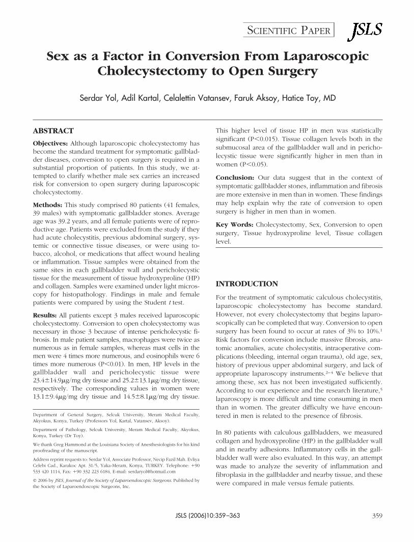
Inflammatory cells were more numerous in the tissuesamples taken from men. Macrophages were twice asnumerous in males compared with macrophages in fe-males. Mast cells were 4 times more numerous, and eo-sinophils were 6 to 7 times more numerous in men. All ofthese differences were statistically significant (P�0.01).No difference existed between the sexes in terms of lym-phocyte count (Figure 1).
HP values in men were 23.4�14.9 �g HP/mg dry tissue inthe gallbladder wall and 25.2�13.1 �g HP/mg dry tissuein pericholecystic adhesions. In women, these valueswere 13.1�9.4 �g HP/mg dry tissue in gallbladder walland 14.5�8.1 �g HP/mg dry tissue in adhesions. Thedifference in HP values between male and female patientswas found to be significant (P�0.015).
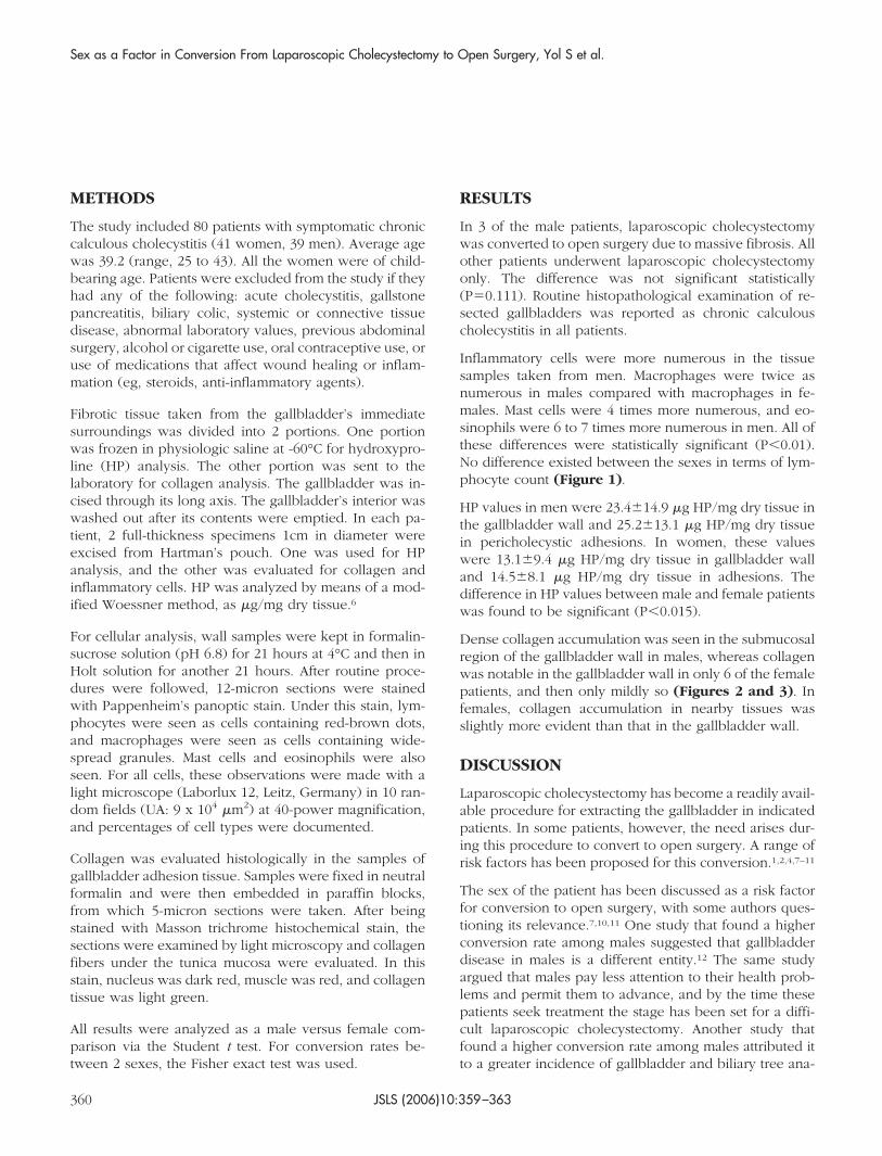
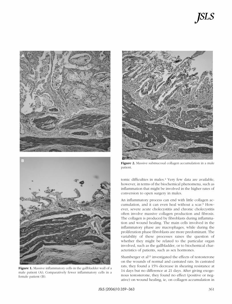
Dense collagen accumulation was seen in the submucosalregion of the gallbladder wall in males, whereas collagenwas notable in the gallbladder wall in only 6 of the femalepatients, and then only mildly so (Figures 2 and 3). Infemales, collagen accumulation in nearby tissues wasslightly more evident than that in the gallbladder wall.
DISCUSSION
Laparoscopic cholecystectomy has become a readily avail-able procedure for extracting the gallbladder in indicatedpatients. In some patients, however, the need arises dur-ing this procedure to convert to open surgery. A range ofrisk factors has been proposed for this conversion.1,2,4,7–11
The sex of the patient has been discussed as a risk factorfor conversion to open surgery, with some authors ques-tioning its relevance.7,10,11 One study that found a higherconversion rate among males suggested that gallbladderdisease in males is a different entity.12 The same studyargued that males pay less attention to their health prob-lems and permit them to advance, and by the time thesepatients seek treatment the stage has been set for a diffi-cult laparoscopic cholecystectomy. Another study thatfound a higher conversion rate among males attributed itto a greater incidence of gallbladder and biliary tree ana-
Sex as a Factor in Conversion From Laparoscopic Cholecystectomy to Open Surgery, Yol S et al.
JSLS (2006)10:359–363360
tomic difficulties in males.4 Very few data are available,however, in terms of the biochemical phenomena, such asinflammation that might be involved in the higher rates ofconversion to open surgery in males.
An inflammatory process can end with little collagen ac-cumulation, and it can even heal without a scar.9 How-ever, severe acute cholecystitis and chronic cholecystitisoften involve massive collagen production and fibrosis.The collagen is produced by fibroblasts during inflamma-tion and wound healing. The main cells involved in theinflammatory phase are macrophages, while during theproliferation phase fibroblasts are more predominant. Thevariability of these processes raises the question ofwhether they might be related to the particular organinvolved, such as the gallbladder, or to biochemical char-acteristics of patients, such as sex hormones.
Shamberger et al13 investigated the effects of testosteroneon the wounds of normal and castrated rats. In castratedrats, they found a 15% decrease in shearing resistance at14 days but no difference at 21 days. After giving exoge-nous testosterone, they found no effect (positive or neg-ative) on wound healing, ie, on collagen accumulation in
Figure 1. Massive inflammatory cells in the gallbladder wall of amale patient (A); Comparatively fewer inflammatory cells in afemale patient (B).
Figure 2. Massive submucosal collagen accumulation in a malepatient.
JSLS (2006)10:359–363 361
normal and castrated rats. Few studies have investigatedeffects of testosterone on wound healing, but a goodnumber of studies have examined the effects of testoster-one on trauma. Whichman et al14 found that after trauma-tization and bleeding of castrated rats, a significant de-crease occurred in synthesis of IL-1 and IL-6 bymacrophages. Angele et al15 induced hemorrhagic shockin male rats that had been given the testosterone antago-nist flutamide, resuscitated them, and then introducedsepsis. They found that immune suppression was pre-vented, and that death due to sepsis was decreased. Thiswas corroborated by a prospective study in humans diag-nosed with sepsis,16 which found a relation between sexhormone levels, inflammatory mediators, and a betterprognosis in women.
Connective tissue and autoimmune diseases like sclero-derma, lupus erythematosus,17 and rheumatoid arthritisare more prevalent in females of reproductive age than inpostmenopausal females or in men. These diseases de-crease in severity when estrogen levels are high.5,18–21 It isalso interesting that estrogen receptors are expressed bymacrophages, monocytes, lymphocytes, and mast cells,
and it is accepted that estrogen directly affects their func-tion and suppresses their cytokine production.8,9,18,22,23
Estrogen replacement has been shown to inhibit perito-neal adhesions in ovariectomized mice, and appears toinhibit macrophage accumulation as well.20 Macrophagesin mice produce fewer cytokines (IL-1, IL-6, and TNFalpha) when exposed to 17-beta estradiol,24 and estrogenalso appears to regulate mRNA expression in fibroblasts.22
The links between sex hormones and inflammation sug-gested by the studies above are consistent with the find-ings of a retrospective study by Kanaan et al,8 whichfound severe inflammation and male sex to be risk factorsfor conversion from laparoscopic to open cholecystec-tomy. In that study, the pericholecystic tissue of inflamedgallbladder showed more severe fibrosis in male patients,and this was associated with difficult dissection and ahigher rate of conversion to open surgery.
The higher levels of collagen and HP found in the samplesfrom male patients in this study suggest a difference in thecellular processes of inflammation. The higher numbers ofmacrophages, mast cells, and eosinophils found in thesamples from male patients corroborate this, and show thedifficulty of trying to pinpoint causality in something ascomplex as the inflammation cascade. The increasednumber of macrophages in men indicates the severity ofinflammation and high levels of cytokines. The cytokinesmediate interactions between macrophages and fibro-blasts in the next phase of the inflammatory process. Moreactive fibroblasts mean more collagen and fibrosis. Mastcells release histamine and chemotactic factors for neutro-phils and eosinophils, thereby expanding the cascade ofcell types involved in inflammation.
Although the rate of conversion to open cholecystectomywas clearly higher in men than in women of reproductiveage, the difference was not statistically significant proba-bly due to the sample size. Also it is unknown whethersuch a difference may exist between postmenopausalwomen and men of comparable age. This is an interestingquestion for further study. If inflammation and fibrosis ingallbladder patients are related to the biochemical aspectsof being male or female, then less of a difference wouldbe expected. However, aging alone is a risk factor forconversion to open surgery, and if we add other systemicdiseases and medications, a study capable of addressingthis question would be difficult to perform. The presentstudy is part of an initiative in our department towardaddressing these broader questions.
Figure 3. Virtual absence of collagen accumulation in a female.
Sex as a Factor in Conversion From Laparoscopic Cholecystectomy to Open Surgery, Yol S et al.
JSLS (2006)10:359–363362
CONCLUSION
The results of this study suggest that in men with symp-tomatic gallbladder stones, inflammation and fibrosis oc-cur more than they do in women with the same disease.This translates into difficult dissections during laparo-scopic cholecystectomy and a higher rate of conversion toopen surgery in male patients.
References:
1. Sanabria JR, Gallinger S, Croxford R, Strasberg SM. Riskfactors in elective laparoscopic cholecystectomy for conversionto open cholecystectomy. J Am Coll Surg. 1994;179:696–704.
2. Lo CM, Fan ST, Liu CL, Lai EC, Wong J. Early decision forconversion of laparoscopic to open cholecystectomy for treat-ment of acute cholecystitis. Am J Surg. 1997;173:513–517.
3. Vatansev C, Kartal A, Calayan O, Vatansev H, Yol S, Tekin A.Why is the conversion rate to open surgery during cholecystec-tomy higher in men than in women? Proceedings of the TurkishNational Surgery Congress, Turkey 2002;p. 177.
4. Zisman A, Gold-Deutch R, Zisman E, Negri M, Halpern Z, LinG, Halevy A. Is male gender a risk factor for conversion oflaparoscopic into open cholecystectomy? Surg Endosc. 1996;10:892–894.
5. Gharaibeh KI, Qasaimeh GR, Al-Heiss H, et al. Effect oftiming of surgery, type of inflammation, and sex on outcome oflaparoscopic cholecystectomy for acute cholecystitis. J Lapa-roendosc Adv Surg Tech A. 2002;12:193–198.
6. Woessner JF Jr. The determination of hydroxyproline intissue and protein samples containing small proportions of thisamino acid. Arch Biochem Biophys. 1961;93:440–447.
7. Alponat A, Kum CK, Koh BC, Rajnakova A, Goh PMY,Mouiel J. Predictive factors for conversions of laparoscopic cho-lecystectomy. World J Surg. 1997;21(6):629–633.
8. Kanaan SA, Murayama KM, et al. Risk factors for conversionof laparoscopic to open cholecystectomy. J Surg Res. 2002;106:20–24.
9. Kartal A, Aksoy F, Vatansev C, et al. Does estrogen causelow conversion rates in laparoscopic cholecystectomies for acuteand chronic cholecystitis in women? JSLS. 2001;5:309–12.
10. Liu CL, Fan ST, Lai EC, Lo CM, Chu KM. Factors affectingconversion of laparoscopic cholecystectomy to open surgery.Arch Surg. 1996;131:98–101.
11. Wiebke EA, Pruitt AL, Howard TJ, et al. Conversion oflaparoscopic to open cholecystectomy. An analysis of risk fac-tors. Surg Endosc. 1996;10:742–745.
12. Russell JC, Walsh SJ, Reed-Fourquet L, Mattie A, Lynch J.Symptomatic cholelithiasis: a different disease in men? Connect-
icut Laparoscopic Cholesystectomy Registry. Ann Surg. 1998;227:195–200.
13. Shamberger RC, Thistlethwaite PA, Thibault LE, Talbot TL,Brennan MF. The effect of testosterone propionate on woundhealing in normal and castrate rats. J Surg Res. 1982;33:58–68.
14. Wichmann MW, Ayala A, Chaudry IH. Male sex steroids areresponsible for depressing macrophage immune function aftertrauma-hemorrhage. Am J Physiol. 1997;273:C1335–C1340.
15. Angele MK, Wichmann MW, Ayala A, Cioffi WG, ChaudryIH. Testosterone receptor blockade after hemorrhage in males.Arch Surg. 1997;132:1207–1214.
16. Schroder J, Kahlke V, Staubach KH, Zabel P, Stuber F.Gender differences in human sepsis. Arch Surg. 1998;133:1200–1205.
17. Inman RD. Immunologic sex differences and the femalepredominance in systemic lupus erythematosus. ArthritisRheum. 1978;21:849–852.
18. Frazier-Jessen MR, Kovacs EJ. Abdominal wall thickness as ameans of assessing peritoneal fibrosis in mice. J Immunol Meth-ods. 1993;162:115–121.
19. Frazier-Jessen MR, Kovacs EJ. Estrogen modulation of JE/monocyte chemoattractant protein-1 mRNA expression in mu-rine macrophages. J Immunol. 1995;154:1838–1845.
20. Frazier-Jessen MR, Mott FJ, Witte PL, Kovacs EJ. Estrogensuppression of connective tissue deposition in a murine modelof peritoneal adhesion formation. J Immunol. 1996;156(8):3036–3042.
21. Hudson I, Hopwood D. Macrophages and mast cells inchronic cholecystitis and “normal” gallbladders. J Clin Pathol.1986;39:1082–1087.
22. Kovacs EJ, Faunce DE, Ramer-Quinn DS, Mott FJ, Dy PW,Frazier-Jessen MR. Estrogen regulation of JE/MCP-1 mRNA ex-pression in fibroblasts. J Leukoc Biol. 1996;59:562–568.
23. Weusten JJ, Blankenstein MA, Gmelig-Meyling FH, Schuur-man HJ, Kater L, Thijssen JH. Presence of oestrogen receptors inhuman blood mononuclear cells and thymocytes. Acta Endocri-nol (Copenh). 1986;112:409–414.
24. Deshpande R, Khalili H, Pergolizzi RG, Michael SD, ChangMD. Estradiol down-regulates LPS-induced cytokine productionand NFkB activation in murine macrophages. Am J Reprod Im-munol. 1997;38:46–54.
JSLS (2006)10:359–363 363