Embed Size (px)
Citation preview
CASE REPORT infectious mononucleosis, complications; neuritis, optic, causes
Severe Optic Neuritis in Infectious Mononucleosis
Because the presentation and clinical features of infectious mononucleosis can be misleading in the elderly, a significant number of infections may go unrecognized. We report an unusual case of infectious mononucleosis in a 61-year-old man in whom marked visual impairment was the presenting complaint and severe optic neuritis was the only prominent finding. Confir- mation of the diagnosis was made by serologic testing for Epstein-Barr virus antibody Recovery of visual function was near complete, but optic atrophy persisted. We reviewed the English literature and collected seven cases of well-documented optic neuritis associated with infectious mononucleosis. A clinical profile of parainfectious optic neuritis is ddiscussed along with the likely pathogenesis for this complication. [Jones J, Gardner W, Newman T: Severe optic neuritis in infectious mononucleosis. Ann Emerg Med April I988;17:361-364.]
INTRODUCTION Infectious mononucleosis is a benign lymphoproliferative disorder that
typically affects young adults. 1 Most individuals more than 50 years old show evidence of previous infection with the Epstein-Barr virus (EBV) and are thus immune to any subsequent exogenous reinfection. 2 When the infec- tion does occur in susceptible older persons, they often have more severe illnesses and complications.g, 4 A significant number of these cases go un- recognized, not only because infectious mononucleosis is considered to be a disease of the young, but also because its presentation can be misleading3,4, 5
The clinical features of classic infectious mononucleosis have been well described, but only isolated reports have dealt with this disease in the geri- atric patient3, 3-6 All of these patients presented with nonspecific constitu- tional symptoms and rarely had pharyngitis, cervical adenopathy, or splenomegaly. Infectious mononucleosis was not initially considered in these patients, because of their age, until atypical lymphocytes were detected on the peripheral blood smear. Heterophil antibody titers confirmed the diag- nosis.
Not infrequently, a complication is the only clinical manifestation of in- fectious mononucleosis in the geriatric patient, s Single symptoms may so dominate the disease picture as to result in a wrong preliminary diagnosis, such as nephritis, hepatitis, or meningitis.7 In such cases, the emergency physician must be aware of the number of complications of the disease to obtain the hematologic and serologic evidence necessary for the diagnosis.
Accordingly, we report a case of infectious mononucleosis in which marked visual impairment was the presenting complaint and moderately se- vere optic neuritis was the only prominent finding. A clinical profile of para- infectious optic neuritis is discussed along with the proposed pathogenesis for this rare complication.
CASE REPORT A 61-year-old man presented with bilateral eye discomfort and blurred vi-
sion of two days duration. Both eyes were affected equally and he had no symptoms of scotoma, diplopia, or difficulty with color vision. The patient had been well until five days before, when he developed an intermittent frontal headache, progressive fatigue, and a low-grade fever of 38.1 C. He had no history of head trauma, mental status changes, neck stiffness, pharyngitis,
Jeffrey Jones, MD William Gardner, MD Tim Newman, MD Akron, Ohio
From the Department of Emergency Medicine and Infectious Disease, Akron General Medical Center, Northeastern Ohio Universities, College of Medicine, Akron, Ohio.
Received for publication April 6, 1987. Revision received November 16, 1987. Accepted for publication January 19, 1988.
Address for reprints: Jeffrey Jones, MD, Akron General Medical Center, Department of Emergency Medicine, 400 Wabash Avenue, Akron, Ohio 44307.
17:4 April 1988 Annals of Emergency Medicine 361/113
OPTIC NEURITIS Jones, Gardner & Newman
or adenopathy. He denied drug inges- tion.
Physical examinat ion revealed a temperature of 36.6 C; blood pressure, 114/70 mm Hg; pulse, 84; and respira- tions, 16. Visual acuity was 20/200 in the right eye and 20/100 in the left eye. Visual fields, color perception, and moti l i ty were normal. Fundu- scopic examination showed slight blurring of disc margins. The remain- ing cranial nerves were normal. Senso- ry, motor, and autonomic systems were normal. The neck was not rigid. The patient's oropharynx showed no evidence of infection. He had no lymphadenopathy, hepatomegaly, or splenomegaly. Jaundice or skin lesions were not evident.
Laboratory values included: erythro- cyte sedimentation rate, 24 mm in first hour; hemoglobin, 14.8 g/dL; leu- kocytes, 10,000/ram 3 (with 29% neu- trophils , 37% lymphocy tes , 25% atypical lymphocytes, and 9% mono- cytes); total serum bilirubin, 0.8 mg/ dL (normal, 0.2 to 0.8 mg/dL); alkaline phosphatase, 472 U/L (normal, < 55 U/L); SGOT, 129 U/L (normal, < 41 U/ L); SGPT, 222 U/L (normal, < 45 U/L); lactate dehydrogenase, 376 U/L (nor- mal, < 200 U/L); and a negative hepa- titis panel. Results were positive from a differential slide test for the hetero- phil antibody to EBV (monospot}, and antibody titer to EBV capsid antigen (EBVCA) was 1:40. A computerized to- mographic scan of the brain was nor- mal.
Two weeks after the onset of fever, visual acuity had decreased to count- ing fingers in the right eye and 20/400 in the left. Each optic disc was swollen with central pallor. Visual fields and motility remained intact. The remainder of the physical exam- ination was normal. Oral prednisone therapy (100 mg/day) was begun, and the dosage was tapered over six weeks. Visual acui ty improved gradually, being 20/40 in the right eye and 20/20 in the left eye two months later. There was a residual pallor of the optic discs bilaterally. Liver function tests, leuko- cyte count, and differential had re- turned to normal.
DISCUSSION Optic neuritis is a broad term de-
noting any inflammation, degenera- tion, or demyelinization of the optic nerve. 8 Loss of vision is the cardinal symptom and serves to differentiate optic neur i t i s f rom papi l ledema,
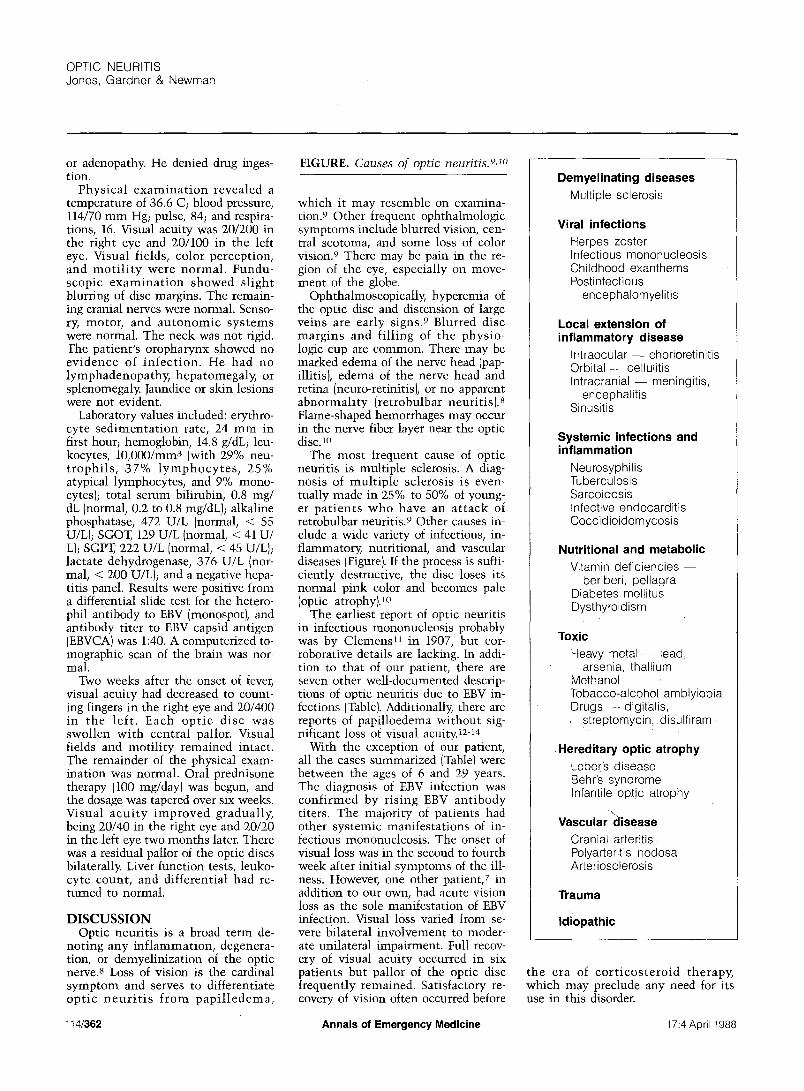
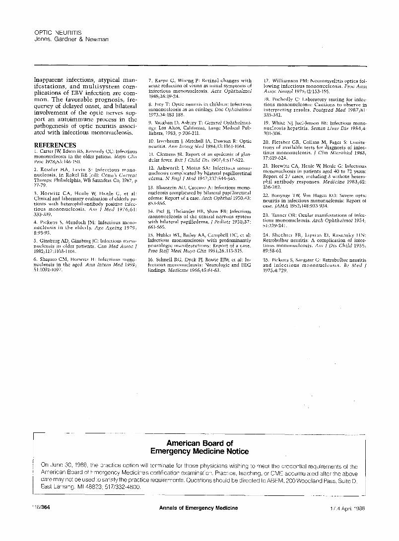
FIGURE. Causes of optic neuritis.9, lo
which it may resemble on examina- tion. 9 Other frequent ophthalmologic symptoms include blurred vision, cen- tral scotoma, and some loss of color vision. 9 There may be pain in the re- gion of the eye, especially on move- ment of the globe.
Ophthalmoscopically, hyperemia of the optic disc and distension of large veins are early signs. 9 Blurred disc margins and filling of the physio- logic cup are common. There may be marked edema of the nerve head (pap- illitis), edema of the nerve head and retina (neuro-retinitis), or no apparent abnormality (retrobulbar neuritis). 8 Flame-shaped hemorrhages may occur in the nerve fiber layer near the optic disc. 10
The most frequent cause of optic neuritis is multiple sclerosis. A diag- nosis of multiple sclerosis is even- tually made in 25% to 50% of young- er patients who have an attack of retrobulbar neuritis. 9 Other causes in- clude a wide variety of infectious, in- flammatory, nutritional, and vascular diseases (Figure). If the process is suffi- ciently destructive, the disc loses its normal pink color and becomes pale (optic atrophy)30
The earliest report of optic neuritis in infectious mononucleosis probably was by Clemens 11 in 1907, but cor- roborative details are lacking. In addi- tion to that of our patient, there are seven other well-documented descrip- tions of optic neuritis due to EBV in- fections (Table}. Additionally, there are reports of papilloedema without sig- nificant loss of visual acuity. 12-14
With the exception of our patient, all the cases summarized (Table) were between the ages of 6 and 29 years. The diagnosis of EBV infection was confirmed by rising EBV antibody titers. The majority of patients had other systemic manifestations of in- fectious mononucleosis. The onset of visual loss was in the second to fourth week after initial symptoms of the ill- ness. However, one other patient, 7 in addition to our own, had acute vision loss as the sole manifestation of EBV infection. Visual 10ss varied from se- vere bilateral involvement to moder- ate unilateral impairment. Full recov- ery of visual acuity occurred in six patients but pallor of the optic disc frequently remained. Satisfactory re- covery of vision often occurred before
Demyelinating diseases Multiple sclerosis
Viral infections Herpes zoster Infectious mononucleosis Childhood exanthems Postinfectious
encephalomyelitis
Local extension of inflammatory disease
In t raocular- chorioretinitis Orbital - - cellulitis Intracranial - meningitis,
encephalitis Sinusitis
Systemic infections and inflammation
Neurosyphilis Tuberculosis Sarcoidosis Infective endocarditis Coccidioidomycosis
Nutritional and metabolic Vitamin def ic ienc ies-
beriberi, pellagra Diabetes mellitus Dysthyroidism
Toxic Heavy metal ~--lead,
arsenia, thallium Methanol Tobacco-alcohol amblyiopia Drugs - - digitalis,
streptomycin,_ disulfiram
Hereditary optic atrophy Leber's disease Behr's syndrome Infantile optic atrophy
Vascular disease Cranial arteritis Polyarteritis nodosa Arteriosclerosis
Trauma
Idiopathic
the era of cor t icosteroid therapy, which may preclude any need for its use in this disorder.
114/362 Annals of Emergency Medicine 17:4 April 1988
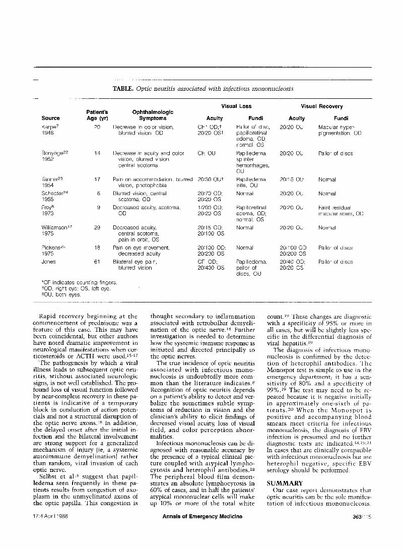
TABLE. Optic neuritis associated with infectious mononucleosis
Visual Loss Patient's
Source Age (yr)
Karpe 7 20 1948
Ophthalmologic Symptoms Acuity
Decrease in color vision, CF,* OD;t blurred vision, ©D 20/20 OSt
Bonynge 22 14 1952
Decrease in acuity and color vision, blurred vision, central scotoma
Tanner 23 17 1954 Schecter 24 6 1955 F~y 8 9 1973
CF, OU
Williamson 17 29 1975
Pickens 25 18 1975
Jones 61
Pain on accommodation, blurred 20/30 OU* vision, photophobia
Blurred vision, central 20/70 OD; scotoma, OD 20/20 OS
Decreased acuity, scotoma, 1/200 OD; OD 20/20 OS
*CF indicates counting fingers. rOD, right eye; OS, left eye. ¢OU, both eyes.
Decreased acuity, 20/15 OD; central scotoma, 20/100 OS pain in orbit, OS
Pain on eye movement, 20/100 OD; decreased acuity 20/200 OS
Bilateral eye pain, CF, OD; blurred vision 20/400 OS
Fundi
Pallor of disc, papilloretinal edema, OD; normal, OS
Papilledema, splinter hemorrhages, OU
Papilledema, iritis, OU Normal
Papilloretina[ edema, OD; normal, OS
Normal
Normal
Papilledema, pallor of discs, OU
Visual Recovery
Acuity Fundi
20/20 OU Macular hyper- pigmentation, OD
20/20 OU Pallor of discs
20/15 OU Normal
20/20 OU Normal
20/20 OU Faint residual macular scars, OD
20/20 OU Normal
20/100 OD; Pallor of discs 20/2O00S
20/40 OD; Pallor of discs 2O/20 OS
Rapid r ecove ry b e g i n n i n g at the c o m m e n c e m e n t of prednisone was a feature of this case. This may have been coincidental , but o ther authors have noted dramatic improvement in neurological manifestat ions when cor- t icosteroids or ACTH were used. lsq7
T h e pathogenesis by which a viral il lness leads to subsequent optic neu- r i t is , w i t h o u t a s soc ia ted neuro log ic signs, is not well established. The pro- found loss of visual function followed by near-complete recovery in these pa- t i en t s is i nd ica t ive of a t e m p o r a r y block in conduct ion of action poten- t ials and not a structural disruption of the optic nerve a x o n s ) 8 In addit ion, the delayed onset after the ini t ial in- fection and the bilateral involvement a r e strong support for a general ized m e c h a n i s m of injury (ie, a sys temic a u t o i m m u n e demye l ina t i on ) ra ther than random, viral invasion of each optic nerve.
Selbst et al t8 suggest t ha t papi l - l edema seen f requent ly in these pa- t ients results from congestion of axo- p lasm in the unmye l ina t ed axons of the optic papilla. This congest ion is
t hough t s econda ry to i n f l a m m a t i o n associated wi th re t robulbar demyel i - na t ion of the optic nerve. 18 Further inves t igat ion is needed to de te rmine how the systemic immune response is in i t ia ted and directed pr incipal ly to the optic nerves.
The true incidence of optic neuri t is a s s o c i a t e d w i t h i n f e c t i o u s m o n o - nucleosis is undoubted ly more com- m o n than the l i t e r a tu re ind ica tes . 8 Recognition of optic neuri t is depends on a pat ient 's abil i ty to detect and ver- bal ize the s o m e t i m e s sub t l e symp- toms of reduct ion in vis ion and the clinician's abil i ty to elicit findings of decreased visual acuity, loss of visual f ie ld , and co lo r p e r c e p t i o n abnor - malit ies.
Infectious mononucleosis can be di- agnosed wi th reasonable accuracy by the presence of a typical clinical pic- ture coupled w i t h a typ ica l l y m p h o - cytos is and he t e roph i l a n t i b o d i e s ) s T h e p e r i p h e r a l b lood f i lm d e m o n - strates an absolute lymphocy tos i s in 60% of cases, and in half the patients ' atypical mononuclear cells wil l make up 10% or more of the total whi te
count. 19 These changes are diagnostic wi th a specificity of 95% or more in all cases, but wil l be slightly less spe- cific in the d i f fe ren t ia l d iagnosis of viral hepa t i t i s ) 9
The diagnosis of infect ious mono- nucleosis is conf i rmed by the detec- t i on of h e t e r o p h i l an t i bod i e s . The Monospot test is simple to use in the emergency department; it has a sen- s i t iv i ty of 80% and a spec i f i c i ty of 99%. 19 The tes t may need to be re- peated because it is negative ini t ial ly in a p p r o x i m a t e l y o n e - s i x t h of pa- t i e n t s . 20 W h e n t h e M o n o s p o t i s p o s i t i v e and a c c o m p a n y i n g b l o o d smears m e e t c r i t e r ia for in fec t ious mononucleosis , the diagnosis of EBV infection is presumed and no further diagnost ic tests are indicated.Is,19, 21 In cases that are clinically compatible wi th infectious mononucleosis but are h e t e r o p h i l n e g a t i v e , spec i f i c EBV serology should be performed.
S U M M A R Y Our case report demons t ra tes that
optic neuri t is can be the sole manifes- t a t ion of in fec t ious m o n o n u c l e o s i s .
17:4 April 1988 Annals of Emergency Medicine 363/115
OPTIC NEURITIS Jones, Gardner & Newman
Inapparent infections, atypical man- ifestations, and mul t i sys tem com- plications of EBV infection are com- mon, The favorable prognosis, fre- quency of delayed onset, and bilateral involvement of the optic nerves sup- port an autoimmune process in the pathogenesis of optic neuritis associ- ated with infectious mononucleosis.
REFERENCES 1. Carter JW, Edson RS, Kennedy CC: Infectious mononucleosis in the older patient. Mayo Clin Proc 1978;53:146-150.
2. Kessler HA, Levin S: Infectious mono- nucleosis, in Rakel RE (ed): Conn's Current Therapy. Philadelphia, WB Saunders Co, 1987, p 77-79.
3. Horwitz CA, Henle W, Henle G, et al: Clinical and laboratory evaluation of elderly pa- tients with heterophil-antibody positive infec- tious mononucleosis . Am J Med 1976;61: 333-339.
4. Pickens S, Murdoch JM: Infectious mono- nucleosis in the elderly. Age Ageing 1979; 8:93-95.
5. Ginsburg AD, Ginsburg JC: Infectious mono- nucleosis in older patients. Can Med Assoc J 1982;127:1103-1104.
6. Shapiro CM, Horwitz H: Infectious mono- nucleosis in the aged. Ann Intern Med 1959; 51:1092-1097.
7. Karpe G, Wising P: Retinal changes with acute reduction of vision as initial symptoms of infectious mononucleosis. Acta Ophthalmol 1948;26:19-24.
8. Frey T: Optic neuritis in children: Infectious mononucleosis as an etiology. Doc Ophthalmol 1973;34:183-188.
9. Vaughan D, Asbury T: General Ophthalmol- ogy. Los Altos, California, Lange Medical Pub- lishers, 1983, p 206-211.
10. Javerbaum J, Mitchell JA, Dawson R: Optic neuritis. Ann Emerg Med 1984;13:1061-1064.
11. Clemens JR: Report of an epidemic of glan- dular fever. Brit J Child Dis 1907;4:517-522.
12. Ashworth J, Motto SA: Infectious mono- nucleosis complicated by bilateral papilloretinal edema. N Engl J Med 1947;237:544-545.
13. Blaustein ALl, Caccavo A: Infectious mono- nucleosis complicated by bilateral papilloretinal edema: Report of a case. Arch Ophthal 1950;43: 853-856.
14. Piel JJ, Thelander HE, Shaw EB: Infectious mononucleosis of the central nervous system with bilateral papilledema. J Pediatr 1950;37: 661-665.
15. Hubler WL, Bailey AA, Campbell DC, et al: Infectious mononucleosis with predominantly neurologic manifestations: Report of a case. Proc Staff Meet Mayo Clin 1951;26:313-315.
16. Schnell RG, Dyck PJ, Bowie EJW, et al: In- fectious mononucleosis: Neurologic and EEG findings. Medicine 1966;45:61-63.
17. Williamson PM: Neuromyelitis optica fol- lowing infectious mononucleosis. Proc Aust Assoc Neurol 1975;12:153-155.
t8. Pochedly C: Laboratory testing for infec- tious mononucleosis: Cautions to observe in interpreting results. Postgrad Med 1987;81: 335-342.
19. White NJ, Juel-Jenson BE: Infectious mono- nucleosis hepatitis. Semin Liver Dis 1984;4: 301-306.
20. Fleisher GR, Collins M, Fager S: Limita- tions of available tests for diagnosis of infec- tious mononucleosis. J Clin Microbiol 1983; 17:619-624.
21. Horwitz CA, Henle W, Henle G: Infectious mononucleosis in patients aged 40 to 72 years: Report of 27 cases, including 3 without hetero- phii-antibody responses. Medicine 1983;62: 256-262.
22. Bonynge TW, Von Hagen KO: Severe optic neuritis in infectious mononucleosis: Report of case. [AMA 1952;148:933-934.
23. Tanner OR: Ocular manifestations of infec- tious mononucleosis. Arch Ophthalmol 1954; 51:229-241.
24. Shechter FR, Lipsius EI, Rasansky HN: Retrobulbar neuritis: A complication of infec- tious mononucleosis. Am J Dis Child 1955; 89:58-61.
25. Pickens S, Sangster G: Retrobulbar neuritis and infec t ious mononuc leos i s . Br Med ] 1975;4: 729.
American Board of Emergency Medicine Notice
On June 30, 1988, the practice option will terminate for those physicians wishing to meet the credential requ=rements of the American Board of Emergency Medicine's certification examination. Practice, teaching, or CME accumulated after the above date may not be used to satisfy the practice requirements. Questions should be directed to ABEM, 200 Woodland Pass, Suite D, East Lansing, MI 48823; 517/332-4800.
116/364 Annals of Emergency Medicine 17:4 April 1988