Embed Size (px)
Citation preview

Severe human infection with a novel avian-origin influenza A(H7N4) virusSevere human infection with a novel avian-origin influenza A(H7N4) virus
Xiang Huo, Lun-biao Cui, Cong Chen, Dayan Wang, Xian Qi, Ming-hao Zhou, Xiling Guo, Fengming Wang, William J. Liu, Weirong Kong, Daxin Ni, Ying Chi,Yiyue Ge, Haodi Huang, Feifei Hu, Chao Li, Xiang Zhao, Ruiqi Ren and Feng-Cai Zhu
Citation: Science Bulletin 6363, 1043 (2018); doi: 10.1016/j.scib.2018.07.003
View online: http://engine.scichina.com/doi/10.1016/j.scib.2018.07.003
View Table of Contents: http://engine.scichina.com/publisher/scp/journal/SB/63/16
Published by the Science China Press
Articles you may be interested inArticles you may be interested in
Environmental connections of novel avian-origin H7N9 influenza virus infection and virus adaptation to the humanSCIENCE CHINA Life Sciences 5656, 485 (2013);
Lessons learnt from the human infections of avian-origin influenza A H7N9 virus: Live free markets and human healthSCIENCE CHINA Life Sciences 5656, 493 (2013);
Human avian influenza A (H5N1) virus infection in ChinaScience in China Series C-Life Sciences 5252, 407 (2009);
Research progress in human infection with avian influenza H7N9 virusSCIENCE CHINA Life Sciences 6060, 1299 (2017);
Human infection with a further evolved avian H9N2 influenza A virus in Sichuan, ChinaSCIENCE CHINA Life Sciences 6161, 604 (2018);
Article
Severe human infection with a novel avian-origin influenza A(H7N4) virus
Xiang Huo a,h,1, Lun-biao Cui a,h,1, Cong Chen d,1, Dayan Wang c, Xian Qi a,h, Ming-hao Zhou a, Xiling Guo a,Fengming Wang d, William J. Liu c, Weirong Kong e, Daxin Ni b, Ying Chi a, Yiyue Ge a, Haodi Huang a,Feifei Hu d, Chao Li b, Xiang Zhao c, Ruiqi Ren b, Chang-jun Bao a,h,⇑, George F. Gao b,c,f,g,⇑, Feng-Cai Zhu a,h,⇑aKey Laboratories of Enteric Pathogenic Microbiology (Ministry of Health), Jiangsu Provincial Center for Disease Control and Prevention, Nanjing 210009, ChinabChinese Center for Disease Control and Prevention (China CDC), Beijing 102206, ChinacNational Institute for Viral Disease Control and Prevention, China CDC, Beijing 102206, ChinadChangzhou Center for Disease Control and Prevention, Changzhou 213022, Chinae Liyang Center for Disease Control and Prevention, Liyang 213300, ChinafCAS Key Laboratory of Pathogenic Microbiology and Immunology, Institute of Microbiology, Chinese Academy of Sciences (CAS), Beijing 100101, ChinagCenter for Influenza Research and Early-Warning (CASCIRE), Chinese Academy of Sciences, Beijing 100101, ChinahKey Laboratory of Infectious Diseases, School of Public Health, Nanjing Medical University, Nanjing 211166, China
a r t i c l e i n f o
Article history:Received 27 June 2018Received in revised form 29 June 2018Accepted 29 June 2018Available online 19 July 2018
Keywords:Avian influenza virus (AIV)Human infectionH7N4EpidemiologyPneumonia
a b s t r a c t
Human infections with influenza H7 subtypes, such as H7N9, have raised concerns worldwide. Here, wereport a human infection with a novel influenza A(H7N4) virus. A 68 years-old woman with cardiovascu-lar and cholecystic comorbidities developed rapidly progressed pneumonia with influenza-like-illness asinitial symptom, recovered after 23 days-hospitalization including 8 days in ICU. Laboratory indicatorsfor liver and blood coagulation dysfunction were observed. Oseltamivir phosphate, glucocorticoids andantibiotics were jointly implemented, with nasal catheterization of oxygen inhalation for this patient.We obtained the medical records and collected serial respiratory and blood specimens from her. We col-lected throat, cloacal and/or feces samples of poultry and wild birds from the patient’s backyard, neigh-borhood, local live poultry markets (LPMs) and the nearest lake. All close contacts of the patient werefollowed up and sampled with throat swabs and sera. Influenza viruses and other respiratory pathogenswere tested by real-time RT-PCR, viral culturing and/or sequencing for human respiratory and bird sam-ples. Micro-neutralizing assay was performed for sera. A novel reassortant wild bird-origin H7N4 virus isidentified from the patient and her backyard poultry (chickens and ducks) by sequencing, which is dis-tinct from previously-reported avian H7N4 and H7N9 viruses. At least four folds increase of neutralizingantibodies to H7N4 was detected in her convalescent sera. No samples from close contacts, wild birds orother poultry were tested positive for H7N4 by real-time RT-PCR.
� 2018 Science China Press. Published by Elsevier B.V. and Science China Press. All rights reserved.
1. Introduction
Avian influenza virus (AIV) H7N9 has raised global concernssince human infections of low pathogenic (LP) and highly patho-genic (HP) H7N9 viruses emerged in 2013 and 2017, respectively[1,2]. The virus has caused five waves of virulent human infectionsin China Mainland [3], and cases have been exported to other dis-tricts in Asia and North America [4–6], with a huge economic loss[7,8]. The H7 subtype HA gene has been found in combination withall nine NA subtype genes [9]. Other influenza A subtype H7
viruses, such as H7N2, H7N3 and H7N7, were documented beingcapable of infecting human as well [10–13], but usually self-limiting.
Both HP and LP strains of influenza A (H7N4) virus have beenidentified from domestic poultry and wild birds in Australia, Korea,Europe and North America [14–17]. Here, we report a human casewith severe pneumonia infected by a novel wild bird-origin H7N4virus.
2. Methods and materials
2.1. Case identification
A surveillance network on hospitalized pneumonia patients wasestablished in Jiangsu Province, Eastern China since 2012, as a part
https://doi.org/10.1016/j.scib.2018.07.0032095-9273/� 2018 Science China Press. Published by Elsevier B.V. and Science China Press. All rights reserved.
⇑ Corresponding authors.E-mail addresses: [email protected] (C.-j. Bao), [email protected] (G.F. Gao),
[email protected] (F.-C. Zhu).1 Contributed equally.
Science Bulletin 63 (2018) 1043–1050
Contents lists available at ScienceDirect
Science Bulletin
journal homepage: www.elsevier .com/locate /sc ib
Downloaded to IP: 192.168.0.24 On: 2019-01-11 11:04:24 http://engine.scichina.com/doi/10.1016/j.scib.2018.07.003
of the China National Unknown Cause Pneumonia SurveillanceNetwork. Patients admitted to a local hospital with diagnosis ofpneumonia, who have rapid progress but lack of identified patho-genic causes, are required to be sampled and sent to laboratorynetworks for pathogenic analyses, including influenza viruses. Asa practice in China, empirical antiviral treatment (neuraminidaseinhibitor) is recommended for suspected influenza patients beforelaboratory confirmation, in order to acquire the most healthbenefits.
2.2. Epidemiological investigation and specimen collection
We acquired patient’s epidemiological information, such asdemographic characteristics, recent direct or indirect exposuresto poultry or other animals, recent travel history and history ofseasonal influenza vaccination, by a face-to-face interview withthe patient and her family members. Patient’s clinical data,including underlying medical conditions, clinical symptoms andsigns, radiographic examinations, clinical laboratory testingresults, clinical treatment, clinical complications and outcome,were derived from her medical records. Serial respiratory andblood samples were taken at an interval of 1–3 days until thepatient’s recovery.
Close contacts were defined as individuals who had pro-vided care to, had been living with, or had potentially beendirectly exposed to respiratory secretions or body fluids ofthe patient during the time span of one day before the illnessonset to the isolation of the patient. We took respiratory andblood samples from all close contacts, and followed them updaily for any signs or symptoms of respiratory illness till the28th day after their last unprotected contact with the patient.No antiviral chemoprophylaxis was implemented. Those whoshared a similar exposure history with the patient, such ashad been exposed to the same animals or environment duringthe 14 days before illness onset of the patient, were defined asco-exposers.
We collected throat, cloacal and/or feces samples of poultry andwild birds from the patient’s backyard, neighborhood, local livepoultry markets (LPMs) and the nearest lake, for tracing sourceand spread range of the virus.
2.3. Laboratory analyses
2.3.1. Nucleic acid extraction and detectionNucleic acids were extracted using Viral DN&RNA Kit
(Tianlong Bio-Technology Co., Ltd, Suzhou, China), accordingto the manufacturer’s instructions. Commercial real-timeRT-PCR kits for detecting H1 to H16 and N1 to N9 subtypeswere performed to detect the influenza subtypes (JiangsuBioperfectus Technologies Co., LTD, Taizhou, China). In addition,the patient’s throat swabs were tested for 28 other respiratorypathogens (Table S1 online) using laboratory-developed specificreal-time PCR assays (conditions and primers are available onrequest).
Supplementary data associated with this article can be found, inthe online version, at https://doi.org/10.1016/j.scib.2018.07.003.
2.3.2. Virus isolationThroat swabs of the patient, cloacal swabs of chicken and ducks
were collected and maintained in a viral transport medium. Theswabs were propagated in the allantoic sac and amniotic cavityof 9-to-11-day-old specific pathogen-free (SPF) embryonatedchicken eggs for 72 h at 36 �C. The virus titer was established witha haemagglutination (HA) test using turkey red blood cells and wasrecorded as the reciprocal of the highest dilution of the virus thatinduced hemagglutination.
2.3.3. Micro-neutralization assayMicro-neutralization (MN) tests (from titers �40) were per-
formed for patient’s serum samples collected during acute andconvalescent stage of illness course, according to WHO manual[18]. The standard antigen used in the assay was produced fromthe A(H7N4) isolate from the chicken fed by the patient (A/Chicken/Jiangsu/1/2018 H7N4).
2.3.4. Genome sequencing and phylogenetic analysisVirus RNA was reversely transcripted into cDNA and viral gen-
omes were amplified [19,20]. Sequencing was done with the Illu-mina MiSeq platform as previously described [21]. Maximum-likelihood phylogenetic analysis was conducted with RAxML-HPC2 on the Extreme Science and Engineering Discovery Environ-ment (XSEDE) version 8.2.10 with GTR+C model and 1000 boot-straps for all the eight genes of the eight H7N4 influenza virusesby using the top 100 BLAST hits of A/Chicken/Jiangsu/103/2018(H7N4), which has the highest identity with A/Jiangsu/1/2018(H7N4), available from GenBank and Global Initiative on SharingAll Influenza Data (GISAID). Each tree was rooted with the earliestisolate among the hits and was visualized by FigTree v1.4.3. Ofnote, five human H7N9 viruses from 2013 to 2017 were added inthe HA tree.
2.4. Ethic issue
The National Health and Family Planning Commission deemedthe data collection for this case to be part of the continuing publichealth outbreak investigation and exempt from institutionalreview board assessment. The identity information of the casewas unrevealed in this report. The institutional review board ofJiangsu Provincial Center for Disease Control and Prevention(JSCDC) approved the assessment of these close contacts. Writteninformed consent was obtained from the close contacts.
3. Results
3.1. Social-demographic and clinical characteristics of the patient
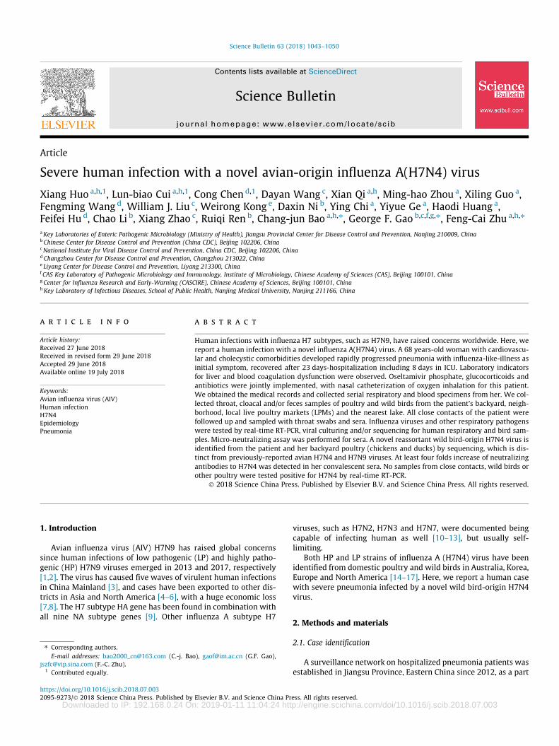
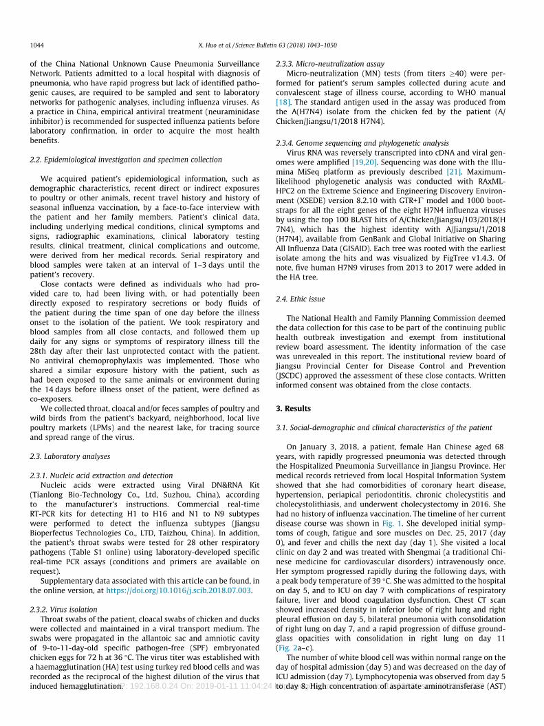
On January 3, 2018, a patient, female Han Chinese aged 68years, with rapidly progressed pneumonia was detected throughthe Hospitalized Pneumonia Surveillance in Jiangsu Province. Hermedical records retrieved from local Hospital Information Systemshowed that she had comorbidities of coronary heart disease,hypertension, periapical periodontitis, chronic cholecystitis andcholecystolithiasis, and underwent cholecystectomy in 2016. Shehad no history of influenza vaccination. The timeline of her currentdisease course was shown in Fig. 1. She developed initial symp-toms of cough, fatigue and sore muscles on Dec. 25, 2017 (day0), and fever and chills the next day (day 1). She visited a localclinic on day 2 and was treated with Shengmai (a traditional Chi-nese medicine for cardiovascular disorders) intravenously once.Her symptom progressed rapidly during the following days, witha peak body temperature of 39 �C. She was admitted to the hospitalon day 5, and to ICU on day 7 with complications of respiratoryfailure, liver and blood coagulation dysfunction. Chest CT scanshowed increased density in inferior lobe of right lung and rightpleural effusion on day 5, bilateral pneumonia with consolidationof right lung on day 7, and a rapid progression of diffuse ground-glass opacities with consolidation in right lung on day 11(Fig. 2a–c).
The number of white blood cell was within normal range on theday of hospital admission (day 5) and was decreased on the day ofICU admission (day 7). Lymphocytopenia was observed from day 5to day 8. High concentration of aspartate aminotransferase (AST)
1044 X. Huo et al. / Science Bulletin 63 (2018) 1043–1050
Downloaded to IP: 192.168.0.24 On: 2019-01-11 11:04:24 http://engine.scichina.com/doi/10.1016/j.scib.2018.07.003
was constantly recorded till day 16. Blood platelet count remaineddecreased until day 10, and D-dimer concentration was elevatedon all tests. Blood concentrations of urea nitrogen and creatininewere normal through all tests, but urine protein was detected onday 6 and day 7. Reduced arterial partial pressure of oxygen wasrecorded the day of admission to hospital prior to the implementa-tion of oxygen inhalation, and remained decreased till day 11(Table 1). Antiviral treatment (Oseltamivir phosphate) was initi-ated empirically on day 5 (75 mg twice a day) and was terminatedafter her discharge from ICU. The dose was doubled during the first4 days of her stay in ICU (Fig. 1). Nasal catheterization of oxygen
inhalation was implemented from the day of admission till her dis-charge from ICU. Glucocorticoids, intravenous albumin andimmune globulin therapy were jointly implemented, with combi-nation of antibiotics for bacteria secondary infection. Methylpred-nisolone sodium succinate was administered from day 5 to day 10.The dose was 20 mg per day for 2 days prior to ICU admission, 40mg twice a day for the first 2 days in ICU and 40 mg per day for thefollowing 2 days. Cefoxitin Sodium (2 g per day) and AzithromycinLactobionate (0.5 g per day) were jointly administered for 2 daysbefore the patient’s ICU admission. Moxifloxacin hydrochloride(0.4 g per day) was implemented during her stay in ICU instead.
Fig. 1. (Color online) Timeline of the clinical course and key indicators of the H7N4 patient.
Fig. 2. (Color online) Chest CT scans of the H7N4 patient. (a–d) Were obtained on day 5, day 7, day 11 and day 21 after illness onset, respectively. a-1, b-1, c-1, d-1: Lungwindow; a-2, b-2, c-2, d-2: Mediastinal window.
X. Huo et al. / Science Bulletin 63 (2018) 1043–1050 1045
Downloaded to IP: 192.168.0.24 On: 2019-01-11 11:04:24 http://engine.scichina.com/doi/10.1016/j.scib.2018.07.003
After that, Cefoperazone Sodium and Sulbactam Sodium (3 g perday) were initiated on day 16 as a substitution, and Linezolid(0.6 g per day) was added the following day. Antibiotics treatmentended on day 24.
Mycoplasma pneumoniae (MP) was cultured from patient’sthroat swab collected on the day of ICU admission (day 7), butthe specific IgM antibody was only weakly positive in patient’sblood specimen obtained on day 8, indicated by indirectimmunofluorescent assay (kits manufactured by VIRCELL, S.L.).During her stay in ICU, patient’s sputum was collected on day 8,9, 12, 13 and 14. From them, Monilia albican was continuously cul-tured and Klebsiella pneumoniae was cultured from the sputumcollected on day 12 only. The patient recovered and was dis-charged from hospital on day 28 (Figs. 1 and 2d).
3.2. Laboratory analyses
Throat swab samples obtained from the patient on day 9 andday 11 were shown to be positive for H7N4, while samplesobtained since day 12 showed negative, by real-time RT-PCR. Allsamples showed negative for other influenza subtypes and 28 res-piratory pathogens, except for herpes simplex virus-1 (HSV-1, pos-itive from day 9 to day 17) and human coronavirus type 229E(HCov 229E, positive from day 20 to day 27) (Table S1). Next gen-eration sequencing (NGS) and genome assembly showed 8 consen-sus gene segments of H7N4 from the throat swab on day 9(designated as A/Jiangsu/1/2018 (H7N4), henceforth, JS1). Theresults were confirmed subsequently by the Chinese NationalInfluenza Center, the WHO Reference Laboratory. Micro-neutralization assay showed that patient’s antibody titer to H7N4was less than 40 on day 9, when she was in ICU with considerableprogress of bilateral pneumonia indicated by CT scan and throatswab positive for H7N4 virus by real-time RT-PCR. On day 12, aneutralizing antibody titer of 40 to H7N4 was detected fromserum, while the patient’s throat swab became negative forH7N4 virus by real-time RT-PCR. The neutralizing antibody titercontinued increasing to 80 on day 14 and to 160 on day 23, which
coincided with the patient’s discharge from ICU and rehabilitationindicated by chest CT scans (Table S2 online). In contrast, HSV-1was detected in patient’s throat swabs even two days after thepatient’s discharge from ICU, and HCov 229E was only detectedduring the patient’s convalescent stage.
As we failed to isolate virus from throat swab of the patient col-lected on the 5th day post antiviral treatment, viral genome wassequenced following influenza virus specific amplification andwas deposited in the GISAID (accession number: EPI1139727-EPI1139730, EPI1139738, EPI1139761, EPI1139780, EPI1139806).Meanwhile, we successfully isolated five and two H7N4 strainsfrom the duck and chicken raised by the patient, respectively,and sequenced the genomes (Fig. 3). Theses viral genomesequences were also deposited in the GISAID (accession number:EPI1139887-EPI1139894, EPI1150038-EPI1150053, EPI1150055 -EPI1150086).
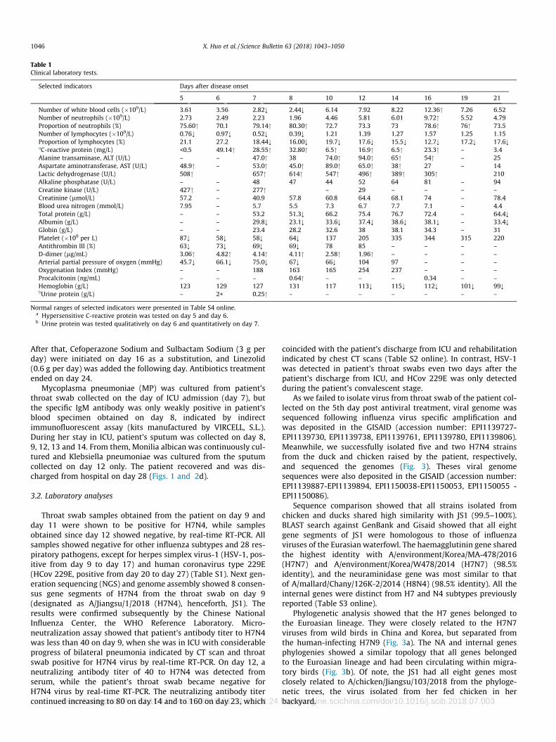
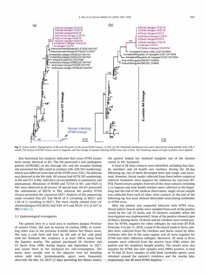
Sequence comparison showed that all strains isolated fromchicken and ducks shared high similarity with JS1 (99.5–100%).BLAST search against GenBank and Gisaid showed that all eightgene segments of JS1 were homologous to those of influenzaviruses of the Eurasian waterfowl. The haemagglutinin gene sharedthe highest identity with A/environment/Korea/MA-478/2016(H7N7) and A/environment/Korea/W478/2014 (H7N7) (98.5%identity), and the neuraminidase gene was most similar to thatof A/mallard/Chany/126K-2/2014 (H8N4) (98.5% identity). All theinternal genes were distinct from H7 and N4 subtypes previouslyreported (Table S3 online).
Phylogenetic analysis showed that the H7 genes belonged tothe Euroasian lineage. They were closely related to the H7N7viruses from wild birds in China and Korea, but separated fromthe human-infecting H7N9 (Fig. 3a). The NA and internal genesphylogenies showed a similar topology that all genes belongedto the Euroasian lineage and had been circulating within migra-tory birds (Fig. 3b). Of note, the JS1 had all eight genes mostclosely related to A/chicken/Jiangsu/103/2018 from the phyloge-netic trees, the virus isolated from her fed chicken in herbackyard.
Table 1Clinical laboratory tests.
Selected indicators Days after disease onset
5 6 7 8 10 12 14 16 19 21
Number of white blood cells (�109/L) 3.61 3.56 2.82; 2.44; 6.14 7.92 8.22 12.36" 7.26 6.52Number of neutrophils (�109/L) 2.73 2.49 2.23 1.96 4.46 5.81 6.01 9.72" 5.52 4.79Proportion of neutrophils (%) 75.60" 70.1 79.14" 80.30" 72.7 73.3 73 78.6" 76" 73.5Number of lymphocytes (�109/L) 0.76; 0.97; 0.52; 0.39; 1.21 1.39 1.27 1.57 1.25 1.15Proportion of lymphocytes (%) 21.1 27.2 18.44; 16.00; 19.7; 17.6; 15.5; 12.7; 17.2; 17.6;aC-reactive protein (mg/L) <0.5 49.14" 28.55" 32.80" 6.5" 16.9" 6.5" 23.3" – 3.4Alanine transaminase, ALT (U/L) – – 47.0" 38 74.0" 94.0" 65" 54" – 25Aspartate aminotransferase, AST (U/L) 48.9" – 53.0" 45.0" 89.0" 65.0" 38" 27 – 14Lactic dehydrogenase (U/L) 508" 657" 614" 547" 496" 389" 305" 210Alkaline phosphatase (U/L) – – 48 47 44 52 64 81 – 94Creatine kinase (U/L) 427" – 277" – 29 – – – –Creatinine (lmol/L) 57.2 – 40.9 57.8 60.8 64.4 68.1 74 – 78.4Blood urea nitrogen (mmol/L) 7.95 – 5.7 5.5 7.3 6.7 7.7 7.1 – 4.4Total protein (g/L) – – 53.2 51.3; 66.2 75.4 76.7 72.4 – 64.4;Albumin (g/L) – – 29.8; 23.1; 33.6; 37.4; 38.6; 38.1; – 33.4;Globin (g/L) – – 23.4 28.2 32.6 38 38.1 34.3 – 31Platelet (�109 per L) 87; 58; 58; 64; 137 205 335 344 315 220Antithrombin III (%) 63; 73; 69; 69; 78 85 – – – –D-dimer (lg/mL) 3.06" 4.82" 4.14" 4.11" 2.58" 1.96" – – – –Arterial partial pressure of oxygen (mmHg) 45.7; 66.1; 75.0; 67; 66; 104 97 – – –Oxygenation Index (mmHg) – – 188 163 165 254 237 – – –Procalcitonin (ng/mL) – – – 0.64" – – – 0.34 – –Hemoglobin (g/L) 123 129 127 131 117 113; 115; 112; 101; 99;bUrine protein (g/L) – 2+ 0.25" – – – – – – –
Normal ranges of selected indicators were presented in Table S4 online.a Hypersensitive C-reactive protein was tested on day 5 and day 6.b Urine protein was tested qualitatively on day 6 and quantitatively on day 7.
1046 X. Huo et al. / Science Bulletin 63 (2018) 1043–1050
Downloaded to IP: 192.168.0.24 On: 2019-01-11 11:04:24 http://engine.scichina.com/doi/10.1016/j.scib.2018.07.003
Key functional loci analyses indicated that avian H7N4 strainswere nearly identical to JS1. The HA possessed a low pathogenicpattern (ELPKGR|G) at the cleavage site, and the receptor bindingsite presented the QSG motif at residues 226–228 (H3 numbering),which was different from that of the H7N9 virus (LSG). No deletionwas detected in the NA stalk. All viruses had H274 (N2 numbering)in NA and S31 in M2, indicative of susceptibility to oseltamivir andadamantane. Mutations of M30D and T215A in M1, and P42S inNS1 were observed in all viruses. Of special note, the JS1 possessedthe substitution of E627K in PB2, whereas the poultry H7N4viruses presented the consensus E627. Analysis of the sequencingreads revealed that JS1 had 98.4% of A (resulting in K627) and1.6% of G (resulting in E627). The most closely related virus (A/chicken/Jiangsu/103/2018) had 0.8% of A and 99.2% of G at 627 inPB2 (Table 2).
3.3. Epidemiological investigation
The patient lives in a rural area in southern Jiangsu Provinceof eastern China. She had no history of visiting LPMs, or travel-ling other area in the previous 4 weeks before her illness onset.She runs a crab farm and lives by the side of her crab pondwith her husband. Her residence is at least 500 m away fromthe hamlets nearby. The patient purchased 26 chickens and23 ducks from LPMs during August and September in 2017,and raised them in her backyard for self-consumption. Theducks were usually sent to the adjacent river for breedingwhere wild birds (predominantly, egret) were frequentlyobserved. On Dec. 23, 2017 (2 days preceding her illness onset),
the patient helped her husband slaughter one of the chickenraised in her backyard.
A total of 28 close contacts were identified, including four fam-ily members and 24 health care workers. During the 28-dayfollowing-up, two of them developed fever and cough, and recov-ered. However, throat swabs collected from them before empiricalantiviral treatment were negative for influenza by real-time RT-PCR. Paired serum samples from ten of the close contacts includinga co-exposer and nine health workers were collected at the begin-ning and the end of the medical observation, single serum samplewas collected from each of other close contacts at the end of thefollowing-up, but none showed detectable neutralizing antibodiesto H7N4 virus.
After the patient was suspected infection with H7N4 virus,throat and/or cloacal swabs were sampled from each of the poultryraised by her (all 19 ducks and 16 chickens available when theinvestigation was implemented). None of the poultry showed signsof illness. Among them, 18 ducks and six chickens were tested pos-itive for H7N4, negative for other subtypes by real-time RT-PCR.From Jan. 6 to Jan 11, 2018, a total of 46 cloacal swab or feces sam-ples were collected from the chickens and ducks raised by otherresidents who live in the same region, and all were negative forH7N4 and other influenza subtypes. Moreover, 20 swab or fecessamples were collected from the nearest local LPMs where thepatient and her neighbors bought poultry. The results were alsonegative for H7N4, but nine samples were H9N2 positive. A totalof 16 and 25 feces samples of wild birds (probable egrets) wereobtained around the patient’s residence and the nearby lake,respectively, but all were H7N4 negative.
Fig. 3. (Color online) Phylogenetics of HA and NA genes of the novel H7N4 viruses. (a) HA; (b) NA. Maximum likelihood trees were inferred by using RAxML with GTR-Cmodel. The branch of H7N4 viruses were in magenta, and the lineage of human-infecting H7N9 virus was in blue. The bootstrap values of major branches were labeled.
X. Huo et al. / Science Bulletin 63 (2018) 1043–1050 1047
Downloaded to IP: 192.168.0.24 On: 2019-01-11 11:04:24 http://engine.scichina.com/doi/10.1016/j.scib.2018.07.003
4. Discussion and conclusion
We report the infections of both human and poultry with anovel reassortant avian influenza A(H7N4) virus, and a poultry-to-human viral transmission route.
The patient was featured with initial symptoms of influenza-like-illness, followed by rapidly progressed pneumonia. No con-junctivitis presented. As many of the H7N9 patients, the H7N4patient also had similar comorbidities, clinical abnormalities andcomplications, as well as bacterial secondary infection. Analogoustreatment [22], such as the combination of antiviral, immunoglob-ulin and antibiotics therapy, showed effectiveness for treatingH7N4 infection in this case as well. The patient’s recovery wasassociated with the negativity of H7N4 virus in throat swabs andthe elevation of neutralizing antibody titers to H7N4 in sera. Bothresults of clinical blood biochemistry tests, such as normal ordecreased number of white blood cells and lymphocytopenia dur-ing acute phase, and fast progressed diffuse ground-glass opacitieswith consolidation indicated by chest CT scans, are the typicalcharacteristics of infection with avian influenza, rather than MP,which is featured with slowly developing syndrome and bibasilarstreaky infiltrates presented on chest CT scan [23]. The detectionof HSV-1 was probably due to the reactivation of the latent virus[24], and the detection of HCov 229E during the patient’s recoveryphase was likely a mild secondary infection. The occasional identi-fication of Klebsiella pneumoniae in patient’s sputum was proba-bly due to a transient nosocomial infection or contamination[25], as the pathogen was consistently negative for PCR in thepatient’s throat swabs collected from day 11 to day 27. Our PCR-testing platform for 28 respiratory pathogens, including virusesand bacteria, provided an efficient alternative for detecting infec-tion or co-infection of pathogenic agents.
Specific antiviral treatment was initiated early in this H7N4case (5 days after onset of disease), compared with the mediantime of H7N9 patients (7 days) [22]. High-dose corticosteroid ther-apy (>150 mg/d methylprednisolone or equivalent) was reportedto significantly increase mortality and viral shedding time ofH7N9 patients [26]. In this case, the dose implemented was lowto moderate (20–80 mg/d).
HP and LP H7N4 viruses have been identified in avian from Aus-tralia, Korea, Europe and North America [14–17], but not fromChina before. However, the genomic analysis indicated that theH7N4 viruses described here differed from those reported previ-ously, especially for the six internal genes. In addition, the H7N4
viruses were in a different subclade from H7N9 viruses. Its appar-ently low pathogenicity could help it to circulate in poultry with-out noticing, which highlighted the importance of hospitalizedpneumonia surveillance in early detection of novel pathogens.
Several features of the H7N4 virus might contribute to the dis-ease severity and recovery of the patient. The binding preference ofavian-like a2, 3-linked sialic acid receptor, which is dominant inhuman lower respiratory tract, makes the virus could result in lungdamage [27]. K627 in PB2 could increase virulence in mammalianhosts [28]. Comparison of the NGS data of JS1 and chicken virusessuggests that virus adaptation caused by E627K conversion hadoccurred during virus replication in the patient. Besides, M30Dand T215A mutations in M1, and P42S mutation in NS1 are associ-ated with increased virulence in mice [29]. The existence of multi-ple comorbidities of the patient could also add to the severity ofthe H7N4 infection. In contrast, the susceptibility of the virus toantivirals (H274 in NA) suggested that the early administrationof Oseltamivir phosphate probably contributed to the fully recov-ery of the patient.
Unlike the H7N2, H7N3 and H7N7 mainly caused only mildsymptoms [10–13], the H7N4 virus led to a severe human infec-tion, as most of the reported H7N9 patients [22]. However, distinctfrom the H7N9, all gene segments of the H7N4 were from influenzaviruses of wild birds, where reassortments might occur. And thehuman infection might have occurred during her butchering thechicken. The molecular data supported the assumption, as one ofthe chicken H7N4 viruses (A/Chicken/Jiangsu/103/2018) sharedthe most similarity to human-infecting H7N4 virus (A/Jiangsu/1/2018).
Some person-to-person transmission of influenza H7 subtypeviruses, such as H7N9 and H7N7 [13,30], has been reported. How-ever, in this study, the medical observation and laboratory testingof close contacts showed no evidence of person-to-person trans-mission of this novel H7N4 virus, in the context of no close con-tacts receiving oseltamivir chemoprophylaxis. Absence of thenovel H7N4 virus in poultry raised by other residents who livedin the same region, and in local LPMs also indicates that the virushas not spread out by poultry. However, considering the virus wasvery likely carried by migratory birds, similar cross-species trans-mission would happen elsewhere at the interface of wild anddomestic birds, and thus infect humans.
The symptomatic surveillance system and pathogen detectionplatform, including NGS, established in Jiangsu Province play animportant role in the early identification and early intervention
Table 2Mutations in human and poultry H7N4 viruses, compared with H7N9 (A/Anhui/1/2013).
Gene Key sites Patient’s strains Poultry strains H7N9 strain
HA Cleavage site PELPKGRGLF PELPKGRGLF PEIPKGRGLFRBS positions (H3 numbering), altered receptor specificityQ226L Q Q LS227N S S SG228S G G G
NA Stalk, 69-73 deletion No No YesAntiviral resistance (oseltamivir)H274Y H H HR292K R R R
PB2 Enhanced polymerase activity and increased virulence in miceL89V V V VE627K K E K
PB1 H5 virus transmissible among ferretsH99Y H H HI368V I I V
PB1-F2 Full length 90aa 90aa 90aaM1 Increased virulence in mice
M30D D D DT215A A A A
M2 Antiviral resistance (amantadine) S31N S S NNS1 Increased virulence in mice P42S S S S
1048 X. Huo et al. / Science Bulletin 63 (2018) 1043–1050
Downloaded to IP: 192.168.0.24 On: 2019-01-11 11:04:24 http://engine.scichina.com/doi/10.1016/j.scib.2018.07.003
on novel influenza viruses which could infect human. Our studyadded that a novel influenza H7 subtype virus could severely infecthuman. Unlike H7N9, the new wild bird-origin H7N4 virus candirectly cause human infection, without reassortments with anyother poultry influenza viruses. These results highlight a potentialthreat on human health that backyard poultry feeding might serveas the media in the human infection with AIVs carried by wildbirds.
Conflict of interest
The authors declare that they have no conflict of interest.
Acknowledgement
This work was supported by National Science and TechnologyMajor Project of China (2015ZX09101044), Science & TechnologyDemonstration Project for Emerging Infectious Diseases Controland Prevention of Jiangsu Province, China (BE2015714 &BE2017749) and Key Medical Discipline of Jiangsu Science & Tech-nology Project of China (epidemiology, ZDXKA2016008). Theauthors would like to thank Dr. Di Liu (Wuhan Institute of Virology,Chinese Academy of Sciences, China) for his assistance in phyloge-netic analysis and manuscript preparation.
References
[1] Gao R, Cao B, Hu Y, et al. Human infection with a novel avian–origin influenzaA (H7N9) virus. N Engl J Med 2013;368:1888–97.
[2] Ke C, Mok C, Zhu W, et al. Human Infection with Highly Pathogenic AvianInfluenza A(H7N9) Virus, China. Emerg Infect Dis 2017;23:1332–40.
[3] Wang X, Jiang H, Wu P, et al. Epidemiology of avian influenza A H7N9 virus inhuman beings across five epidemics in mainland China, 2013–17: anepidemiological study of laboratory–confirmed case series. Lancet Infect Dis2017;17:822–32.
[4] William T, Thevarajah B, Lee SF, et al. Avian influenza (H7N9) virus infection inChinese tourist in Malaysia, 2014. Emerg Infect Dis 2015;21:142–5.
[5] Skowronski DM, Chambers C, Gustafson R, et al. Avian influenza A(H7N9) virusinfection in 2 travelers returning from China to Canada, January 2015. EmergInfect Dis 2016;22:71–4.
[6] Kile JC, Ren R, Liu L, et al. Update: increase in human infections with novelAsian lineage avian influenza A(H7N9) viruses during the fifth epidemic –China, October 1, 2016–August 7, 2017. MMWR Morb Mortal Wkly Rep2017;66:928–32.
[7] Qi X, Jiang D, Wang H, et al. Calculating the burden of disease of avian–originH7N9 infections in China. BMJ Open 2014;4:e4189.
[8] Huo X, Chen LL, Hong L, et al. Economic burden and its associated factors ofhospitalized patients infected with A (H7N9) virus: a retrospective study inEastern China, 2013–2014. Infect Dis Poverty 2016;5:79.
[9] Abdelwhab EM, Veits J, Mettenleiter TC. Prevalence and control of H7 avianinfluenza viruses in birds and humans. Epidemiol Infect 2014;142:896–920.
[10] Marinova-Petkova A, Laplante J, Jang Y, et al. Avian influenza A(H7N2) virus inhuman exposed to sick cats, New York, USA, 2016. Emerg Infect Dis2017;23:2046–9.
[11] Tweed SA, Skowronski DM, David ST, et al. Human illness from avian influenzaH7N3. British Columbia. Emerg Infect Dis 2004;10:2196–9.
[12] Puzelli S, Rossini G, Facchini M, et al. Human infection with highly pathogenicA(H7N7) avian influenza virus, Italy, 2013. Emerg Infect Dis 2014;20:1745–9.
[13] Du Ry VBHM, Meijer A, Koopmans M, de Jager CM. Human–to–humantransmission of avian influenza A/H7N7, The Netherlands, 2003. Euro Surveill2005;10:264–8.
[14] Selleck PW, Arzey G, Kirkland PD, et al. An outbreak of highly pathogenic avianinfluenza in Australia in 1997 caused by an H7N4 virus. Avian Dis2003;47:806–11.
[15] Terregino C, Cattoli G, De Nardi R, et al. Isolation of influenza A viruses subtypeH7N7 and H7N4 from waterfowl in Italy. Vet Rec 2005;156:292.
[16] Kim YI, Kim SW, Si YJ, et al. Genetic diversity and pathogenic potential of lowpathogenic H7 avian influenza viruses isolated from wild migratory birds inKorea. Infect Genet Evol 2016;45:268–84.
[17] Xu Y, Bailey E, Spackman E, et al. Limited antigenic diversity in contemporaryH7 avian-origin influenza A viruses from North America. Sci Rep2016;6:20688.
[18] World Health Organization. Laboratory Procedures: Serological detection ofavian influenza A(H7N9) infections by microneutralization assay; 2013.
<http://www.who.int/influenza/gisrs_laboratory/cnic_serological_diagnosis_microneutralization_a_h7n9.pdf> [accessed March 12, 2018].
[19] Hoffmann E, Stech J, Guan Y, Webster RG, Perez DR. Universal primer set forthe full–length amplification of all influenza A viruses. Arch Virol2001;146:2275–89.
[20] Zhou B, Jerzak G, Scholes DT, Donnelly ME, Li Y, Wentworth DE. Reversegenetics plasmid for cloning unstable influenza A virus gene segments. J VirolMethods 2011;173:378–83.
[21] Cui L, Liu D, Shi W, et al. Dynamic reassortments and genetic heterogeneity ofthe human–infecting influenza A (H7N9) virus. Nat Commun 2014;5:3142.
[22] Gao HN, Lu HZ, Cao B, et al. Clinical findings in 111 cases of influenza A (H7N9)virus infection. N Engl J Med 2013;368:2277–85.
[23] Atkinson TP, Balish MF, Waites KB. Epidemiology, clinical manifestations,pathogenesis and laboratory detection of Mycoplasma pneumoniae infections.FEMS Microbiol Rev 2008;32:956–73.
[24] Saleh D, Herpes Bermudez R. Simplex, Type 1. StatPearls [Internet]. TreasureIsland (FL): StatPearls Publishing; 2018.
[25] Zhang Y, Yao Z, Zhan S, et al. Disease burden of intensive care unit–acquiredpneumonia in China: a systematic review and meta–analysis. Int J Infect Dis2014;29:84–90.
[26] Cao B, Gao H, Zhou B, et al. Adjuvant corticosteroid treatment in adults withinfluenza A (H7N9) viral pneumonia. Crit Care Med 2016;44:e318–28.
[27] van Riel D, Munster VJ, de Wit E, et al. H5N1 virus attachment to lowerrespiratory tract. Science 2006;312:399.
[28] Hatta M, Gao P, Halfmann P, Kawaoka Y. Molecular basis for high virulence ofHong Kong H5N1 influenza A viruses. Science 2001;293:1840–2.
[29] To KK, Tsang AK, Chan JF, Cheng VC, Chen H, Yuen KY. Emergence in China ofhuman disease due to avian influenza A(H10N8)––cause for concern? J Infect2014;68:205–15.
[30] Qi X, Qian YH, Bao CJ, et al. Probable person to person transmission of novelavian influenza A (H7N9) virus in Eastern China, 2013: epidemiologicalinvestigation. BMJ 2013;347:f4752.
Xiang Huo received his Bachelor of Medicine and M.Sc.Degree from School of Public Health, Nanjing MedicalUniversity in 2004 and 2007. He works in Departmentof Acute Infectious Disease, Jiangsu Provincial Center forDisease Control and Prevention thereafter. His researchinterests focus on the epidemiology, control and eval-uation of acute infectious diseases, especially respira-tory diseases, including influenza and other respiratorypathogens.
Chang-Jun Bao received his M.Sc. degree from SoutheastUniversity. He serves as the chief of Department ofAcute Infectious Diseases Control and Prevention inJiangsu Province Center for Disease Control andPrevention. His research interests include epidemiologyand control strategies of acute infectious disease andemerging infectious diseases.
George F. Gao is the Director-General of Chinese Centerfor Disease Control and Prevention; and a Professor atthe Institute of Microbiology, Chinese Academy of Sci-ences. Dr. Gao’s research interests include envelopedviruses and molecular immunology, mainly focusing onthe enveloped virus entry and release, esp. interspeciestransmission (host jump) of influenza virus and coron-aviruses. His research has recently expanded on publichealth policy and global health strategy. He has beenelected as a member/fellow of several academies,including Chinese Academy of Sciences, the Third WorldAcademy of Sciences (TWAS), European MolecularBiology Organization (EMBO) and Royal Society ofEdinburgh (RSE).
X. Huo et al. / Science Bulletin 63 (2018) 1043–1050 1049
Downloaded to IP: 192.168.0.24 On: 2019-01-11 11:04:24 http://engine.scichina.com/doi/10.1016/j.scib.2018.07.003

Feng-Cai Zhu is the deputy Director of Jiangsu ProvincialCenter for Disease Control and Prevention, a member ofNational Drug Review Expert and of the National ExpertAdvisory Committee on Immunization Program. Hismain research interests are infectious disease controland clinical trial of vaccines.
1050 X. Huo et al. / Science Bulletin 63 (2018) 1043–1050
Downloaded to IP: 192.168.0.24 On: 2019-01-11 11:04:24 http://engine.scichina.com/doi/10.1016/j.scib.2018.07.003


![MODELING SEASONALITY IN AVIAN INFLUENZA H5N1Breban et al. [4] model seasonality and environmental transmission of low pathogenic avian influenza (LPAI) viruses in wild birds with](https://img.pdfslide.us/doc/110x75/5e4ff4b6a699067e2614dfde/modeling-seasonality-in-avian-influenza-h5n1-breban-et-al-4-model-seasonality.jpg)


![The pandemic potential of avian influenza A(H7N9) virus: a ......the Hong Kong Center for Health Protection] were reviewed for pertinent information. Background and epidemiology The](https://img.pdfslide.us/doc/110x75/5ec59525e671092f9e18a83b/the-pandemic-potential-of-avian-iniuenza-ah7n9-virus-a-the-hong-kong.jpg)