Embed Size (px)
Citation preview

1
BC530
Protein Structure I“Seven Levels – part I”
October 2011
Wim G.J. Holwww.bmsc.washington.edu/WimHol
PROTEINS
Proteins are wonderful molecules of life which perform an enormous number of different functions in each organism.
They are complicated, dynamic and gregarious
In order to understand them, it is crucial to realize that:
- Most, but not all, proteins assume at least one well defined "native state"
- Many proteins adopt several quite different states while performing their function
- Many proteins form multimeric assemblies, or protein-nucleic acid complexes
- About 20-30% of all proteins are, at least partially, embedded in a lipid bilayer
- Many proteins have covalently modified side chains, or even modified main chains
- Many proteins bind metal ions or organic molecules ("cofactors")

2
The complexity of functions performed by proteins is unbelievable
Proteins:· carry out the most wonderful chemistry as enzymes · perform exquisite feats of recognition in:
- signal transduction, - immune recognition, etc.
· capture and emit light· pump small and large molecules, and protons, across membranes· form cellular scaffolds, like microtubules and actin fibers· run along highways, usually burning ATP as fuel· pamper, regulate, repair, chop up and copy RNA and DNA· form hair, nail, cartilage, and silk· etc.
Several proteins perform multiple, totally unrelated, functions. For instance, aldolase in the malaria parasite is:1. a key glycolytic enzyme2. a component of the host cell invasion machinery
Proteins
A Very Quick Initial View

3
Gly
Ser
ASP
His
Ala
Val
Ile
Leu
Met
Phe
Tyr
Pro
Gln
Cys
Thr
AsnGlu
Lys
Arg
Trp
The Building Blocks of Proteins
Twenty Amino Acids
A Polypeptide Chain
Link Amino Acids Together

4
The Chain Adopts a “Fold”
The B-subunit of Cholera Toxin
Proteins adopt usually a (quite) compact structure
The A-subunit of Cholera Toxin

5
Basic Principles of Protein Structure
Thou shalt:
1. Bury (sufficient) hydrophobics
2. Not create (too) many cavities
3. Satisfy (most) hydrogen bond donors and acceptors
4. Pamper thy buried charges
5. Avoid (most) side chain and main chain strain
PROTEIN STRUCTURE HIERARCHY
THE SEVEN LEVELS
7
6
5
4
3
2
1
0
MULTI-MACROMOLECULAR ASSEMBLIES
MULTI-SUBUNIT COMPLEXES
MULTI-DOMAIN PROTEINS
DOMAINS
MOTIFS
BASIC FOLDS
CHAINS
BUILDING BLOCKS
7
6
5
4
3
2
1
0

6
Proteins
Level 1:
“Building Blocks”
PROTEIN BUILDING BLOCKS
Proteins are made of:
• 20 natural amino acids
• Trans-peptide units
• Cis-peptide units - quite rare
• Numerous, often complex, inorganic and organic co-factors
• Many, many, many covalent modifications of sidechains, andsometimes even the main chain.• These modifications are often, but not always, made by
other proteins.• Many of these modifications are permanent, others are
transient.

7
Diagram showing a polypeptide chain with a side group (Ser;’i’ is its number in the chain).
The peptide units are outlined.
The main-chain angles of rotation (Φ,Ψ,ω) and that of the side chain (χ1) are presented.
Arrows show the direction of rotation of the part of the chain closest to the viewer about its remote part that increases the rotation angle.
(From Finkelstein and Ptitsyn - “Protein Physics”)
Peptide units, side chains and dihedral angles
The transpeptide unit
The cis peptide unit
G.E. Schulz & R.H. Schirmer. Principles of Protein Structure.. 1979: Figure 2.1
Two variants of the Peptide Unit
The partial double bond character of the C-N peptide bond makes the peptide unit quite planar

8
For non-Gly and non-Pro only: Red and Yellow :OK Pale Yellow : “Generously Allowed” White : Special or Problem
Rama: for Pro much more restricted; for Gly much less restricted; for cis-trans and for trans-cis –peptides: a nice exercise
The Ramachandran plot(for trans peptides only)
For recent fine tuning of peptide group geometry as function of phi-psi combination see:
Berkholz, Structure 17:1316 (2009)
The side chains of twenty standard amino acid residues (projecting from the main-chain Cα atoms).
Atoms forming the amino acids are shown on the right
(From Finkelstein and Ptitsyn - “Protein Physics”)
The Amino Acid Side Chains

9
STRUCTURE OF HEME and its attachments to the protein chain of cytochrome c. The face view (left) shows how the central iron atom is attached to four nitrogen atoms within the planar heme ring: as is shown in the edge view
(right), the iron also has bonds to the sulfur atom of a methionine side chain on the protein and to the nitrogen atom of a histidine. The heme is also attached to the protein chain covalently through the sulfur atoms of two
cysteines, electrons are “delocalized” within the porphyrin skeleton (colored region of left).
Dickerson, (March 1980) Scient. American, 242 2:137
A Complex Co-factor..
…and also a post-translational modification!
Proteins
Level 2:
“Secondary Structure Elements”
10
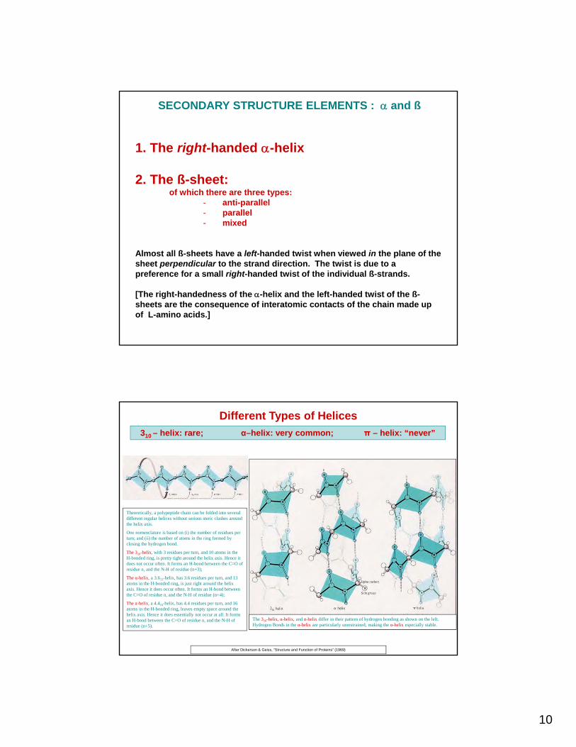
1. The right-handed -helix
2. The ß-sheet:of which there are three types:
- anti-parallel- parallel- mixed
Almost all ß-sheets have a left-handed twist when viewed in the plane of the sheet perpendicular to the strand direction. The twist is due to a preference for a small right-handed twist of the individual ß-strands.
[The right-handedness of the -helix and the left-handed twist of the ß-sheets are the consequence of interatomic contacts of the chain made up of L-amino acids.]
SECONDARY STRUCTURE ELEMENTS : and ß
310 – helix: rare; α–helix: very common; π – helix: “never”
Different Types of Helices
Theoretically, a polypeptide chain can be folded into several different regular helices without serious steric clashes around the helix axis.
One nomenclature is based on (i) the number of residues per turn; and (ii) the number of atoms in the ring formed by closing the hydrogen bond.
The 310-helix, with 3 residues per turn, and 10 atoms in the H-bonded ring, is pretty tight around the helix axis. Hence it does not occur often. It forms an H-bond between the C=O of residue n, and the N-H of residue (n+3);
The α-helix, a 3.613-helix, has 3.6 residues per turn, and 13 atoms in the H-bonded ring, is just right around the helix axis. Hence it does occur often. It forms an H-bond between the C=O of residue n, and the N-H of residue (n+4);
The π-helix, a 4.416-helix, has 4.4 residues per turn, and 16 atoms in the H-bonded ring, leaves empty space around the helix axis. Hence it does essentially not occur at all. It forms an H-bond between the C=O of residue n, and the N-H of residue (n+5).
The 310-helix, α-helix, and π-helix differ in their pattern of hydrogen bonding as shown on the left. Hydrogen Bonds in the α-helix are particularly unrestrained, making the α-helix especially stable.
After Dickerson & Geiss, “Structure and Function of Proteins” (1969)

11
(Adapted from Finkelstein and Ptitsyn - “Protein Physics”)
The α-Helix
Helix characteristics:- 100 rotation per residue- 3.6 residues per turn- 1.5 Å shift parallel to helix axis per residue- 5.4 Å shift parallel to helix axis per turn
An approximate charge distribution of a peptide unit
The actual charge distribution is different since:
there is some charge on the C
every peptide unit is in a different environment
hydrogen bonding changes charge distribution
electric fields change charge distribution
atomic polarizibility
The canonical value of pep = 3.5 Debye = 0.728 e.Å
(For comparison H20 = 1.85 Debye)
The Dipole of the Peptide Unit
The key point for what follows is that the peptide dipole moment can be approximated as a positive point charge of 0.728 elementary units and a negative point charge of -
0.728 units, separated by 1 Å;
AND ALSO by two half elementary charges of opposite sign separated by 1.5 Å.
After all, the dipole moment remains the same: 0.728x1.0 ≈ 0.5x1.5 ≈ 0.73 e.Å

12
1. In an -helix the peptide dipole moments are essentially parallel to the helix axis;
2. The positive end of each peptide unit’s dipole is near the N-terminus of the helix;
3. Each peptide unit is shifted by 1.5 Å parallel to the helix axis;
4. Each peptide dipole can be projected onto the helix axis as two half charges of opposite sign, separated by 1.5 Å;
5. Hence all half charges cancel out EXCEPT:
- one positive half charge near the N-terminus, and
- one negative half charge near the C-terminus.
The α–helix dipole
The -helix dipole and the properties of proteins. Nature 273, 443-446. Hol, W. G. J., van Duijnen, P. T. & Berendsen, H. J. C. (1978).
The role of the -helix dipole in protein structure and function. Hol, W. G. J. Prog. Biophys. Mol. Biol. 45, 149-195 (1985).
The alpha-helix as an electric macro-dipole Wada, A. In Adv Biophys (Kotani, M., ed.), (1976).
1. In an -helix the peptide dipole moments are essentially parallel to the helix axis;
2. The positive end of each peptide unit’s dipole is near the N-terminus of the helix;
3. Each peptide unit is shifted by 1.5 Å parallel to the helix axis;
4. Each peptide dipole can be projected onto the helix axis as two half charges of opposite sign, separated by 1.5 Å;
5. Hence all half charges cancel out EXCEPT:
- one positive half charge near the N-terminus, and
- one negative half charge near the C-terminus.

13
The α–helix dipole
The -helix dipole and the properties of proteins. Nature 273, 443-446. Hol, W. G. J., van Duijnen, P. T. & Berendsen, H. J. C. (1978).
The role of the -helix dipole in protein structure and function. Hol, W. G. J. Prog. Biophys. Mol. Biol. 45, 149-195 (1985).
The alpha-helix as an electric macro-dipole Wada, A. In Adv Biophys (Kotani, M., ed.), (1976).
-1/2
+1/2
1. In an -helix the peptide dipole moments are essentially parallel to the helix axis;
2. The positive end of each peptide unit’s dipole is near the N-terminus of the helix;
3. Each peptide unit is shifted by 1.5 Å parallel to the helix axis;
4. Each peptide dipole can be projected onto the helix axis as two half charges of opposite sign, separated by 1.5 Å;
5. Hence all half charges cancel out EXCEPT:
- one positive half charge near the N-terminus, and
- one negative half charge near the C-terminus.
The α–helix dipole
The -helix dipole and the properties of proteins. Nature 273, 443-446. Hol, W. G. J., van Duijnen, P. T. & Berendsen, H. J. C. (1978).
The role of the -helix dipole in protein structure and function. Hol, W. G. J. Prog. Biophys. Mol. Biol. 45, 149-195 (1985).
The alpha-helix as an electric macro-dipole Wada, A. In Adv Biophys (Kotani, M., ed.), (1976).
-1/2
-1/2
-1/2
-1/2
-1/2
-1/2
-1/2
-1/2
-1/2
-1/2
+1/2
+1/2
+1/2
+1/2
+1/2
+1/2
+1/2
+1/2
+1/2
+1/21. In an -helix the peptide dipole moments are essentially parallel to the helix axis;
2. The positive end of each peptide unit’s dipole is near the N-terminus of the helix;
3. Each peptide unit is shifted by 1.5 Å parallel to the helix axis;
4. Each peptide dipole can be projected onto the helix axis as two half charges of opposite sign, separated by 1.5 Å;
5. Hence all half charges cancel out EXCEPT:
- one positive half charge near the N-terminus, and
- one negative half charge near the C-terminus.

14
The α–helix dipole
The -helix dipole and the properties of proteins. Nature 273, 443-446. Hol, W. G. J., van Duijnen, P. T. & Berendsen, H. J. C. (1978).
The role of the -helix dipole in protein structure and function: Hol, W. G. J. Prog. Biophys. Mol. Biol. 45, 149-195 (1985).
The alpha-helix as an electric macro-dipole: Wada, A. In Adv Biophys (Kotani, M., ed.), (1976).
-1/2
+1/21. In an -helix the peptide dipole moments are essentially parallel to the helix axis;
2. The positive end of each peptide unit’s dipole is near the N-terminus of the helix;
3. Each peptide unit is shifted by 1.5 Å parallel to the helix axis;
4. Each peptide dipole can be projected onto the helix axis as two half charges of opposite sign, separated by 1.5 Å;
5. Hence all half charges cancel out EXCEPT:
- one positive half charge near the N-terminus, and
- one negative half charge near the C-terminus.
Electrostatic energy and the α–helix dipole
(1) 1 eV = 23.06 kcal mol-1
(2) Uel = q x = charge x potential (in eV)
For Helix Dipole:
At 5 Å from N-term of a 2-turn helix and assuming = 2: 0.5 Volts
Then the energy after bringing a negative point charge from infinity through a medium of constant dielectric properties to the point with potential is:
For = 2: kcal mol-1
For = 80: kcal mol-1
Hence, bringing a doubly charged negative ion from infinity to 5 Å from the N-terminus of an -helix: Uel - 0.6 kcal mol-1 (assuming = 80)
3040
12
40
50
40
501 .eV
..Uel
1250501 eV..Uel

15
The anti-parallel beta-sheet
(From Branden and Tooze - “Introduction to Protein Structure, 1st ed.”)
The parallel beta-sheet
(From Branden and Tooze - “Introduction to Protein Structure, 1st ed.”)
16
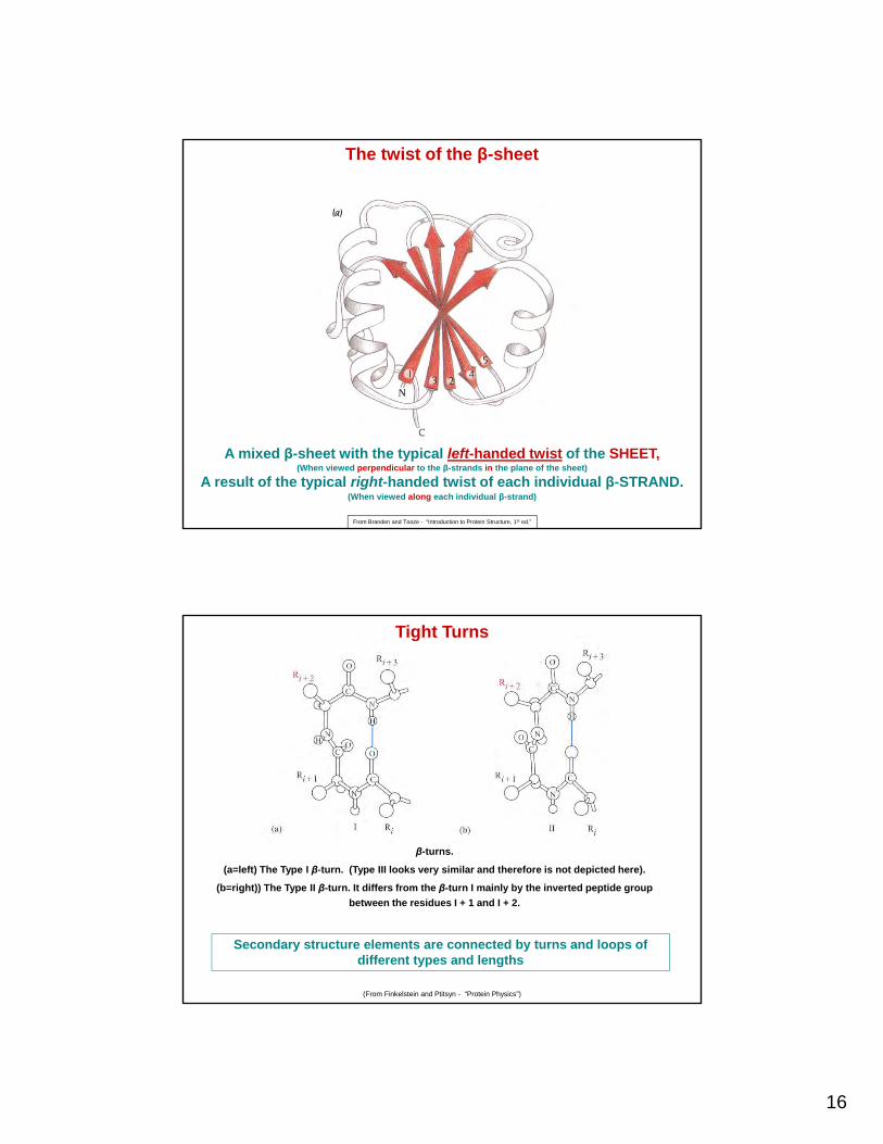
A mixed β-sheet with the typical left-handed twist of the SHEET,(When viewed perpendicular to the β-strands in the plane of the sheet)
A result of the typical right-handed twist of each individual β-STRAND.(When viewed along each individual β-strand)
From Branden and Tooze - “Introduction to Protein Structure, 1st ed.”
The twist of the β-sheet
β-turns.
(a=left) The Type I β-turn. (Type III looks very similar and therefore is not depicted here).
(b=right)) The Type II β-turn. It differs from the β-turn I mainly by the inverted peptide group
between the residues I + 1 and I + 2.
(From Finkelstein and Ptitsyn - “Protein Physics”)
Secondary structure elements are connected by turns and loops of different types and lengths
Tight Turns

17
Proteins
Level 3:
“Motifs”
Structural motifs, also called "folding units" or "supersecondary structure elements", arecompact entities composed of only a few secondary structure elements.
The most elemental structural motifs are:
1. The unit - two anti-parallel helices linked by a shorn turn
2. The ßß unit - two anti-parallel ß strands linked by a short turn
3. The ßß unit - Note that the "right handed ßß unit" is much more common than the"left handed ßß unit" for reasons which are not entirely clear.
With these elemental structural motifs, larger modules can easily be formed. Forinstance:
1. two ßß units, with a share middle ß strand "ßßß"
2. two ßß units "W" or "ß propeller" motif
3. one ßß unit and an -helix Zn-finger
4. and many more
MOTIFS

18
35
βαβ-motif βββ-motif αα-motif
Greek Key motifFormed by folding over two β-hairpins
Looks all simple but…
Supersecondary structure elements
Two adjacent parallel β strands are usually connected by an helix from three C-terminus of strand 1 to the N-terminus of strand 2. Most protein structures that contain parallel β sheets are built up from combinations of such β--β motifs. Beta strands are red, and helices are yellow.
Arrows represent β strands, and cylinders represent helices.
(a) Schematic diagram of the path of the main chain.
(b) Topological diagrams of the β--β motif.
(From Branden and Tooze - “Introduction to Protein Structure, 1st ed.”)
…in actual proteins the β-α-β unit is right-handed (!)(When looking perpendicular to the β-strands in the plane of the β-sheet)
…the β-α-β unit has a surprise
19
Level 4:
“Domains”
Proteins
• Domains can be defined as compact folded units ofcontiguous polypeptide chain.
• They are quite fundamental units of protein structure.
• Nature "juggles" with domains.
• But plenty proteins are not easily assembled fromdomains, and are more complex.
DOMAINS

20
Topology diagrams are shown below
each Ribbon diagram.
With some simplification, these structures can be considered to be
layered, with each layer composed of either -helices or β-strands but not both secondary
structure elements.
The main classes of Domains: all-, all-β, /β and +β
(From Finkelstein and Ptitsyn -“Protein Physics”)
All-
/β +β
All- β
Myohemerythrin Cytochrome b562
Cytochrome C’ Tobacco Mosaic Virus Protein
Hemoglobin β subunit
Thermolysin domain 2
Phage T4 Lysosome domain 2Papain domain 1
All-α Domains
The anatomy and taxonomy of protein structure, J.S Richardson Adv, Protein Chem. 34, 167- 339 (1981)

21
Three all -proteins that are similar in architecture (“four-helix bundle”) but entirely different in function.
Both the protein chain and co-factors are shown: wire models represent the heme (in cytochrome) and an RNA fragment (in virus coat protein), orange spheres are for iron ions (in the cytochrome heme and in hemerythrin), and the red sphere is for iron-bound oxygen (in hemerythrin). The overall architecture of such “bundles” resembles the
co-linear packing of β-sheets.
The topological diagram (right) shows all these proteins as viewed (in the same orientation) from their lower butt-ends. The circles represent the ends of -helices. The cross corresponds to the N-end of the segment (i.e., the
segment goes away for the view); the dot corresponds to its C-end (i.e., the segment comes towards the viewer).
SAME FOLD YET ENTIRELY DIFFERENT FUNCTIONS
Finkelstein and Ptitsyn - “Protein Physics”
Cytocrome c’ Hemerythrin TMV Coat protein(TMV=Tobacco Mosaic Virus)
Triose Phosphate Isomerase
Pyruvate Kinase domain I KDPG Aldolase (?)
Lactate Dehydrogenase domain I
Alcohol Dehydrogenase domain 2
Aspartate Transcarbamylase catalytic domain 2
Phosphoglycerate Kinasedomain 2
Parallel β-barrels Parallel/ Mixed β-sheets
α/β domains
The anatomy and taxonomy of protein structure, J.S Richardson Adv, Protein Chem. 34, 167- 339 (1981)

22
The common “TIM-barrel” – a famous α/β protein fold
Branden, C. & Tooze, J. (1991). Introduction to Protein Science.
Schematic diagram of the structure of the
ribonuclease inhibitor.
The molecule, which is built up by repetitive β-
loop- motifs, resembles a horseshoe
with a 17-stranded parallel β sheet on the inside and 16 helices
on the outside.
The β sheet is light red, - helices are blue, and loops that are part of the
β-loop- motifs are orange.
(Adapted from B. Kobe et al., Nature 366: 751-
756, 1993)
A quite different α/β protein with a “β-loop-α” motif repeated
Branden, C. & Tooze, J. Introduction to Protein Science.

23
Thermolysin domain 1 Glyceraldehyde P Dehydrogenase domain 2
Bacteriochlorophyll Protein
α+β domains
The anatomy and taxonomy of protein structure, J.S Richardson Adv, Protein Chem. 34, 167- 339 (1981)
Trypsin domain 1 Pyruvate Kinase domain 2
Prealbumin
Plastocyanin Immunoglobulin VL domain
Staphylococcal Nuclease Cu, Zn Superoxide Dismutase
All-β domains
The anatomy and taxonomy of protein structure, J.S Richardson Adv, Protein Chem. 34, 167- 339 (1981)

24
The orthogonal (a) and aligned (b) packing of β-sheets viewed face on (top) and from their lower end (bottom). In the face view, the β-strands are wider as they approach the viewer. The dashed line
shows the axis of the orthogonal β-barrel to which both “open” corners belong. Here the two β-sheets are most splayed. At the two “closed” corners the sheets are extremely close together; here the chain
bends and passes from one layer to the next. In the orthogonal packing the hydrophobic core is almost cylindrical. In contrast, in the aligned packing, the core is flat, the distance between the
twisted sheets remains virtually unchanged, and the relative arrangement of the sheets allows the hydrophobic faces of twisted β-strands to make contact over a great length.
Adapted from Chothia C., Finkelstein A.V. Annu. Rev. Biochem. (1990) 59: 1007-1039
Packing of β-sheets in simple all-β proteins
From Finkelstein and Ptitsyn - “Protein Physics”
Left: β-structures forming a “six-blade propeller” in influenza virus neuraminidase
Right: topological diagram of this protein
Six anti-parallel β4-motifs in a circular arrangement
From Finkelstein and Ptitsyn - “Protein Physics”

25
The β-prism in acyl transferase (a) and in pectate lyase (b).
Notice the handedness of the chain’s coiling around the axis of the prism: (a) left-handed, which is unusual, (b) right-handed, which is common. (Ignore the N-to-C-direction of the chain!)
Also note that when the chain’s coiling is left-handed, as in (a), the common twist of the β-sheet is absent.
This common twist, i.e., the left-handed twist, (viewed perpendicular to the β-strands in the plane of the sheet) is seen in (b).
From Finkelstein and Ptitsyn - “Protein Physics”
All-ß proteins: Parallel ß-helices
Proteins
Level 5:“Multi-domain proteins”

26
Single-Domain Proteins obviously coincide with Level 4: "Domains"
Multi-Domain Proteins can be classified as Proteins with:· Multiple copies of similar domains
As seen in:- rhodanese: sequence similarity lost, folds of the two
domains very similar- immunoglobulins: a true multidomain game
· Multiple dissimilar domainsAs seen in many proteins, for instance:- tyrosine kinases- pyruvate kinase- phosphoglycerate kinase
And of course also: Multiple copies of similar domains plus multiple dissimilar domains, etc.
Multi-Domain Proteins
The two domains of rhodanese resemble each other very much in structureBut are quite different in sequence
A theme seen many times since 1978…
Multi-domain proteins with domains of similar foldYet the sequences in the domains can be very different
The anatomy and taxonomy of protein structure, J.S Richardson Adv, Protein Chem. 34, 167- 339 (1981)

27
Immunoglobulins G (IgG)Combinatorial juggle of V and C domains
“C-domains” “V-domains”
(From Finkelstein and Ptitsyn - “Protein Physics”)Branden, C. & Tooze, J. (1991). Introduction to Protein Science
Multi-domain proteins with dissimilar domains
Pyruvate Kinase domain 1 Pyruvate Kinase domain 2 Pyruvate Kinase domain 3
Pyruvate kinase domains 1, 2, and 3 as an example of a protein whose domains show no structural resemblance whatsoever.
The anatomy and taxonomy of protein structure, J.S Richardson Adv, Protein Chem. 34, 167- 339 (1981)

28
55
The Multi-domain Game(Combining domains is e.g. a rapid way to combine functions in a single chain protein)
The order of the symbols indicates
the order of the domains
Domains are compact folded “nodules” of a
protein chain
Do NOT forget: Many proteins have a complex fold(A “novel” Thymidylate Synthase - not simply “Domains-on-a-string”
ThyX from Mycobacterium tuberculosis has a complex fold:
The Top and Bottom Domains are made up of a “contiguous piece of polypeptide chain.
But look at the Central Domain: the stretch α6-α7-β5-β6 is contiguous, but strands β1-β2-β3-β4 of the central β-sheet are very non-contiguous.
Parthasarathy Sampathkumar.

29
Branden, C. & Tooze, J. (1991). Introduction to Protein Science.
Influenza Virus Haemagglutinin(i. e. again not simply “Domains-on-a-string”
Just following the chain in this structure is quite a challenge….
Proteins
“The Conformational Change”

30
Proteins, e.g.:- phosphoglycerate kinase- adenylate kinase- GTPases like Elongation Factor Tu- Influenza virus haemagglutinin- Oxy vs deoxy hemoglobin- F1 ATPase- Protein kinases
DNA, e.g.- as bound, in a very kinked manner, to TATA-box binding
protein
RNA- where the same RNA molecule can choose different base-
pairing schemes to end up with very different structures.
CONFORMATIONAL CHANGESA crucial property of biomacromolecules
An interesting database on Protein Motions is: http://molmovdb.mbb.yale.edu/MolMovDB/
The bilobal structure of calmodulin before binding a cognate peptide
Calmodulin
N-terminal domain
C-terminal domain
Calcium ion

31
The compact structure of calmodulin after binding a cognate peptide
A Calmodulin/ Peptide complex
N-terminal domain
C-terminal domain
Calmodulin-binding peptide
The functional conformational change of calmodulin
Protein structure is important.Yet, without functional conformational changes of proteins life
would be pretty miserable.
The Conformation Change of Calmodulin
Before peptide binding After peptide binding

32
The newest book on protein structure is:“Text book of Strutural Biology”
by Liljas, Liljas, Piskur, Lindblom, Nissen and KjelgaardWorld Scientific 2009
An excellent book on protein structure is:“Introduction to Protein Structure”
by C. Branden and John ToozeNew York: Garland Publishing, 1st Ed 1991, 2nd Ed 1999
Another book is:“Protein Structure and Function”By G Petsko and Dagmar Ringe
New Science Press 2004
A beautiful much older book is:"The Structure and Action of Proteins"by Richard E. Dickerson & Irving Geiss
New York: Harper & Row, 1969
Physical principles are emphasized in:"Biophysical Chemistry"
by Charles R. Cantor & Paul R. SchimmelSan Francisco: W.H. Freeman, 1980
`"Proteins, Structure and Molecular Properties"by Thomas E. Creighton
Oxford; Boston: Blackwell Scientific Publications, 1993
A older review on protein domains, a true classic, is:"The anatomy and taxonomy of protein structure"
by Jane S. RichardsonAdv. Prot. Chemistry 34:167-339 (1981)
Now also on the web: http://kinemage.biochem.duke.edu/teaching/anatax/index.html
LITERATURE
A good book with emphasis on biophysics:“Protein Physics”
Alexei Finkelstein and Oleg B PtitsynAcademic Press (2002)
A book giving a broad perspective:"From Cells to Atoms"by Rees and Sternberg
Blackwell Scientific Publications, 1984
An award winning website on Principles of Protein Structure:http://www.cryst.bbk.ac.uk/PPS2/
(But this site does not appear to have been updated since mid-1996…)
Structural Classifications of Proteins SCOP and CATH:http://scop.mrc-lmb.cam.ac.uk/scop/
http://www.cathdb.info/
Proteopedia:http://www.proteopedia.org
Protein motion:http://molmovdb.org/
LITERATURE (Ctd)

33
The Endof
Seven Levels Part I