Embed Size (px)
Citation preview

Serum Levels of Trace Elements and Heavy Metals in Patientswith Acute Hemorrhagic Stroke
Sevdegul Karadas • Refah Sayın • Mehmet Aslan •
Hayriye Gonullu • Celal Katı • Recep Dursun •
Latif Duran • Edip Gonullu • Halit Demir
Received: 24 July 2013 / Accepted: 5 December 2013 / Published online: 18 December 2013
� Euratom: Turkey; European Union 2013
Abstract Trace elements are essential components of
biological structures, but alternatively, they can be toxic at
concentrations beyond those necessary for their biological
functions. Changes in the concentration of essential trace
elements and heavy metals may affect acute hemorrhagic
stroke. The aim of this study was to measure serum levels
of essential trace elements [iron (Fe), zinc (Zn), manganese
(Mn), copper (Cu), and magnesium (Mg)] and heavy
metals [cobalt (Co), cadmium (Cd), and lead (Pb)] in
patients with acute hemorrhagic stroke. Twenty-six
patients with acute hemorrhagic stroke and 29 healthy
controls were enrolled. Atomic absorption spectropho-
tometry (UNICAM-929) was used to measure serum Fe,
Cu, Pb, Cd, Zn, Co, Mn and Mg concentrations. Serum Cd,
Pb and Fe levels were significantly higher in patients with
acute hemorrhagic stroke than controls (p \ 0.001), while
serum Cu, Zn, Mg and Mn levels were significantly lower
(all p \ 0.001). However, there was no significant differ-
ence between the groups with respect to serum Co levels
(p [ 0.05). We first demonstrate increased Cd, Pb, and Fe
levels; and decreased Cu, Zn, Mg, and Mn levels in
patients with acute hemorrhagic stroke. These findings may
have diagnostic and prognostic value for acute hemor-
rhagic stroke. Further studies are required to elucidate the
roles of trace elements and heavy metals in patients with
acute hemorrhagic stroke.
Keywords Acute hemorrhagic stroke � Trace element �Heavy metals
Introduction
Stroke is recognized as a leading cause of death and severe
neurological disability across the world (No authors listed
1990). Ischemic and hemorrhagic stroke are the two pri-
mary subtypes of stroke (Adams et al. 1993). A stroke
occurs when blood vessels that deliver oxygen to the brain
either rupture or become clogged, causing brain cells/
neurons to die (Demirdogen et al. 2009). Acute stroke must
be considered an emergency of the highest priority. Early
brain imaging is essential to discriminate between ischemic
and hemorrhagic stroke (Lichy and Hacke 2010). Several
conventional risk factors, such as hypertension, diabetes,
and genetic factors, have been identified in the etiopatho-
genesis of stroke (Benmoyal-Segal et al. 2005).
S. Karadas (&) � H. Gonullu
Department of Emergency Medicine, Medical Faculty, Yuzuncu
Yıl University, Van, Turkey
e-mail: [email protected]
R. SayınDepartment of Neurology, Medical Faculty, Yuzuncu YılUniversity, Van, Turkey
M. Aslan
Department of Internal Medicine, Medical Faculty, Yuzuncu YılUniversity, 65000 Van, Turkey
C. Katı � L. Duran
Department of Emergency Medicine, Ondokuz MayısUniversity, Samsun, Turkey
R. Dursun
Department of Emergency Medicine, Van Training Education
and Research Hospital, Van, Turkey
E. Gonullu
Department of Anesthesiology and Reanimation, Van Training
Education and Research Hospital, Van, Turkey
H. Demir
Department of Chemistry, Faculty of Science and Art, Yuzuncu
Yıl University, Van, Turkey
123
J Membrane Biol (2014) 247:175–180
DOI 10.1007/s00232-013-9621-0

Trace elements at optimal levels are required for
numerous metabolic and physiological processes in the
human body (Mertz 1981). Moreover, trace metals have
important influences on brain development and function
(Hickenbottom and Grotta 1998). Zinc (Zn), copper (Cu),
and manganese (Mn) are important cofactors for several
enzymes that play a role in maintaining DNA integrity
(Mahabir et al. 2007; Leach 1971). Cu, Zn, and Mn also act
as antioxidants (Shenkin 1997). Moreover, Fe metabolism
plays a key role in maintaining normal brain function. Fe is
the most abundant transitional metal in the brain, and the
brain has higher Fe content than most organs (Moos 2002).
Fe is essential for normal neuronal function and activity.
The synthesis of several neurotransmitters is Fe-dependent
(Beard et al. 1993). On the other hand, magnesium (Mg) is
the fourth most abundant cation in the body and plays a
pivotal role as an enzyme cofactor in biosynthesis of pro-
teins and mineral administration. Its metabolism is con-
nected with bone health, as it is indispensable for
osteogenesis and mineralization of bones (Rahnama and
Marcınıak 2002)..
The essential trace element cobalt (Co) is an integral
part of vitamin B12, which is essential for folate and fatty
acid metabolism (Anderson et al. 1992). On the other hand,
cadmium (Cd) is one of the most dangerous occupational
and environmental toxins. It is found in drinking water,
atmospheric air, and even food. Products of vegetable
origin are the main carrier of Cd compounds in food
(Klos 2001). Furthermore, lead (Pb) increases oxidative
stress, affects endothelial function, promotes inflammation,
downregulates nitric oxide production, and induces renal
dysfunction (Sokolov et al. 2002).
Several studies have assessed trace elements such as Zn,
Fe, and Cu in cerebral hemorrhage and stroke patients, but
the results have been inconsistent (Munshi et al. 2010;
Uza et al. 1995). However, to the best of our knowledge,
serum levels of heavy metals have not previously been
evaluated in patients with acute hemorrhagic stroke.
The aim of this study was to measure the serum levels of
essential trace elements (Fe, Zn, Mn, Cu, and Mg) and
heavy metals (Co, Cd, and Pb) in patients with acute
hemorrhagic stroke.
Materials and Methods
Subjects
Twenty-six patients (12 females and 14 males) with acute
hemorrhage stroke and 29 healthy volunteers (14 females
and 15 males) were enrolled in this prospective study.
Patients admitted to the emergency department were
examined neurologically, and they were assessed using the
Glasgow Coma Scale (Teasdale and Jenett 1974). The
diagnosis was also confirmed via brain computerized
tomography.
Patients were examined neurologically. All hemorrhagic
stroke patients were treated similarly in the emergency
department. The treatment provided depended on the
preservation of vital functions and the brain. The intra-
cranial pressure was stabilized by administering intrave-
nous mannitol or furosemide.
The control group was selected from 29 healthy volun-
teers (14 females and 15 males). All of the control subjects
were asymptomatic with unremarkable medical histories
and normal physical examinations. All of the control sub-
jects were nonsmokers.
The study protocol was conducted in accordance with
the Declaration of Helsinki as revised in 2000 and was
approved by the local ethics committee. All of the subjects
were informed about the study, and written consent was
obtained from each one.
Exclusion Criteria
Exclusion criteria included a history of alcohol abuse,
habitual smoking, intravenous drug abuse, pregnancy, use
of antioxidant supplements, hypertension, diabetes melli-
tus, liver or renal disease, rheumatoid arthritis, pulmonary
disease, and coronary artery disease.
Blood Samples
Blood samples were collected before treatment and were
immediately stored at 4 �C. Serum samples were then
separated from the blood cells by centrifugation at
3,000 rpm for 10 min. Serum samples prepared for mea-
surement of trace element levels and heavy metals were
maintained at -80 �C until they were used.
Measurement of Trace Elements in Serum
Serum concentrations of Zn, Cu, Fe, Cd, Pb, and Mn were
determined via atomic absorption spectrophotometry using
a UNICAM-929 spectrophotometer (Unicam Ltd, York
Street, Cambridge, UK).
Statistical Analysis
The results were expressed as the mean ± standard
deviation. Parametric variables were compared using
Student’s t test. Qualitative variables were assessed using
Chi square tests. The results were considered to be sta-
tistically significant when the p value was\0.05. The data
were analyzed using SPSS� for Windows Version 11.0
software.
176 S. Karadas et al.: Trace Elements in Acute Hemorrhagic Stroke
123
Results
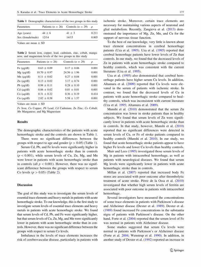
The demographic characteristics of the patients with acute
hemorrhagic stroke and the controls are shown in Table 1.
There were no significant differences between the
groups with respect to age and gender (p [ 0.05) (Table 1).
Serum Cd, Pb, and Fe levels were significantly higher in
patients with acute hemorrhagic stroke than in controls
(p \ 0.001), while serum levels of Cu, Zn, Mg, and Mn
were lower in patients with acute hemorrhagic stroke than
in controls (all p \ 0.001). However, there was no signif-
icant difference between the groups with respect to serum
Co levels (p [ 0.05) (Table 2).
Discussion
The goal of this study was to investigate the serum levels of
essential trace elements and heavy metals in patients with acute
hemorrhagic stroke. To our knowledge, this is the first study to
investigate serum levels of essential trace elements and heavy
metals in patients with acute hemorrhagic stroke. We found
that serum levels of Cd, Pb, and Fe were significantly higher,
but that serum levels of Cu, Zn, Mg, and Mn were significantly
lower in patients with acute hemorrhagic stroke than in con-
trols. However, there was no significant difference between the
groups with respect to serum Co levels.
Imbalance in the levels of trace elements increases the
risk of cerebrovascular disease, particularly in patients with
ischemic stroke. Moreover, certain trace elements are
necessary for maintaining various aspects of neuronal and
glial metabolism. Recently, Zangieva et al. (2013) dem-
onstrated the importance of Mg, Zn, Mn, and Cu for the
support of nervous tissue function.
To the best of our knowledge, very little is known about
trace element concentrations in cerebral hemorrhage
patients (Uza et al. 1995). Uza et al. (1995) reported that
cerebral hemorrhage patients have lower levels of Zn than
controls. In our study, we found that the decreased levels of
Zn in patients with acute hemorrhagic stroke compared to
healthy controls, which was consistent with the current
literature (Uza et al. 1995).
Uza et al. (1995) also demonstrated that cerebral hem-
orrhage patients have higher serum Cu levels. In addition,
Altamura et al. (2009) reported that Cu levels were ele-
vated in the serum of patients with ischemic stroke. In
contrast, we found that the decreased levels of Cu in
patients with acute hemorrhagic stroke compared to heal-
thy controls, which was inconsistent with current literature
(Uza et al. 1995; Altamura et al. 2009).
Munshi et al. (2010) demonstrated that the serum Zn
concentration was lower in stroke patients than in healthy
subjects. We found that serum levels of Zn were signifi-
cantly lower in patients with acute hemorrhagic stroke than
in controls. In that study, however, Munshi et al. (2010)
reported that no significant differences were detected in
serum levels of Cu, or Fe of stroke patients compared to
healthy controls (Munshi et al. 2010). In our study, we
found that acute hemorrhagic stroke patients appear to have
higher Fe levels and lower Cu levels than healthy controls.
Muir and Lees (1995) investigated those serum levels of
Mg in patients with intracerebral hemorrhage, as well as
patients with neurological diseases. We found that serum
Mg levels were significantly lower in patients with acute
hemorrhagic stroke than in controls.
Millan et al. (2007) reported that increased body Fe
stores are associated with poor outcome after thrombolytic
treatment of acute stroke. Perez de la Ossa et al. (2010)
investigated that whether high serum levels of ferritin are
associated with poor outcome in patients with intracerebral
hemorrhage.
Several investigators have measured the concentrations
of some trace elements in patients with Parkinson’s disease
and Alzheimer disease (Dexter et al. 1989). Dexter et al.
(1989) found increased Fe concentrations in the substantia
nigra of patients with Parkinson’s disease. On the other
hand, Forte et al. (2004) reported that the serum level of Fe
was normal in patients with Alzheimer disease.
Some studies suggested that serum Cu levels were
normal in patients with Parkinson’s or Alzheimer disease
(Forte et al. 2004; Wenstrup et al. 1990). In addition, in
another study of Dexter et al. (1992) reported an increase in
Table 1 Demographic characteristics of the two groups in this study
Parameters Patients (n = 26) Controls (n = 29) p
Age (years) 44 ± 6 41 ± 3 0.211
Sex (female/male) 12/14 14/15 0.805
Values are mean ± SD
Table 2 Serum iron, copper, lead, cadmium, zinc, cobalt, manga-
nese, and magnesium levels of the two groups in this study
Parameters Patients (n = 26) Controls (n = 29) p
Fe (lg/dI) 0.63 ± 0.09 0.17 ± 0.06 0.001
Mg (lg/dI) 10.70 ± 0.97 24.56 ± 1.96 0.001
Mn (lg/dI) 0.11 ± 0.02 0.27 ± 0.04 0.001
Zn (lg/dI) 0.13 ± 0.02 3.17 ± 0.74 0.001
Pb (lg/dI) 0.98 ± 0.11 0.28 ± 0.10 0.001
Cd (lg/dI) 0.06 ± 0.02 0.01 ± 0.01 0.001
Co (lg/dI) 0.31 ± 0.22 0.36 ± 0.19 0.414
Cu (lg/dI) 2.82 ± 0.58 5.58 ± 1.57 0.001
Values are mean ± SD
Fe Iron; Cu Copper; Pb Lead; Cd Cadmium; Zn Zinc; Co Cobalt;
Mn Manganese; and Mg Magnesium
S. Karadas et al.: Trace Elements in Acute Hemorrhagic Stroke 177
123

Zn levels in substantia nigra, lateral putamen, and caudate
nucleus in Parkinson’s disease patients.
In humans, Mn, Fe, Cu, and Zn fulfill decisive functions
to maintain human health. Zn, Fe, Mn, and Cu are essential
micronutrients incorporated into many metalloenzymes
and proteins involved in cell metabolism, production of
neurotransmitters, and regulatory pathways controlling
oxidative stress (Tapiero et al. 2003). Excessive accumu-
lation or depletion of trace elements may have significant
clinical implications, including increased risk of cardio-
vascular disease, immune deficiency, cancer, and bone
disease (Fraga 2005). To cause physiological or patho-
physiological effects in the brain, the metals must either
traverse the blood–brain barrier to act directly on the
neurons or glial cells, or alternatively, they could have an
indirect effect. Trace elements might also have indirect
effects by influencing the transport or regulation of other
substances at the blood–brain barrier. (Bradbury 1992)
Fe is an essential trace element for all organisms that is
crucial for normal cell function, and its deficiency, or
excess is associated with several disease states. It is known
that excess Fe and Fe deficiency also lead to oxidative
DNA damage (Dayani et al. 2004).
Mn is essential for normal physiologic function in
humans and animals, but is toxic at higher levels of
exposure (Bureau et al. 2002). Mn also plays a role in the
free radical scavenging activity of superoxide dismutase
(SOD).
Cd is a dangerous occupational and environmental toxin
that accumulates in humans primarily in the liver and the
kidneys (Kowalczyk et al. 2003).
Cu is an essential element that plays a role in the pro-
duction of hemoglobin, myelin, collagen, and melanin
(Aggett 1999). Recent evidence also suggests that adequate
uptake of Cu is necessary for normal immune function
(Ertekin et al. 2006). Cu deficiency affects various physi-
ological functions that may be important in the immuno-
logical defense to pathogenic challenge (Stabel et al. 1993).
Cu also functions as a cofactor of SOD and catalase in the
antioxidant redox system. (Ozkaya et al. 2011; Kayan et al.
2010) However, Cu is also a highly toxic metal that has
been associated with several neurodegenerative disorders
(Levenson 2005). Moreover, high blood Cu concentration
is thought to be an independent risk factor for cardiovas-
cular disease (Kang 2011).
Zn is an essential nutrient that is a component of several
metalloenzymes (Cartwright and Wintrobe 1964). In
addition, Zn plays an important role in brain metabolism
and has antioxidant and anti-inflammatory properties in the
brain (Arslan et al. 2011), protecting cells from free radical
injury. Moreover, Zn plays an anticarcinogenic role by
stabilizing the structure of DNA, RNA, and ribosomes
(Wu et al. 2004). Zn deficiency has been implicated as an
explanation for the central nervous system (CNS) symp-
toms in liver cirrhosis, fetal alcohol syndrome, malab-
sorption, and acrodermatitis enteropathica. Low serum or
plasma concentrations of Zn have been found in patients
with multiple sclerosis and in chronic alcoholics with liver
disease. High concentrations of Zn have been found in
patients with multiple sclerosis and Pick’s disease (Dexter
et al. 1993).
Organisms have developed mechanisms to utilize vital
trace elements such as zinc and copper, while minimizing
the toxic influence of heavy metals such as Cd and Pb
(Solioz et al. 1994).
Pb serves no useful function in the human body, and its
presence in the body can lead to toxic effects. Recent
research appears to indicate that even a low level of Pb
exposure has a number of negative consequences on health.
These consequences include impairment of renal tubular
cell function, inhibition of sperm formation, fetal damage,
slowing of motor nerve velocity, CNS dysfunction,
hypertension, and other cardiovascular diseases (Meller
et al. 1992). Pb is an indicator of oxidative stress and
affects endothelial function, promotes inflammation,
downregulates nitric oxide production, and induces renal
dysfunction (Lustberg and Silbergeld 2002). Pb has been
shown to permeate the blood–brain barrier. A high Pb level
in the human body may lead to irreversible damage to the
CNS (Donma and Donma 2002).
Physiological doses of Cd increase endothelial perme-
ability via DNA damage-induced inhibition of endothelial
proliferation and cell death induction (Messner et al. 2009).
Cd is a ubiquitous toxic heavy metal and, unlike organic
compounds, it is not biodegradable, and has a very long
biological half-life (Donma and Donma 2002). Based on
data from animals and humans, soon after Cd exposure, Cd
in blood is present in plasma, whereas later, it becomes
bound to erythrocytes (Nordberg 1978). Cd exposure may
induce lipid peroxidation in the heart, the lungs, the liver,
and the spleen (Manca et al. 1994), as well as increase
oxidative stress in tissues (Kirchvink et al. 2006). Cd is an
established toxic and carcinogenic heavy metal (Nawrot
et al. 2002). Although Cd does not generate free radicals
directly, it may contribute to the formation of reactive
oxygen species indirectly. Cd has also a role in the inhi-
bition of gene expression and signal transduction (Wais-
berg et al. 2003).
Co is a natural element found throughout the environ-
ment. Co is known to perform a vital role in hemoglobin
biosynthesis and is an integral part of vitamin B12, which is
an essential for folate and fatty acid metabolism (Anderson
et al. 1992). Excess Co in the body is characterized by
polycythemia (Atasoy et al. 2011). Co may also induce
DNA damage and mediate free radical generation (Jomova
and Valko 2011).
178 S. Karadas et al.: Trace Elements in Acute Hemorrhagic Stroke
123

There were several limitations in the present study. First,
this study is cross-sectional in nature. Second, the number
of patients with acute hemorrhagic stroke who were
included in the study was relatively small, and a larger
sample size would have increased the power to detect
differences in serum levels of essential trace elements and
heavy metals in patients with acute hemorrhagic stroke.
Third, serum levels of essential trace elements and heavy
metals levels were not measured after treatment in patients
with acute hemorrhagic stroke.
In conclusion, we demonstrate that the increased serum
levels of Cd, Pb, and Fe and decreased serum levels of Cu,
Zn, Mg, and Mn in patients with acute hemorrhagic stroke.
These findings may have diagnostic and prognostic value
for acute hemorrhagic stroke. Further studies are needed to
elucidate the roles of trace elements and heavy metals in
patients with acute hemorrhagic stroke.
Acknowledgments The authors do not report any conflicts of
interest regarding this work.
References
Adams H, Bendixen B, Kappelle L, Biller J, Love B, Gordon D,
Marsh EE 3rd (1993) Classification of subtype of acute ischemic
stroke. Definitions for use in a multicenter clinical trial. TOAST.
Trial of Org 10172 in acute stroke treatment. Stroke 24:35–41
Aggett PJ (1999) An overview of the metabolism of copper. Eur J
Med Res 4:214–216
Altamura C, Squitti R, Pasqualetti P, Gaudino C, Palazzo P, Tibuzzi
F, Lupoi D, Cortesi M, Rossini PM, Vernieri F (2009)
Ceruloplasmin/Transferrin system is related to clinical status in
acute stroke. Stroke 40:1282–1288
Anderson MB, Pedigo NG, Katz RP, George WJ (1992) Histopa-
thology of testes from mice chronically treated with cobalt.
Reprod Toxicol 6:41–50
Arslan M, Demir H, Arslan H, Gokalp AS, Demir C (2011) Trace
elements, heavy metals and other biochemical parameters in
malignant glioma patients. Asian Pac J Cancer Prev 12(2):
447–451
Atasoy N, Mercan U, Alacabey I, Kul AR (2011) Levels of heavy
metals and certain macro elements in potable and tap water at
Van City Center. Hacettepe J Biol Chem 39:391–396
Beard J, Connor J, Jones B (1993) Iron in the brain. Nutr Rev
51:157–170
Benmoyal-Segal L, Vander T, Shifman S, Bryk B, Ebstein RP,
Marcus EL, Stessman J, Darvasi A, Herishanu Y, Friedman A,
Soreq H (2005) Acetylcholinesterase/paraoxonase interactions
increase the risk of insecticide-induced Parkinson’s disease.
FASEB J 19:452–454
Bradbury MW (1992) An approach to study of transport of trace
metals at the blood-brain barrier. Prog Brain Res 91:133–138
Bureau I, Anderson RA, Arnaud J, Raysiguier Y, Favier AE, Roussel AM
(2002) Trace mineral status in post menopausal women: impact of
hormonal replacement therapy. J Trace Elem Med Biol 16:9–13
Cartwright GE, Wintrobe MM (1964) Copper metabolism in normal
subjects. Am J Clin Nutr 14:224–232
Dayani PN, Bishop MC, Black K, Zeltzer PM (2004) Desferoxamine
(DFO)-mediated iron chelation: rationale for a novel approach to
therapy for brain cancer. J Neurooncol 67:367–377
Demirdogen BC, Demirkaya S, Turkanoglu A, Bek S, Arinc E, Adali
O (2009) Analysis of paraoxonase 1 (PON1) genetic polymor-
phisms and activities as risk factors for ischemic stroke in
Turkish population. Cell Biochem Funct 27(8):558–567
Dexter DT, Wells FR, Lees AJ, Agid F, Agid Y, Jenner P, Marsden
CD (1989) Increased nigral iron content and alterations in other
metal ions occuring in brain in Parkinson’s disease. J Neurochem
52:1830–1836
Dexter DT, Jenner P, Schapira AH, Marsden CD (1992) Alterations in
levels of ıron, ferritin, and other trace metals in neurodegene-
retive diseases affecting the basal ganglia. Ann Neurol 32:
94–100
Dexter DT, Sian J, Jenner P, Marsden CD (1993) Implications of
alterations in trace element levels in brain in Parkinson’s disease
and other neurological disorders affecting the basal ganglia. Adv
Neurol 60:273–281
Donma O, Donma MM (2002) Association of headaches and the
metals. Biol Trace Elem Res 90:1–14
Ertekin A, DeGer Y, Mert H, Mert N, Yur F, Dede S, Demir H (2006)
An investigation of the effects of alpha-tocopherol on the levels
Fe, Cu, Zn, Mn and carbonic anhydrase in rats with bleomycin-
induced pulmonary fibrosis. Biol Trace Elem Res 114:1–12
Forte G, Bocca B, Senofonte O, Petrucci F, Brusa L, Stanzione P,
Zannino S, Violante N, Alimonti A, Sancesario G (2004) Trace
and major elements in whole blood, serum, cerebrospinal fluid
and urine of patients with Parkinson’s disease. J Neural Transm
111:1031–1040
Fraga Cesar G (2005) Relevance, essentiality and toxicity of trace
elements in human health. Mol Aspect Med 26:235–244
Hickenbottom SL, Grotta J (1998) Neuroprotective therapy. Sem
Neurol 18:485–492
Jomova K, Valko M (2011) Advances in metal-induced oxidative
stress and human disease. Toxicology 283:65–87
Kang YJ (2011) Copper and homocysteine in cardiovascular diseases.
Pharmacol Ther 129(3):321–331
Kayan M, Nazıroglu M, Barak C (2010) Effects of vitamin C and E
combination on trace element levels in blood of smokers and
nonsmokers radiology X-ray technicians. Biol Trace Elem Res
136:140–148
Kirchvink N, Martin N, Fievez L, Smith N, Marlin D, Gustin P (2006)
Airway inflammation in cadmium-exposed rats is associated
with pulmonary oxidative stress and emphysema. Free Radic Res
40:241–250
Klos A (2001) Lead, cadmium and mercury content in meals planned
for consumption in selected kindergartens in Warsaw: IV
International Scientific-Technical Conference, Warsaw, p 4–5
Kowalczyk E, Kopff A, Fijalkowski P, Kopff M, Niedworok J,
Blaszczyk J, Kedziora J, Tyoelerowicz P (2003) Effect of
anthocyanins on selected biochemical parameters in rats exposed
to cadmium. Acta Bio Pol 50:543–548
Leach RM Jr (1971) Role of manganese in mucopolysaccharide
metabolism. Fed Proc 30:991–994
Levenson CW (2005) Trace metal regulation of neuronal apoptosis:
from genes to behavior. Physiol Behav 15:399–406
Lichy C, Hacke W (2010) Stroke. Internist (Berl) 51(8):1003–1011
Lustberg M, Silbergeld E (2002) Blood lead levels and mortality.
Arch Intern Med 162:2443–2449
Mahabir S, Spitz MR, Barrera SL, Beaver SH, Etzel C, Forman MR
(2007) Dietary zinc, copper and selenium, and risk of lung
cancer. Int J Cancer 120:1108–1115
Manca D, Ricard AC, Tra HV, Chevalier G (1994) Relation between
lipid peroxidation and inflammation in the pulmonary toxicity of
cadmium. Arch Toxicol 68:364–369
Meller L, Tage S, Kristensen TS (1992) Blood lead as a cardiovas-
cular risk factor. Am J Epidemiol 136:1091–1100
Mertz W (1981) The essential trace elements. Science 213:1332–1338
S. Karadas et al.: Trace Elements in Acute Hemorrhagic Stroke 179
123

Messner B, Knoflach M, Seubert A, Ritsch A, Pfaller K, Henderson
B, Shen YH, Zeller I, Willeit J, Laufer G, Wick G, Kiechl S,
Bernhard D (2009) Cadmium is a novel and independent risk
factor for early atherosclerosis mechanisms and in vivo rele-
vance. Arterioscler Thromb Vasc Biol 29:1392–1394
Millan M, Sobrino T, Castellanos M, Nombela F, Arenillas JF, Riva
E, Cristobo I, Garcıa MM, Vivancos J, Serena J, Moro MA,
Castillo J, Davalos A (2007) Increased body iron stores are
associated with poor outcome after thrombolytic treatment in
acute stroke. Stroke 38(1):90–95
Moos T (2002) Brain iron homeostasis. Dan Med Bull 49:279–301
Muir KW, Lees KR (1995) A randomized, double-blind, placebo-
controlled pilot trial of ıntravenous magnesium sulfate in acute
stroke. Stroke 26:1183–1188
Munshi A, Babu S, Kaul S, Shafi G, Rajeshwar K, Alladi S, Jyothy A
(2010) Depletion of serum zinc in ischemic stroke patients.
Methods Find Exp Clin Pharmacol 32(6):433–436
Nawrot TS, Thijs L, Den Hond EM, Roels HA, Staessen JA (2002)
An epidemiological re-appraisal of the association between
blood pressure and blood lead: a meta-analysis. J Hum Hyper-
tens 16:123–131
No authors listed (1990) Special report From The National Institute of
Neurological Disorders and Stroke. Classification of cerebro-
vascular diseases III. Stroke 21:637–676
Nordberg M (1978) Studies on metallothionein and cadmium.
Environ Res 15:381–404
Ozkaya MO, Nazıroglu M, Barak C, Berkkanoglu M (2011) Effects of
multivitamin/mineral supplementation on trace element levels in
serum and follicular fluid of women undergoing in vitrofertiliza-
tion (IVF). Biol Trace Elem Res 139:1–9
Perez de la Ossa N, Sobrino T, Silva Y, Blanco M, Millan M, Gomis
M, Agulla J, Araya P, Reverte S, Serena J, Davalos A (2010)
Iron-related brain damage in patients with intracerebral hemor-
rhage. Stroke 41(4):810–813
Rahnama M, Marcınıak A (2002) Influence of Estrogen Deficiency on
the level of magnesium in rat mandible and teeth. Bull Vet Inst
Pulawy 46:267–271
Shenkin A (1997) Micronutrients and outcome. Nutrition 13:825–828
Sokolov DL, Bailey MR, Crum LA, Blomgren PM, Connors BA,
Evan AP (2002) Prefocal alignment improves stone comminu-
tion in shockwave lithotripsy. J Endourol 16:709–715
Solioz M, Odermatt A, Krapf R (1994) Copper pumping ATPases:
common concept in bacteria and man. FEBS Lett 346:44–47
Stabel JR, Spears JW, Brown TT (1993) Effect of copper deficiency
on tissue, blood characteristics and immune function of calves
challenged with infectious bovine rhinotracheitis virus and
Pasteurella hemolytica. J Anim Sci 71:1247–1255
Tapiero H, Townsend DM, Tew KD (2003) Trace elements in human
physiology and pathology. Biomed Pharmacother 57:399–411
Teasdale G, Jenett B (1974) Assessment of coma and impaired
consciousness: a practical scale. Lancet 2:81–83
Uza G, Comes L, Uza D, Pop O (1995) Serum zinc and copper in
patients with cerebral vascular disease. Rom J Intern Med
33:19–26
Waisberg M, Joseph P, Hale B, Beyersmann D (2003) Molecular and
cellular mechanisms of cadmium carcinogenesis. Toxicology
192:95–117
Wenstrup D, Ehmann WD, Markesbery WR (1990) Trace element
imbalances in isolated subcellular fractions of Alzheimer’s
disease brains. Brain Res 533:125–131
Wu T, Sempos CT, Freudenheim JL, Muti P, Smit E (2004) Serum
iron, copper and zinc concentrations and risk of cancer mortality
in US adults. Ann Epidemiol 14:195–201
Zangieva ZK, Torshin IIu, Gromova OA, Nikonov AA (2013) Trace
elements in the nervous tissue and ischemic stroke. Zh Nevrol
Psikhiatr Im S S Korsakova 113:30–36
180 S. Karadas et al.: Trace Elements in Acute Hemorrhagic Stroke
123


![Alterations of Several Serum Parameters Are Associated with ...downloads.hindawi.com/journals/dm/2020/7815214.pdfand prognosis evaluation of PE [20]. Changes of serum trace elements](https://img.pdfslide.us/doc/110x75/5fa10d12182a6e0c36358aa6/alterations-of-several-serum-parameters-are-associated-with-and-prognosis-evaluation.jpg)