Embed Size (px)
Citation preview
M I N E R A L O G I C A L M A G A Z I N E , M A R C H I 9 7 9, VOL. 43 , PP. I 3 5 - 4 o
Serpentine minerals from veins in serpentinite rocks
N. MORANDI AND G. FELICE
Istituto di Mineralogia e Petrografia dell'Universit~t di Bologna, Italy
SUMMARY. Crystal-chemical analyses of the serpentine polymorphs, sampled from macroscopic veins in the serpentinite rocks from Sasso della Mantesca and Sasso di S. Zanobi, are reported. The samples were examined by X-ray diffraction, thermal analysis (TG and DTG), scan- ning and transmission electron microscopy, IR absorp- tion spectroscopy, microprobe, and chemical tests.
The results show: chrysotile 20rcl in tubular fibrils placed perpendicularly to walls of iso-oriented 'lenticular veins'; and polygonal serpentine formed by flat layers of lizardite 2H and/or I T, arranged as an outer shell of a central core of chrysotile 2Mcx fibres; this type of serpentine with high crystallinity and unusual morpho- logical development is included in 'green veins', which show variable A1, Fe, and Mg contents.
A genetic mechanism correlating the petrographic observations with the mineralogical results is discussed.
THIS paper is an attempt to interpret the charac- teristic features and the significance of the serpen- tine polymorphs sampled from different sorts of veins in the serpentinite rocks from Sasso della Mantesea and Sasso di S. Zanobi, northern Apen- nine, near Bologna, Italy (Tav. I S E - - F . 9 8 - - I.G.M.).
Mineralogical and petrographic studies on these outcrops (each extended for about 3oo m 2) were made recently by Pellizzer (I96 I) and by Bocchi et at. (I976) .
Materials. The serpentinite rock from Sasso della Mantesca (samples prefixed by the number 8) is a serpentinized peridotite with pseudomorphic tex- tures beginning to transform to non-pseudo- morphic textures. It consists mainly of serpentine mesh textures (dark coloured in hand specimen) after olivine and clino- and ortho-pyroxenes. It contains iso-oriented 'lenticular veins' with fine fibres of bright serpentine elongated perpendicular to the vein walls. On a microscopic scale the veins contain iso-oriented fibrils of y-serpentine (in the sense of Wicks and Zussman's i975 terminology), variously curved and displaced; these cross the pseudomorphs and the mesh texture and gradually disappear in the latter (fig. I). This type o f vein is related to a late stage of serpentinization.
The serpentinite rocks from Sasso di S. Zanobi (samples prefixed by the number I) are serpentin- ized peridotites with big bastite pseudomorphs
FIG. I FIG. 2
FIGS. I and 2: FIG. 1. Thin section of a slip vein of 7-serpentine crossing the bastite pseudomorph (right) and disappearing in the mesh texture of the serpentinite rock from Sasso della Mantesca. Crossed polars. • 5 o. FIG. 2. Thin
section of a 'green vein' showing stripes with different coarseness of crystallinity. Crossed polars, x 5 o.
~) Copyright the Mineralogical Society
I36 N. MORANDI AND G. FELICE
FIG. 3. Thin section of a 'green vein' showing a 'cone-in- cone' development of crystals. Crossed polars, x 5 o.
electron microscope investigations show the typical fibrous morphology of crystals, giving invariably ortho-type electron diffraction patterns. The X-ray results (fig. 9), the thermal behaviour (fig. IO), the IR absorption spectra (fig. I I) and the chemical data (Table 1) are on the whole consistent with this characterization.
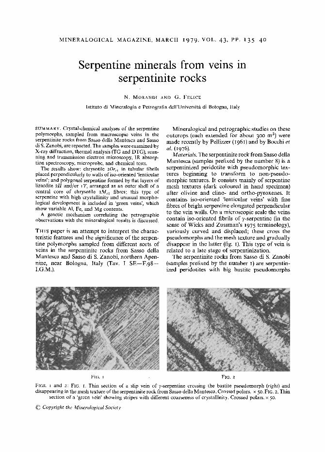
The samples (8-MA, 8-22b, i - i ib, 8-MV, I-I3a, 8-24d ) from the 'green veins' contain, from EM observations, rare platy crystals of lizardite IT mixed with more frequent tubular individuals. The latter show clearly distinctive morphological fea- tures in the high crystallinity, hollow tube (fig. 4)
included in an or-serpentine (Wicks and Zussman, I975) mesh and hour-glass texture.
The whole of the serpentinite rocks contain macroscopic 'green veins' consisting of non-fibrous serpentine in hand specimen. On a microscopic scale the 'green veins' show a zoned texture with stripes characterized by different coarseness of crystallinity (fig. 2). Frequently an unusual'cone-in- cone' development of the serpentine crystals (fig. 3) is visible.
The 'green veins' show normally green to dark green coloured stripes; if the material is weathered (for example the sample 8-MA) the colour changes into a pale yellow-green, if it is sheared a pseudo- fibrous texture is apparent. This type of vein cuts the pseudomorphs, the mesh or hour-glass textures and the 'lenticular veins' and is related to a final stage of serpentinization.
Experimental methods. X-ray diffraction studies were made with a Philips diffractometer on ran- domly oriented powder specimens with a silicon internal standard. Thermal analyses (TG and DTG) were made with a Du Pont apparatus in air and a heating rate of 5o ~ The infra-red absorption spectra were recorded on a 467 Perkin Elmer spectrophotometer, randomly oriented specimens being prepared by the KBr pressed- disc technique (Russell, 1974). The electron micro- graphs and the electron diffraction patterns were recorded using an AEI-EM6 instrument and an ultrasonic cleaner was used to disaggregate and disperse serpentine specimens. The electron-probe microanalyses were made on selected areas of polished samples using an $4 Cambridge Stereo- scan with a semifocusing X-ray spectrometer attachment.
Results. The sample 8-24c , from 'slip veirW, is characterized by pure chrysotile, 1 20r~1. The
1 The notations used here for the serpentine poly- morphs are those used by Wicks and Whittaker (I975).
FIG. 4 FIG. 5
FIGS. 4 and 5: FIG. 4. Electron micrograph of the 8-MA sample. FIG. 5. Electron micrograph of the 8-MV sample showing some examples of the tube-in-tube development.
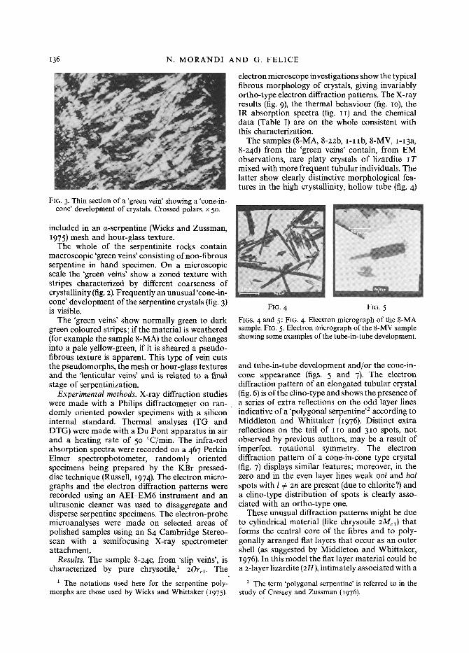
and tube-in-tube development and/or the cone-in- cone appearance (figs. 5 and 7)- The electron diffraction pattern of an elongated tubular crystal (fig. 6) is of the clino-type and shows the presence of a series of extra reflections on the odd layer lines indicative of a 'polygonal serpentine '2 according to Middleton and Whittaker (1976). Distinct extra reflections on the tail of IIO and 31o spots, not observed by previous authors, may be a result of imperfect rotational symmetry. The electron diffraction pattern of a cone-in-cone type crystal (fig. 7) displays similar features; moreover, in the zero and in the even layer lines weak ool and hol spots with 1 @ 2n are present (due to chlorite?) and a clino-type distribution of spots is clearly asso- ciated with an ortho-type one.
These unusual diffraction patterns might be due to cylindrical material (like chrysotile 2Mcl) that forms the central core of the fibres and to poly- gonally arranged fiat layers that occur as an outer shell (as suggested by Middleton and Whittaker, I976 ). In this model the fiat layer material could be a 2-layer lizardite (2H), intimately associated with a
2 The term 'polygonal serpentine' is referred to in the study of Cressey and Zussman (i976).
SERPENTINE MINERALS IN VEINS 137
FIG. 6 FIG. 7 FIG. 8 FIGS. 6 to 8: FIG. 6. Electron diffraction pattern of an elongated tubular crystal of the 8-MA sample. FIG. 7. Electron diffraction pattern of a cone-in-cone crystal, coming from the 8-MV sample. FIG. 8. Electron diffraction pattern of a
fibre arranged parallel to the electron beam (sample 8-MV).
I-layer lizardite (IT). The presence of tubular crystals with a large diameter, randomly oriented in the EM specimens, made it possible to obtain the electron diffraction pattern of the b-c plane of a fibre (fig. 8) (the fibre axis, parallel to a, is approxi- mately parallel to the electron beam). This diffrac- tion pattern is characterized by incomplete ool and oko rings and the stronger spots are radially distributed. This result suggests that the outer shell
1-11b [ L I
I
I
�9 Y s ' '
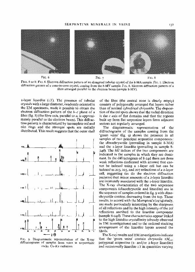
FIG. 9. Diagrammatic representation of the X-ray diffractograms of samples from veins in serpentinite
rocks. Cu-Kc~ radiation.
of the fibre (the central zone is dearly empty) consists of polygonally arranged flat layers rather than of normal cylindrical chrysotile. The disposi- tion of the ool spots shows that the radial direction is the c axis of flat domains and that the regions built up from fiat serpentine layers form adjacent sectors not regularly arranged.
The diagrammatic representation of the diffractograms of the samples coming from the 'green veins' (fig. 9) shows the presence in all samples of two principal serpentine components: the clinochrysotile (prevailing in sample 8-MA) and the i-layer lizardite (prevailing in sample 8- 24d ). The hkl indices of the two components are indicated in the samples in which they are domi- nant. In the diffractogram of 8-24d there a re three weak reflections (indicated with arrows) that can- not be indexed using a I-layer cell but can be indexed as 203, 205, and 207 reflections of a z-layer cell, suggesting (as do the electron diffraction patterns) that minor amounts of a z-layer lizardite are intimately associated with the I-layer lizardite. The X-ray characteristics of the two serpentine components (clinochrysotile and lizardite) are in the sequence of samples ordered in fig. 9 with dino- chrysotile content decreasing from the top. Those results, in accord with the Mumpton's (1974) study, are made particularly interesting by the sharpness of all reflections and by the high intensity of the 2ol reflections ascribed to the lizardite component (sample 8-z4d ). These characteristics appear linked to the high lizardite crystallinity (already observed in EM investigations) and to the ordered stacking arrangement of the lizardite layers around the fibrils.
The X-r@ results and EM investigations indicate that the 'green veins' contain chrysotile 2Me1, polygonal serpentine (I- and/or z-layer lizardite) and occasionally lizardite I T in quantities varying
I38 N. MORANDI AND G. FELICE
from sample to sample, the proportion ofchrysotile being greater in the weathered samples.
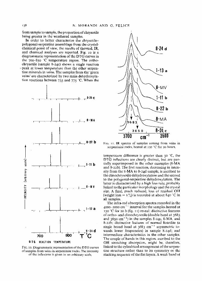
In order to better characterize the chrysotile- polygonal-serpentine assemblage from the crystal- chemical point of view, the results of thermal, IR, and chemical analyses are reported. Fig. IO is a diagrammatic representation of the DTG curves in the 7oo-85o ~ temperature region. The ortho- chrysotile (sample 8-24c ) shows a single reaction
--peak at lower temperature than the other serpen- tine minerals in veins. The samples from the 'green veins' are characterized by two main dehydroxyla- tion reactions between 735 and 775 ~ When the
, ,1 , , , , I ,_8-24C
,- I ,1, , , i 8-MA i I
,8-22 b
n
:>,,
z
, , , ! , I , , 4 ,1-11b
I_,L i i i I r
J I i i t ~ I I i
I , 8-MV
,1-13 a
I I 8-24d 7()0 . . . . 8bo' t ,Oc "1"
OTG REACTION 'TEMPERATURE FIG. I o. Diagrammatic representation of the DTG curves of samples from veins in serpentinite rocks. The intensity
of the inflexions is given in an arbitrary scale,
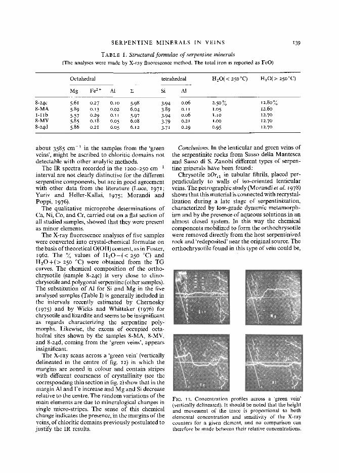
L oo~u I U f-~l ~
3700 cm -1 3500 FIG. I I. IR spectra of samples coming from veins in
serpentinite rocks, heated at I5O ~ for 2o hours.
temperature difference is greater than 3 ~ ~ the DTG inflections are clearly distinct, but are par- tially superimposed in the other examples (8-MA and 8-22b). The first reaction, decreasing in inten- sity from the 8-MA to 8-24d sample, is ascribed to the clinochrysotile dehydroxylation and the second to the polygonal-serpentine dehydroxylation. The latter is characterized by a high loss rate, probably linked to the particular morphology and the crystal size. A final, much reduced, loss of residual OH (weight loss = I ~o) is recorded at about 84o ~ in all samples.
The infra-red absorption spectra recorded in the 4ooo-2ooo cm- 1 interval for the samples heated at 150 eC for 20 h (fig. I I) reveal: distinctive features of ortho- and clinochrysotile (double band at 3685 and 365o cm-1) in the samples 8-24c , 8-MA, and 8-22b; distinctive features of normal lizardite (a single broad band at 3683 cm 1 asymmetric to- wards lower frequencies) in sample 8-24d; and intermediate characteristics in the other samples. The couple of bands in this region, ascribed to the OH stretching absorption, might be, therefore, linked to the cylindrical arrangement of the serpen- tine structure rather than to its symmetry or the stacking sequence of the flat layers. A weak band at
SERPENTINE MINERALS IN VEINS
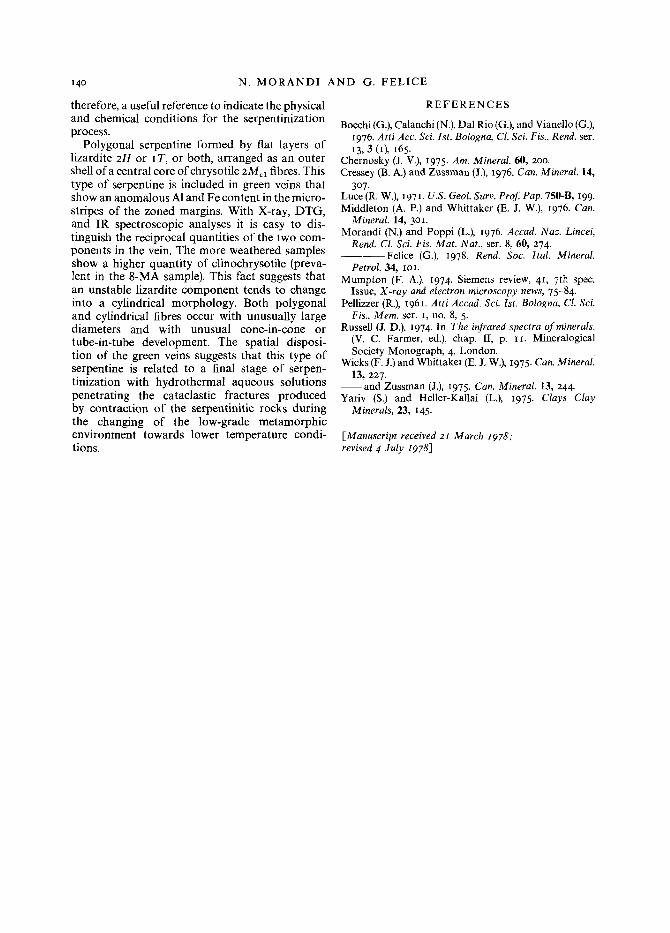
TAB L E I. Structural formulae of serpentine minerals (The analyses were made by X-ray fluorescence method. The total iron is reported as FeO)
139
Octahedral tetrahedral H20 (< 250 ~
Mg Fe 2 + A1 X Si A1
H20(> 25o ~
8-24c 5.6I 0.27 O.IO 5.98 3.94 0.06 2.50~o 8-MA 5.89 o. 13 0.02 6.04 3.89 o. I I I.O 5 I-IIb 5.57 0.29 o.ii 5.97 3.94 0.06 I.iO 8-MV 5.85 o.i8 o.o5 6.08 3.79 o.2~ 1.oo 8-24d 5.86 o.2I o.o 5 6.12 3.71 0.29 0.95
12.80% 12.6o 12.7o 12.7o 12.70
about 3585 cm-1 in the samples from the 'green veins', might be ascribed to chloritic domains not detectable with other analytic methods.
The IR spectra recorded in the I2OO-25o c m - 1
interval are not clearly distinctive for the different serpentine components, but are in good agreement with other data from the literature (Luce, I97I ; Yariv and Heller-Kallai, I975; Morandi and Poppi, 1976 ).
The qualitative microprobe determinations of Ca, Ni, Co, and Cr, carried out on a flat section of all studied samples, showed that they were present as minor elements.
The X-ray fluorescence analyses of five samples were converted into crystal-chemical formulae on the basis of theoretical O(OH) content, as in Foster, 1962. The ~o values of H 2 0 - ( < 25o ~ and H 2 0 + ( > 250 ~ were obtained from the TG curves. The chemical composition of the ortho- chrysotile (sample 8-24c ) is very close to clino- chrysotile and polygonal serpentine (other samples). The substitution of A1 for Si and Mg in the five analysed samples (Table I) is generally included in the intervals recently estimated by Chernosky (I975) and by Wicks and Whittaker (i976) for chrysotile and lizardite and seems to be insignificant as regards characterizing the serpentine poly- morphs. Likewise, the excess of occupied octa- hedral sites shown by the samples 8-MA, 8-MV, and 8-24d, coming from the 'green veins', appears insignificant.
The X-ray scans across a 'green vein' (vertically delineated in the centre of fig. i2) in which the margins are zoned in colour and contain stripes with different coarseness of crystallinity (see the corresponding thin section in fig. 2) show that in the margin A1 and Fe increase and Mg and Si decrease relative to the centre. The random variations of the main elements are due to mineralogical changes in single micro-stripes. The sense of this chemical change indicates the presence, in the margins of the veins, of chloritic domains previously postulated to justify the IR results.
Conclusions. In the lenticular and green veins of the serpentinite rocks from Sasso della Mantesca and Sasso di S. Zanobi different types of serpen- tine minerals have been found:
Chrysotile 20rex in tubular fibrils, placed per- pendicularly to walls of iso-oriented lenticular veins. The petrographic study (Morandi et al. I978) shows that this material is connected with recrystal- lization during a late stage of serpentinization, characterized by low-grade dynamic metamorph- ism and by the presence of aqueous solutions in an almost closed system. In this way the chemical components mobilized to form the orthochrysotile were removed directly from the host serpentinized rock and 'redeposited' near the original source. The orthochrysotile found in this type of vein could be,
\2 s,: -':i !I"
"v% : {~;X:,
.
FIG. I2. Concentration profiles across a 'green vein' (vertically delineated). It should be noted that the height and movement of the trace is proportional to both elemental concentration and sensitivity of the X-ray counters for a given element, and no comparison can therefore be made between their relative concentrations.
I40 N. M O R A N D I AND G. F E L I C E
therefore, a useful reference to indicate the physical and chemical conditions for the serpentinization process.
Polygonal serpentine formed by fiat layers of lizardite 2H or I T, or both, arranged as an outer shell of a central core of chrysotile 2Me i fibres. This type of serpentine is included in green veins that show an anomalous A1 and Fe content in the micro- stripes of the zoned margins. With X-ray, DTG, and IR spectroscopic analyses it is easy to dis- tinguish the reciprocal quantities of the two com- ponents in the vein. The more weathered samples show a higher quantity of clinochrysotile (preva- lent in the 8-MA sample). This fact suggests that an unstable lizardite component tends to change into a cylindrical morphology. Both polygonal and cylindrical fibres occur with unusually large diameters and with unusual cone-in-cone or tube-in-tube development. The spatial disposi- t ion of the green veins suggests that this type of serpentine is related to a final stage of serpen- tinization with hydrothermal aqueous solutions penetrating the cataclastic fractures produced by contraction of the serpentinitic rocks during the changing of the low-grade metamorphic environment towards lower temperature condi- tions.
R E F E R E N C E S
Bocchi (G.), Calanchi (N.), Dal Rio (G.), and Vianello (G.), 1976. Atti Acc. Sci. 1st. Bologna, Cl. Sci. Fis., Rend. ser. 13, 3 (I), 165.
Chernosky (J. V.), 1975. Am. Mineral. 60, 2oo. Cressey (B. A.) and Zussman (J.), 1976. Can. Mineral. 14,
3o7 . Luce (R. W.), I971. U.S. Geol. Surv. Prof. Pap. 750-B, 199. Middleton (A. P.) and Whittaker (E. J. W.), 1976. Can.
Mineral. 14, 3oi. Morandi (N.) and Poppi (L.), 1976. Accad. Naz. Lincei,
Rend. Cl. Sci. Fis. Mat. Nat., ser. 8, 60, 274. - - - - F e l i c e (G.), 1978. Rend. Soc. Ital. Mineral.
Petrol. 34, IOI. Mumpton (F. A.), I974. Siemens review, 4 I, 7th spec.
Issue, X-ray and electron microscopy news, 75-84. Pellizzer (R.), I96I. Atti Accad. Sci. 1st. Bologna, Cl. Sci.
Fis., Mere. ser. I, no. 8, 5. Russell (J. D.), I974. In The infrared spectra of minerals.
(V. C. Farmer, ed.), chap. lI, p. I1. Mineralogical Society Monograph, 4, London.
Wicks (F. J.) and Whittaker (E. J. W.), I975. Can. Mineral 13, 227.
- - a n d Zussman (J.), 1975. Can. Mineral. 13, 244. Yariv (S.) and Heller-Kallai (L.), 1975. Clays Clay
Minerals, 23, I45.
[Manuscript received 21 March I978; revised 4 July I978]


![Geochemistry of Chromitites in Eastern Part of …ophiolite rocks [48] [49], serpentinite alteration of peridotite rocks has occurred in varying degrees (10%- 90% serpentine) in the](https://img.pdfslide.us/doc/110x75/5facd6422f2b5827be4a48d7/geochemistry-of-chromitites-in-eastern-part-of-ophiolite-rocks-48-49-serpentinite.jpg)