Embed Size (px)
Citation preview
Central Journal of Hematology & Transfusion
Cite this article: Karaman S, Gülkilik G, Vural S, Aydın B, Polat N, et al. (2014) Serous Retinal Detachment in a Pediatric Case with Acute Lymphoblastic Leukemia as a First Sign of Leukemic Relapse. J Hematol Transfus 2(1): 1014.
Corresponding authorSerap Karaman, Clinic of Pediatrics, Sisli Etfal Education and Research Hospital, Street No: 9/4 Kücükcekmece-Istanbul , Istanbul, Turkey, Tel: +90-212-6244607; Email:
Submitted: 29 January 2014
Accepted: 12 February 2014
Published: 14 February 2014
Copyright© 2014 Karaman et al.
OPEN ACCESS
Keywords•Leukemia•Retinal detachement•Child
Case Report
Serous Retinal Detachment in a Pediatric Case with Acute Lymphoblastic Leukemia as a First Sign of Leukemic RelapseSerap Karaman1*, Gökhan Gülkilik2, Sema Vural1, Betul Aydın1, Nedim Polat3, Orhan Kizilkaya4 and Banu Arslan2
1Department of Pediatric Hematology-Oncology, Sisli Etfal Training and Research Hospital, Turkey 2Department of Ophthalmology, Istanbul Medipol University, Turkey3Department of Pathology, Sisli Etfal Training and Research Hospital, Turkey4Department of Radiation Oncology, Sisli Etfal Training and Research Hospital, Turkey
Abstract
A 7 year-old boy with T-cell acute lymphoblastic leukemia (T-ALL) was under treatment according to the ALL BFM 2000 protocol. Cerebrospinal fluid analysis did not reveal any blast cells at the beginning of the therapy. The patient developed bilateral visual loss at the third month of therapy. In the fundus examination there was bilateral optic disc edema, and serous retinal detachment with subretinal infiltrates. The patient with his bone marrow in remission was treated by systemic and intratechal chemotherapy combined with craniospinal and orbital radiotherapy. Subretinal fluid began to resolve and visual acuity improved, 24 hours after initiation of systemic and intratecal chemotherapy. The patient died of bone marrow relapse during preparation period for bone marrrow transplantation. Acute visual loss due to serous retinal detachment may be the first sign of relapse in ALL. Detailed ophthalmic examination should be performed in patients with leukemia as early diagnosis and prompt treatment is critical.
INTRODUCTION Ophthalmologic findings can be seen in acute lymphoblastic
leukemia due to leukemic cell invasion or hematological disturbances. Although these findings are detected in the active phase of the disease, they can also be the first sign of a leukemic relapse. All parts of the eye including retina, optic nerve, and choroid can be effected [1]. While microvascular changes, intraretinal and vitreous hemmorrhage and leukemic infiltrates are commonly seen, serous retinal detachment is a rare finding [2,3]. In this case, we report a 7-year-old boy with T cell ALL who was under chemotherapy and presented with acute visual loss due to serous retinal (SRD) detachment as the first sign of leukemic relapse after achieving hematological remission.
CASE REPORTA 7 year-old boy who had been diagnosed with T-ALL
was under remission with ALL BFM 2000 protocol induction treatment. During this time his ophthalmological examination was normal and he had no central nervous system involvement. At the third month of treatment, he presented with acute bilateral
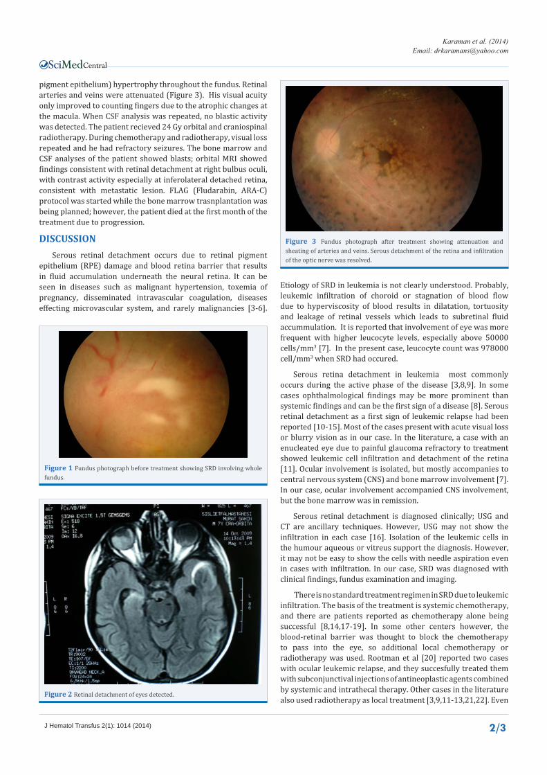
visual loss. He was referred to ophthalmology department for evaluation. On ophthalmological examination, visual acuities were hand motions in both eyes with normal pupil reaction without an afferent pupillary defect. Intraocular pressures (IOPs) were 18 mm Hg in the right eye and 17 mm Hg in the left eye by applanation tonometry. Anterior segment examination was normal. Dilated fundus examination showed bilateral optic disc swelling with subretinal infiltrates and microvascular changes throughout the fundus. Both veins and arteries were attenuated with venous dilatation and segmentation. Retinas were opaque and elevated with subretinal shifting fluid in both eyes (Figure 1). Diagnosis of serous retinal detachment was confirmed with magnetic resonance imaging (Figure 2). Other systemic examination findings were normal and blood tests were within normal range. There was no leukemic blasts in bone marrow aspirate. Cerebro spinal fluid (CSF) analysis revealed leukemic blast cells. Chemotherapy with intensive intrathecal therapy was initiated. His fundoscopic findings improved dramatically 2 days after the initiation of the intrathecal chemotherapy Serous retinal detachment and optic disc edema resolved. There were focal hyperpigmentation areas consistent with RPE (retinal
Central
Karaman et al. (2014)Email:
J Hematol Transfus 2(1): 1014 (2014) 2/3
pigment epithelium) hypertrophy throughout the fundus. Retinal arteries and veins were attenuated (Figure 3). His visual acuity only improved to counting fingers due to the atrophic changes at the macula. When CSF analysis was repeated, no blastic activity was detected. The patient recieved 24 Gy orbital and craniospinal radiotherapy. During chemotherapy and radiotherapy, visual loss repeated and he had refractory seizures. The bone marrow and CSF analyses of the patient showed blasts; orbital MRI showed findings consistent with retinal detachment at right bulbus oculi, with contrast activity especially at inferolateral detached retina, consistent with metastatic lesion. FLAG (Fludarabin, ARA-C) protocol was started while the bone marrow trasnplantation was being planned; however, the patient died at the first month of the treatment due to progression.
DISCUSSIONSerous retinal detachment occurs due to retinal pigment
epithelium (RPE) damage and blood retina barrier that results in fluid accumulation underneath the neural retina. It can be seen in diseases such as malignant hypertension, toxemia of pregnancy, disseminated intravascular coagulation, diseases effecting microvascular system, and rarely malignancies [3-6].
Etiology of SRD in leukemia is not clearly understood. Probably, leukemic infiltration of choroid or stagnation of blood flow due to hyperviscosity of blood results in dilatation, tortuosity and leakage of retinal vessels which leads to subretinal fluid accummulation. It is reported that involvement of eye was more frequent with higher leucocyte levels, especially above 50000 cells/mm3 [7]. In the present case, leucocyte count was 978000 cell/mm3 when SRD had occured.
Serous retina detachment in leukemia most commonly occurs during the active phase of the disease [3,8,9]. In some cases ophthalmological findings may be more prominent than systemic findings and can be the first sign of a disease [8]. Serous retinal detachment as a first sign of leukemic relapse had been reported [10-15]. Most of the cases present with acute visual loss or blurry vision as in our case. In the literature, a case with an enucleated eye due to painful glaucoma refractory to treatment showed leukemic cell infiltration and detachment of the retina [11]. Ocular involvement is isolated, but mostly accompanies to central nervous system (CNS) and bone marrow involvement [7]. In our case, ocular involvement accompanied CNS involvement, but the bone marrow was in remission.
Serous retinal detachment is diagnosed clinically; USG and CT are ancillary techniques. However, USG may not show the infiltration in each case [16]. Isolation of the leukemic cells in the humour aqueous or vitreus support the diagnosis. However, it may not be easy to show the cells with needle aspiration even in cases with infiltration. In our case, SRD was diagnosed with clinical findings, fundus examination and imaging.
There is no standard treatment regimen in SRD due to leukemic infiltration. The basis of the treatment is systemic chemotherapy, and there are patients reported as chemotherapy alone being successful [8,14,17-19]. In some other centers however, the blood-retinal barrier was thought to block the chemotherapy to pass into the eye, so additional local chemotherapy or radiotherapy was used. Rootman et al [20] reported two cases with ocular leukemic relapse, and they succesfully treated them with subconjunctival injections of antineoplastic agents combined by systemic and intrathecal therapy. Other cases in the literature also used radiotherapy as local treatment [3,9,11-13,21,22]. Even
Figure 1 Fundus photograph before treatment showing SRD involving whole fundus.
Figure 2 Retinal detachment of eyes detected.
Figure 3 Fundus photograph after treatment showing attenuation and sheating of arteries and veins. Serous detachment of the retina and infiltration of the optic nerve was resolved.
Central
Karaman et al. (2014)Email:
J Hematol Transfus 2(1): 1014 (2014) 3/3
there is no standard dose for radiotherapy had been described, cases who had no or less than 10 Gy ocular radiotherapy were found to have higher relapse rates, and recommended to recieve minimum 30 Gy [11, 21]. In some cases, ocular findings may be refractory, even the haematologic remission is achieved, and these cases need more aggressive approaches [9]. Enucleation is also recommended if infiltrates are resistant to chemotherapy and radiotherapy [11]. Despite the multimodal treatments, the prognosis of the ocular involvement is poor [1]. In our case, the symptoms and findings regressed in 24 hours after systemic and intrathecal chemotherapy. Radiotherapy was started after induction chemotherapy, but bone marrow relapse occured and CNS and ocular findings relapsed before the end of the treatment. The patient died at the 8th month of ALL diagnosis, and at the 2nd month of his ocular involvement with progressive disease.
As a result, in diagnosis of childhood leukemias, even rare, ocular involvement during therapy should be kept in mind, and patients with ocular findings should be examined for relapse of the disease.
REFERENCES1. Sharma T, Grewal J, Gupta S, Murray PI. Ophthalmic manifestations of
acute leukaemias: the ophthalmologist’s role. Eye (Lond). 2004; 18: 663-672.
2. Reddy SC, Jackson N. Retinopathy in acute leukaemia at initial diagnosis: correlation of fundus lesions and haematological parameters. Acta Ophthalmol Scand. 2004; 82: 81-85.
3. Stewart MW, Gitter KA, Cohen G. Acute leukemia presenting as a unilateral exudative retinal detachment. Retina. 1989; 9: 110-114.
4. Stropes LL, Luft FC. Hypertensive crisis with bilateral bullous retinal detachment. JAMA. 1977; 238: 1948-1949.
5. Jabs DA, Hanneken AM, Schachat AP, Fine SL. Choroidopathy in systemic lupus erythematosus. Arch Ophthalmol. 1988; 106: 230-234.
6. Bjerknes T, Askvik J, Albrechtsen S, Skulstad SM, Dalaker K. Retinal detachment in association with preeclampsia and abruptio placentae. Eur J Obstet Gynecol Reprod Biol. 1995; 60: 91-93.
7. Russo V, Scott IU, Querques G, Stella A, Barone A, Delle Noci N. Orbital and ocular manifestations of acute childhood leukemia: clinical and statistical analysis of 180 patients. Eur J Ophthalmol. 2008; 18: 619-623.
8. Kim J, Chang W, Sagong M. Bilateral serous retinal detachment as a presenting sign of acute lymphoblastic leukemia. Korean J Ophthalmol. 2010; 24: 245-248.
9. Paydas S, Soylu MB, Disel U, Yavuz S, Sahin B, Ersoz C, et al. Serous
retinal detachment in a case with chronic lymphocytic leukemia: no response to systemic and local treatment. Leuk Res. 2003; 27: 557-559.
10. Birinci H, Albayrak D, Oge I, Acar E. Bilateral exudative retinal detachement during maintenance therapy in a child patient with acute lymphoblastic leukemia. Ret-vit. 2000; 8: 96-101
11. Primack JD, Smith ME, Tychsen L. Retinal detachment in a child as the first sign of leukemic relapse: histopathology, MRI findings, treatment, and tumor-free follow up. J Pediatr Ophthalmol Strabismus. 1995; 32: 253-256.
12. Yoshida K, Hasegawa D, Takusagawa A, Kato I, Ogawa C, Echizen N, et al. Bullous exudative retinal detachment due to infiltration of leukemic cells in a child with acute lymphoblastic leukemia. Int J Hematol. 2010; 92: 535-537.
13. Curto ML, D’Angelo P, Jankovic M, Fugardi MG, Ziino O, Casale F. Isolated ocular relapse in childhood acute lymphoblastic leukemia during continuing complete remission. Haematologica. 1996; 81: 47-50.
14. Miyamoto K, Kashii S, Honda Y. Serous retinal detachment caused by leukemic choroidal infiltration during complete remission. Br J Ophthalmol. 2000; 84: 1318-1319.
15. Wu L, Calderón M, Hernández G, Marbis J, Ramírez V. Bilateral exudative retinal detachment as the first sign of relapsing acute myelogenous leukaemia. Clin Experiment Ophthalmol. 2006; 34: 623-625.
16. Wallace RT, Shields JA, Shields CL, Ehya H, Ewing M. Leukemic infiltration of the optic nerve. Arch Ophthalmol. 1991; 109: 1027.
17. Tang RA, Vila-Coro AA, Wall S, Frankel LS. Case report. Acute leukemia presenting as a retinal pigment epithelium detachment. Arch Ophthalmol. 1988; 106: 21-22.
18. Wolff-Kormann PG, Hasenfratz BC, Heinrich B, Kormann B. Successful treatment with hydroxyurea of ocular involvement in chronic myelomonocytic leukemia. N Engl J Med. 1991; 325: 209-210.
19. Malik R, Shah A, Greaney MJ, Dick AD. Bilateral serous macular detachment as a presenting feature of acute lymphoblastic leukemia. Eur J Ophthalmol. 2005; 15: 284-286.
20. Rootman J, Gudauskas G. Treatment of ocular leukemia with local chemotherapy. Cancer Treat Rep. 1985; 69: 119-122.
21. Curto ML, Zingone A, Acquaviva A, Bagnulo S, Calculli L, Cristiani L, et al. Leukemic infiltration of the eye: results of therapy in a retrospective multicentric study. Med Pediatr Oncol. 1989; 17: 134-139.
22. Abdallah E, Hajji Z, Mellal Z, Belmekki M, Bencherifa F, Berraho A. [Macular serous detachment revealing acute lymphoblastic leukemia]. J Fr Ophtalmol. 2005; 28: 39-44.
Karaman S, Gülkilik G, Vural S, Aydın B, Polat N, et al. (2014) Serous Retinal Detachment in a Pediatric Case with Acute Lymphoblastic Leukemia as a First Sign of Leukemic Relapse. J Hematol Transfus 2(1): 1014.
Cite this article