Embed Size (px)
Citation preview

Serology as a Diagnostic
Technique

Characteristics of Any Diagnostic
Techniques
Any useful detection strategy must be: Specific: yield a positive response for only the target
organism or molecule.
Sensitive: identify very small amount of the target the target organism or molecule, even in the presence of other potentially interfering organisms or substances
Simple: to be run efficiently, effectively, and inexpensively on a routine basis
Replicable (or Reliability ): to be repeated different times with different technicians under the same experimental conditions and leading for the same results

Diagnostic Methods
Biological assays: Symptomatology
Microscope: Observation
Serological Assays: Antibodies
Molecular Diagnostic Tools: DNA

Serological Assays
Immunology (sometimes called serology ): is the
study of the serum, and the properties and functions
of its components.
Immuno-technology, is an important arm of
biotechnology, constituting the industrial scale
application of immunological procedures to produce
vaccines, for mass immunisation to prevent prevalent
diseases and/or producing immunological therapeutic
agents to cure the afflicted.
When an infecting organism gains entry into the
mammalian system for the first time, the immune
system of the mammal reacts, mainly in response to
the proteins of the invading organism, generally

Serological Assays As A Diagnostic Tool
Antiserum: Blood serum containing antibodies arising out of immunisation or after an infectious disease.
Production of antibodies: Antibodies are produced by the lymphocytes. The process of antibody production and immune response are complex and both the lymphatic and the blood systems are very closely involved: Polyclonal antibodies: antibodies produced by molecules with
several different antigenic determinants (epitopes) and/or several different cell populations.
Monoclonal antibodies: antibodies produced against a single antigenic determinant (epitope) and/or by a single cell population; hence are very specific.

Antigen-binding sitesAntibody A
Antigen
Antibody BAntibody C
Epitopes(antigenicdeterminants)
Antigen Recognition by Lymphocytes An antigen is any foreign molecule that is specifically
recognized by lymphocytes and elicits a response from
them
A lymphocyte actually recognizes and binds to just a small,
accessible portion of the antigen called an epitope.

Antigen Recognition by
Lymphocytes Epitope: a part of a protein
molecule that acts as an
immunogenic/antigenic
determinant [and so
determines specificities]
a macromolecule, such as a
protein, may contain many
different epitopes, each
capable of stimulating the
production of specific
antibodies, each with a
correspondingly specific
binding site.
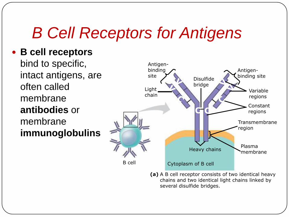
B Cell Receptors for Antigens B cell receptors
bind to specific,
intact antigens, are
often called
membrane
antibodies or
membrane
immunoglobulins
Antigen-bindingsite
Antigen-binding site
Disulfidebridge
Lightchain
Heavy chains
Cytoplasm of B cell
A B cell receptor consists of two identical heavy chains and two identical light chains linked by several disulfide bridges.
(a)
Variableregions
Constantregions
Transmembraneregion
Plasmamembrane
B cell
C C

Antibodies: (also known as immunoglobulins, abbreviated
Ig) are gamma globulin proteins that are produced by the
immune system of an organism in response to exposure to a
foreign molecule and characterised by its specific binding to a
site, related to an epitope of that molecule; induced response
proteins.
The antibodies, like all proteins, are formed of chains of
amino acids, which undergo very complex packing, giving the
proteins a specific and functionally significant final shape
(tertiary configuration), which determines most of the
characteristics of the protein.

Molecular Structure of Antibodies The conventional model of the
Ig molecules is a ‘Y’ shapedconfiguration, with two heavy chains and two light chains, with two open arms containing the antigen combining sites, which occur on both the light and the heavy chains.
The two heavy chains are bound together by disulphide bonds.
At any point, the molecule has two chain sections, parallel to each other.

Clonal Selection
of Lymphocytes
In a primary immune
response binding of
antigen to a mature
lymphocyte induces the
lymphocyte’s
proliferation and
differentiation, a process
called clonal selection
Clonal selection of B
cells generates a clone of
short-lived activated
effector cells and a clone of

Antigen molecules
Antigenreceptor
B cells thatdiffer inantigen
specificity
Antibodymolecules
Clone of memory cells
Clone of plasma cells
Antigen moleculesbind to the antigenreceptors of only oneof the three B cellsshown.
The selected B cellproliferates, forminga clone of identicalcells bearingreceptors for theselecting antigen.
Some proliferatingcells develop intoshort-lived plasmacells that secreteantibodies specificfor the antigen.
Some proliferating cells develop into
long-lived memory cells that can
respond rapidly uponsubsequent exposureto the same antigen.
Clonal Selection of Lymphocytes

In the secondary immune response memory cells
facilitate a faster, more efficient responseAntibody c
oncentr
ation
(arb
itra
ry u
nits)
104
103
102
101
100
0 7 14 21 28 35 42 49 56
Time (days)
Antibodiesto A
Antibodiesto B
Primaryresponse toantigen Aproduces anti-bodies to A
2Day 1: First exposure toantigen A
1 Day 28: Second exposureto antigen A; firstexposure to antigen B
3 Secondary response to anti-gen A produces antibodiesto A; primary response to anti-gen B produces antibodies to B
4

Antibody
Classes
These are the five
major classes of
antibodies, or
immunoglobulins
differ in their
distributions and
functions within the
body
IgM: primary response
and 10 active site, 5 Y-
shape
IgG: one single Y-
shape 2 active sites,
secondary response
First Ig class produced after initial exposure to antigen; then its concentration in the blood declines
Most abundant Ig class in blood; also present in tissue fluids
Only Ig class that crosses placenta, thus conferring passive immunity on fetus
Promotes opsonization, neutralization, and agglutination of antigens; less effective in complement activation than IgM
Present in secretions such as tears, saliva, mucus, and breast milk
Triggers release from mast cells and basophils of histamine and other chemicals that cause allergic reactions
Present primarily on surface of naive B cells that havenot been exposed to antigens
IgM(pentamer)
IgG(monomer)
IgA(dimer)
IgE(monomer)
J chain
Secretorycomponent
J chain
Transmembraneregion
IgD(monomer)
Promotes neutralization and agglutination of antigens; very effective in complement activation
Provides localized defense of mucous membranes byagglutination and neutralization of antigens
Presence in breast milk confers passive immunity onnursing infant
Acts as antigen receptor in antigen-stimulated proliferation and differentiation of B cells (clonal selection)

Binding of antibodies to antigensinactivates antigens by
Viral neutralization(blocks binding to host)
and opsonization (increasesphagocytosis)
Agglutination ofantigen-bearing particles,
such as microbes
Precipitation ofsoluble antigens
Activation of complement systemand pore formation
Bacterium
Virus Bacteria
Solubleantigens Foreign cell
Complementproteins
MAC
Pore
Enhances
Phagocytosis
Leads to
Cell lysis
Macrophage
Antibody-mediated mechanisms of antigen disposal

Antiserum PreparationObtaining PAb for the diagnostic purposes:
1. Choose of the animal: decide wither polyclonal or
monoclonal
2. Preparation of antigen
3. Immunization: the antigen is emulsified sometimes
with with an equal volume of complete adjuvant
(Freund’s adjuvant- mannitol monoleate mixed
with paraffin oil)
4. Collection of sera: (the supernatant)
5. Titration….
6. Storage: (addition of antiseptic substances (NaN3);
lyophilized; at temperature below -20°C)

Typical Serological Profile After Acute
Infection
Note that during reinfection, IgM may be absent or present at a low level transiently
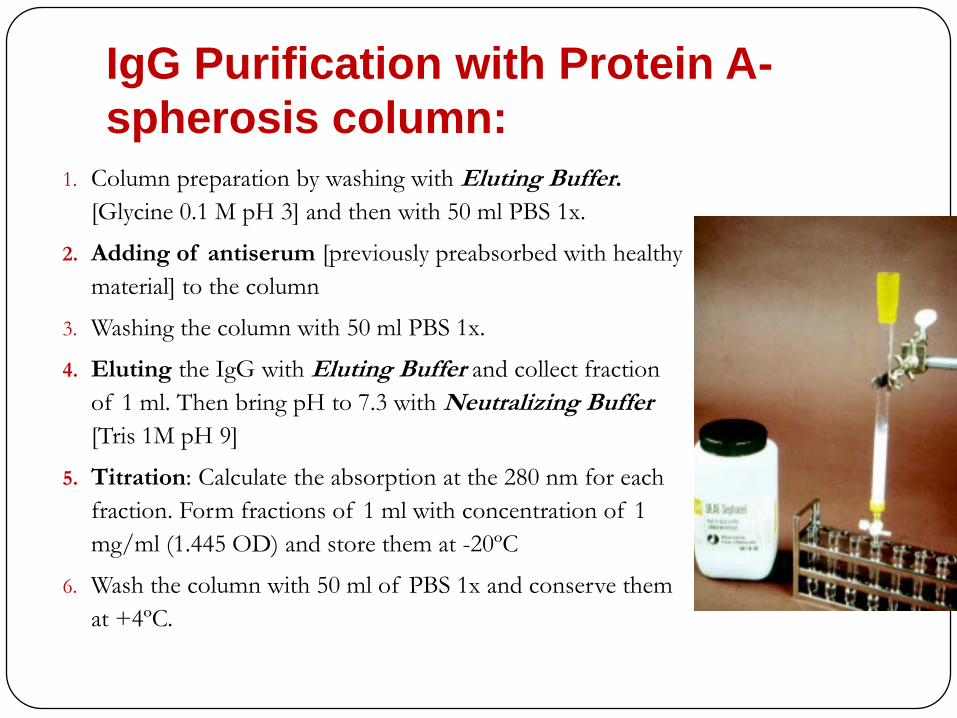
IgG Purification with Protein A-
spherosis column:1. Column preparation by washing with Eluting Buffer.
[Glycine 0.1 M pH 3] and then with 50 ml PBS 1x.
2. Adding of antiserum [previously preabsorbed with healthy
material] to the column
3. Washing the column with 50 ml PBS 1x.
4. Eluting the IgG with Eluting Buffer and collect fraction
of 1 ml. Then bring pH to 7.3 with Neutralizing Buffer
[Tris 1M pH 9]
5. Titration: Calculate the absorption at the 280 nm for each
fraction. Form fractions of 1 ml with concentration of 1
mg/ml (1.445 OD) and store them at -20ºC
6. Wash the column with 50 ml of PBS 1x and conserve them
at +4ºC.

Monoclonal Antibodies Immunochemical techniques are extremely useful for the
rapid and accurate routine detection of host pathogens
and ultimately the diagnosis of host disease and their
relatedness
The introduction of hybridoma technology has provided
methods for the production of homologous and biochemically
defined immunological reagents of identical specificity
Hybridoma are produced by a single cell line and are directed
against a unique epitop of the immunizing antigen.

Antibodies-
Production
1. Spleen cells are removed washed
minced and gently agitated to
release individual cells, some of
which are
antibody-producing B cells.
2. Myeloma cells (immortal B-cell Line)
are genetically defective for the
enzyme:
“Hypoxanthine-Guanine
PhosphoRibosylTransferase
(HGPRT-)
3. The combined cells are mixed with
35% PEG (to facilitate fusion
between cells) for few minutes to
form hybridoma, then transferred to
a growth medium containing:
Hypoxanthine, Aminopterin, and
Thymidine (HAT medium).
4. The HAT medium allow only the

Explanation of use HAT
medium
The myeloma (HGPRT-) will not be able to synthesize
purines in HAT medium:
The (HGPRT-) myeloma alone or as myeloma-myeloma fusion
cells cannot use the Hypoxanthine as a precursor for
biosynthesis of purines (adenine and guanine), which of
course essential for nucleic acid synthesis.
They may use the other pathway for purine synthesis by the
enzyme dihydrofolate reductase, but the enzyme
Aminopterin which included in the media will inhibit the
enzyme dihydrofolate reductase activity.
Spleen+myeloma fused cells would be the only
survivors in HAT medium as they contain HGPRT
which allow the use of Hypoxanthine for biosynthesis
of purines.
Although Thymidine will overcome the blockage of
dihydrofolate reductase activity by Aminopterin

Monoclonal Antibodies
Production

Serological Techniques In serology Ab acts as a probe for detection of Ag => ppt
Direct Methods: it is possible to visualize the Antigen
Antibody reaction without the help of other reactions
Precipitation or agglutination
Immunoelectron microscopy
Agar precipitation
Indirect Methods:
ELISA

Comparison among the two
Antiserum Preparation
Polyclonal Antibodies (Pabs) has two drawbacks:
The prepared amount of PAbs produced each time
could be varied.
PAbs couldn't be used to distinguish two similar
target protein.
Monoclonal Antibodies (MAbs) are useful for their:
Reproducibility
Specificity
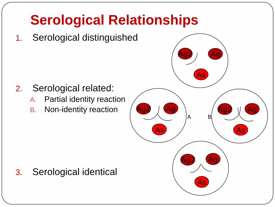
Serological Relationships1. Serological distinguished
2. Serological related:A. Partial identity reaction
B. Non-identity reaction
3. Serological identical
Ag2 Ag
As
Ag2 Ag
As
Ag2 Ag
As
Ag2 Ag
As
A B
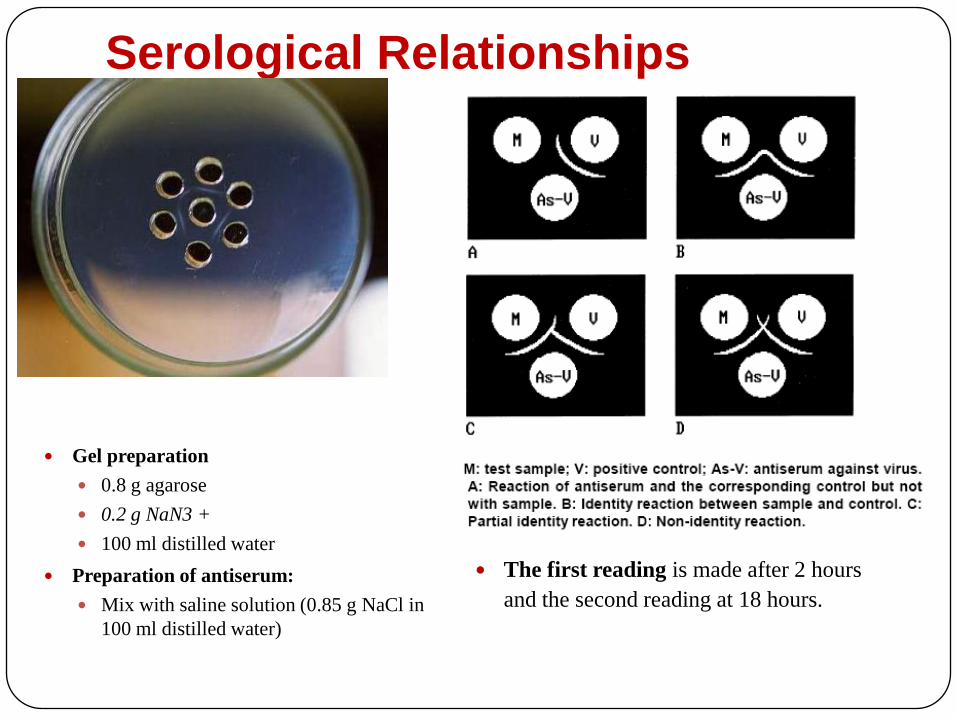
Serological Relationships
Gel preparation
0.8 g agarose
0.2 g NaN3 +
100 ml distilled water
Preparation of antiserum:
Mix with saline solution (0.85 g NaCl in
100 ml distilled water)
The first reading is made after 2 hours
and the second reading at 18 hours.

Immunological Diagnosis: ELISA
ELISA Diagnostic Kits – Some
Characteristics
A number of disease detection kits have been developed for use at the site where a disease is suspected.
These kits, which in most cases do not require laboratory equipment, are very useful
Some tests only take five minutes to perform.
The diagnostic kits are based on a method that uses proteins called antibodies to detect disease causal agents called antigen.
One of the technique used is called ELISA (enzyme-linked immunosorbent assay).

ELISA Diagnostic Kits This assay is based on the ability of an antibody to recognize and
bind to a specific antigen, a substance associated with a host pathogen.
The antibodies used in the diagnostic kits are highly purified proteins produced by injecting a warm-blooded animal (like a rabbit) with an antigen associated with one particular host disease.
The animal reacts to the antigen and produces antibodies. The antibodies produced recognize and react only with the proteins associated with the causal agent of that host disease.
Color changes on the unit’s surface indicate a positive (disease present) reaction.

ELISA Diagnostic Kits

ELISA-test
Sample preparations

Generalized ELISA-test protocol1) Coating of the plates
(wells):
with IgG diluted in
coating buffer
2) Adding of the extract
: Coating antibody
: specific Protein
: non specific proteins
Incubation at 37°C
Incubation at 37°C

Generalized ELISA-test
protocol
5) Observe or measure the amount of colored product
3) Adding of conjugated IgG
diluted in conjugating buffer.
: Labeled antibody with Alkaline phosphatase
4) Adding of substrate solution
p-nitrophenyl-phosphate
diluted in substrate buffer.
: Substrate Enzyme
Incubation at 37°C

ELISA-test
Light Filter 405
Healthy O.D * constant (2.5) >
Positive control

Different Types of ELISA-test
Double Antibody
Sandwich ELISA (DAS)
Triple Antibody
Sandwich-Indirect
ELISA (TAS)
Enhanced Double
Antibody Sandwich-
Indirect ELISA (DAS-I)
: Coating antibody
: Antigen
: Labeled antibody
: Substrate Enzyme
: The intermediate antibody
: The labeled anti-mouse IgG
: Biotinylated anti-body
: Streptavidine-enzyme
conjugates

F(ab')2 Antibodies Production
Papain cleavage: (Fc)
Pepsin cleavage [F(ab)2]

F(ab')2 Antibodies Production
• A system has been devised to carry out DAS-I using asingle antiserum
• The antibodies are treated with the enzyme pepsin toremove the Fc portion of the molecule.
• The remaining F(ab')2, fragment still has the antigenbinding sites and will bind to the immunosorbent, but willnot bind to protein A.
• The F(ab')2 fragments are used as trapping “antibody”and the whole antibody is used as the intermediateantibody.
• It is possible to use specific substances that recognize Fcportion for increase the reaction sensitivity.