Embed Size (px)
Citation preview
Serological Tools For Investigating Problems In
Immunohaematology
Dr.K.Vasantha
Deputy Director (Retd.)
National Institute of Immunohaematology
(ICMR)
Parel, Mumbai
• Immunohaematology is an application of
the principles of Immunology to the study
of red cell antigens and their corresponding
antibodies in blood for resolving the
problems
Purpose
Important overall purpose of the field of
Immunohaematology is to predict a successful
transfusion outcome
Outcome
Pretransfusion testings to ensure the best
possible results in blood transfusion i.e the
transfused red blood cells will have an
acceptable survival rate
Detection of antigens or antibodies
• Agglutination
Sensitization
Visible agglutination
• Neutralization
Used in secretory studies
Basic Laboratory Investigations
• ABO and Rh Blood Grouping
• Crossmatching
• Detection of unexpected antibodies
• Selection of compatible units
• Early part of 20th century, many important
discoveries leading to safer compatibility
procedures
• Latter part of the century saw developments
in serological methodology.
• Molecular biology in Immunohaematology.
Importance of blood group systems
• Blood group antibodies play an important role in
transfusion medicine - in blood transfusion and pregnancy.
Though all antibodies are not clinically significant many
have the potential to be clinically significant
• Importance of the system is based on whether the
antibodies can facilitate accelerated destruction of red
cells having the corresponding antigen
• Unlike ABO & Rh, other blood group antigens are not
tested for in blood banks as their antibodies are not of
frequent occurrence
ABO GROUPING
Cell and serum grouping is a must
Cell grouping with standard antisera and
serum grouping with pooled A, B and O cells
Any discrepancy to be resolved
Problems due to discrepancies
in cell & serum grouping
Technical errors
• clerical errors
• dysfunction of reagents or equipments
• procedural errors
RBC grouping & serum grouping
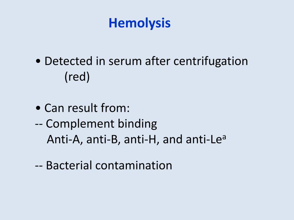
• Dete ted i seru after e trifugatio (red)
• Ca result fro : -- Complement binding
Anti-A, anti-B, anti-H, and anti-Lea
-- Bacterial contamination
Hemolysis

Problems due to discrepancies
in cell & serum grouping
* Problems in RBC grouping
a. Weak reacting or missing antigens
b. Unexpected antigen reactions
* Problems in serum grouping
a. Weak reacting or missing antibody
b. Unexpected antibody reaction
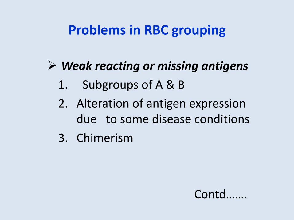
Problems in RBC grouping
Weak reacting or missing antigens
1. Subgroups of A & B
2. Alteration of antigen expression
due to some disease conditions
3. Chimerism
Contd…….
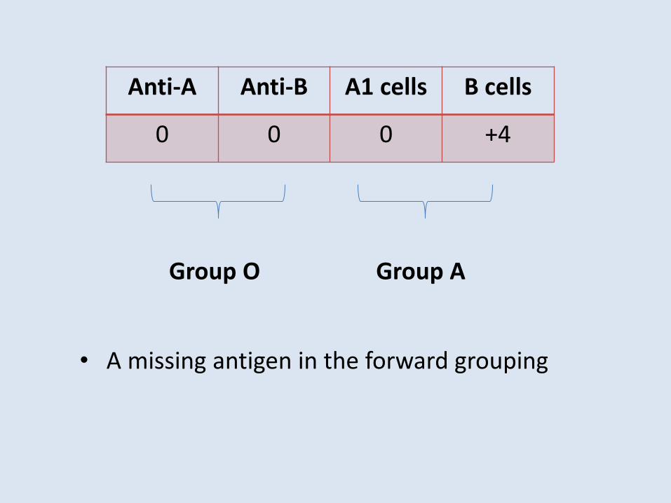
Anti-A Anti-B A1 cells B cells
0 0 0 +4
Group O Group A
• A missing antigen in the forward grouping
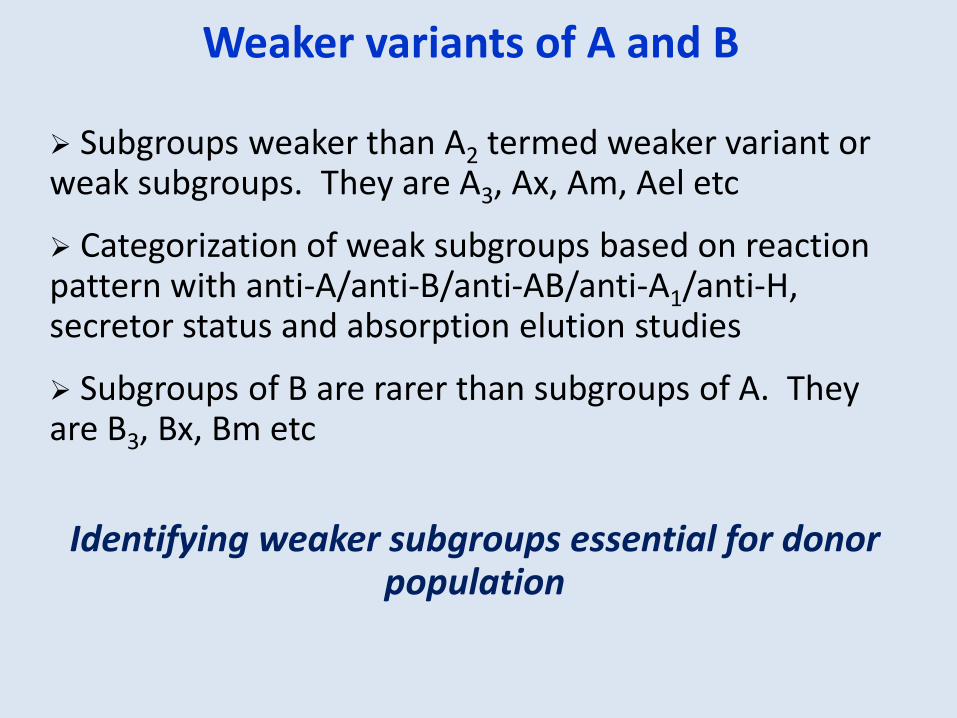
Subgroups weaker than A2 termed weaker variant or weak subgroups. They are A3, Ax, Am, Ael etc
Categorization of weak subgroups based on reaction pattern with anti-A/anti-B/anti-AB/anti-A1/anti-H, secretor status and absorption elution studies
Subgroups of B are rarer than subgroups of A. They are B3, Bx, Bm etc
Identifying weaker subgroups essential for donor
population
Weaker variants of A and B
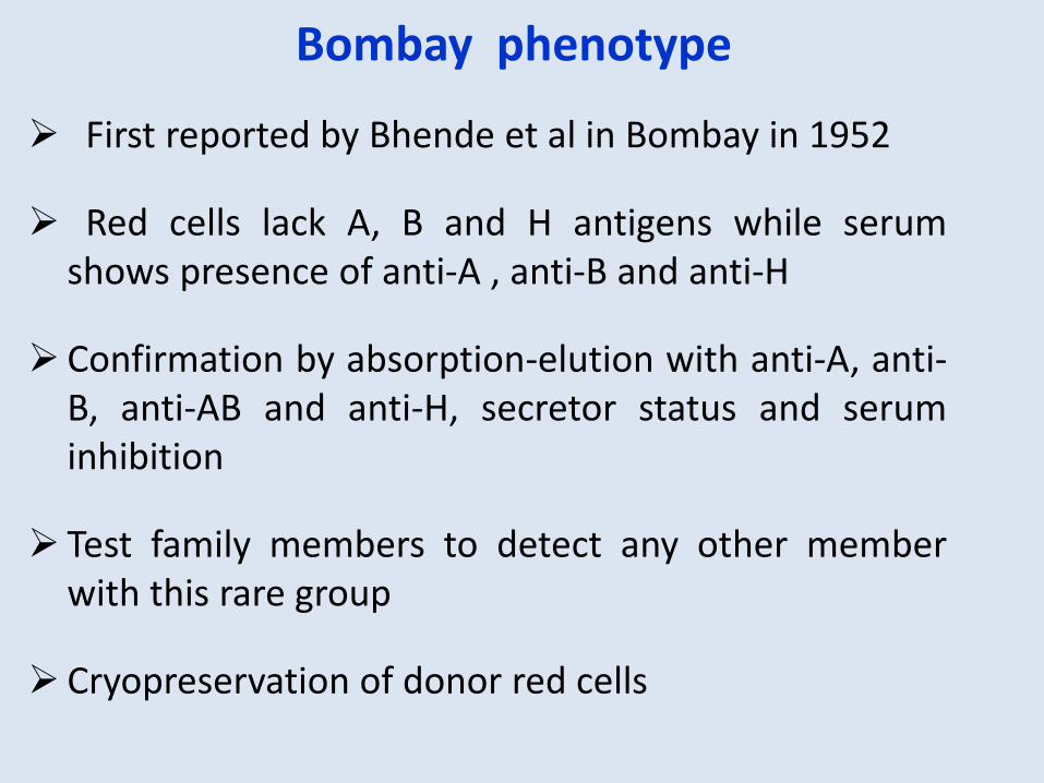
Bombay phenotype
First reported by Bhende et al in Bombay in 1952
Red cells lack A, B and H antigens while serum
shows presence of anti-A , anti-B and anti-H
Confirmation by absorption-elution with anti-A, anti-
B, anti-AB and anti-H, secretor status and serum
inhibition
Test family members to detect any other member
with this rare group
Cryopreservation of donor red cells
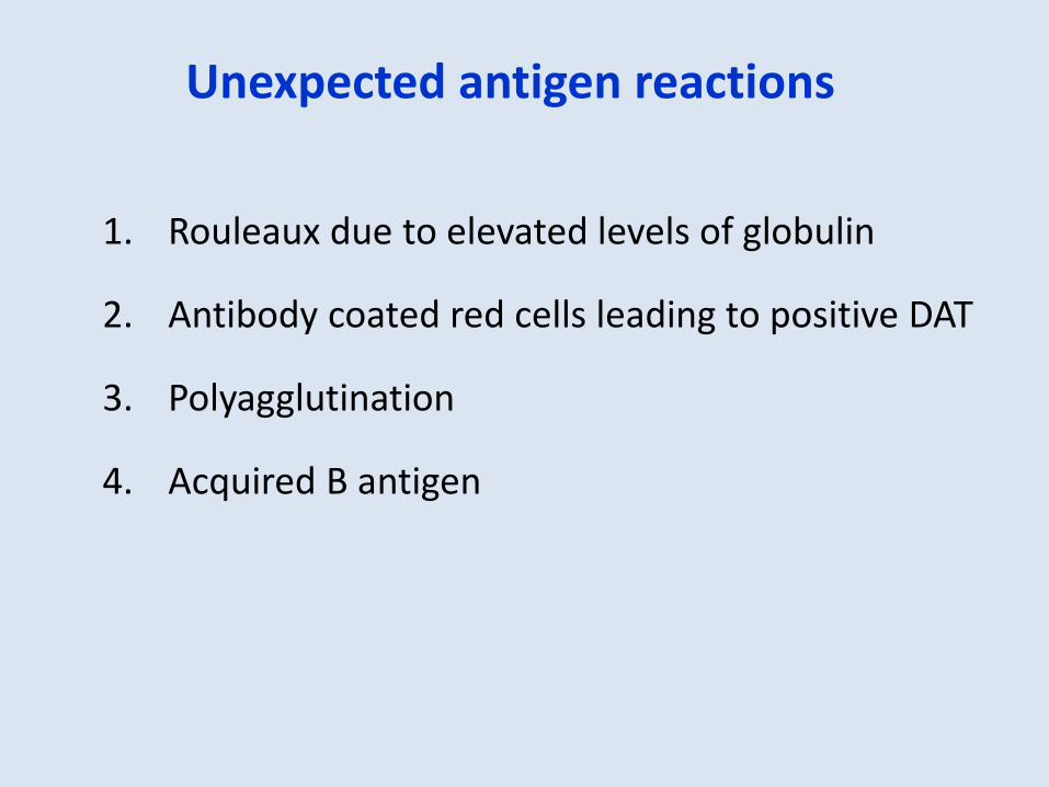
1. Rouleaux due to elevated levels of globulin
2. Antibody coated red cells leading to positive DAT
3. Polyagglutination
4. Acquired B antigen
Unexpected antigen reactions
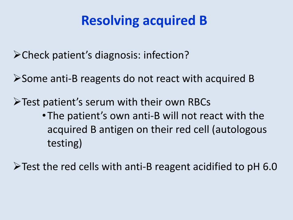
Che k patie t’s diag osis: i fe tio ?
Some anti-B reagents do not react with acquired B
Test patie t’s seru ith their o RBCs
•The patie t’s o a ti-B will not react with the
acquired B antigen on their red cell (autologous
testing)
Test the red cells with anti-B reagent acidified to pH 6.0
Resolving acquired B
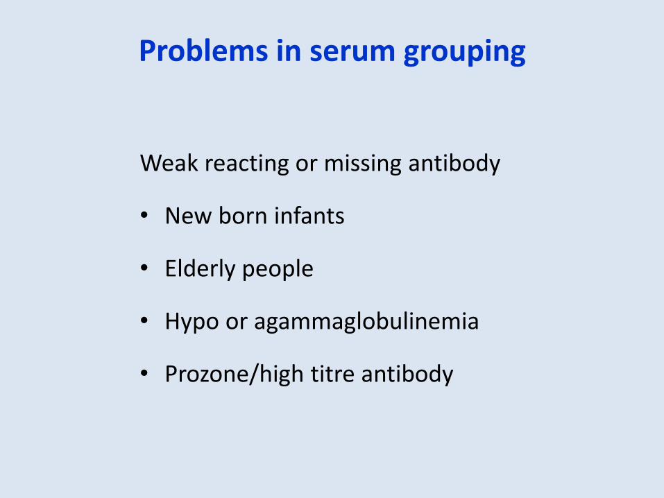
Problems in serum grouping
Weak reacting or missing antibody
• New born infants
• Elderly people
• Hypo or agammaglobulinemia
• Prozone/high titre antibody
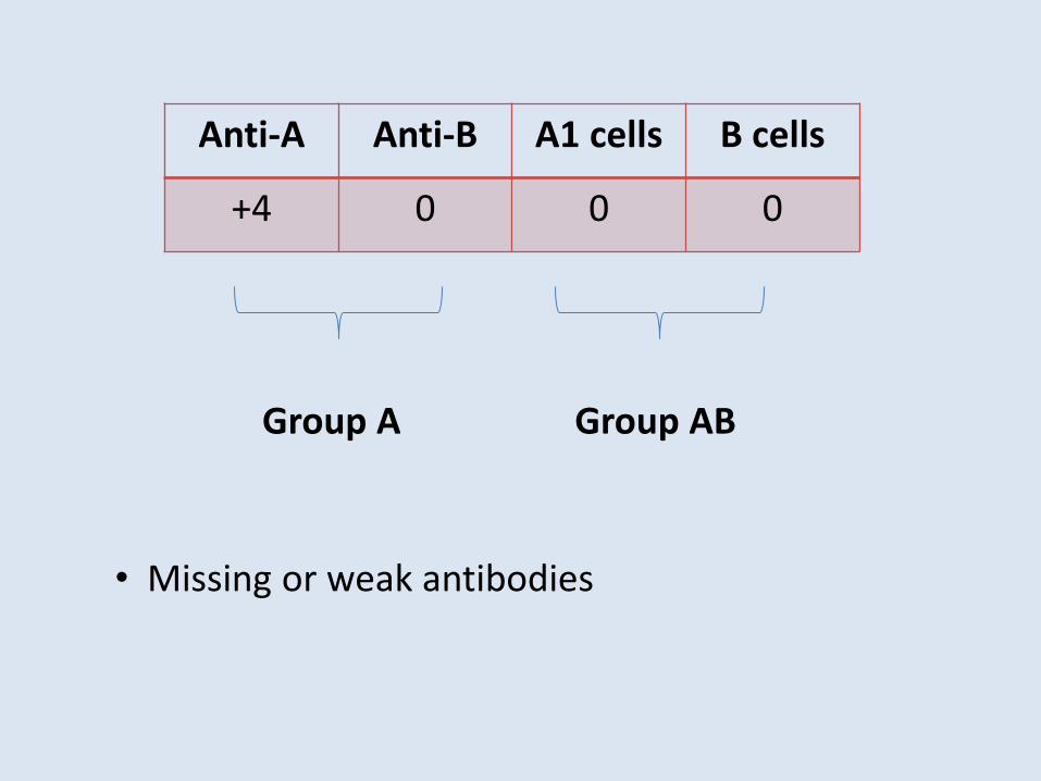
Anti-A Anti-B A1 cells B cells
+4 0 0 0
Group A Group AB
• Missing or weak antibodies

• Rouleaux
• A2 and A2B with naturally occurring anti-A1
• Cold autoantibodies like anti-I
• Cold alloantibodies like anti-M, anti-N etc
• Passively acquired antibodies
Unexpected antibody reactions
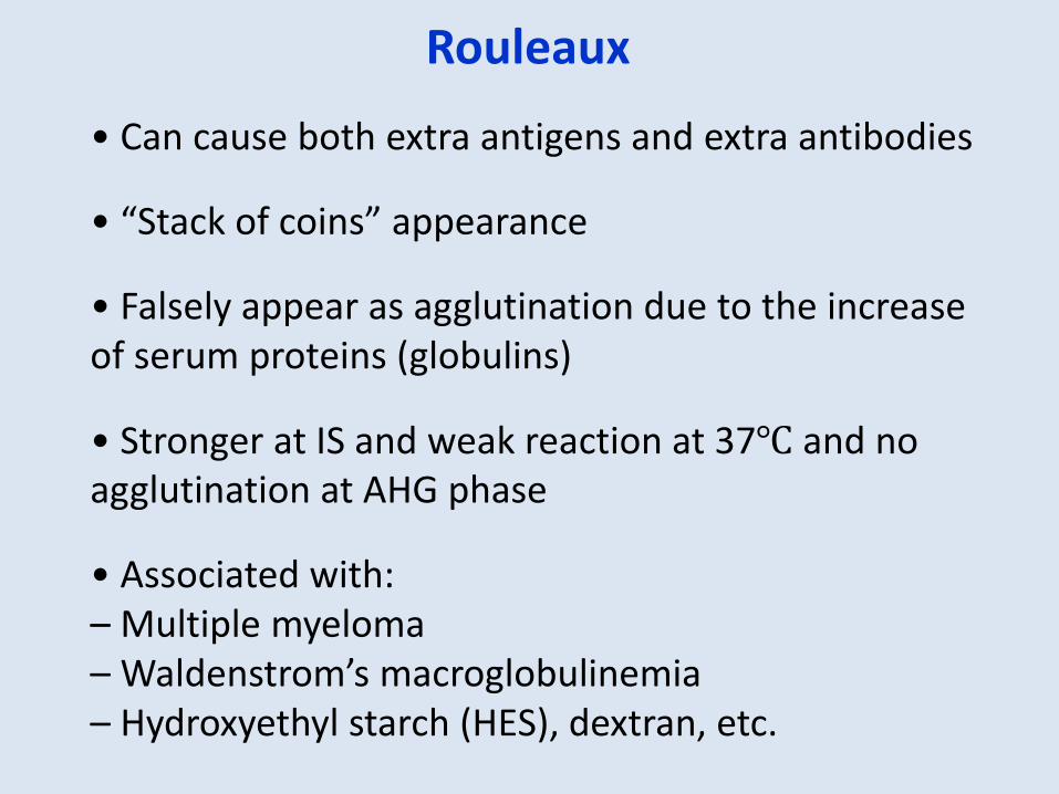
• Ca ause oth e tra a tige s a d e tra a ti odies
• “ta k of oi s appeara e
• Falsel appear as aggluti atio due to the i rease of serum proteins (globulins)
• “tro ger at I“ a d eak rea tio at 37℃ and no
agglutination at AHG phase
• Asso iated ith: – Multiple myeloma
– Walde stro ’s macroglobulinemia
– Hydroxyethyl starch (HES), dextran, etc.
Rouleaux
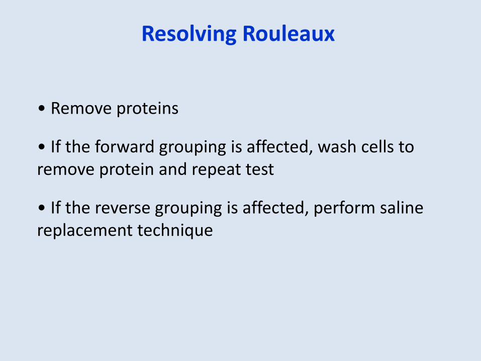
• Re o e protei s
• If the for ard groupi g is affe ted, ash ells to remove protein and repeat test
• If the re erse groupi g is affe ted, perfor sali e
replacement technique
Resolving Rouleaux
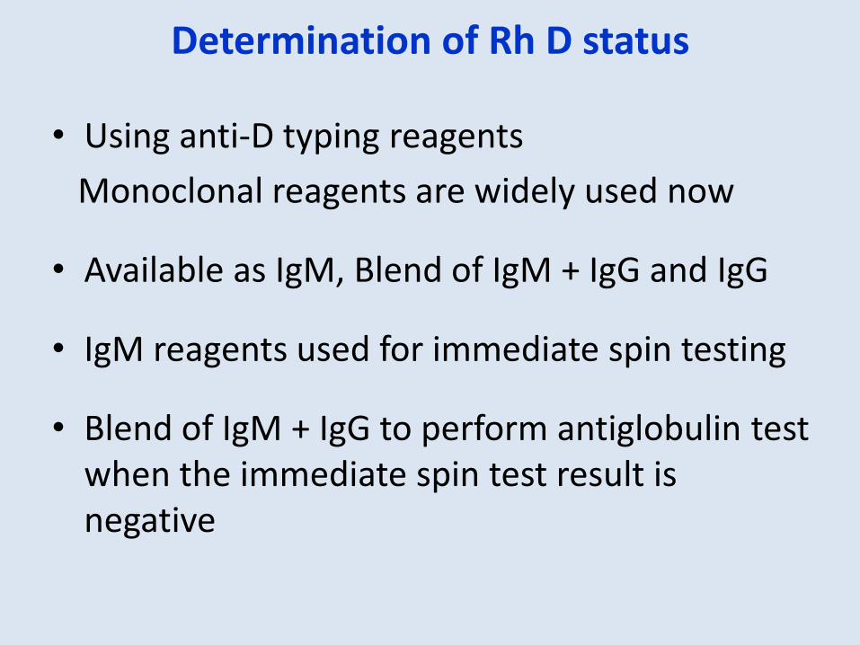
Determination of Rh D status
• Using anti-D typing reagents
Monoclonal reagents are widely used now
• Available as IgM, Blend of IgM + IgG and IgG
• IgM reagents used for immediate spin testing
• Blend of IgM + IgG to perform antiglobulin test
when the immediate spin test result is
negative
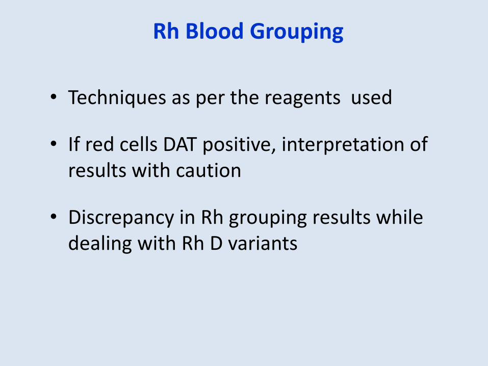
Rh Blood Grouping
• Techniques as per the reagents used
• If red cells DAT positive, interpretation of
results with caution
• Discrepancy in Rh grouping results while
dealing with Rh D variants
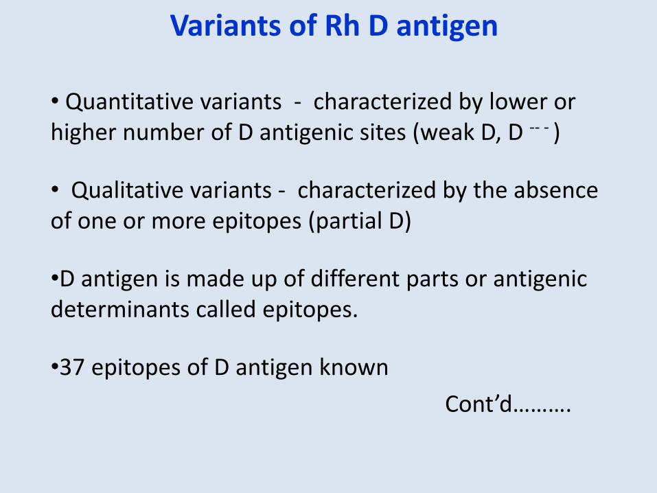
Variants of Rh D antigen
• Quantitative variants - characterized by lower or
higher number of D antigenic sites (weak D, D -- - )
• Qualitative variants - characterized by the absence
of one or more epitopes (partial D)
•D antigen is made up of different parts or antigenic
determinants called epitopes.
•37 epitopes of D antigen known
Co t’d……….
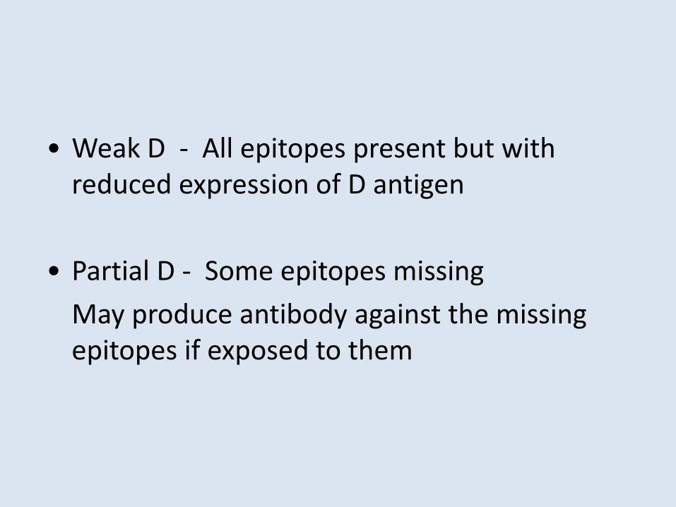
• Weak D - All epitopes present but with
reduced expression of D antigen
• Partial D - Some epitopes missing
May produce antibody against the missing
epitopes if exposed to them
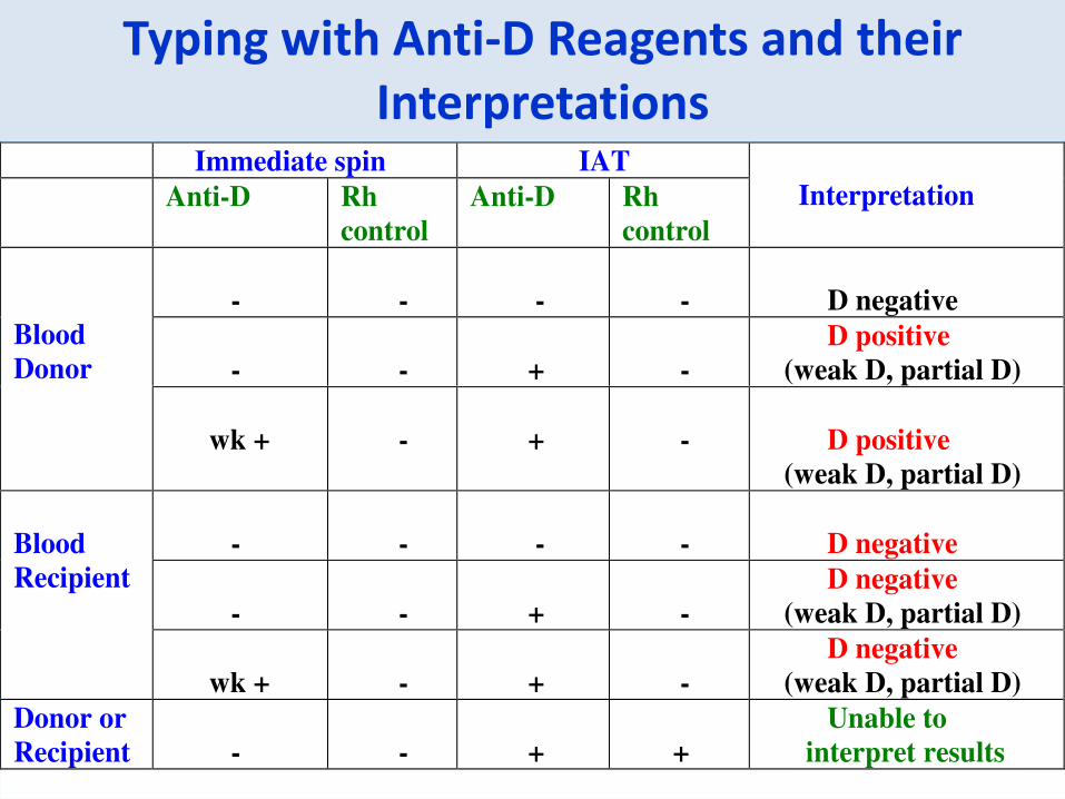
Typing with Anti-D Reagents and their
Interpretations Immediate spin IAT
Anti-D Rh
control
Anti-D Rh
control
Interpretation
- - - - D negative
- - + -
D positive
(weak D, partial D)
Blood
Donor
wk + - + - D positive
(weak D, partial D)
- - - - D negative
- - + -
D negative
(weak D, partial D)
Blood
Recipient
wk + - + -
D negative
(weak D, partial D)
Donor or
Recipient - - + +
Unable to
interpret results
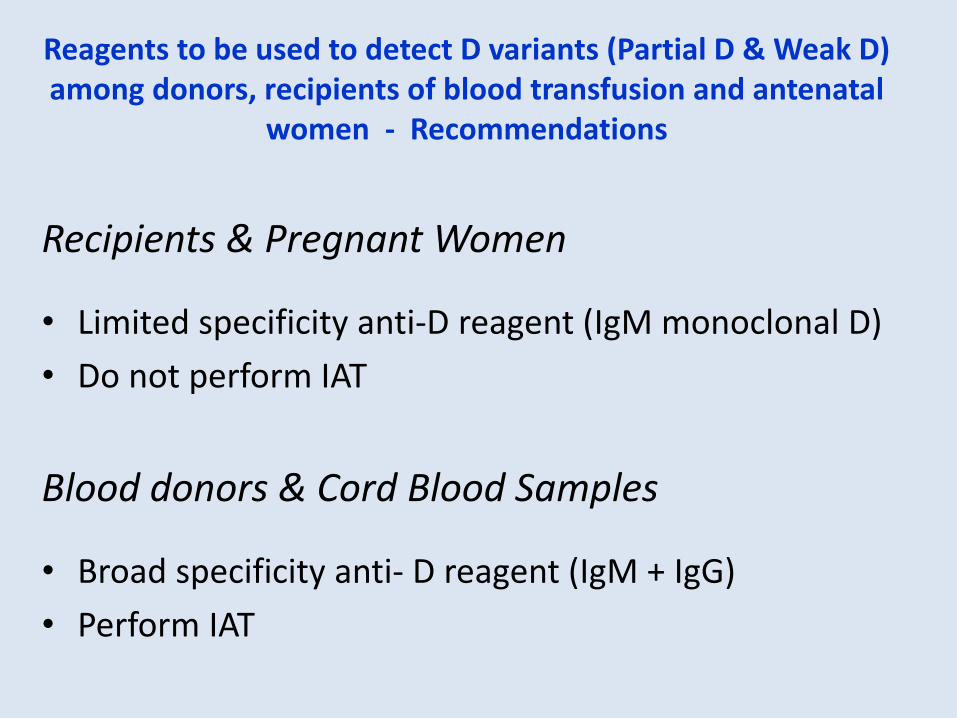
Reagents to be used to detect D variants (Partial D & Weak D)
among donors, recipients of blood transfusion and antenatal
women - Recommendations
Recipients & Pregnant Women
• Limited specificity anti-D reagent (IgM monoclonal D)
• Do not perform IAT
Blood donors & Cord Blood Samples
• Broad specificity anti- D reagent (IgM + IgG)
• Perform IAT
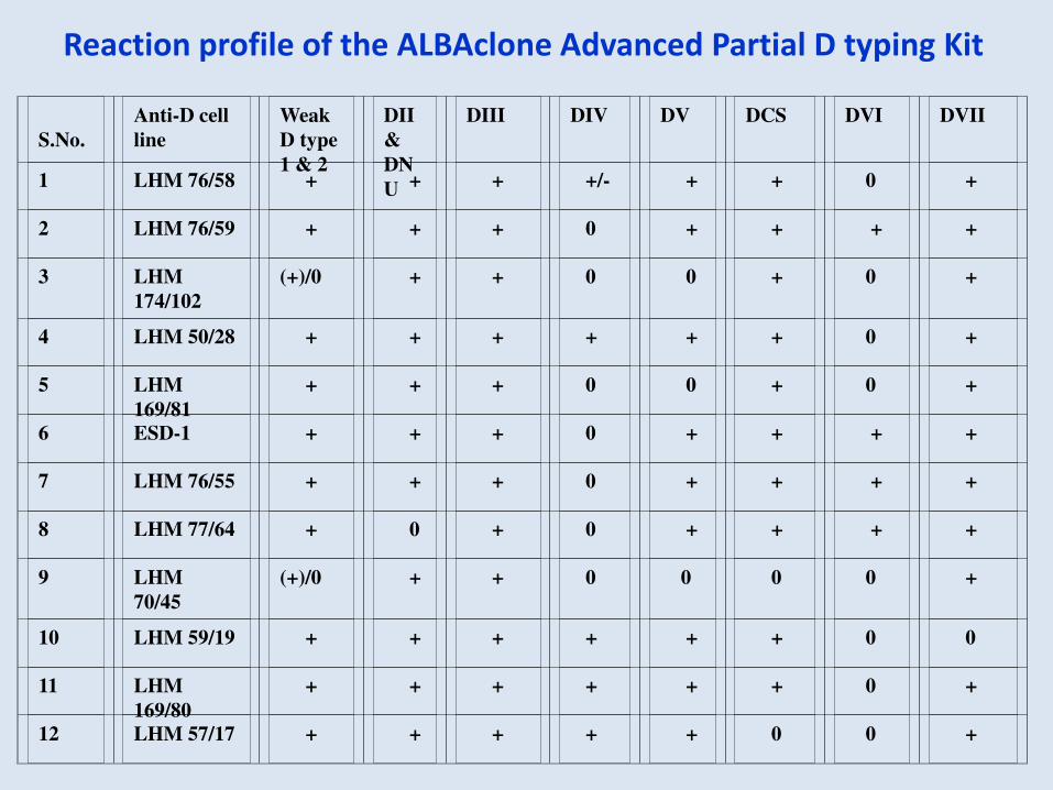
S.No.
Anti-D cell
line
Weak
D type
1 & 2
DII
&
DN
U
DIII
DIV
DV
DCS
DVI
DVII
1
LHM 76/58
+
+
+
+/-
+
+
0
+
2
LHM 76/59
+
+
+
0
+
+
+
+
3
LHM
174/102
(+)/0
+
+
0
0
+
0
+
4
LHM 50/28
+
+
+
+
+
+
0
+
5
LHM
169/81
+
+
+
0
0
+
0
+
6
ESD-1
+
+
+
0
+
+
+
+
7
LHM 76/55
+
+
+
0
+
+
+
+
8
LHM 77/64
+
0
+
0
+
+
+
+
9
LHM
70/45
(+)/0
+
+
0
0
0
0
+
10
LHM 59/19
+
+
+
+
+
+
0
0
11
LHM
169/80
+
+
+
+
+
+
0
+
12
LHM 57/17
+
+
+
+
+
0
0
+
Reaction profile of the ALBAclone Advanced Partial D typing Kit
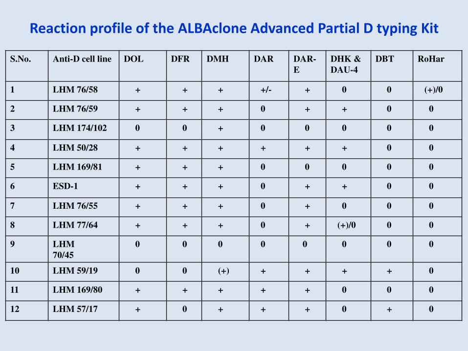
S.No. Anti-D cell line DOL DFR DMH DAR DAR-
E DHK &
DAU-4 DBT RoHar
1 LHM 76/58 + + + +/- + 0 0 (+)/0
2 LHM 76/59 + + + 0 + + 0 0
3 LHM 174/102 0 0 + 0 0 0 0 0
4 LHM 50/28 + + + + + + 0 0
5 LHM 169/81 + + + 0 0 0 0 0
6 ESD-1 + + + 0 + + 0 0
7 LHM 76/55 + + + 0 + 0 0 0
8 LHM 77/64 + + + 0 + (+)/0 0 0
9 LHM
70/45 0 0 0 0 0 0 0 0
10 LHM 59/19 0 0 (+) + + + + 0
11 LHM 169/80 + + + + + 0 0 0
12 LHM 57/17 + 0 + + + 0 + 0
Reaction profile of the ALBAclone Advanced Partial D typing Kit
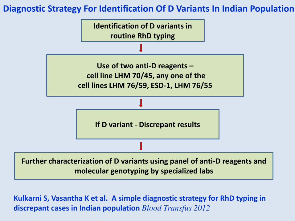
Diagnostic Strategy For Identification Of D Variants In Indian Population
Further characterization of D variants using panel of anti-D reagents and
molecular genotyping by specialized labs
If D variant - Discrepant results
Use of two anti-D reagents –
cell line LHM 70/45, any one of the
cell lines LHM 76/59, ESD-1, LHM 76/55
Identification of D variants in
routine RhD typing
Kulkarni S, Vasantha K et al. A simple diagnostic strategy for RhD typing in
discrepant cases in Indian population Blood Transfus 2012
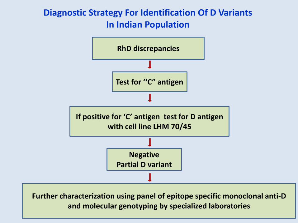
RhD discrepancies
Test for C” a tige
If positive for C a tige test for D a tige with cell line LHM 70/45
Negative
Partial D variant
Further characterization using panel of epitope specific monoclonal anti-D
and molecular genotyping by specialized laboratories
Diagnostic Strategy For Identification Of D Variants
In Indian Population
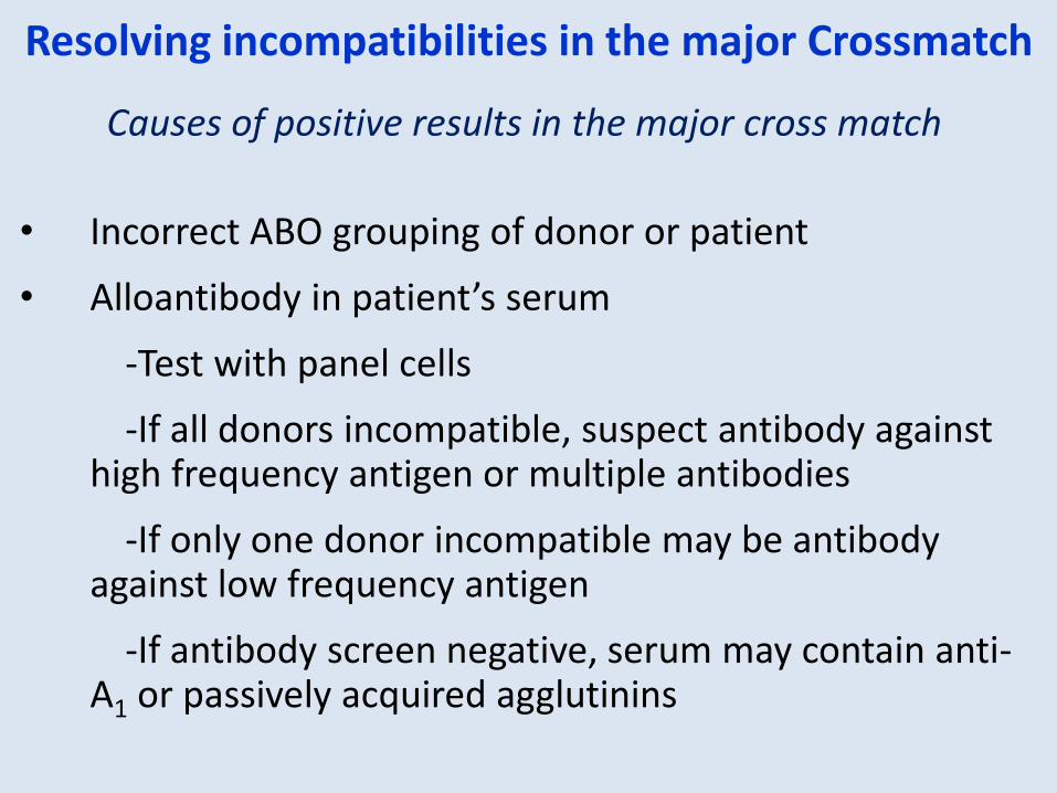
Resolving incompatibilities in the major Crossmatch
Causes of positive results in the major cross match
• Incorrect ABO grouping of donor or patient
• Alloa ti od i patie t’s seru
-Test with panel cells
-If all donors incompatible, suspect antibody against high frequency antigen or multiple antibodies
-If only one donor incompatible may be antibody against low frequency antigen
-If antibody screen negative, serum may contain anti-A1 or passively acquired agglutinins
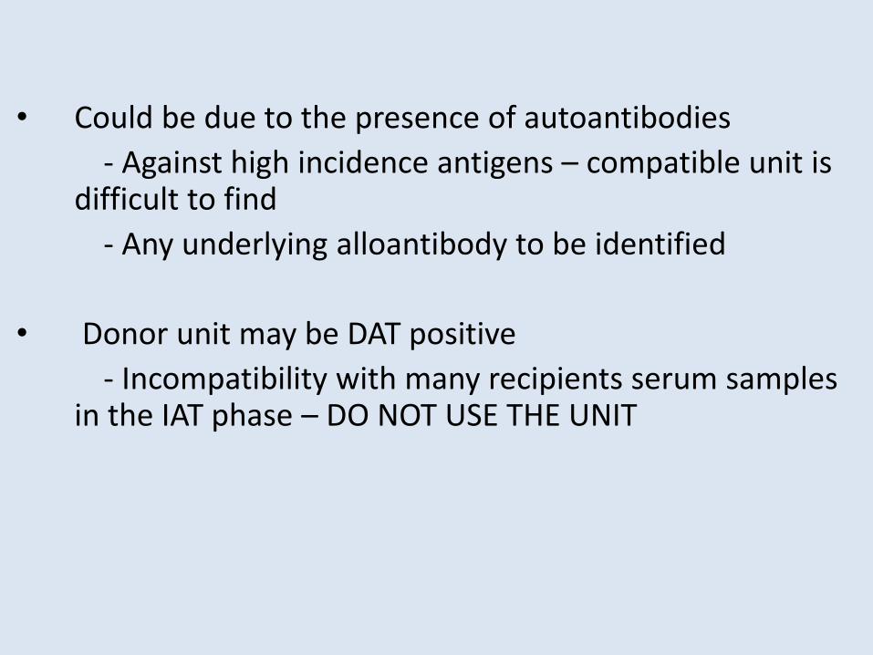
• Could be due to the presence of autoantibodies
- Against high incidence antigens – compatible unit is difficult to find
- Any underlying alloantibody to be identified
• Donor unit may be DAT positive
- Incompatibility with many recipients serum samples in the IAT phase – DO NOT USE THE UNIT
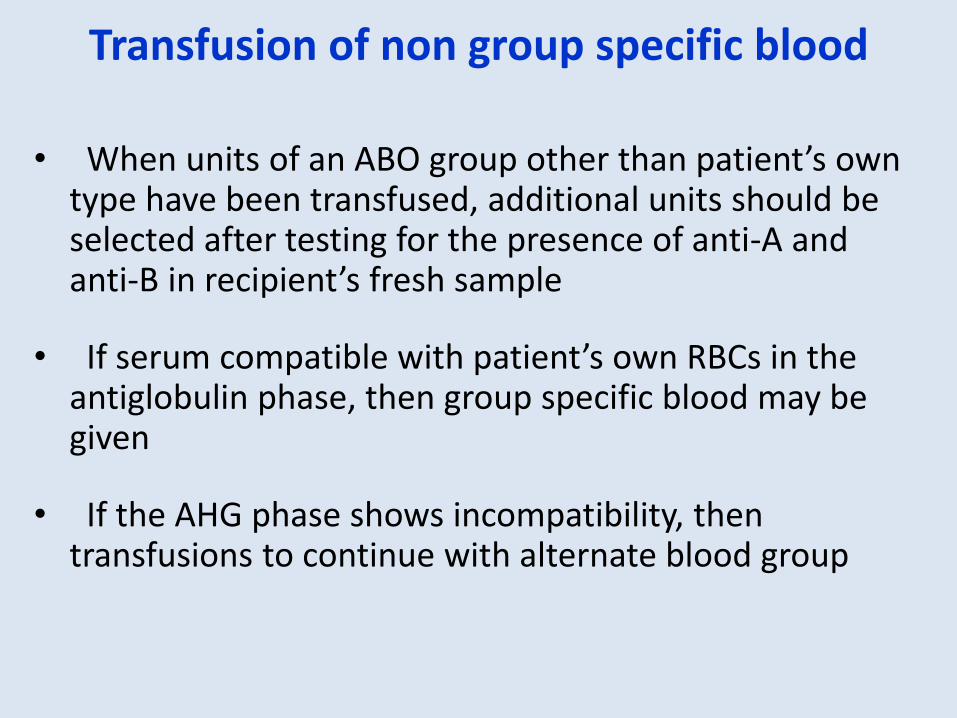
Transfusion of non group specific blood
• Whe u its of a ABO group other tha patie t’s o type have been transfused, additional units should be selected after testing for the presence of anti-A and anti-B i re ipie t’s fresh sa ple
• If seru o pati le ith patie t’s o RBCs i the antiglobulin phase, then group specific blood may be given
• If the AHG phase shows incompatibility, then transfusions to continue with alternate blood group
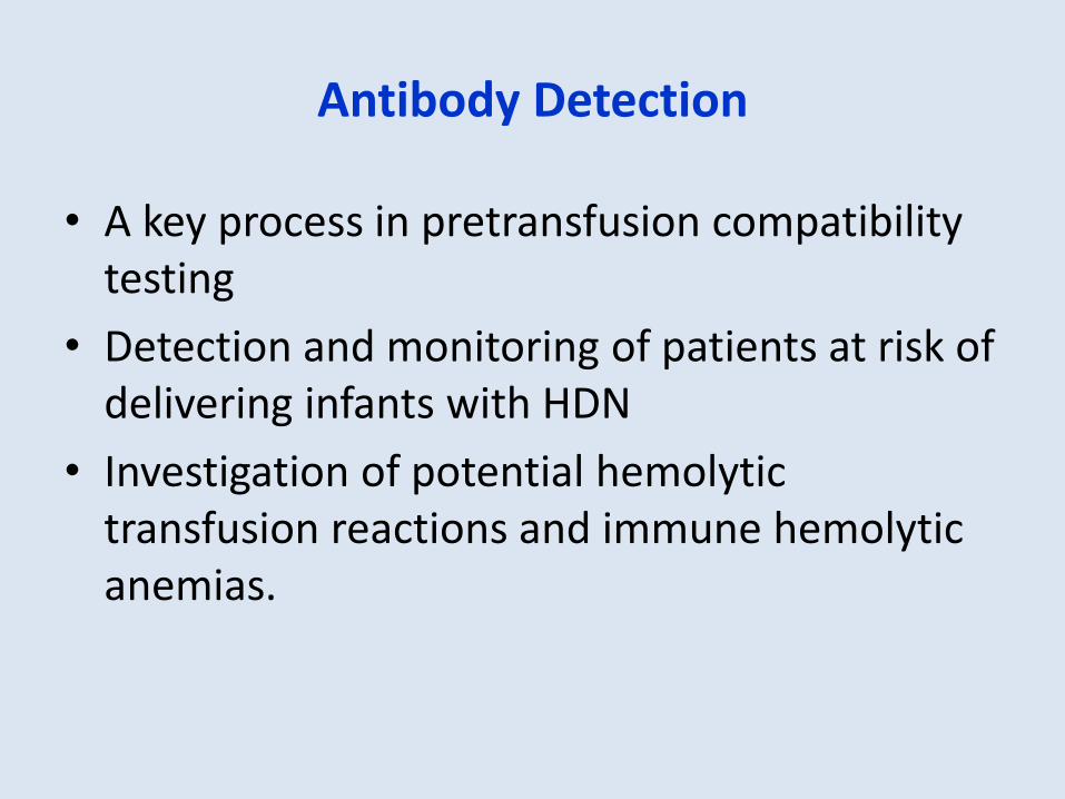
Antibody Detection
• A key process in pretransfusion compatibility
testing
• Detection and monitoring of patients at risk of
delivering infants with HDN
• Investigation of potential hemolytic
transfusion reactions and immune hemolytic
anemias.
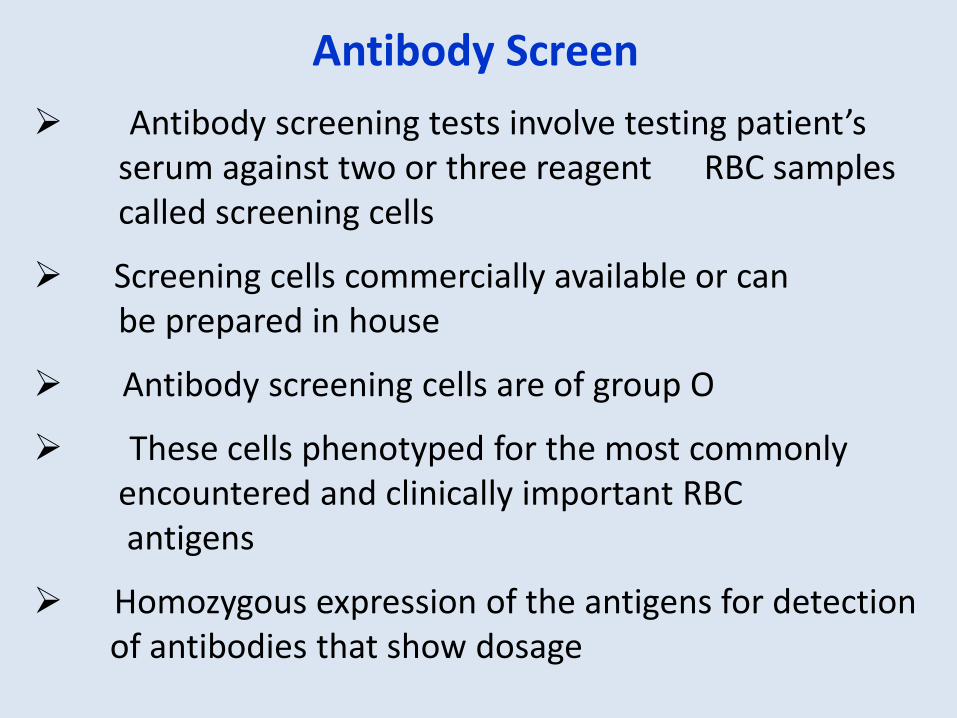
Antibody Screen
Antibody screening tests involve testing patie t’s
serum against two or three reagent RBC samples
called screening cells
Screening cells commercially available or can
be prepared in house
Antibody screening cells are of group O
These cells phenotyped for the most commonly
encountered and clinically important RBC
antigens
Homozygous expression of the antigens for detection
of antibodies that show dosage
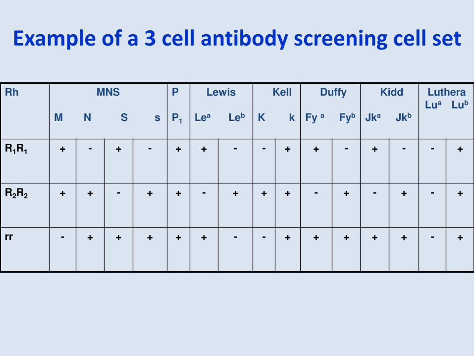
Example of a 3 cell antibody screening cell set
Rh MNS
M N S s
P
P1
Lewis
Lea Leb
Kell
K k
Duffy
Fy a Fyb
Kidd
Jka Jkb
Luthera
Lua Lub
R1R1 + - + - + + - - + + - + - - +
R2R2 + + - + + - + + + - + - + - +
rr - + + + + + - - + + + + + - +
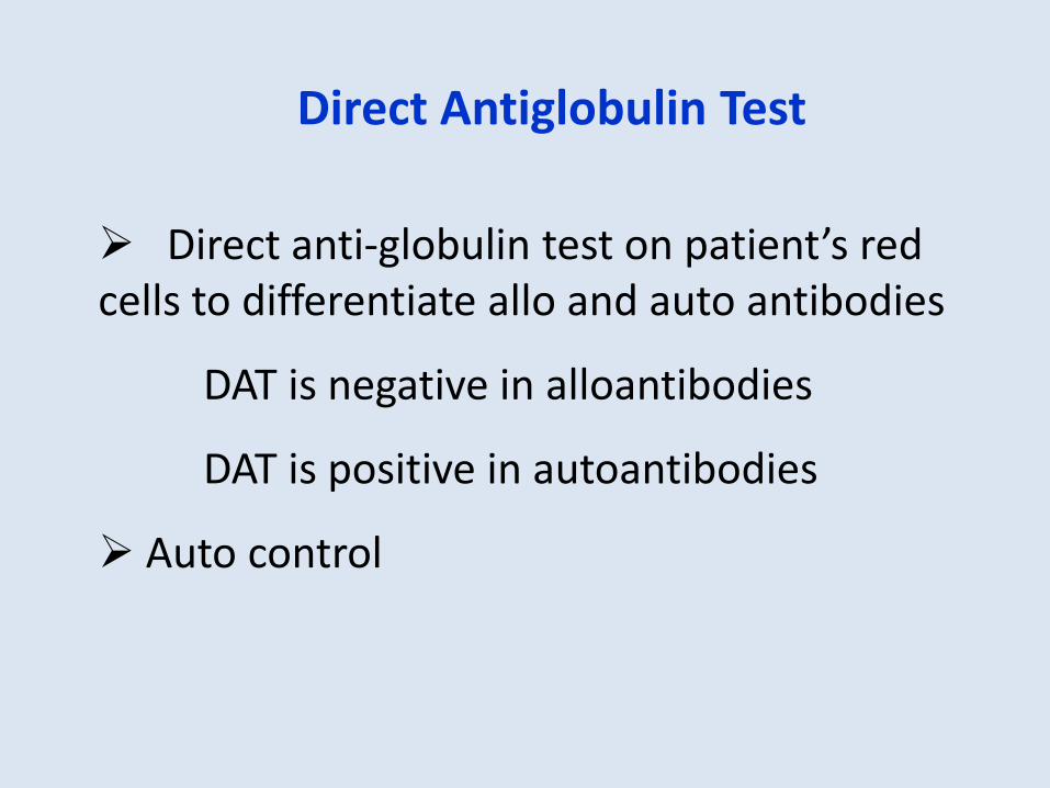
Direct anti-glo uli test o patie t’s red cells to differentiate allo and auto antibodies
DAT is negative in alloantibodies
DAT is positive in autoantibodies
Auto control
Direct Antiglobulin Test
Interpretation of the Antibody Screening results
based on:
• Phase of reaction
• Autocontrol
• With how many screening cells the reaction
was seen
Methods for Antibody Screening
• Manual
• Semiautomation
• Automation
Antibody Identification
• When antibodies are detected, they must be identified
• Antibody identification needed for transfusion purposes
and it is an important part of compatibility testing
• A person with antibody when exposed to donor cells
with the corresponding antigen serious side effects can
occur
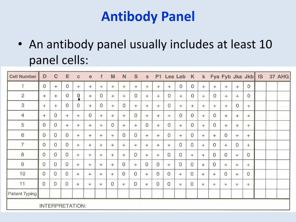
Antibody Panel
• An antibody panel usually includes at least 10
panel cells:
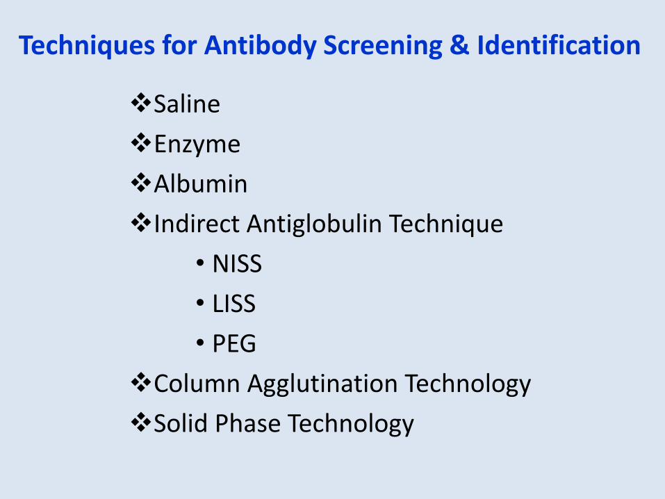
Techniques for Antibody Screening & Identification
Saline
Enzyme
Albumin
Indirect Antiglobulin Technique
• NISS
• LISS
• PEG
Column Agglutination Technology
Solid Phase Technology
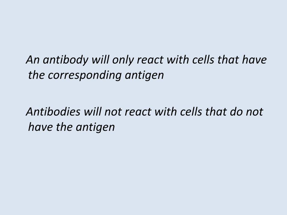
An antibody will only react with cells that have
the corresponding antigen
Antibodies will not react with cells that do not
have the antigen
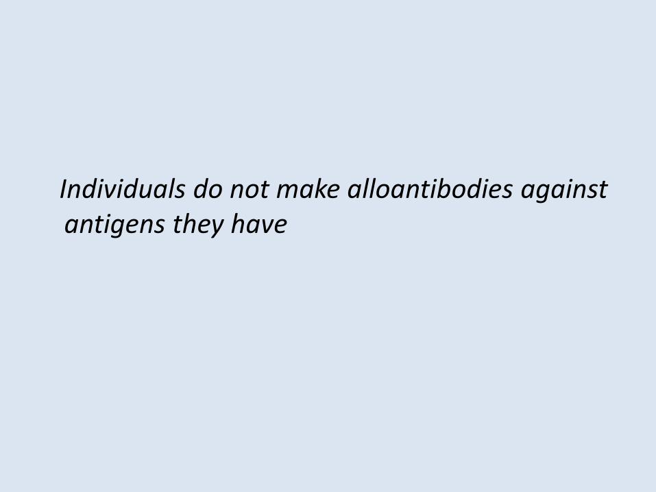
Individuals do not make alloantibodies against
antigens they have
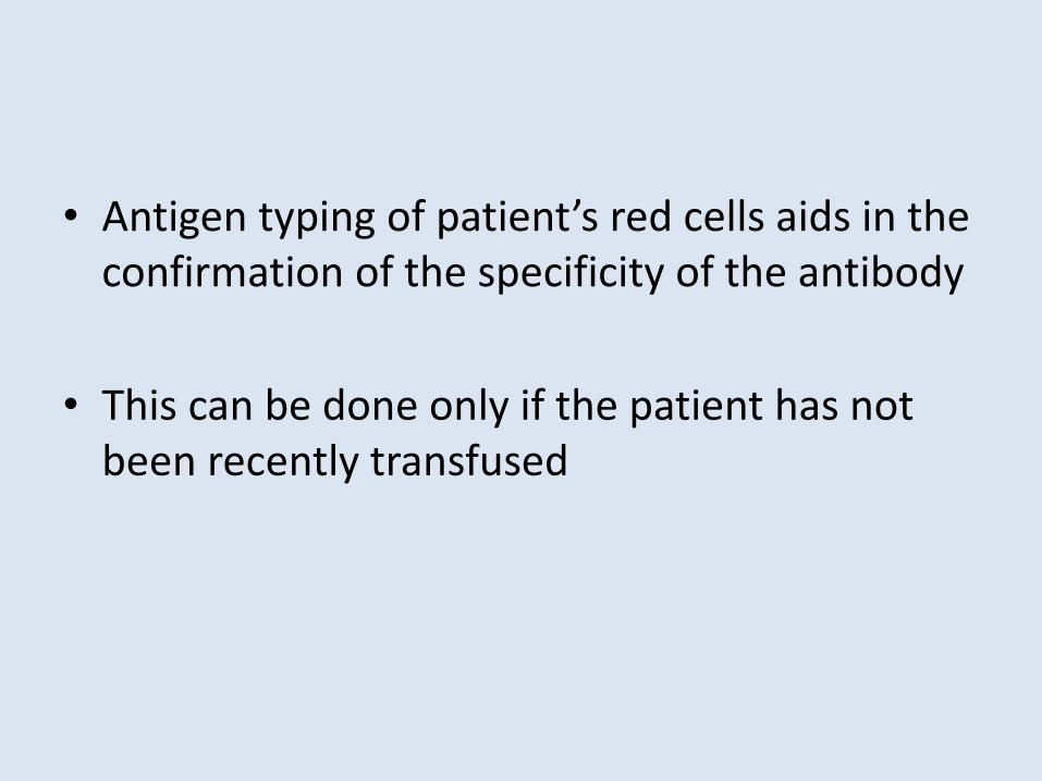
• A tige t pi g of patie t’s red ells aids i the confirmation of the specificity of the antibody
• This can be done only if the patient has not
been recently transfused
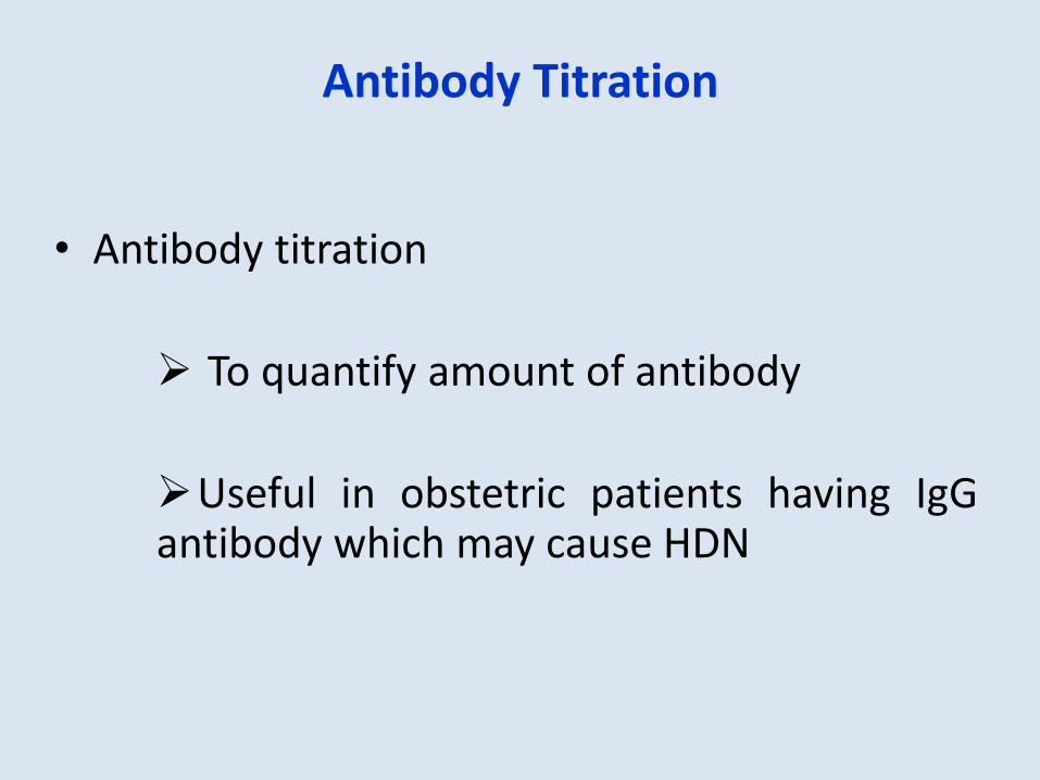
Antibody Titration
• Antibody titration
To quantify amount of antibody
Useful in obstetric patients having IgG antibody which may cause HDN
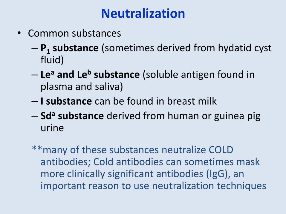
Neutralization
• Common substances
– P1 substance (sometimes derived from hydatid cyst fluid)
– Lea and Leb substance (soluble antigen found in plasma and saliva)
– I substance can be found in breast milk
– Sda substance derived from human or guinea pig urine
**many of these substances neutralize COLD antibodies; Cold antibodies can sometimes mask more clinically significant antibodies (IgG), an important reason to use neutralization techniques
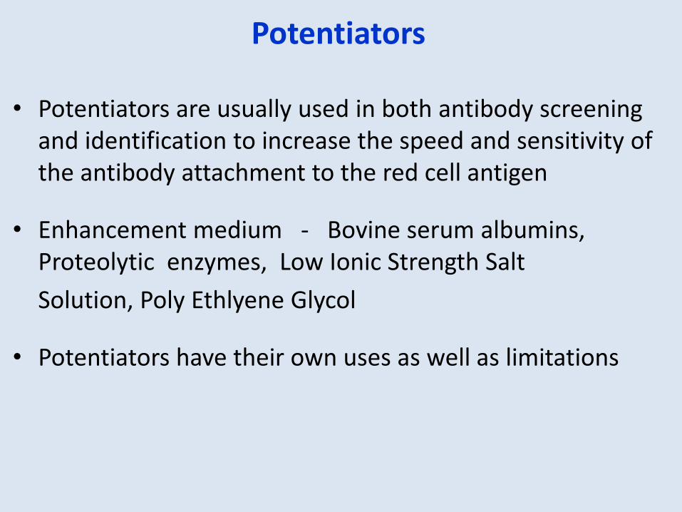
Potentiators
• Potentiators are usually used in both antibody screening
and identification to increase the speed and sensitivity of
the antibody attachment to the red cell antigen
• Enhancement medium - Bovine serum albumins,
Proteolytic enzymes, Low Ionic Strength Salt
Solution, Poly Ethlyene Glycol
• Potentiators have their own uses as well as limitations
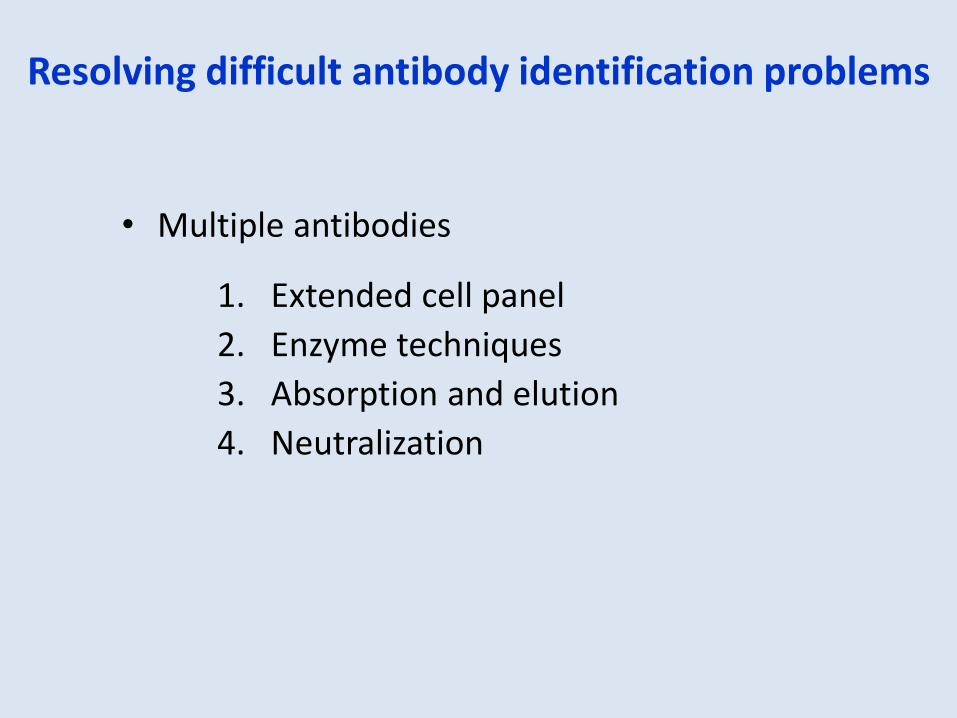
Resolving difficult antibody identification problems
• Multiple antibodies
1. Extended cell panel
2. Enzyme techniques
3. Absorption and elution
4. Neutralization
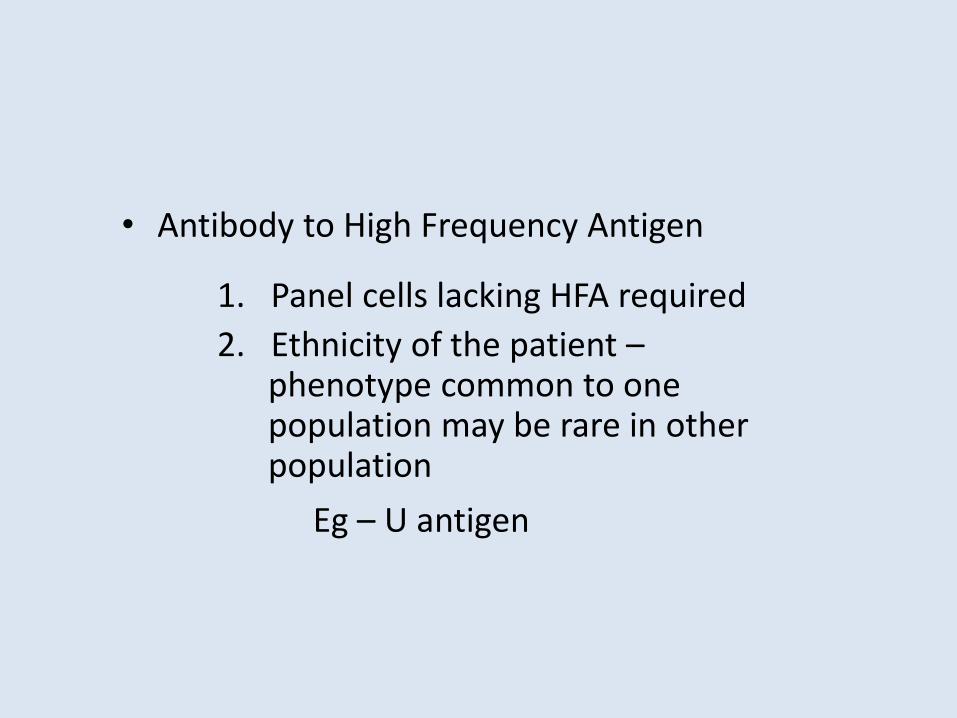
• Antibody to High Frequency Antigen
1. Panel cells lacking HFA required
2. Ethnicity of the patient – phenotype common to one population may be rare in other population
Eg – U antigen
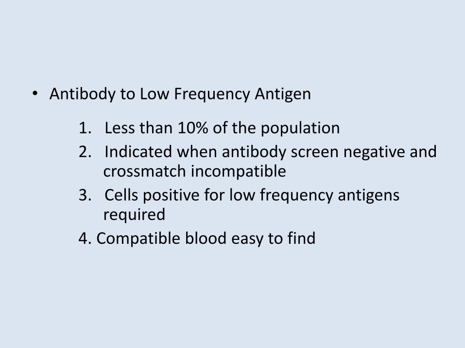
• Antibody to Low Frequency Antigen
1. Less than 10% of the population
2. Indicated when antibody screen negative and crossmatch incompatible
3. Cells positive for low frequency antigens required
4. Compatible blood easy to find
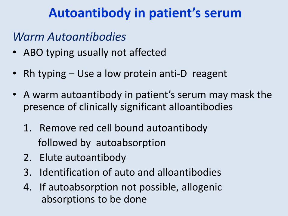
Warm Autoantibodies
• ABO typing usually not affected
• Rh typing – Use a low protein anti-D reagent
• A ar autoa ti od i patie t’s seru a ask the presence of clinically significant alloantibodies
1. Remove red cell bound autoantibody
followed by autoabsorption
2. Elute autoantibody
3. Identification of auto and alloantibodies
4. If autoabsorption not possible, allogenic absorptions to be done
Autoa tibody i patie t s seru
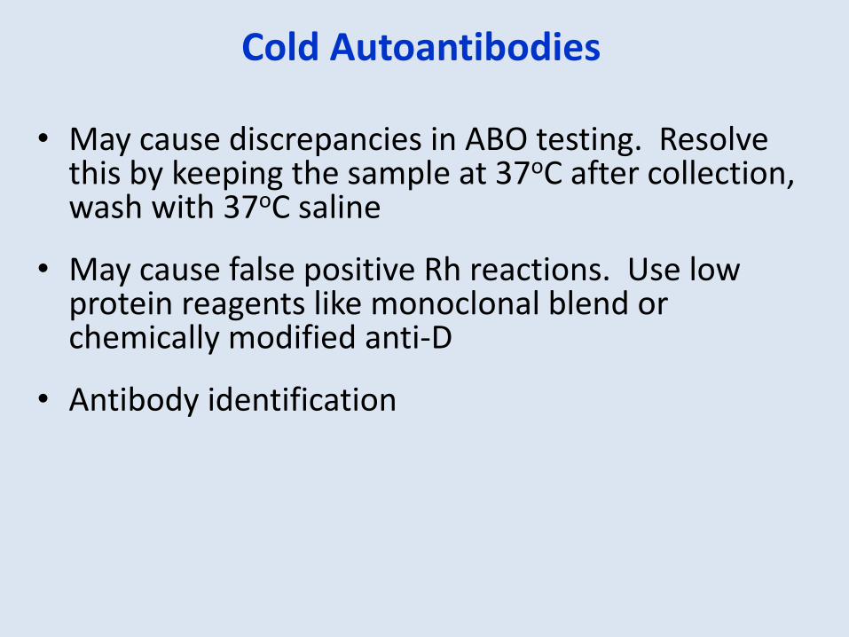
Cold Autoantibodies
• May cause discrepancies in ABO testing. Resolve this by keeping the sample at 37oC after collection, wash with 37oC saline
• May cause false positive Rh reactions. Use low protein reagents like monoclonal blend or chemically modified anti-D
• Antibody identification

Thank you…!