Embed Size (px)
Citation preview

CLINICAL MICROBIOLOGY REVIEWS,0893-8512/98/$04.0010
Jan. 1998, p. 121–141 Vol. 11, No. 1
Copyright © 1998, American Society for Microbiology
Serologic Response to Cell Wall Mannoproteins andProteins of Candida albicans
JOSE P. MARTINEZ,1* M. LUISA GIL,1 JOSE L. LOPEZ-RIBOT,2 AND W. LAJEAN CHAFFIN3
Departamento de Microbiologıa y Ecologıa, Facultad de Farmacia, Universitat de Valencia,Valencia, Spain,1 and Division of Infectious Diseases, Department of Medicine,The University of Texas Health Sciences Center at San Antonio, San Antonio,2
and Department of Microbiology and Immunology, Texas TechUniversity Health Sciences Center, Lubbock,3 Texas
INTRODUCTION .......................................................................................................................................................121C. ALBICANS CELL WALL ......................................................................................................................................122
Composition, Structure, and Functions...............................................................................................................122Variability and Dynamics of Cell Wall Antigens................................................................................................123
CARBOHYDRATE COMPONENT ..........................................................................................................................124Mannan ....................................................................................................................................................................124Nonmannan Carbohydrates...................................................................................................................................127
PROTEIN COMPONENT .........................................................................................................................................127Secreted Proteins ....................................................................................................................................................127
Secreted aspartyl proteinase .............................................................................................................................128Immunosuppressive and B-cell mitogenic protein .........................................................................................128
Heat Shock Proteins...............................................................................................................................................128hsp90.....................................................................................................................................................................129hsp70.....................................................................................................................................................................129Other heat shock proteins .................................................................................................................................129
Glycolytic Enzymes .................................................................................................................................................129Enolase .................................................................................................................................................................130Other glycolytic enzymes....................................................................................................................................131
Receptors and Binding Proteins for Host Ligands............................................................................................132ANTIBODIES TO CELL WALL ANTIGENS: PROTECTIVE AND DIAGNOSTIC VALUE..........................132SUMMARY AND OUTLOOK...................................................................................................................................134ACKNOWLEDGMENTS ...........................................................................................................................................135REFERENCES ............................................................................................................................................................135
INTRODUCTION
The dimorphic fungus Candida albicans is both a commensaland opportunistic pathogen of humans. Predisposing factorsfor candidiasis include immunosuppressive, antibiotic, and cy-totoxic therapies; the presence of intravenous catheters andindwelling devices; very low birth weight; AIDS; diabetes; anddrug abuse. Depending on the underlying host defect, thismicroorganism is able to cause a variety of infections thatrange from mucosal candidiasis to life-threatening dissemi-nated candidiasis (15, 61, 214).
C. albicans pathogenicity also depends on a complex array ofmicroorganism-related putative virulence factors. These in-clude the yeast-to-mycelium transition, antigenic variability,phenotypic switching, adhesion to host cells and tissues, cellsurface hydrophobicity, molecular mimicry, and production ofextracellular enzymes (24–26, 50, 64, 113, 233, 246, 277). Mostof the biological functions related to pathogenicity and viru-lence reside in the fungal cell wall, whose main features arepresented later in greater detail.
As the outermost cellular structure, the cell wall plays anessential role in the interactions with the host, including the
triggering and modulation of the anti-Candida host immuneresponses, which appear to rely on a complex interplay be-tween natural and adaptive immunity (89). Fungal antigensmay stimulate specific cell-mediated and humoral immune re-sponses. The importance of cellular defense mechanisms forprotection against fungal infections is supported by the clinicalobservation that most invasive fungal infections occur in pa-tients with defective cellular immunity (81, 155). The role ofantibodies in resistance to candidiasis is poorly understood,and reports exist in the literature providing evidence both infavor of and against the importance of antibody immunity as amain host defense mechanism against fungi. However, severalauthors have suggested the need to reexamine the contributionof antibodies in fungal infections (28, 60, 185, 189), and thereis renewed interest in the study of the host antibody responseto Candida and the possibility of immunointervention as afeasible prophylactic or therapeutic approach for the manage-ment of candidiasis.
Another controversial aspect is the laboratory diagnosis ofCandida infections. The diagnosis of invasive candidiasis isusually difficult to establish by clinical criteria; therefore, cul-ture techniques and serodiagnostic tests for antigen and anti-body detection have been used as laboratory aids to diagnosis.However, blood cultures for Candida species generally exhibitlow sensitivity (129, 214, 240), and the tests for determinationof marker antigens need further fine-tuning to improve theirsensitivity and specificity so that they will be valuable in guiding
* Corresponding author. Mailing address: Departamento de Micro-biologıa y Ecologıa, Facultad de Farmacia, Room 3-70, Universitat deValencia, Avda. Vicente Andres Estelles, s/n, 46100-Burjasot, Valencia,Spain. Phone and fax: 34-6-3864770. E-mail: [email protected].
121
on February 3, 2019 by guest
http://cmr.asm
.org/D
ownloaded from
clinical treatment decisions (203, 238, 240). Likewise, standardimmunological tests to detect anti-Candida antibodies usuallyhave low specificity and/or sensitivity (i.e., in most cases theyfailed to discriminate between disseminated and superficialcandidiasis), since they mainly recognize antibodies againstCandida cell wall mannan, which are ubiquitous in human sera(129, 306, 318). Furthermore, the crude preparations of can-didal antigens cannot be standardized enough to allow goodtest reproducibility among laboratories (129). A simple, reli-able, and easy-to-perform serological assay for the diagnosis ofsystemic Candida infections is still not available but is urgentlyneeded. In this context, the detection of defined fungal anti-gens and/or antibodies to such antigens may provide a suitableprocedure for diagnosis of invasive candidiasis.
In this review, we will focus specifically on the antibodyresponse to defined protein and glycoprotein C. albicans cellwall components in humans and in animal models of experi-mentally induced candidiasis (Table 1), with special emphasison the implications for developing novel diagnostic, prophy-lactic, and therapeutic techniques for candidiasis. Since theseproteins and glycoproteins are integral candidal components,we will first consider their main biological features in the con-text of their natural environment within the fungal cell. Read-ers who wish to know more about basic aspects of cell wall
biology are referred to a number of excellent recent reviewsdiscussing the composition and structure, physiological role,and antigenic composition of the cell wall of C. albicans (26, 36,41, 62, 88, 210, 233, 259, 260, 265, 266).
C. ALBICANS CELL WALL
The cell wall may be envisaged as a dynamic and plastic,multilayered structure located external to the plasma mem-brane. The presence of the wall is essential to several aspectsof the biology and, as noted above, the pathogenicity of C.albicans. Thus, the cell wall is the structure responsible formaintaining the shape that characterizes each growth form(yeast and hyphae) of the fungus, and it acts as a permeabilitybarrier that protects the protoplast against physical and os-motic injuries. In addition, the cell wall plays nutritional rolesand is the structure that mediates the initial interaction be-tween the microorganism and the environment.
Composition, Structure, and Functions
The major components (80 to 90%) of the cell wall of C.albicans are carbohydrates: (i) mannan or polymers of man-nose covalently associated with proteins to form glycoproteins,
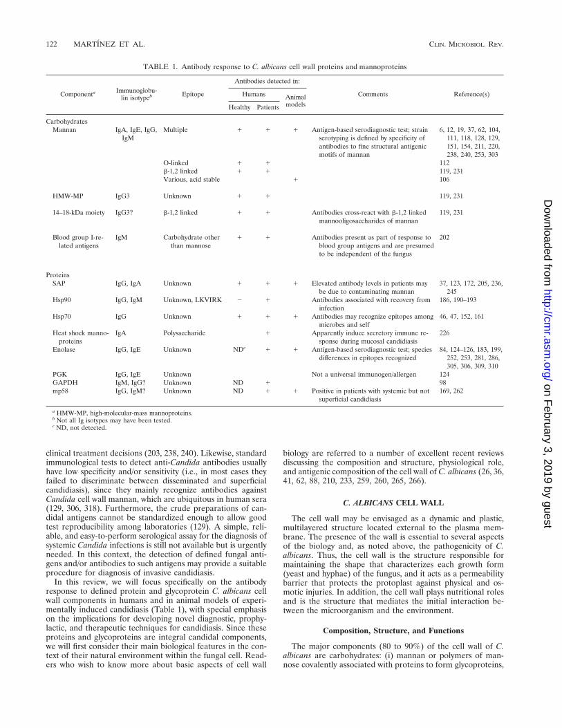
TABLE 1. Antibody response to C. albicans cell wall proteins and mannoproteins
Componenta Immunoglobu-lin isotypeb Epitope
Antibodies detected in:
Comments Reference(s)Humans AnimalmodelsHealthy Patients
CarbohydratesMannan IgA, IgE, IgG,
IgMMultiple 1 1 1 Antigen-based serodiagnostic test; strain
serotyping is defined by specificity ofantibodies to fine structural antigenicmotifs of mannan
6, 12, 19, 37, 62, 104,111, 118, 128, 129,151, 154, 211, 220,238, 240, 253, 303
O-linked 1 1 112b-1,2 linked 1 1 119, 231Various, acid stable 1 106
HMW-MP IgG3 Unknown 1 1 119, 231
14–18-kDa moiety IgG3? b-1,2 linked 1 1 Antibodies cross-react with b-1,2 linkedmannooligosaccharides of mannan
119, 231
Blood group I-re-lated antigens
IgM Carbohydrate otherthan mannose
1 1 Antibodies present as part of response toblood group antigens and are presumedto be independent of the fungus
202
ProteinsSAP IgG, IgA Unknown 1 1 1 Elevated antibody levels in patients may
be due to contaminating mannan37, 123, 172, 205, 236,
245Hsp90 IgG, IgM Unknown, LKVIRK 2 1 Antibodies associated with recovery from
infection186, 190–193
Hsp70 IgG Unknown 1 1 1 Antibodies may recognize epitopes amongmicrobes and self
46, 47, 152, 161
Heat shock manno-proteins
IgA Polysaccharide 1 Apparently induce secretory immune re-sponse during mucosal candidiasis
226
Enolase IgG, IgE Unknown NDc 1 1 Antigen-based serodiagnostic test; speciesdifferences in epitopes recognized
84, 124–126, 183, 199,252, 253, 281, 286,305, 306, 309, 310
PGK IgG, IgE Unknown Not a universal immunogen/allergen 124GAPDH IgM, IgG? Unknown ND 1 98mp58 IgG, IgM? Unknown ND 1 1 Positive in patients with systemic but not
superficial candidiasis169, 262
a HMW-MP, high-molecular-mass mannoproteins.b Not all Ig isotypes may have been tested.c ND, not detected.
122 MARTINEZ ET AL. CLIN. MICROBIOL. REV.
on February 3, 2019 by guest
http://cmr.asm
.org/D
ownloaded from

also referred to as mannoproteins; (ii) b-glucans that arebranched polymers of glucose containing b-1,3 and b-1,6 link-ages; and (iii) chitin, which is an unbranched homopolymer ofN-acetyl-D-glucosamine (GlcNAc) containing b-1,4 bonds.Proteins (6 to 25%) and lipids (1 to 7%) are present as minorwall constituents (26, 36, 265, 266). Yeast cells and germ tubesare similar in their cell wall composition, although the relativeamounts of b-glucans, chitin, and mannan may vary with themorphology (26, 259).
b-Glucans and chitin are the structural components of thewall. Quantitatively, b-glucans contribute to 47 to 60% byweight of the cell wall. Chitin is a minor (0.6 to 9%) butstructurally important component, particularly associated withcell-cell connections in the ring between parent and daughtercells, in the bud scar, and in the septa of dividing independentcells.
Mannose polymers do not exist as such but are found incovalent association with proteins (mannoproteins) (26, 36,259, 265, 266). They are the main material of the amorphouscell wall matrix in which the structural polymers (b-glucans andchitin) are embedded, and they represent 40% of the total cellwall carbohydrate. The mannoproteins of C. albicans shareseveral general features with the mannoproteins of Saccharo-myces cerevisiae, a model organism that is one of the mostthoroughly investigated yeasts in this regard. The structure ofthe carbohydrate component of C. albicans mannoproteins isdescribed in detail below. Although mannose is the mainmonosaccharide component of C. albicans cell wall glycopro-teins, there is evidence that other sugar residues are alsopresent in some wall glycoprotein species (see below).
Electron microscopy studies have shown the existence ofseveral layers in the cell wall of C. albicans. The structuralappearance is variable and seems to be related to the strainexamined, growth conditions, morphology, and the experimen-tal conditions used to prepare the specimens (26, 36, 233).Several cytochemical and cytological studies indicate that thecell wall layering is essentially due to the distribution of man-noproteins at various levels within the wall structure (36). Themicrofibrillar polysaccharides, glucan and chitin, appear to bemore concentrated in the inner cell wall layer, adjacent to theplasma membrane. Proteins and glyco(manno)proteins fill thenetwork of structural polymers and appear to be dominant inthe outermost cell wall layer, which has a fibrillar or flocculentaspect (36, 259).
The different cell wall components interact with each otherto give rise to the overall architecture of the cell wall. Besideshydrogen and hydrophobic bonds, there is also experimentalevidence for the presence of covalent linkages between differ-ent components (249, 260). In this context, Surarit et al. (288)reported the presence of glycosidic linkages between glucanand chitin in the nascent wall of C. albicans. On the other hand,recent evidence indicates that mannoproteins may establishcovalent associations with b-1,3- and b-1,6-glucans throughphosphodiester linkages (139, 140, 251), suggesting that suchassociations may play a key role in configuring the final cellwall structure characteristic for each growth form (yeast andmycelium) of C. albicans (249, 257–260). Finally, interactionsbetween mannoproteins and chitin also appear to exist in thewall of C. albicans cells (176). Hence, it is possible that theso-called layering, as concluded mainly from electron micros-copy observations, is the result of quantitative differences inthe proportions of the individual wall components (b-glucans,chitin, and mannoproteins) in each layer rather than absolutequalitative differences (212). Nevertheless, since proteins andmannoproteins appear to be involved in a variety of essentialprocesses for C. albicans such as morphogenesis and several
pathogenicity-related aspects, and since they may be responsi-ble for the hydrophobic or hydrophilic status of the cell surface(24–26, 36, 65, 66, 114–116, 121, 162, 249, 257–260), an asym-metric distribution of wall protein and glycoprotein constitu-ents as a consequence of the physiologic role played by eachmoiety should not be ruled out (41).
A complex array of protein-containing components has beensolubilized from isolated cell wall preparations and from intactcells of both candidal growth forms by different treatments(29–31, 33, 39, 71, 162, 176, 178, 227–229, 285). Analysis ofthese components has revealed quantitative and qualitativedifferences in the protein composition of yeast and mycelialcell walls. Some components have been characterized as high-molecular-mass (.120-kDa) mannoproteins that appear to becovalently linked to the structural polysaccharides. These spe-cies, which contain large amounts of carbohydrate and conse-quently could be major elicitors of anti-candidal host immu-nity, seem to play also an important and active role inmodulating the organization of the different cell wall constit-uents to obtain the final supramolecular structure of the wallthat is specific for each morphologic form of C. albicans (31,34, 70, 95, 96, 164, 167, 179, 180, 228, 259, 285). Several studieshave identified 20 to 40 polypeptide species in the medium-to-low-molecular-mass (from 80 to 15 kDa) range (33, 39, 71).Most molecules that appear to be present in the outermost walllayers and that exhibit receptor-like activities and adhesinproperties are medium- and low-molecular-mass species (41).The high-molecular-mass species seem to be homogeneouslydistributed on the cell surface (31, 34, 95), whereas protein andmannoprotein moieties that may represent receptors for dif-ferent host ligands exhibit clustering or asymmetric cell surfacedistribution (32, 163, 181). This further supports the conten-tion, noted above, that differences in the distribution or topo-logical location of wall mannoproteins appear to be related tothe distinct functional or structural role played by individualspecies.
Variability and Dynamics of Cell Wall Antigens
The expression, distribution, chemical characteristics, andbehavior observed in vitro and the biological properties of C.albicans cell wall-bound proteins and glycoproteins appear tobe dependent on multiple environmental and organism-relatedfactors. These include growth conditions, growth state, mor-phology of the cells, strain and serotype, phenotypic switching,cell surface hydrophobic or hydrophilic status, and the natureof the biological specimens (intact cells or isolated wall prep-arations) that are subjected to analysis (3, 5, 14, 21, 29, 35, 59,96, 114, 115, 120, 122, 164, 167, 179, 229, 233, 278). Particu-larly, differential expression of antigens at the cell surface levelof both C. albicans growth forms has received considerableattention from numerous research groups. A number of dif-ferent possibilities have been suggested to explain the changesin the cell wall composition observed during the morphologictransition. Possible explanations are as follows: (i) form-spe-cific components reflect de novo synthesis of proteins; (ii)topological rearrangement of preexisting cell wall constituentsoccurs; (iii) differences are due to major quantitative variationsin the composition of surface components on both fungal mor-phologies; (iv) changes in the glycosylation pattern of the samemolecule occur depending on the cell morphology considered;and (v) a combination of all the above could occur (21, 31, 33,42, 215, 287, 301, 308). Since the ability to grow in the fila-mentous form appears to be one of the important virulencetraits in C. albicans, and since the presence of hyphal elementsin deep tissues is related to infection (214), special emphasis
VOL. 11, 1998 SEROLOGIC RESPONSE TO C. ALBICANS CELL WALL ANTIGENS 123
on February 3, 2019 by guest
http://cmr.asm
.org/D
ownloaded from

has been directed towards the characterization of hypha-spe-cific moieties and their possible use as antigen markers in theserodiagnosis of systemic candidiasis (208, 209, 230, 235, 307).In this context, the ideal marker antigen should be expressedby all C. albicans strains under all different environmentalconditions. However, since the cell wall appears to be a highlydynamic “organelle” that exhibits both the capacity of differ-entially expressing variable constituents useful for switchingbetween the commensal and pathogenic lifestyles and the ca-pacity for modulating and/or evading the immune host defensebarriers, this variability must be taken into consideration whentrying to identify potential marker antigens of infection. There-fore, this variability accounts, at least partly, for the poor spec-ificity and sensitivity displayed by some of the methods cur-rently available for immunodiagnosis of systemic candidiasis.
Structural peculiarities in the cell wall mannan (see below)are responsible for the antigenic specificity of C. albicans se-rotype A and B strains (111). Antigenic variability in theepitopes that accounts for this specificity has been detectedalso. In fact, there is evidence suggesting that both serotypesare determined phenotypically rather than genotypically. Pou-lain et al. (234) showed that serotype B yeast cells and germtubes express antigen 6 (the factor characteristic for serotype Astrains) in infected tissues and that germ tubes of serotype Bstrains, but not the mother yeast cells from which the germtubes emanate, express the antigen in vitro. Kobayashi et al.(144) have shown that serotype A strains behave as serotype Bstrains at pH 2 since they do not express both factors 5 and 6,the latter being specific for serotype A C. albicans cells. Bar-turen et al. (14) reported that the expression of antigens re-sponsible for serotype A specificity is modulated by the pH ofthe culture medium. These observations may be related to theactivity of the enzyme 1,2-b-mannosyltransferase, which ap-pears to be suppressed or lowered at low pH (148).
This antigenic variability detected for epitopes conferringserotype specificity is also extended to other polysaccharideepitopes. The surface antigen makeup of C. albicans is ingeneral variable both in vitro and in vivo, as already indicated.Recently, De Bernardis et al. (56) studied the expression of apolysaccharide epitope in yeast and mycelial cells in a model ofcandidal vaginitis in rats. This epitope is shared by a family ofstrongly antigenic cell wall mannoproteins including a 65-kDacomponent, which is recognized as a main target of cell-medi-ated anti-Candida immunity in humans (99, 294, 296). Theyshowed that this epitope is efficiently incorporated in all layersof the cell wall and abundantly expressed on the yeast cellsurface soon (1 h) after the vaginal challenge but is no longerexpressed on the surface of most of the cells harvested after24 h of intravaginal growth. This represents the first clearindication of cell surface antigenic variation of C. albicansduring active infection. Since mannans are strongly immuno-genic and immunomodulatory constituents of C. albicans,changes associated with the composition of these wall poly-mers and epitope expression on the cell surface may theoret-ically represent a mechanism that modulates the host immuneresponse.
CARBOHYDRATE COMPONENT
As stated above, b-glucans, chitin, and mannan are the threemajor polysaccharide components of the cell wall of C. albi-cans. Although b-glucans are present in greater abundancethan mannan in the wall of this fungal species (44), they areimmunologically less active (233).
Most of the mannan is found as large N-linked polymerscontaining several hundred mannose residues associated with
high-molecular-mass species, yet smaller N-linked moietiesand/or O-linked mannooligosaccharides are associated withsmaller glycoproteins. Antibodies to all these carbohydrateimmunodeterminants are readily detectable in serum samples.Although mannose appears to be the major monosaccharideconstituent, the potential for other complex carbohydrates thatmay be also present in candidal cell wall glycoproteins to elicitan antibody response has been reported.
Mannan
Initial studies focusing on the characterization of antigenpatterns for the serologic classification or identification ofmedically important Candida species demonstrated that cellwall mannan is the main candidal antigen responsible for thespecificity of the different serologic reactions (91, 111, 282, 302,303). Consequently, considerable effort has been expended todetermine the specific chemical structure of mannan and todefine the epitopes present in this wall component that mayaccount for serospecificity (for a review, see reference 210).
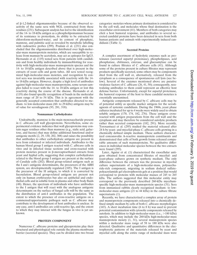
As mentioned above, mannan does not exist as such in thewall structure but exists in covalent association with proteins(mannoproteins) (36, 62, 265, 266). However, the term “man-nan” has also been used to refer to the main soluble immuno-dominant component present in the outer cell wall layer of C.albicans. This component, also called phosphomannoproteinor phosphopeptidemannan complex (Fig. 1), contains ho-mopolymers of D-mannose (as the main component), 3 to 5%protein, and 1 to 2% phosphate (239). A detailed structure ofthis cell wall constituent has emerged from several studies (10,144–149, 267–274). The mannose polymers are linked to pro-teins by N-glycosidic bonds (through two GlcNAc [di-N-ace-tylchitobiose] units) to asparagine residues and by O-glyco-sidic, alkali-labile linkages to threonine or serine residues. TheN-glycosidically linked carbohydrate has a backbone chain ofa-1,6-linked mannopyranosyl residues with oligosaccharidebranches containing mannopyranosyl residues with a-1,2, a-1,3, b-1,2, b-1,6, and single a-1,6-linked mannose units andphosphodiester bonds (87, 90, 145, 150, 271). Single mannoseresidues and short, unbranched mannose oligosaccharides con-stitute the O-glycosidically linked sugar component (222).
The antigenic specificity of serotypes A and B of C. albicans,described by Hasenclever and Mitchell (111), appears to bedetermined by structural peculiarities of the carbohydrate moi-ety of mannan present in the cell wall of strains belonging to aparticular serotype. In this context, Kobayashi et al. (147)showed that oligomannosyl side chains containing both b-1,2and a-1,2 linkages were serotype A-specific epitopes (factor 6)whereas b-1,2-linked oligomannosyl side chains, which are at-tached to phosphate, serve as major common epitopes (factor5) for both C. albicans serotypes (267). Thus, individual oligo-mannosides obtained by depolymerization of the phosphopep-tidemannan complex, represent epitopes that have been de-scribed as the basis of rabbit polyclonal antibody specificities(267). Further characterization of epitopes recognized by an-tisera to other C. albicans antigenic factors revealed that anti-serum to factor 1 is directed to the O-linked mannose chains ofthe cell wall mannoproteins whereas the epitope recognized byantisera to factor 9 is the a-1,6-linked mannose backbone ofthe outer chain of the N-linked oligosaccharide moiety (10).Single mannose units linked by a-1,6 bonds to the oligosaccha-ride branches containing a-1,2- and a-1,3-linked mannopy-ranosyl residues make the epitope corresponding to antigenicfactor 4 (271). The structures defining the different antigenicfactors mentioned above are depicted in Fig. 1.
The mannan component appears to be involved in the ad-
124 MARTINEZ ET AL. CLIN. MICROBIOL. REV.
on February 3, 2019 by guest
http://cmr.asm
.org/D
ownloaded from

herence of C. albicans cells to macrophages located in thesplenic marginal zone and in the subcapsular and medullarysinuses of peripheral lymph node tissue of mice (40, 51, 109,135–137, 157). Kanbe et al. (136) showed that the mannanportion of the phosphomannoprotein isolated from yeast-form
C. albicans cells by b-mercaptoethanol extraction and con-canavalin A-agarose affinity chromatography contained the ad-hesins responsible for attachment to macrophages in bothtypes of tissues. Li and Cutler (157) identified one adhesin siteon the acid-labile portion of the mannoprotein as a b-1,2-
FIG. 1. Structures of C. albicans mannan (M, D-mannopyranose units; GlcNAc, N-acetylglucosamine units; Asn, asparagine residue). (A) Representative structureof serotype A strain mannan. Side chains shown by the outlined letter M indicate putative serotype A-specific structures containing b-1,2-linked mannopyranosylresidues at the nonreducing terminal (i.e., antigenic factor 6); other side chains depicted are common in mannans from both serotypes. Reprinted from reference 267with permission of the publisher. (B) Representative structure of serotype B strain mannan. Single mannose units linked by a-1,6 bonds to the oligosaccharide branchescontaining a-1,2- and a-1,3-linked mannopyranosyl residues make a serotype B-specific epitope (i.e., antigenic factor 4); a-1,6-linked mannose residues in bracketsindicate the partial absence of these units in mannan from C. albicans B cells. Reprinted from reference 271 with permission. Structures corresponding to otherantigenic factors present in mannans from both C. albicans serotypes are described in the text.
VOL. 11, 1998 SEROLOGIC RESPONSE TO C. ALBICANS CELL WALL ANTIGENS 125
on February 3, 2019 by guest
http://cmr.asm
.org/D
ownloaded from

linked tetramannosyl residue (Ag10G). However, the resultsindicated that additional adhesins in the phosphomannopro-tein complex were involved in the adherence to macrophages.In fact, Kanbe and Cutler (135) later reported evidence ofstrong adhesin activity in the acid-stable moiety of the phos-phomannoprotein complex. The adhesin site implicated is acarbohydrate but is devoid of epitopes for factors 5 and 6 or forAg10G, which contains the b-1,2-linked oligomannosyl adhe-sin site. The diagnostic implications of using these carbohy-drate epitopes or monoclonal antibodies against them to detectspecific antibody or antigen, respectively, in sera from patientswith disseminated candidiasis need to be investigated. In anycase, immunization of mice with the mannan adhesin fractionimplicated in adhesion to macrophages elicits protective anti-body responses against experimental Candida infection (106)(see below).
Anti-mannan antibodies have been shown to be ubiquitousin human sera, presumably because the immune system can bestimulated as a result of colonization by C. albicans in theabsence of disease (45, 62, 129). In any defined population,levels of anti-mannan antibodies are usually distributed abouta mean; sera with the highest levels give positive precipitintests when tested against cell wall mannan (128). When anti-mannan antibodies were measured in serially drawn sera fromneutropenic patients, a frequency distribution plot showed thatthe levels of antibodies from patients with invasive candidiasiswere elevated and tended to skew the normal distributioncurve to the right. However, a clear bimodal distribution ofthese antibody levels was not observed; therefore, after estab-lishing a cutoff value for anti-cell wall mannan antibodies, thebest sensitivity value was about 65% (102).
The applications for the diagnosis of disseminated candidi-asis by detection of (i) mannan that circulates during infectionor (ii) anti-mannan antibodies have been discussed elsewhere(129, 238, 240). Although most of the recent serological testsfor invasive candidiasis are directed to the detection of specificcirculating cell wall-bound or cytoplasmic candidal antigensother than mannan, special emphasis has been put on thedetection of mannan, as a major cell wall antigen, by differentprocedures including counterimmunoelectrophoresis, radioim-munoassay, enzyme-linked immunosorbent assay (ELISA),and latex agglutination (11, 12, 19, 23, 57, 74, 82, 85, 92, 94,131, 143, 154, 156, 198, 211, 220, 256, 311–313). Sensitivities of23 to 100% and specificities of 92 to 100% have been reportedfor ELISAs for mannan detection (57). Although early studiesshowed good sensitivity and specificity of latex-agglutinationtests (92), recent studies have been less favorable (19, 23, 154),particularly for sera from patients with malignancies (74). Acommercial test for mannan detection, the Cand-Tec system(Ramco Laboratories Inc., Houston, Tex.) has been shown tobe relatively insensitive (30 to 50%) compared to other meth-ods (6, 154, 220). Another commercial system to detectCandida mannan in serum, the Pastorex Candida test (SanofiDiagnostics Pasteur, Marnes-la-Coquette, France [118]), ap-peared promising, but Gutierrez et al. (104), who conducted aprospective clinical trial, found a 0% sensitivity for this test.The sensitivity of the Pastorex system was improved whenserial assays with multiple consecutive serum samples wereused (103). In a recent study, Savolainen et al. (253) examinedthe reactivity of immunoglobulin E (IgE), IgG, and IgM anti-bodies to mannan by a radioallergosorbent test (RAST) andELISA and to proteins (some of which are in the cell wall) byimmunoblotting and concluded that (i) the IgE titer rose earlyand was more useful for detection than were IgG and IgMtiters, (ii) RAST and immunoblotting analyses were both re-quired for maximum sensitivity and specificity, and (iii) inva-
sive disease could be distinguished from mucosal colonizationby this procedure.
Most serologic studies used whole phosphopeptidemannancomplexes as antigenic preparations and did not identify indi-vidual epitopes recognized by different antibodies. To deter-mine the molecular basis of recognition of oligomannosideantigenic motifs by human and animal (in experimentally in-duced candidiasis models) immune systems, Faille et al. (76)developed a method of rendering oligomannosides antigenicby coupling them to a carrier molecule that was able to bind toconventional substrates for immunoanalysis. The oligomanno-sides were coupled to a lipid, 4-hexadecylaniline by a methodleading to a mole-to-mole binding whose efficiency can bevisualized and assessed by thin-layer chromatography. The hy-brid molecules (neoglycolipids [NGL]) exhibited both immu-nogenicity and antigenicity.
Trinel et al. (298) constructed NGL from three families ofoligomannosides released by sequential depolymerization of C.albicans phosphopeptidemannan by acid hydrolysis (NGLH),b-elimination (NGLO), and acetolysis (NGLA) and studiedtheir reactivity by ELISA with six monoclonal antibodies re-acting with polysaccharide moieties of C. albicans mannopro-teins. The six monoclonal antibodies were also tested by im-munoblotting against germ tube antigens obtained by alkaliextraction under reducing conditions. By this extraction meth-od, the authors obtained highly standardized profiles of C.albicans germ tube cell wall and cytoplasmic proteins and man-noproteins ranging from 300 to 10 kDa (119). Comparing theepitope mapping of antibodies by ELISA with NGL and byimmunoblotting with the extract, they provided evidence thatthe reactivity of monoclonal antibodies with NGLH (corre-sponding to homopolymers of b-1,2-linked mannopyranosylunits [77, 268]) was correlated with reactivity with a 14- to18-kDa C. albicans antigen. Conversely, monoclonal antibodiesreacting with NGLA also reacted with polydisperse C. albicanshigh-molecular-mass mannoprotein species (298). Oligoman-nosides released after mild acetolysis have been described asbeing composed of from 1 to more than 11 mannosyl residues(75) linked mainly through a-1,2 and a-1,3 bonds (146, 150,268, 273) and to a lesser extent through b-1,2 bonds (145, 268).
By using the method of NGL construction, an analysis of thehuman antibody response to C. albicans-derived oligomanno-sides was undertaken to define the molecular basis of therecognition of the mannan molecule by human Igs (75, 112,231, 299). Construction of NGL from C. albicans O-linkedoligomannosides provided evidence that human antibodiesreacted with these residues (112). O-linked oligomannosidesreleased by mild-alkali degradation contained one to sevenmannose residues, among which the quantitatively major com-ponents, mannobiose and mannotriose, were shown to containexclusively a-1,2 linkages (112). The pool of oligomannosideswas converted to NGL and tested by ELISA with human sera.The sera were from patients who had seroconverted during thecourse of a mycological and serological survey for candidiasisas detected by indirect immunofluorescence and co-counter-immunoelectrophoresis assays (232). The results of ELISA-NGLO tests appeared to correlate with the results of ELISAon the original phosphopeptidemannan molecule and with theresults of routine serologic tests (112). Subsequently, Poulainet al. (231) selected sera with different IgG3 levels againstNGLH and NGLA from patients with candidiasis to check thepreviously correlated reactivity between NGLH and the 14- to18-kDa antigen and between NGLA and high-molecular-massmannoprotein species (298). Patient sera recognized saccha-ride epitopes distributed over the 14- to 18-kDa antigen. Theseepitopes, which were identified by IgG3, are thought to consist
126 MARTINEZ ET AL. CLIN. MICROBIOL. REV.
on February 3, 2019 by guest
http://cmr.asm
.org/D
ownloaded from
of b-1,2-linked oligomannosides because of the observed re-activity of the same sera with NGL constructed from theseresidues (231). Subsequent analysis has led to the identificationof the 14- to 18-kDa antigen as a phospholipomannan becauseof its resistance to proteolysis, its ability to be extracted bychloroform-methanol-water, and its content of phosphate,mannose, and palmitic acid as revealed by metabolic labellingwith radioactive probes (299). Poulain et al. (231) also con-cluded that the oligomannosides distributed over high-molec-ular-mass mannoprotein moieties, which are susceptible to re-lease from mannan by acetolysis, may act as epitopes for IgG3.Hernando et al. (119) tested sera from patients with candidi-asis and from healthy individuals by immunoblotting for reac-tivity with high-molecular-mass mannoprotein species and the14- to 18-kDa antigen described above that is present in theextracts of yeast cells and germ tubes. All sera tested recog-nized high-molecular-mass moieties, and recognition by con-trol sera was invariably associated with reactivity with the 14-to 18-kDa antigen. However, despite a high level of antibodiesagainst high-molecular-mass mannoproteins, some serum sam-ples failed to react with the 14- to 18-kDa antigen or lost thisreactivity during the course of the disease. Hernando et al.(119) also found specific recognition of bands in the molecularmass range of 20 to 54 kDa. This is in agreement with thegenerally accepted contention that antibodies directed to me-dium- to low-molecular-mass (60- to 29-kDa) antigens may beof value for serodiagnosis (93, 281, 318).
Nonmannan Carbohydrates
Undoubtedly, mannose is the main monosaccharide presentin C. albicans cell wall glycoproteins. Nevertheless, some ex-perimental evidence indicates that wall glycoproteins may con-tain sugar residues other than mannose (e.g., sialic acid, galac-tose, and fucose) that may define additional functional and/orantigenic motifs (2, 29, 130, 202, 308) and raises the possibilitythat antibodies to other carbohydrate antigens are present insera from colonized or infected individuals. Antibodies to thehuman blood group I antigen reacted with C. albicans cells invitro and in infected tissue sections and cross-reacted withprotein moieties present in b-mercaptoethanol extracts fromyeast and hyphal cells, suggesting that complex carbohydratesrelated to the blood group I antigen are present at the surfaceof Candida cells (202). Blood group-related antigens such asthe I and i antigenic determinants, the precursors of the ABHsystem, are developmentally regulated (105). The I antigen isthe precursor of the H antigen, to which it is converted byfucosylation. Blood group-related antigens are present notonly on human erythrocytes but also on epithelial and endo-thelial cells and in soluble form in plasma and other body fluids(48). Hence, the spectrum of individuals who have antibodiesto the I antigen that will react with the analogous antigenicdeterminants on the surface of fungal cells will be the same asthe distribution of anti-I antibodies in the population. Theextent to which the presence of cross-reactive antigens on acommensal/opportunistic pathogen such as C. albicans maycontribute to the development of host antibodies is unclear. Inany case, anti-I antibodies are cold-reactive Igs, and the extentto which they may interact with the fungus in vivo is yet un-known.
PROTEIN COMPONENT
Numerous C. albicans proteins and glycoproteins play theirstructural and physiological role outside the plasma membranebarrier (secreted species). They can be divided into two broad
categories: moieties whose primary destination is considered tobe the cell wall, and molecules whose final destination is theexocellular environment (41). Members of both categories mayelicit a host humoral response, and antibodies to several se-creted candidal proteins have been detected in sera from bothhuman patients and animals with experimentally induced can-didiasis (Table 1).
Secreted Proteins
A complex assortment of hydrolytic enzymes such as pro-teinases (secreted aspartyl proteinase), phospholipases, acidphosphatase, chitinases, esterase, and glucoamylase can befound in culture filtrates of C. albicans cells (41). Althoughsome of the proteins present in culture filtrates may representproducts specifically secreted, some others may be componentsshed from the cell wall or, alternatively, released from thecytoplasm as a consequence of spontaneous cell lysis (see be-low). Several of the enzymes mentioned above are putativevirulence factors of C. albicans (26, 41, 50); consequently, neu-tralizing antibodies to them could represent an effective hostdefense barrier. Unfortunately, except for aspartyl proteinase,the humoral response of the host to these candidal moieties isessentially unexamined.
Antigenic components released by C. albicans cells may beof potential utility as specific marker antigens for the serodi-agnosis of systemic candidiasis. During the 1960s, yeast phasecultures incubated for up to 4 weeks were widely used to obtainsoluble antigens (170, 174, 182, 276). These antigens cross-reacted with antigen preparations from the cell wall and thecytoplasm and may therefore be considered autolytic productsrather than secreted components (182, 291). More recently,Torosantucci et al. (295) studied the material released after24 h by yeast- and mycelial-phase C. albicans cells growing in achemically defined simple medium. These authors character-ized concanavalin A-reactive mannoprotein constituents anddemonstrated that cells of both growth forms released compa-rable amounts of such mannoproteins. No qualitative differ-ences in individual molecular species between the two extractswere found (295).
Later, Aguiar et al. (1) characterized the exocellular anti-gens obtained from concentrated filtrates of mycelial- andyeast-phase cultures grown on synthetic medium. The onlydifference between the extracts was the presence in mycelialculture supernatants of a high-molecular-mass, polysaccha-ride-rich component, migrating in sodium dodecyl sulfate-polyacrylamide gel electrophoresis gels at a position that wouldcorrespond to proteins with molecular masses of 245 to 265kDa. The authors suggested that this molecular entity maycorrespond to the previously described 260-kDa mycelium-specific high-molecular-mass mannoprotein species (31). Serafrom immunized rabbits clearly recognized medium- to low-molecular-mass antigens (21 to 44 kDa) in the culture filtratesupernatants (1).
Recently, we have characterized a complex array of proteinand mannoprotein components released into a chemically de-fined simple medium by cells of both C. albicans morphologies(165). A short incubation time (6 to 8 h) was used to preventpotential contamination with cytosolic components released byautolysis. In addition to high-molecular-mass (i.e., .180 kDa)species, which may include the 260-kDa high-molecular-massmannoprotein moiety (1, 31), several mannoprotein specieswithin a molecular mass range of 58 to 180 kDa were alsodetected. Qualitative and quantitative differences in the elec-trophoretic patterns of the materials released by yeast andmycelial cells along the entire range of molecular mass were
VOL. 11, 1998 SEROLOGIC RESPONSE TO C. ALBICANS CELL WALL ANTIGENS 127
on February 3, 2019 by guest
http://cmr.asm
.org/D
ownloaded from
resolved by the 5 to 15% slab gradient gels used in this study(165). The soluble material cross-reacted with polyclonal anti-sera to known cell wall components, including the 58-kDafibrinogen-binding mannoprotein (mp58) that we described(32, 33, 168).
Other candidal cell surface-related moieties that have beenpurified and characterized from culture supernatants are thereceptor for the complement C3d fragment (255), a fucoside-binding adhesin with lectin-like properties (49, 63, 297), anexo-b-glucanase activity (43, 171), and enolase (284). Exceptfor enolase and preliminary observations in the case of themp58 receptor (see below), the literature contains no reportsof studies on the serologic response in humans and/or animalmodels to other candidal moieties present in supernatant fil-trates of C. albicans cultures. The presence of cell wall-boundcomponents in the extracellular medium could be a conse-quence of an imbalance in degradation and synthesis occurringduring cell wall expansion (101, 259, 260) and/or of fibril shed-ding. This imbalance could occur during changes in hydropho-bicity related to environmental and nutritional influences (114,115), which may result in the liberation of minor but significantamounts of protein and mannoprotein wall components (165).If this situation also occurs in vivo, the release of wall-associ-ated antigenic moieties may trigger a complex humoral re-sponse in the host.
Secreted aspartyl proteinase. An extracellular proteinaseactivity from C. albicans, reported by Staib (279), Remold et al.(241), and Ruchel (243), has been characterized as a carboxylproteinase. Among putative virulence factors in candidiasis,this secreted aspartyl proteinase (SAP) ranks with those whichare most widely investigated (246), since several observationssuggest a major role for SAP in the pathogenesis of candidiasis(16, 17, 52, 54, 55, 68, 69, 132–134, 173, 216, 237, 244, 247, 248).
MacDonald and Odds (172) compared the titers of antibod-ies to candidal SAP and to cytoplasmic antigens in the sera ofhealthy individuals, patients with candidiasis, and other hospi-talized patients without candidiasis. The levels of anti-SAPantibodies in human sera were higher in patients with candi-diasis than in healthy individuals, while the antibody titeragainst candidal cytoplasmic antigens was almost equal in thetwo groups. The authors concluded that SAP could be a spe-cific antigen for the serological diagnosis of candidiasis (172).Detection of anti-SAP antibodies for serodiagnosis has beenattempted (213). Elevated levels of IgG antibodies to SAPwere also observed in patients with invasive or disseminatedcandidiasis (236, 245).
Ishiguro et al. (123) studied the serological characteristics ofpatients with C. albicans vaginal infection. The frequency ofdetection of antibodies and their binding patterns against cel-lular antigens did not differ greatly between healthy femalesand patients with vaginitis. In contrast, IgG antibodies againstSAP purified from culture medium were found at a higherfrequency among patients who carried C. albicans in theirvaginal discharge than among healthy individuals. Vaginal in-fection by C. albicans might contribute to a rise in the level ofanti-proteinase antibodies. However, antibody production maybe stimulated as a result of gastrointestinal colonization orother types of infection, and this may explain why 18 and 13%of healthy females and patients in whom the absence of Can-dida was assessed by microbiological culture and supported byclinical signs and symptoms, respectively, had anti-proteinaseantibodies.
The purified enzyme used in the studies by Ishiguro et al.(123) was contaminated with mannan. Later, Morrison et al.(204) purified SAP and removed mannan by column chroma-tography with sequential anion-exchange, gel permeation, and
linear gradient anion-exchange steps. The polyclonal antibod-ies prepared against this purified SAP were used in a compet-itive binding-enzyme immunoassay in an immunosuppressed-rabbit model of disseminated candidiasis (205). This assay wasable to detect SAP antigenuria within 24 h of intravenouschallenge and could discriminate between gastrointestinal C.albicans colonization and disseminated candidiasis.
In a study by Cassone et al. (37) demonstrating that anti-bodies mediate protection in rat vaginal candidiasis (see be-low), anti-proteinase IgA antibodies were found to be presentin the vaginal fluid from most of the infected animals. Thepresence of anti-proteinase antibodies in the vaginal fluid maybe expected, taking into account that this enzyme is activelyproduced in vivo by C. albicans cells during infection (52).Preadsorption of the vaginal fluid from infected rats with C.albicans cells previously grown in vitro under conditions inwhich synthesis of SAP was not induced significantly reducedthe ability of the vaginal fluid to transfer protection to healthyanimals. Hence, it seems likely that a major protective role inthis model is played by anti-mannan antibodies (37). However,the possibility of a protective effect exerted by specific anti-SAP antibodies cannot be completely ruled out (53).
Immunosuppressive and B-cell mitogenic protein. Tavareset al. (293) have purified from the supernatants of C. albicanscultures an immunosuppressive B-cell mitogenic (ISM) proteinthat plays an important role in the survival of the microorgan-ism in the host. They showed that the immunosuppressive andB-cell mitogenic properties of this protein, designated p43,were quantitatively associated with the host susceptibility to C.albicans infection. Moreover, treatment of C57BL/6 mice withp43 decreases the resistance of the host and subsequently fa-cilitates fungal growth. The loss of the capacity to produce p43by C. albicans correlates with the loss of the fungal virulence.Immunosuppressive B-cell mitogenic proteins produced bybacteria and viruses have been described previously (8, 9, 80).These ISM proteins are virulence factors because the immu-nosuppression, a consequence of B-cell overstimulation in-duced by such proteins, is crucial for the survival of the pro-ducing microorganisms in the host (158). These observationssuggest that a strategy of vaccine development may involve theneutralization of microbial metabolites or substances that arevirulence factors like the ISM protein. Tavares et al. (292)investigated whether neutralization of p43 by previous selec-tive immunization of the host with this protein would protectagainst C. albicans infection. They found that immunization ofBALB/c mice with p43 fully protected the mice against thefungal infection and that passive administration of specificanti-p43 antibodies significantly protected the animals againstchallenge with living microorganisms. However, antibodies top43 have been not detected in naturally infected human indi-viduals.
Heat Shock Proteins
All living organisms have the ability to respond to suddenchanges of temperature by increasing their production of a setof proteins, the so-called heat shock proteins (hsps). C. albi-cans is no exception to this rule (317). hsps are also immuno-dominant antigens and major targets of host immune responseduring different types of infections (141, 142, 177, 316). Aninteresting feature of some hsps of C. albicans is that they arenot confined to the intracellular compartment but are alsopresent at the cell (wall) surface. Members of the hsp70 familyof proteins of this fungus, along with the 47-kDa fragment ofhsp90, have been reported as being present in the cell wall(161, 197). As components of fungal cell walls, hsps may play
128 MARTINEZ ET AL. CLIN. MICROBIOL. REV.
on February 3, 2019 by guest
http://cmr.asm
.org/D
ownloaded from

roles similar to those described for hsps in the cytoplasm (160).This surface location implies that the antigenic determinantsare naturally exposed and may be responsible for the immu-nodominant nature of C. albicans hsps. Moreover, the pres-ence of hsps at the candidal cell surface may facilitate potentialprotective roles of host antibodies.
In any case, the ubiquitous nature of hsps, along with thehigh degree of homology among them, poses interesting chal-lenges to the immune system of the host. First, the presence ofepitopes shared by a number of infectious agents may providethe immune system with a universal signal for infection, andantibodies to these conserved regions could provide some nat-ural resistance to infection, bridging the gap between innateand acquired immunity. Second, epitopes shared by the para-site and the host may trigger deleterious autoimmune re-sponses (141). This could also be true for the immune re-sponses against highly conserved immunodominant glycolyticenzymes (see below).
hsp90. Immunoblotting experiments with sera from patientswith systemic candidiasis showed the presence of a 47-kDaimmunodominant antigen in whole-cell extracts of the fungus(186, 190, 191). This 47-kDa antigen was further identified asa heat-stable breakdown product of hsp90 (187). Monovalentantibodies against the 47-kDa fragment were used in immu-noelectron microscopy to demonstrate its cell wall location(197). Antibody to the 47-kDa antigen is present in serumsamples from a high proportion of patients with chronic mu-cocutaneous candidiasis and with AIDS (20, 186, 193). Patientswho recover from systemic candidiasis produce a major anti-body response to the 47-kDa component, whereas patientswith fatal cases have little antibody or falling titers (186, 190,191, 193). In a mouse model of systemic candidiasis, prioradministration of sera from two infected patients containingantibodies to hsp90 resulted in decreased mortality rates (194).Epitope mapping of C. albicans hsp90 with patient sera re-vealed that patients recovering from systemic candidiasis pro-duce antibodies against both fungus-specific and conservedepitopes of hsp90 (192). In particular, a highly conserved oli-gopeptide epitope (LKVIRK) was recognized by sera from allpatients with antibody to the 47-kDa antigen (192). Moreover,when given prophylactically, a murine monoclonal antibodyraised against this epitope reduced mortality in a mouse modelof invasive candidiasis (194). The LKVIRK epitope is centralto the proposed protein-binding site of human hsp90, suggest-ing a possible mechanism whereby the monoclonal antibodywas protective. If extraneous circulating candidal hsp90 bindsto serum proteins, causing them to malfunction, an antibodypreventing this would be beneficial (185). Thus, it was con-cluded that autoantibodies against hsp90 can protect againstinfection. “Human recombinant antibodies” are entirely hu-man in origin, thus avoiding problems inherent to the humananti-mouse antibody response; therefore they are the antibod-ies of choice for an Ig-based therapy for candidiasis. In thiscontext, the protective potential of a human recombinant an-tibody to the LKVIRK motif was assessed in two models ofmurine invasive candidiasis. A statistically significant improve-ment in survival (acute model) or renal clearance of infection(chronic model) was apparent in both experiments (195, 196).
C. albicans hsp90 circulates in body fluids of patients withdisseminated candidiasis, and the 47-kDa antigen was isolatedfrom patient sera by affinity chromatography (191). An en-zyme-linked immunodot assay with affinity-purified antibody tothe 47-kDa moiety was capable of detecting circulating antigenin the patient sera (186). By using this assay, systemic candi-diasis was detected in 77% of neutropenic patients and in 87%of nonneutropenic patients. The sensitivity and specificity of
detection were improved over those of other commerciallyavailable products.
In another study, Zoller et al. (318) used purified somaticantigens of C. albicans, including a 47-kDa component, inenzyme immunoassays for antibody detection in sera frompatients with confirmed disseminated candidiasis. The assayhad a sensitivity of 81.5% and a specificity of 97%. However,the identity of this 47-kDa antigen remains to be resolved,since it could actually be enolase (see below).
hsp70. Two members of the hsp70 family of proteins (Ssa1pand Ssa2p) have been found in C. albicans (72, 152, 161). A cellwall location of at least one member of this family of proteinshas been shown for both S. cerevisiae and C. albicans (160,161). The presence of hsp70s in the cell wall and surfaceimplies that the antigen is present in intact cells, without thenecessity for cellular disruption, and is readily exposed to thehost immune defenses. La Valle et al. (152) reported that serafrom three healthy individuals contained antibodies to C. albi-cans Ssa1p and suggested that the presence of such antibodiescould contribute to protection against infection. It was alsopointed out that the variability in this generally conservedprotein was predominantly in the C-terminal region, which isalso the immunodominant region (142). In the case of theproduct of C. albicans SSA2 gene, it was reported that serumsamples from both healthy people and patients with candidiasiscontained antibodies to the C-terminal portion of Ssa2p (161).Constantino et al. (47) have analyzed the humoral response ofCBA/H mice to systemic infection with C. albicans. The majorantibody response identified was directed against a proteinantigen of 96 kDa (which was not induced by heat shock) andagainst a 75-kDa candidal hsp, which, according to these au-thors, appears to be a member of the hsp70 family. Due to thehigh homology among the different members of the hsp70family, the antibody responses reported by the different groupscould result from a combination of antibodies to the differenthsp70s present in C. albicans, as well as to hsp70s from otherorganisms. These reactivity patterns are also likely to be theresult of combination of antibodies to conserved epitopes andepitopes unique to individual C. albicans hsp70s.
Other heat shock proteins. Swoboda et al. (290) probed hsps(with molecular masses of 38 to 42, 66 to 68, 70 to 72, and 74to 76 kDa) of C. albicans isolated by ATP affinity chromatog-raphy in Western immunoblots and ELISA experiments withsera from patients with oral and/or esophageal C. albicansinfections. These hsps were recognized by the different serumsamples assayed; in addition, the levels of IgA correlated withthe severity of candidal infection in each case. Heat shockmannoproteins with approximate molecular masses of 180 to200, 130 to 150, 90 to 110, and 60 to 70 kDa have beenidentified as being involved in the secretory immune responseduring mucosal candidiasis (226). These molecules were over-expressed after a temperature shift from 25 to 37°C. The an-tigenic determinants recognized by secretory IgA antibodies insaliva and vaginal washings were polysaccharide in nature. The180- to 200-kDa component also enhanced tumor necrosisfactor secretion by a murine macrophage cell line (221).
Glycolytic Enzymes
Glycolytic enzymes are abundant immunodominant antigensduring C. albicans infections, and C. albicans enolase (2-phos-pho-D-glycerate hydrolase; EC 4.2.1.11) phosphoglyceratekinase (PGK; EC 2.7.2.3), alcohol dehydrogenase (ADH;EC 1.1.1.1), pyruvate kinase (PYK; EC 2.7.1.40), aldolase(EC 4.1.2.13), and glyceraldehyde-3-phosphate dehydroge-nase (GAPDH; EC 1.2.1.12) have been described as major
VOL. 11, 1998 SEROLOGIC RESPONSE TO C. ALBICANS CELL WALL ANTIGENS 129
on February 3, 2019 by guest
http://cmr.asm
.org/D
ownloaded from

allergens or immunogens during candidiasis (97, 98, 104, 123,124, 199, 263, 281, 286, 289, 304–306, 310). All of these en-zymes appear to be highly conserved.
The presence of glycolytic enzymes in the cell wall of C. al-bicans has been reported recently. Thus, enolase was found tobe associated with glucan in the inner layers of the cell wall aswell as in culture supernatants (7). PGK was found in the cellwall and at the outermost cell surface (4). The presence ofADH in the wall of C. albicans has been suggested also (219).Finally, our group has recently found evidence indicating thepresence in C. albicans of a wall-associated, enzymatically ac-tive form of GAPDH, which appears to be located at the mostexternal cell surface layer (97, 98). The cell wall location ofother glycolytic enzymes such as PYK or aldolase has not beenexamined.
It has recently been suggested that the cytoplasm may be theorigin of several protein species (including enolase and hsp70)found in the cell wall of C. albicans and that these moieties maymask other essential cell surface antigens, thus subverting spe-cific host immune responses to such antigens (73). However,although the localization of presumably “intracellular” glyco-lytic enzymes in the wall of C. albicans cells is intriguing, thepresence of glycolytic enzymes on microbial surfaces is notunprecedented. Thus, GAPDH has been identified as a con-stitutive protein component of the cell wall of Streptococcuspyogenes, where it not only is enzymatically active but alsoserves as a binding protein for fibronectin, lysozyme, myosin,and actin (218, 314), and in the cell wall of another yeastspecies, Kluyveromyces marxianus, where its concentration in-creases substantially upon induction of flocculation (78, 79). Inaddition, Goudot-Crozet et al. (100) presented evidence for anactive form of GAPDH on the surface of Schistosoma mansoniand showed that sera from subjects with low susceptibility toinfection by this parasite reacted with this protein while sera ofsusceptible individuals had little or no reactivity. ADH is alsoa surface protein of Entamoeba histolytica (315). Finally, PGKand triose-phosphate isomerase have been found on the sur-face of S. mansoni (153, 275) and have been suggested aspotential candidates for vaccine development against this par-asite.
Glycolytic enzymes of C. albicans are immunogens duringcandidiasis. Of these, enolase seems to be the most immuno-dominant antigen in humans. Enolase stimulates both humoraland cellular immune responses in mice, and these responsesare indicative of C. albicans proliferation in the host. Anti-bodies to other glycolytic enzymes (PYK, ADH, PGK, andGAPDH) have also been found in the sera of patients withdifferent forms of candidiasis. Enolase, PGK, aldolase, andADH of C. albicans are also allergens for allergic patients.
Enolase. Despite the considerable heterogeneity of the hu-moral responses to antigens of C. albicans in humans (175, 190,281), several immunodominant antigens have been identified.These include enolase, the glycolytic enzyme that catalyzes thereversible dehydration of 2-phospho-D-glycerate to high-en-ergy phosphoenolpyruvate. Strockbine et al. (281) character-ized the antigenic components in cytoplasmic extracts of C.albicans that were recognized by sera from patients with dis-seminated candidiasis. They found that these patients had cir-culating antibodies directed against a 48-kDa protein antigenpurified by anion-exchange chromatography from the candidalextract. Individuals colonized with C. albicans or without evi-dence of candidiasis did not have detectable levels of antibod-ies to this antigen, which was subsequently identified as enolase(84, 183). Circulating anti-enolase antibodies may have poten-tial value for the diagnosis of candidiasis (199, 305, 306). Thus,an ELISA with purified C. albicans enolase as the target was
devised to detect antibodies in sera from patients with provencandidiasis. Statistical analysis of the results obtained indicatedthat the assay was able to discriminate between invasive infec-tion and simple colonization (305). However, the test sufferedfrom low sensitivity. Using purified candidal enolase as theantigen in immunoblotting experiments, another group de-tected anti-enolase IgG antibodies in serial samples drawnfrom 92.5% of the patients with systemic candidiasis examined,with a specificity of 95% (199).
Antigenemia with the 48-kDa antigen as detected by ELISAwas observed in a murine model of disseminated candidiasis inthe absence of fungemia and correlated with deep tissue in-fection (309). To investigate the expression of this candidalcytoplasmic antigen in the serum of patients with cancer, whoare at high risk for deep invasive candidiasis, Walsh et al. (310)conducted a prospective clinical trial with patients from fourmedical oncology centers over a 2-year period. They concludedthat C. albicans enolase antigenemia is a marker for deeptissue invasion even in the absence of fungemia. The serumenolase immunoassay complemented rather than replacedblood cultures for the diagnosis of such infections (240, 310).The assay was very specific (96%), but the sensitivity was low(only 54% in patients with proven deep tissue invasion). Test-ing of multiple samples improved the sensitivity for antigendetection to 85% for patients with proven deep tissue infectionand to 64% for patients with proven candidemia (310). Acommercial assay (Directigen; Becton Dickinson) for the de-tection of candidal enolase has been evaluated, in comparisonwith other serodiagnostic reagents for invasive Candida infec-tion, by Gutierrez et al. (104). They showed that the mostuseful markers in patients with first-time C. albicans sepsis areIgM antibodies to blastoconidial antigens. However, for ade-quate detection of invasive candidiasis, they proposed thatlevels of both IgG and IgM antibodies and of circulating 48-kDa antigen should be determined.
Some major protein allergens of C. albicans have been iden-tified by immunoblotting with anti-human IgE antibodies. Inone initial study, 77% of the serum samples from asthmaticpatients reacted with fractions containing a 46-kDa protein(252). Ishiguro et al. (124) also focused on the identificationand characterization of IgE binding of Candida antigens. Sev-eral moieties with molecular masses of 175, 125, 46, 43, and 37kDa were detected. The 46-, 43-, and 37-kDa antigens werepurified, and their polypeptide sequences were found to havesignificant levels of homology to the S. cerevisiae glycolyticenzymes enolase, PGK, and aldolase, respectively. Althoughenolase from S. cerevisiae, in addition to the C. albicans en-zyme, is a major allergen (125, 151), S. cerevisiae enolase didnot react with IgE of patient sera, suggesting that IgE antibod-ies whose levels are elevated in allergic patients recognized alimited set of epitopes. In this context, characterization ofcandidal enolase antigenic motifs eliciting IgE responses re-vealed that the C-terminal portion of the protein was the moreimmunogenic (126). The molecular mass and cytoplasmic lo-calization point to the possibility that the 46-kDa allergendetected by Savolainen et al. (252) was also enolase.
Recently, the reactivity toward mannan of antibodies (IgE,IgG, and IgM) present in postoperative patients with invasivecandidiasis has been examined by different methods includingimmunoblotting (253). Several protein antigens were detectedby this technique, none by all patient sera, although a 46-kDaband, believed to be enolase was the most conspicuous im-munogen detected (253). Enolase thus appears to be an im-munodominant protein both in allergic reactions to fungalantigens and in fungal infections. In this context, Breitenbachet al. (22) have reported that serum from a patient allergic to
130 MARTINEZ ET AL. CLIN. MICROBIOL. REV.
on February 3, 2019 by guest
http://cmr.asm
.org/D
ownloaded from

the molds Alternaria alternata and Cladosporium herbarum re-acted equally well with the enolases from Alternaria, Saccha-romyces, and Candida, concluding that enolases are highly con-served major fungal allergens.
Since a Th2 cell response that predominates over a Th1response has been suggested to underlie the IgE response inatopic individuals (58), and since disseminated candidiasis inmurine models is associated with the Th2 response (242), IgEsmay be a better indicator of infection than other Ig classes. Inone study (253), IgE titers rose early during infection and thegreatest sensitivity (93%) and specificity (73%) were seen withthe combined use of two immunoassays (RAST and immuno-blotting) for IgE detection, while IgG and IgM detection re-sulted in lower sensitivities and specificities. Whether IgE re-sponses are similar and potentially useful for diagnosis inimmunocompromised patients has not been examined.
The humoral and cellular immune responses to enolase havebeen studied in mice (286). Both lymphocyte activation andantibody production to enolase were evident in germ-free micecolonized with C. albicans. Immune responses arising as aresult of gastrointestinal tract colonization can be studied inthis model because the alimentary tracts of germ-free adultmice are easily colonized by oral administration of C. albicansand the microorganism does not persist in the bloodstream orinvade internal organs (13). Moreover, lymphocytes from in-travenously challenged mice responded to enolase, and inthese animals enolase was the immunodominant humoral an-tigen. This study demonstrated that enolase stimulates cellularand humoral responses and that specific immune responses toenolase are sensitive indicators of the presence of proliferatingC. albicans in mice (286).
Sundstrom and Aliaga (283) isolated and sequenced a clonecoding for C. albicans enolase from a cDNA library. Compar-ison of the predicted C. albicans enolase sequence with that ofcrystallized S. cerevisiae enolase (280) showed extensive homol-ogy in regions of secondary structure, thus illustrating the sim-ilarity in the architecture of the two enzymes. The proteinproduct of the cloned cDNA has been purified as a recombi-nant protein fused to glutathione S-transferase and has beenshown to have enolase enzymatic activity. Consistent with thepresumed cytoplasmic location of this enzyme, no reactivity ofwhole cells with antiserum to the fusion protein was observedby immunofluorescence and no enolase activity was detectedwhen intact cells were used in the enzymatic assays (284).These results indicated that enolase was not present on thesurface of C. albicans, however, enolase was detected by ra-dioimmunoprecipitation in cell wall extracts obtained after di-gestion of the wall b-glucan network with b-glucanases (Zy-molyase). The enzyme was also found in culture supernatants.The presumed cytoplasmic location of enolase suggests thatenolase released from fungal cells spontaneously or as a con-sequence of damage inflicted by host cells and factors mayaccount for the circulating levels of the enzyme in the blood-stream of patients with disseminated candidiasis. Moreover,the release of enolase may be important in defining the selec-tive stimulation of the host antifungal responses during infec-tion. In this context, it has been recently reported that anti-enolase antibodies may have an immunoprotective effect inmice (304).
Although the cytoplasmic location of enolase appeared to beestablished, Angiolella et al. (7) have recently shown that thisenzyme is also present in the cell wall of C. albicans. Theseauthors studied the effect of cilofungin, a lipopeptide antibioticaffecting b-1,3-D-glucan synthesis, and showed that subinhibi-tory, nonlytic doses of this antibiotic inhibited the incorpora-tion of a 46- to 48-kDa glucan-associated protein (46K pro-
tein), which seems to be a cell wall-associated form of enolase,into the growing cell wall. Several lines of evidence supportedthis contention: (i) the 46K protein exhibited cross-reactivitytoward anti-enolase antibodies; (ii) two internal peptide frag-ments of the purified 46K protein were identical in amino acidsequence to the peptide sequence of the recombinant enolaseprotein; and (iii) by immunoelectron microscopy with anti-enolase antibodies, the 46K protein was clearly detected in theinner layers of the fungal cell wall (7, 83, 184, 283). The pres-ence of the protein in the inner layers but not in the outermostregions of the cell wall is consistent with previous results ofSundstrom and Aliaga (284). Results reported by Angiolella etal. (7) strongly suggested that enolase is a bona fide cell wallprotein, in contrast to previous results obtained by other au-thors (73, 261). In any case, it remains to be demonstratedwhether the cell wall-associated form of enolase is enzymati-cally active, what its role in the cell wall could be, if thepostulated cell-wall location of the enzyme contributes to itsantigenic properties, and/or if the wall-associated form mayaccount for the enolase found in the external milieu.
Other glycolytic enzymes. A number of recent reports indi-cate that in addition to enolase, several other candidal glyco-lytic enzymes act as elicitors of the host immune response. IgEantibodies to candidal antigens present in 41% of serum sam-ples from allergic patients reacted with PGK (124). The IgEresponse seemed to be directed against specific epitopes of theC. albicans PGK, since the antibodies did not cross-react withthe homologous enzyme from S. cerevisiae (124). Similar toenolase (7), evidence indicating that PGK is also a bona fidecomponent of the cell wall of C. albicans has recently beenreported (4).
A dual location (cytoplasm and cell wall) has been suggestedfor the candidal ADH (219). Although the presence of thisenzyme in the wall of C. albicans cells requires further confir-mation, ADH has been shown to be a major allergen and toelicit an immune response during candidiasis. Thus, IgE anti-bodies from allergic patients reacted with ADH (263), andantibodies to candidal ADH have been reported to be presentin the sera of patients with superficial candidiasis (289). Animmunoblot analysis of C. albicans components cross-reactingwith human IgE antibodies revealed the existence of an immu-nodominant candidal allergen that was recognized by 77% ofserum samples obtained from asthmatic patients (264) and thatwas identified as ADH (263).
Recently, a cDNA clone coding for a protein exhibiting 76%homology to the GAPDH from S. cerevisiae has been isolatedby immunoscreening of a cDNA library with a pooled anti-serum preparation obtained by mixing sera from two patientswith systemic candidiasis confirmed by blood culture and fiveneutropenic patients with a high level of anti-C. albicans IgMantibodies (97, 98). The most reactive isolated clone containeda 0.9-kb cDNA encoding a polypeptide immunoreactive onlywith sera from patients with confirmed systemic candidiasis.The deduced amino acid sequence from the cDNA clone cor-responded to the GAPDH of C. albicans, and the enzyme wasfound to be present and enzymatically active both at the outersurface of the wall and in the cytoplasm of C. albicans cells (97,98).
Finally, IgE antibodies present in sera from allergic patientshave also been found to react with aldolase (124), and serumfrom individuals affected by superficial candidiasis containsantibodies to candidal PYK (289). However, as noted above,these proteins are not known to be in the wall of C. albicanscells.
VOL. 11, 1998 SEROLOGIC RESPONSE TO C. ALBICANS CELL WALL ANTIGENS 131
on February 3, 2019 by guest
http://cmr.asm
.org/D
ownloaded from

Receptors and Binding Proteins for Host Ligands
C. albicans displays a large repertoire of cell wall-boundreceptor-like molecules that mediate the interaction betweenthe fungus and the host cells and tissues. Although there isdetailed information about the biochemical characteristics andfunctional features of most of these candidal adhesins andreceptors (24–26, 41, 65, 66, 121, 219), information on thecell-mediated and/or humoral host responses to these moietiesis scant. Among the molecules displaying receptor-like char-acteristics, perhaps two of the best characterized are the C3dreceptor mannoprotein (CR2) and the 58-kDa fibrinogenbinding mannoprotein (mp58). Although several similaritiesappear to exist between these two candidal cell wall man-noproteins (27, 32, 159, 255), the receptors appear to bebiochemically and functionally independent entities (166).
The ability of C. albicans cells to bind serum proteins such asfibrinogen was initially described by different authors (18, 217,300). Later, our group identified, in b-mercaptoethanol ex-tracts from intact C. albicans yeast and hyphae, mp58 whichspecifically binds fibrinogen (32) and which appears to be het-erogeneously distributed on the surface of cells in vivo (181).This molecule has N- and O-glycosidically linked mannose;O-linked sugar residues seem to be involved in the interactionof the molecule with fibrinogen (32). By using a polyclonalmonospecific antiserum raised against the mp58 species (32) asa probe, it was found that the mp58 species is expressed by C.albicans cells both in culture and in infected human tissues (32,166, 168, 181). In addition, a large number of C. albicansclinical isolates examined contained cell wall-bound functional(i.e., as detected by their ability to bind fibrinogen) mp58-likemoieties (169, 262). The presence of antibodies to the mp58species in sera from patients with different types of candidiasishas also been investigated by immunoblotting. All sera frompatients with confirmed systemic candidiasis reacted with thisantigen, whereas none of the sera from control individuals andpatients with superficial candidiasis contained detectable levelsof antibodies to the mp58 (169, 262). These results suggest thepotential usefulness of mp58 as a marker antigen for the se-rodiagnosis of systemic candidiasis.
The ability of C. albicans to rosette antibody-sensitizederythrocytes coated with complement fragments C3d and iC3bwas initially reported by Heidenreich and Dierich (117) andlater reported by other investigators (for a review of this topic,see references 26 and 121). With respect to candidal cell sur-face moieties that may represent receptors for C3d, a 60-kDacomponent that exhibits the ability to bind C3d has been de-tected in both growth forms of the fungus (27, 138, 159, 255,308). The candidal receptor for C3d is expressed in vivo byfungal cells in kidney tissue sections and peritoneal lavage fluidfrom infected mice (138) and also appears to be immunogenicin vivo, eliciting a cell-mediated response (86). However, aserologic host response to this C. albicans cell wall component,which may play a role in virulence and pathogenesis, has notyet been evaluated.
ANTIBODIES TO CELL WALL ANTIGENS: PROTECTIVEAND DIAGNOSTIC VALUE
Clinical observations indicate that antibodies appear to playan important role in host defense against disseminated candi-diasis, because individuals with defects in cell-mediated immu-nity mechanisms are particularly prone to superficial but notdisseminated candidiasis (for reviews, see references 185 and189). The role of the host humoral response to C. albicans inproviding protection against disease, however, remains conten-
tious. In this context, the literature is full of conflicting datathat either support or refute the importance of anti-Candidaantibodies as effective mechanisms for fighting infection. In arecent review, Casadevall (28) pointed out that the antibodyresponse to Candida is complex, with the presence in immunesera of protective, nonprotective, and deleterious (infection-enhancing) antibodies that may be isotype and epitope specific.As a result, immunoprotection experiments with polyclonalantibodies that may differ in their isotype and in their recog-nition of epitopes are difficult to interpret and usually lead toinconclusive observations. Also, a number of variables must beconsidered when searching for and assessing protective anti-body effects. Adding further complexity to this problem is thefact that antibodies may exert protective effects by an assort-ment of mechanisms (which may be difficult to evaluate) andthat the fungus may use a variety of “tricks” to evade the hostimmune defenses (mainly antigenic variability and immuno-modulation). However, in recent years, there has been increas-ing evidence that some Candida-specific antibodies can beimmunoprotective during infection, thus suggesting the viabil-ity of an immunotherapy and/or vaccine approach for the treat-ment and management of candidiasis (28, 60, 185, 189).
All fungal antigens that appear to be potential elicitors ofthese immunoprotective antibody responses are secreted pro-teins or C. albicans cell wall-bound components (Table 2).These include several adhesins, most of which are wall man-noproteins, which play a key role in attachment of the fungusto host tissues and cells or to inert surfaces or materials (re-viewed in references 24–26, 41, 50, 65, 66, and 121).
Several authors have found that the carbohydrate portion ofmany candidal cell surface adhesins and receptors is importantin fungal adherence to host epithelial cells and proteins (32, 40,51, 65, 66, 109, 135–137, 157, 200, 201, 250). Han and Cutler(106) immunized mice with a mannan fraction (previously en-capsulated into liposomes) implicated in the adhesion of C.albicans cells to macrophages (see the section on carbohydratecomponent, above) (135) to induce protective antibody re-sponses (106). Circulating agglutinins specific for this fractioncorrelated with an increased resistance to disseminated candi-diasis. Specific circulating antibodies that recognized the acid-labile mannan portion of the phosphomannoprotein adhesincomplex were at least partly responsible for the protection.Encapsulation of the antigen in lipid was necessary to producethe protective response, thus suggesting that some undeter-mined coadjuvant factors may participate in triggering the hosthumoral response.
In the same study, these authors tested two monoclonalantibodies specific for different mannan epitopes in the adhe-sin fraction. Both antibodies agglutinated Candida cells, butonly one of them protected mice against disseminated candi-diasis. The protective antibody recognized the acid-labile ad-hesin, while the nonprotective antibody recognized a differentepitope in the fraction. Although further experimentation isnecessary to determine the mechanism by which anti-adhesinantibodies protect the host against disseminated candidiasis,the authors suggest that such antibodies may alter the adher-ence of yeast cells in vivo or may enhance phagocytosis (106).Taking into account that patients with disseminated candidia-sis vary not only in whether they express a good humoralresponse to candidal antigens but also in the antigen specificityand class of antibodies produced and the epitopes they recog-nize on particular antigens (189), these observations under-score the importance of defining precisely which fungal epi-topes can induce protective responses and indicate that onlyantibodies with certain specificities are protective. In this con-text, it has been reported recently that a mannan vaccine (li-
132 MARTINEZ ET AL. CLIN. MICROBIOL. REV.
on February 3, 2019 by guest
http://cmr.asm
.org/D
ownloaded from

posome-encapsulated cell surface mannans) and the protectivemonoclonal antibody previously described by Han and Cutler(106, 107) protect mice against Candida vaginitis (108) but thata mannan-protein conjugate vaccine can also induce a protec-tive immune response against the same infection in mice (110).Hence, the diagnostic implications of using these carbohydrateepitopes or monoclonal antibodies against them to detect spe-cific antibody or antigen, respectively, in sera from patientswith disseminated candidiasis needs to be investigated.
Polonelli et al. (224) demonstrated that secretory anti-idio-type IgA elicited by intravaginal vaccination with a monoclonalantibody with specificity for a yeast killer toxin from Pichiaanomala (which kills Candida cells) protected rats from infec-tious challenge with C. albicans by molecular mimicking ofyeast killer toxin activity as its internal image. In this case, theprotective antibodies were not anti-Candida antibodies. Con-sistent with this observation was a previous report from thisgroup showing that yeast killer toxin-like anti-idiotypic anti-bodies specifically react with the C. albicans killer toxin recep-tor (223). The killer toxin receptor is b-1,6 glucan and not awall-associated protein or glycoprotein (254). Later, Polonelliet al. (225) showed that exposure to C. albicans bearing the
killer toxin receptor stimulated the production of antibodiesthat were functionally equivalent to anti-idiotypic antibodiesbecause they were also able to confer a significant protectionagainst Candida cells in a model of rat vaginitis. Antibodiesthat mimicked killer toxin were found in unvaccinated micethat were exposed to the fungus. Such antibodies were recov-ered from the vaginal fluid of women with candidiasis, and,once purified, they were able to afford passive protection in theanimal model. The authors speculated that the host immuneresponse may exploit the killer toxin receptor of microbialpathogens to produce microbicidal antibodies (38). In anycase, additional clinical studies (with a large number of sub-jects) are required to demonstrate a correlation between in-fection or simple colonization by C. albicans and levels ofantibodies to anti-killer toxin.
In another study, antibodies to the cell wall carbohydratecomponent were also implicated in mediating protection in ratvaginal candidiasis. Cassone et al. (37) showed that the spon-taneous clearing of the initial infection rendered the animalshighly resistant to a second vaginal challenge with the fungus.The vaginal fluid of these Candida-resistant rats containedantibodies to mannan constituents and SAP and was capable of
TABLE 2. Putative protective antibodies to Candida cell wall components: models and strategies
Component Infection or vaccination Antibodies Model Comments Reference(s)
Mannan Infection Polyclonal Rat vaginitis Passive transfer with antibodies tomannan and SAP
37, 53
b-1,2 linked oligo-mannoside
Vaccination with liposomal-packed component
Polyclonal Mouse and mousevaginitis
Correlation between agglutinatingantibodies and resistance
106, 108
Passive transfer Monoclonal (IgG) Mouse and mousevaginitis
Apparent epitope and antibody spec-ificity; agglutination not sufficientfor protection
106, 108
Vaccination with mannan-protein conjugate
Polyclonal Mouse vaginitis Correlation between agglutinatingantibodies and resistance
110
Yeast killer toxin recep-tor (b-1,6 glucan)
Infection; vaccination withanti-idiotypic antibodies
Polyclonal (IgA) Rat vaginitis Direct candidacidal activity 38, 224, 225
SAP Infection Polyclonal (IgA) Rat vaginitis Polyclonal antibodies contained bothanti-mannan and anti-SAP immu-noglobulins; anti-SAP antibodiesmay contribute to resistance
37, 53
hsp90 Human infection Polyclonal Human andmouse
Presence of antibody correlates withprotection; passive transfer reducesmortality rate
188, 193, 194
LKVIRK epitope Monoclonal (IgG) Mouse Prophylaxis reduced mortality 194Human infection Human recombinant
antibodyMouse Reduced mortality in acute infection
and reduced organ burden inchronic model
196
hsp70 Human infection Polyclonal Human Proposed as a general intermediatemechanism between innate andspecific immunity
141
hsp75 Infection Polyclonal (IgG) Mouse High levels of antibodies correlatedwith resolution of infection
46, 47
Non-hsp 96-kDa moiety Infection Polyclonal (IgG) Mouse High levels of antibodies correlatedwith resolution of infection
47
Enolase Vaccination Polyclonal Mouse Passive transfer in a lethal but notacute model
304
p43 immunosup-ressive factor
Vaccination Polyclonal Mouse Neutralization of immunosuppressiveproperties; no lethal model
292
VOL. 11, 1998 SEROLOGIC RESPONSE TO C. ALBICANS CELL WALL ANTIGENS 133
on February 3, 2019 by guest
http://cmr.asm
.org/D
ownloaded from

transferring a degree of protection against Candida cells tonaive rats. This protection was mediated by the immunoglobu-lins present in the vaginal fluid. These results suggest thatacquired anti-candidal protection in this vaginitis model is me-diated at least in part by antibodies, among which those di-rected against the mannan antigen(s) might play a key role.
Recently, a protective role for anti-enolase antibodies hasbeen suggested (304). Thus, passive protection against a sub-acute but lethal challenge with C. albicans cells in a murinesystem was provided by repeated administration of immuneserum. The protective effect appeared not to cause the initialclearance of fungi but to afford protection at some later stagein infection.
As noted in a previous section of this review, antibody to thep43 immunosuppressive factor provided protection against afungal challenge following vaccination with the factor or pas-sive transfer of anti-factor serum (292).
As discussed above, a protective role for antibodies to hsp90has been suggested on the basis of both correlates of antibodytiters in patients recovering from candidiasis and passive-pro-tection experiments in murine models (186, 188, 190–193, 196).A human recombinant monoclonal antibody to an epitoperecognized by all positive sera has been found to provide pas-sive protection in mice (196). hsp70s are immunodominantantigens that elicit an antibody response in different types ofmicrobial infection (141, 142, 177). Such responses to con-served epitopes may confer a level of cross-species protectionintermediate between innate and acquired immunity (141).
The potential of additional candidal cell wall proteins toprovide targets for protective antibodies has been pointed outby Casadevall (28) and Matthews and colleagues (185, 189).We have previously shown that Fab fragments of a monoclonalantibody to a putative structural protein of the wall of C.albicans germ tubes (31) inhibited morphogenesis in vitro (34).Casadevall (28) also emphasized that protective antibodies areepitope and isotype specific. On the one hand, this raises ques-tions about whether complex antigen sources such as intactcells or cell extracts will be successful in producing a generalprotective antibody response; on the other hand, it suggeststhat specific antigens and epitopes may be successful in thisregard. In addition, antibodies may be able to enhance theefficacy of antifungal drugs. Certain monoclonal antibodies tothe glucuronoxylomannan of Cryptococcus neoformans not onlyprovide passive protection against infection but also enhancethe efficacy of antifungal drugs (67, 206, 207). Preliminaryexperiments with C. albicans suggest that a monoclonal anti-body to the acid-labile mannan adhesin can prolong survival ininfected mice receiving nontherapeutic doses of amphotericinB and fluconazole (107).
Another controversial issue is the diagnosis of candidiasis,mostly in its invasive form. Systemic candidiasis is difficult todiagnose antemortem because of the lack of a typical clinicalpicture; in addition, although advances in blood culture meth-ods have improved the detection of fungemia, routine bloodcultures are relatively insensitive and make take several days toestablish the diagnosis of candidemia (129, 240). The need fora rapid noninvasive test for the diagnosis of invasive Candidainfections has focused the interest of many research groups formore than a quarter of a century (reviewed in references 57,129, 209, and 240). In this context, the potential of severalcandidal antigens and antibodies to them, including several cellwall-associated antigenic species, has been examined. Theprincipal cell wall components that have been used in thesetests are mannan and enolase in both antigen and antibodydetection schemes. Other antigens have also been detected inimmunoblot analyses. The diagnostic applications of these spe-
cies were described more extensively in previous sections. Asdiscussed elsewhere in this review, some of these tests in thehands of their developers provided both sensitivity and speci-ficity. Thus, the answer to the general question whether sero-logic tests for candidiasis can be diagnostic is yes. However, adistinct question is whether these tests are clinically useful andfeasible to perform. In this area, the answer is more ambigu-ous. The predictive value of serologic tests generally increaseswhen serial serum samples are obtained, tests for more thanone antigen or antibody are used, or serological tests are usedin conjunction with blood culture procedures. A successfulcommercially available test, which simply does not exist todate, will require both high specificity and high sensitivity todiscriminate between colonization and infection. New testsmust improve on previous methods in these areas and be moretimely, allowing a more rapid diagnosis of the invasive forms ofthe disease. This is clearly an aspect in which our increasingunderstanding of the antibody response to Candida could havedirect implications. Thus, detection of specific immunodomi-nant antigens, rather than undefined antigenic preparations,and/or detection of antibodies to such antigens may provesuitable for diagnosis of invasive candidiasis. The ideal “anti-genic marker” for deep infection needs to meet certain criteriain order to be useful in the diagnosis of candidiasis. First, it hasto be expressed in all strains of C. albicans and under differentgrowing conditions. Second, it has to be expressed and triggeran immune response during infection, preferably at the earlystages. Finally, it should not elicit a major immune responseduring the interactions of C. albicans with the host as a com-ponent of the normal human flora. As mentioned earlier in thisreview in the discussion of individual cell wall molecules, anumber of cell wall antigens with potential utility as antigenicmarkers have been identified, characterized, cloned, and se-quenced. With their amino acid sequences readily availableand the application of new epitope-mapping techniques, it isnow easier to identify specific epitopes within these antigensand to adapt this knowledge to the development of improvedserologic tests for the diagnosis of candidiasis.
SUMMARY AND OUTLOOK
Morbidity and mortality rates associated with systemic in-fections by C. albicans remain unacceptably high, the mainreasons being the difficulties in the diagnosis and treatment ofthis type of infection. Many research laboratories have exam-ined the utility of detecting antigenemia with candidal moietiesor host antibodies produced to fungal antigens for diagnosis ofdisseminated candidiasis, and studies towards this objectivecontinue in several laboratories (127, 253, 305). However, al-though urgently needed, an early, accurate, and reliable serol-ogy-based procedure for diagnosis is not commercially avail-able and in general use, nor are alternative approaches for thetherapy and prophylaxis of candidiasis available. Clearly, thedevelopment of these innovative tools can directly benefit froma better understanding of the mechanisms of pathogenicitydisplayed by C. albicans, especially the role of the antibodyresponse during disseminated candidiasis. As stated above, cellwall proteins and mannoproteins of C. albicans are major elic-itors of the host immune response, and our increasing knowl-edge of the identity and expression of these components mayassist in the development of innovative tools leading to a bettermanagement of candidiasis.
The demonstration of protective antibody effects has bene-fited from major advances in different fields such as (i) ourincreasing knowledge of the pathogenicity mechanisms associ-ated with Candida infections, which can help identify oppor-
134 MARTINEZ ET AL. CLIN. MICROBIOL. REV.
on February 3, 2019 by guest
http://cmr.asm
.org/D
ownloaded from

tunities for immunointervention; (ii) the identification andcharacterization of individual cell wall moieties displaying an-tigenic properties, which may represent potential candidatesfor vaccine development; and (iii) the advent of the monoclo-nal antibody and antibody engineering technologies, whichhave renewed interest in serum therapy as a safe, inexpensivealternative to treat infections. As stated by Matthews andBurnie (189), a number of questions need to be answered whenassessing the therapeutic potential of antibodies to candidalantigens (e.g., which types of candidal infections and groups ofpatients would respond to antibodies, which Candida speciesare potentially treatable with which antibodies, and whetherthere is synergism between antibodies to different antigens andbetween antibodies and antifungal agents). However, it is ex-pected that the currently expanding knowledge in these areascould ultimately result in the design of innovative prophylacticand therapeutic strategies for these infections.
ACKNOWLEDGMENTS
Work in the laboratory of J.P.M. was supported by grants fromDIGICyT (PM92-0246) and CICyT (SAF95-0595), Ministerio de Edu-cacion y Ciencia, and FISSS (93/0801), Ministerio de Sanidad y Con-sumo, Spain. Work in the laboratory of W.L.C. has been supported byPublic Health Service grant AI23416 from the National Institutes ofHealth. The support of grant CRG931457 (NATO Collaborative Re-search Grants Programme) to W.L.C. and J.P.M. is also acknowl-edged. J.L.L.R. acknowledges support from Public Health Servicegrant 1R29 AI42401-01 and a Pfizer Medical Education grant.
REFERENCES
1. Aguiar, J. M., F. Baquero, and J. M. Jones. 1993. Candida albicans exocel-lular antigens released into a synthetic culture medium: characterizationand serological response in rabbits. J. Gen. Microbiol. 139:3005–3010.
2. Alaei, S., C. Larcher, C. Ebenbichler, W. M. Prodinger, J. Janatova, andM. P. Dierich. 1993. Isolation and biochemical characterization of the iC3breceptor of Candida albicans. Infect. Immun. 61:1395–1399.
3. Alloush, H. M., J. L. Lopez-Ribot, and W. L. Chaffin. 1996. Dynamicexpression of cell wall proteins of Candida albicans revealed by probes fromcDNA clones. J. Med. Vet. Mycol. 34:91–97.
4. Alloush, H. M., J. L. Lopez-Ribot, B. J. Masten, and W. L. Chaffin. 1997.3-Phosphoglycerate kinase: a glycolytic enzyme present in the cell wall ofCandida albicans. Microbiology 143:321–330.
5. Anderson, J., R. Mihalik, and D. R. Soll. 1990. Ultrastructure and antige-nicity of the unique cell wall pimple of the Candida opaque phenotype. J.Bacteriol. 172:224–235.
6. Anderson, T. J., H. E. Bryant, and D. L. Church. 1992. Evaluation of aCandida antigen detection method (Cand-Tec): experience from a univer-sity teaching hospital. Can. J. Infect. Dis. 3:167–172.
7. Angiolella, L., M. Facchin, A. Stringaro, B. Maras, N. Simonetti, and A.Cassone. 1996. Identification of a glucan-associated enolase as a main cellwall protein of Candida albicans and an indirect target of lipopeptideantimycotics. J. Infect. Dis. 173:684–690.
8. Arala-Chaves, M. P., A. S. Ribeiro, M. M. G. Santarem, and A. Coutinho.1986. Strong mitogenic effect for murine B lymphocytes of an immunosup-pressor substance released by Streptococcus intermedius. Infect. Immun.54:543–548.
9. Arala-Chaves, M. P., A. S. Ribeiro, M. Vilanova, M. T. Porto, M. M. G.Santarem, and M. Lima. 1988. Correlation between B-cell mitogenicity andimmunosuppressor effects of a protein released by porcine monocytes in-fected with African swine fever virus. Am. J. Vet. Res. 49:1955–1961.
10. Ataoglu, H., J. Zueco, and R. Sentandreu. 1993. Characterization ofepitopes recognized by Candida factor 1 and 9 antisera by use of Saccha-romyces cerevisiae mnn mutants. Infect. Immun. 61:3313–3317.
11. Axelsen, N. H., and C. H. Kirkpatrick. 1973. Simultaneous characterizationof free candida antigens and candida precipitins in a patient’s serum bymeans of crossed immunoelectrophoresis with intermediate gel. J. Immu-nol. Methods 2:245–249.
12. Bailey, J. W., E. Sada, C. Brass, and J. E. Bennet. 1985. Diagnosis ofsystemic candidiasis by latex agglutination for serum antigen. J. Clin. Mi-crobiol. 21:749–752.
13. Balish, E., M. J. Balish, C. A. Salkowski, K. W. Lee, and K. R. Bartizal.1984. Colonization of congenitally athymic, gnotobiotic mice by Candidaalbicans. Appl. Environ. Microbiol. 47:647–652.
14. Barturen, B., J. Bikandi, R. San Millan, M. D. Moragues, P. Regulez, G.Quindos, and J. Ponton. 1995. Variability in expression of antigens respon-
sible for serotype specificity in Candida albicans. Microbiology 141:1535–1543.
15. Bodey, G. P. 1993. Candidiasis. Pathogenesis, diagnosis and treatment.Raven Press, New York, N.Y.
16. Borg, M., and R. Ruchel. 1988. Expression of extracellular acid proteinaseby proteolytic Candida spp. during experimental infection of oral mucosa.Infect. Immun. 56:626–631.
17. Borg, M., and R. Ruchel. 1990. Demonstration of fungal proteinase duringphagocytosis of Candida albicans and Candida tropicalis. J. Med. Vet. My-col. 28:3–14.
18. Bouali, A., R. Robert, G. Tronchin, and J. M. Senet. 1987. Characterizationof binding of human fibrinogen to the surface of germ-tubes and myceliumof Candida albicans. J. Gen. Microbiol. 133:545–551.
19. Bougnoux, M. E., C. Hill, D. Moissenet, M. Feuilhade de Chauvin, M.Bonnay, I. Vicens-Sprauel, F. Pietri, M. McNeil, L. Kaufman, J. Dupouy-Camet, C. Bohuon, and A. Andremont. 1990. Comparison of antibody,antigen, and metabolite assays for hospitalized patients with disseminatedor peripheral candidiasis. J. Clin. Microbiol. 28:905–909.
20. Burford-Mason, A. P., R. C. Matthews, and J. R. B. Williams. 1987. Tran-sient abrogation of immunosuppression in a patient with chronic mucocu-taneous candidiasis following vaccination with Candida albicans. J. Infect.14:147–157.
21. Brawner, D. L., and J. E. Cutler. 1986. Variability in expression of cellsurface antigens of Candida albicans during morphogenesis. Infect. Immun.51:337–343.
22. Breitenbach, M., B. Simon, G. Probst, H. Oberkofler, F. Ferreira, P. Briza,G. Achatz, A. Unger, C. Ebner, D. Kraft, and R. Hirschwehr. 1997. Enolasesare highly conserved fungal allergens. Int. Arch. Allergy Immunol. 113:144–117.
23. Cabezudo, I., M. Pfaller, T. Gerarden, F. Koontz, R. Wentzel, R. Gingrinch,K. Heckman, and C. P. Burns. 1989. Value of the Cand-Tec candidaantigen assay in the diagnosis and therapy of systemic candidiasis in high-risk patients. Eur. J. Clin. Microbiol. Infect. Dis. 8:770–777.
24. Calderone, R. A. 1993. Molecular interactions at the interface of Candidaalbicans and host cells. Arch. Med. Res. 24:275–279.
25. Calderone, R. A. 1993. Recognition between Candida albicans and hostcells. Trends Microbiol. 1:55–58.
26. Calderone, R. A., and P. C. Braun. 1991. Adherence and receptor relation-ships of Candida albicans. Microbiol. Rev. 55:1–20.
27. Calderone, R. A., L. Linehan, E. Wadsworth, and A. L. Sandberg. 1988.Identification of C3d receptors on Candida albicans. Infect. Immun. 56:252–258.
28. Casadevall, A. 1995. Antibody immunity and invasive fungal infections.Infect. Immun. 63:4211–4218.
29. Casanova, M., and W. L. Chaffin. 1991. Cell wall glycoproteins of Candidaalbicans as released by different methods. J. Gen. Microbiol. 137:1045–1051.
30. Casanova, M., and W. L. Chaffin. 1991. Phosphate-containing proteins andglycoproteins of the cell wall of Candida albicans. Infect. Immun. 59:808–813.
31. Casanova, M., M. L. Gil, L. Cardenoso, J. P. Martınez, and R. Sentandreu.1989. Identification of wall-specific antigens synthesized during germ tubeformation by Candida albicans. Infect. Immun. 57:262–271.
32. Casanova, M., J. L. Lopez-Ribot, C. Monteagudo, A. Llombart-Bosch, R.Sentandreu, and J. P. Martınez. 1992. Identification of a 58-kilodalton cellsurface fibrinogen-binding mannoprotein from Candida albicans. Infect.Immun. 60:4221–4229.
33. Casanova, M., J. L. Lopez-Ribot, J. P. Martınez, and R. Sentandreu. 1992.Characterization of cell wall proteins from yeast and mycelial cells ofCandida albicans by labelling with biotin: comparison with other tech-niques. Infect. Immun. 60:4898–4906.
34. Casanova, M., J. P. Martınez, and W. L. Chaffin. 1990. Fab fragments froma monoclonal antibody against a germ tube mannoprotein block the yeast-to-mycelium transition in Candida albicans. Infect. Immun. 58:3810–3812.
35. Casanova, M., J. P. Martınez, and W. L. Chaffin. 1991. Identification ofgerm tube cell wall antigens of Candida albicans. J. Med. Vet. Mycol.29:269–272.
36. Cassone, A. 1989. Cell wall of Candida albicans: its functions and its impacton the host. Curr. Top. Med. Mycol. 3:248–314.
37. Cassone, A., M. Boccanera, D. Adriani, G. Santoni, and F. De Bernardis.1995. Rats clearing a vaginal infection by Candida albicans acquire specific,antibody-mediated resistance to vaginal reinfection. Infect. Immun. 63:2619–2624.
38. Cassone, A., S. Conti, F. De Bernardis, and L. Polonelli. 1997. Antibodies,killer toxins and antifungal immunoprotection: a lesson from nature. Im-munol. Today 18:164–169.
39. Chaffin, W. L., and D. M. Stocco. 1983. Cell wall proteins of Candidaalbicans. Can. J. Microbiol. 29:1438–1444.
40. Chaffin, W. L., B. Collins, J. N. Marx, G. T. Cole, and K. J. Morrow, Jr.1993. Characterization of mutant strains of Candida albicans deficient inexpression of a surface determinant. Infect. Immun. 61:3449–3458.
41. Chaffin, W. L., J. L. Lopez-Ribot, M. Casanova, D. Gozalbo, and J. P.
VOL. 11, 1998 SEROLOGIC RESPONSE TO C. ALBICANS CELL WALL ANTIGENS 135
on February 3, 2019 by guest
http://cmr.asm
.org/D
ownloaded from

Martinez. Cell wall and associated proteins of Candida albicans: identifi-cation, function, and expression. Microbiol. Mol. Biol. Rev., in press.
42. Chaffin, W. L., J. Skudlarek, and K. J. Morrow. 1988. Variable expressionof a surface determinant during proliferation of Candida albicans. Infect.Immun. 56:302–309.
43. Chambers, R. S., M. J. Broughton, R. D. Cannon, A. Carne, G. W. Emer-son, and P. A. Sullivan. 1993. An exo-b-(1,3)-glucanase of Candida albi-cans: purification of the enzyme and molecular cloning of the gene. J. Gen.Microbiol. 139:325–334.
44. Chattaway, F. W., M. R. Holmes, and A. J. E. Barlow. 1968. Cell wallcomposition of the mycelial and blastospore forms of Candida albicans.J. Gen. Microbiol. 51:367–376.
45. Chew, W. H., and T. L. Theus. 1967. Candida precipitins. J. Immunol. 98:220–224.
46. Constantino, P. J., N. F. Gare, and J. P. Warmington. 1995. Humoralimmune responses to systemic Candida albicans infection in inbred mousestrains. Immunol. Cell Biol. 73:125–133.
47. Constantino, P. J., K. M. Franklyn, N. F. Gare, and J. R. Warmington.1994. Production of antibodies to antigens of Candida albicans in CBA/Hmice. Infect. Immun. 62:1400–1405.
48. Coon, J. S., and R. S. Weinstein. 1986. Blood group-related antigens asmarkers of malignant potential and heterogeneity in human carcinomas.Hum. Pathol. 17:1089–1106.
49. Critchley, I. A., and L. J. Douglas. 1987. Isolation and partial characteriza-tion of an adhesin from Candida albicans. J. Gen. Microbiol. 133:629–636.
50. Cutler, J. E. 1991. Putative virulence factors of Candida albicans. Annu.Rev. Microbiol. 45:187–218.
51. Cutler, J. E., D. L. Brawner, K. C. Hazen, and M. A. Jutila. 1990. Charac-teristics of Candida albicans adherence to mouse tissue. Infect. Immun.58:1902–1908.
52. De Bernardis, F., L. Agatensi, I. K. Ross, G. W. Emerson, R. Lorenzini,P. A. Sullivan, and A. Cassone. 1990. Evidence for a role for secretedaspartate proteinase of Candida albicans in vulvovaginal candidiasis. J. In-fect. Dis. 161:1276–1283.
53. De Bernardis, F., M. Boccanera, D. Adriani, G. Santoni, and A. Cassone.1997. Protective role of antibodies in experimental candidal vaginitis, abstr.F70. In Abstracts of the 97th General Meeting of the American Society forMicrobiology 1997. American Society for Microbiology, Washington, D.C.
54. De Bernardis, F., P. Chiani, M. Ciccozzi, G. Pellegrini, T. Ceddia, G.D’Offizzi, I. Quinti, P. A. Sullivan, and A. Cassone. 1996. Elevated asparticproteinase secretion and experimental pathogenicity of Candida albicansisolates from oral cavities of subjects infected with human immunodefi-ciency virus. Infect. Immun. 64:466–471.
55. De Bernardis, F., F. Lorenzini, R. Verticchio, L. Agatensi, and A. Cassone.1989. Isolation, acid proteinase secretion, and experimental pathogenicityof Candida parapsilosis from outpatients with vaginitis. J. Clin. Microbiol.27:2598–2603.
56. De Bernardis, F., A. Molinari, M. Boccanera, A. Stringaro, R. Robert, J. M.Senet, G. Arancia, and A. Cassone. 1994. Modulation of cell surface asso-ciated mannoprotein antigen expression in experimental candidal vaginitis.Infect. Immun. 62:509–519.
57. De Repentigny, L., and E. Reiss. 1984. Current trends in immunodiagnosisof candidiasis and aspergillosis. Rev. Infect. Dis. 6:301–312.
58. Del Prete, G. 1992. Human Th1 and Th2 lymphocytes: their role in thepathophysiology of atopy. Allergy 47:450–455.
59. Deslauriers, N., J. Michaud, B. Carre, and C. Leveillee. 1996. Dynamicexpression of cell-surface antigens probed with Candida albicans-specificmonoclonal antibodies. Microbiology 142:1239–1248.
60. Dixon, D. M., R. Cox, J. Cutler, and G. Deepe. 1996. Researchers usemolecular immunology and technology to combat fungal pathogens. ASMNews 62:81–84.
61. Dixon, D. M., M. M. McNeil, M. L. Cohen, B. G. Gellin, and J. R. La-Montagne. 1996. Fungal infections: a growing threat. Public Health Rep.111:226–235.
62. Domer, J. E. 1989. Candida cell wall mannan: a polysaccharide with diverseimmunologic properties. Crit. Rev. Microbiol. 17:33–51.
63. Douglas, L. J. 1987. Adhesion of Candida species to epithelial surfaces.Crit. Rev. Microbiol. 15:27–43.
64. Douglas, L. J. 1988. Candida proteinases and candidosis. Crit. Rev. Bio-technol. 8:121–129.
65. Douglas, L. J. 1992. Mannoprotein adhesins of Candida albicans, p. 34–50.In J. E. Bennet, R. J. Hay, and P. K. Peterson (ed.), New strategies in fungaldisease. Churchill Livingstone, Edinburgh, United Kingdom.
66. Douglas, L. J. 1995. Adhesin-receptor interactions in the attachment ofCandida albicans to host epithelial cells. Can. J. Bot. 73(Suppl. 1):S1147–S1153.
67. Dromer, F., and J. Charreire. 1991. Improved amphotericin B activity by amonoclonal anti-Cryptococcus neoformans antibody: study during murinecryptococosis and mechanism of action. J. Infect. Dis. 163:114–120.
68. Edison, A. M., and M. Manning-Zweerink. 1988. Comparison of the extra-cellular proteinase activity produced by a low-virulence mutant of Candidaalbicans and its wild-type parent. Infect. Immun. 56:1388–1390.
69. El-Maghrabi, E. A., D. M. Dixon, and J. W. Burnett. 1990. Characterizationof Candida albicans epidermolytic proteases and their role in yeast-celladherence to keratinocytes. Clin. Exp. Dermatol. 15:183–191.
70. Elorza, M. V., S. Mormeneo, F. Garcıa de la Cruz, C. Gimeno, and R.Sentandreu. 1989. Evidence for the formation of covalent bonds betweenmacromolecules in the domain of the wall of Candida albicans mycelialcells. Biochem. Biophys. Res. Commun. 162:1118–1125.
71. Elorza, M. V., A. Murgui, and R. Sentandreu. 1985. Dimorphism in Can-dida albicans: contribution of mannoproteins to the architecture of yeastand mycelial cells. J. Gen. Microbiol. 131:2209–2216.
72. Eroles, P., M. Sentandreu, M. V. Elorza, and R. Sentandreu. 1995. Cloningof a cDNA fragment encoding part of a 70 kDa heat shock protein ofCandida albicans. FEMS Microbiol. Lett. 128:95–100.
73. Eroles, P., M. Sentandreu, M. V. Elorza, and R. Sentandreu. 1997. Thehighly immunogenic enolase and Hsp70p are adventitious Candida albicanscell wall proteins. Microbiology 143:313–320.
74. Escuro, R. S., M. Jacobs, S. L. Gerson, A. R. Machicao, and H. M. Lazarus.1989. Prospective evaluation of a candida antigen detection test for invasivecandidiasis in immunocompromised adult patients with cancer. Am. J. Med.87:621–627.
75. Faille, C., D. W. R. Mackenzie, J. C. Michalski, and D. Poulain. 1992. Theevaluation of an enzyme immunoassay using neoglycolipids constructedfrom Candida albicans oligomannosides to define the specificity of anti-mannan antibodies. Eur. J. Clin. Microbiol. Infect. Dis. 11:438–446.
76. Faille, C., J. C. Michalski, G. Strecker, D. W. R. Mackenzie, D. Camus, andD. Poulain. 1990. Immunoreactivity of neoglycolipids constructed fromoligomannosidic residues of Candida albicans cell wall. Infect. Immun.58:3537–3544.
77. Faille, C., J. M. Wieruszeski, G. Lepage, J. C. Michalski, D. Poulain, andG. Strecker. 1991. 1H-NMR spectroscopy of mannooligosaccharides of theb-1,2-linked series released from the phosphopeptidomannan of Candidaalbicans VW32 (serotype A). Biochem. Biophys. Res. Commun. 181:1251–1258.
78. Fernandes, P. A., J. N. Keen, J. B. C. Findlay, and P. Moradas-Ferreira.1992. A protein homologous to glyceraldehyde-3-phosphate dehydrogenaseis induced in the cell wall of flocculent Kluyveromyces marxianus. Biochim.Biophys. Acta 1159:67–73.
79. Fernandes, P. A., M. Sousa, and P. Moradas-Ferreira. 1993. Flocculation ofKluyveromyces marxianus is induced by a temperature upshift. Yeast 9:859–866.
80. Ferreira, P., R. Soares, A. Ribeiro, and M. P. Arala-Chaves. 1988. Corre-lation between specific immunosuppression and polyclonal B cell activationinduced by a protein secreted by Streptococcus mutans. Scand. J. Immunol.27:549–554.
81. Fidel, P. A., Jr., and J. D. Sobel. 1996. Immunopathogenesis of recurrentvulvovaginal candidiasis. Clin. Microbiol. Rev. 9:335–348.
82. Filice, G., B. Yu, and D. Armstrong. 1977. Immunodiffusion and agglutina-tion tests for Candida in patients with neoplastic disease. J. Infect. Dis.135:349–357.
83. Franklyn, K. M., and J. R. Warmington. 1993. Cloning and nucleotidesequence analysis of the Candida albicans enolase gene. FEMS Microbiol.Lett. 111:101–108.
84. Franklyn, K. M., J. R. Warmington, A. K. Ott, and R. B. Ashman. 1990. Animmunodominant antigen of Candida albicans shows homology to the en-zyme enolase. Immunol. Cell Biol. 68:173–178.
85. Fujita, S., F. Matsubara, and T. Matsuda. 1986. Enzyme-linked immu-nosorbent assay measurement of fluctuations in antibody titer and antigen-emia in cancer patients with and without candidiasis. J. Clin. Microbiol.23:568–575.
86. Fukayama, M., E. Wadsworth, and R. A. Calderone. 1992. Expression of theC3d-binding protein (CR2) from Candida albicans during experimentalcandidiasis as measured by lymphoblastogenesis. Infect. Immun. 60:8–12.
87. Fukazawa, Y. 1989. Antigenic structure of Candida albicans, p. 37–62. In E.Kurstak, G. Marquis, P. Auger, L. de Repentigny, and S. Montplaisir (ed.),Immunology of fungal diseases. Marcel Dekker, Inc., New York, N.Y.
88. Fukazawa, Y., and K. Kagaya. 1997. Molecular bases of adhesion of Can-dida albicans. J. Med. Vet. Mycol. 35:87–99.
89. Fukazawa, Y., A. Cassone, F. Bistoni, D. H. Howard, K. Kagaya, J. W.Murphy, E. Cenci, I. E. Lane, A. Menacci, P. Puccetti, L. Romani, R.Spaccapelo, L. Tonneti, and B. A. Wu-Hsieh. 1994. Mechanisms of cell-mediated immunity in fungal infections: a review. J. Med. Vet. Mycol.32:123.
90. Fukazawa, Y., A. Nishikawa, M. Suzuki, and T. Shinoda. 1980. Immuno-chemical basis of the specificity of the yeast: immunochemical determinantsof several antigenic factors of yeast, p. 126–136. In H. J. Preusser (ed.),Medical mycology. Gustav Fischer Verlag, Stuttgart, Germany.
91. Fukazawa, Y., T. Shinoda, and T. Tsuchiya. 1968. Response and specificityof antibodies for Candida albicans. J. Bacteriol. 95:754.
92. Fung, J. C., S. T. Donta, and R. C. Tilton. 1986. Candida detection system(CAND-TEC) to differentiate between Candida albicans colonization anddisease. J. Clin. Microbiol. 24:542–547.
93. Gatermann, S., J. Heesemann, and R. Laufs. 1986. Identification of Can-
136 MARTINEZ ET AL. CLIN. MICROBIOL. REV.
on February 3, 2019 by guest
http://cmr.asm
.org/D
ownloaded from
dida albicans antigens recognized by sera of patients with candidiasis. My-kosen 29:343–354.
94. Gentry, L. O., I. D. Wilkinson, A. S. Lea, and M. F. Price. 1983. Latexagglutination test for detection of Candida antigen in patients with dissem-inated disease. Eur. J. Clin. Microbiol. 2:122–128.
95. Gil, M. L., M. Casanova, and J. P. Martınez. 1994. Changes in the cell wallglycoprotein composition of Candida albicans associated to the inhibition ofgerm tube formation by EDTA. Arch. Microbiol. 161:489–494.
96. Gil, M. L., M. Casanova, J. P. Martınez, and R. Sentandreu. 1991. Anti-genic cell wall mannoproteins in Candida albicans isolates and in otherCandida species. J. Gen. Microbiol. 137:1053–1061.
97. Gil-Navarro, I., M. L. Gil, A. M. Cervera, M. Casanova, J. P. Martınez, andD. Gozalbo. 1997. The glycolytic enzyme glyceraldehyde-3-phosphate de-hydrogenase of Candida albicans is a surface antigen, abstr. F51. In Ab-stracts of the 97th General Meeting of the American Society for Microbi-ology 1997. American Society for Microbiology, Washington, D.C.
98. Gil-Navarro, I., M. L. Gil, M. Casanova, J. E. O’Connor, J. P. Martınez,and D. Gozalbo. 1997. The glycolytic enzyme glyceraldehyde-3-phosphatedehydrogenase of Candida albicans is a surface antigen. J. Bacteriol. 179:4992–4999.
99. Gomez, M. J., A. Torosantucci, S. Arancia, B. Maras, L. Parisi, and A.Cassone. 1996. Purification and biochemical characterization of a 65-kilo-dalton mannoprotein (MP65), a main target of anti-Candida cell-mediatedimmune responses in humans. Infect. Immun. 64:2577–2584.
100. Goudot-Crozel, V., D. Caillol, M. Djabali, and A. J. Dessein. 1989. Themajor parasite surface antigen associated with human resistance to schis-tosomiasis is a 37-kDa glyceraldehyde-3P-dehydrogenase. J. Exp. Med.170:2065–2080.
101. Gozalbo, D., M. V. Elorza, R. Sanjuan, A. Marcilla, E. Valentın, and R.Sentandreu. 1993. Critical steps in fungal cell wall synthesis: strategies fortheir inhibition. Pharmacol. Ther. 60:337–345.
102. Greenfield, R. A., M. J. Bussey, J. L. Stephens, and J. M. Jones. 1983. Serialenzyme-linked immunosorbent assays for antibody to Candida antigensduring induction chemotherapy for acute leukemia. J. Infect. Dis. 148:275–283.
103. Gutierrez, J., and J. Liebana. 1993. Immunological methods for the detec-tion of structural components and metabolites of bacteria and fungi inblood. Clinical effectiveness. Ann. Biol. Clin. 51:83–90.
104. Gutierrez, J., C. Maroto, G. Piedrola, E. Martın, and J. A. Perez. 1993.Circulating Candida antigens and antibodies: useful markers of candidemia.J. Clin. Microbiol. 31:2550–2552.
105. Hakomori, S. 1984. Philip Levine Award Lecture. Blood group glycolipidantigens and their modifications as human cancer antigens. Am. J. Clin.Pathol. 82:635–648.
106. Han, Y., and J. E. Cutler. 1995. Antibody response that protects againstdisseminated candidiasis. Infect. Immun. 63:2714–2719.
107. Han, Y., and J. E. Cutler. 1996. Monoclonal antibody B6.1 protects neu-tropenic mice and enhances therapeutic potential of antifungal drugsagainst disseminated candidiasis, abstr. F87. In Abstracts of the 96th Gen-eral Meeting of the American Society for Microbiology 1996. AmericanSociety for Microbiology, Washington, D.C.
108. Han, Y., and J. E. Cutler. 1997. Antibodies induced by Candida albicanssurface mannans and monoclonal antibody B6.1 specific for the mannanshelp mice resist Candida vaginitis, abstr. F72. In Abstracts of the 97thGeneral Meeting of the American Society for Microbiology 1997. AmericanSociety for Microbiology, Washington, D.C.
109. Han, Y., N. Rooijen, and J. E. Cutler. 1993. Binding of Candida albicansyeast cells to mouse popliteal lymph node tissue is mediated by macro-phages. Infect. Immun. 61:3244–3249.
110. Han, Y., M. A. Ulrich, and J. E. Cutler. 1997. Candida albicans surfacemannan-protein conjugates induce a protective immune response againstcandidiasis, abstr. F71. In Abstracts of the 97th General Meeting of theAmerican Society for Microbiology 1997. American Society for Microbiol-ogy, Washington, D.C.
111. Hasenclever, H. F., and W. O. Mitchel. 1961. Antigenic studies of Candida.I. Observation of two antigenic groups in Candida albicans. J. Bacteriol.82:570–573.
112. Hayette, M. P., G. Strecker, C. Faille, D. Dive, D. Camus, D. W. R. Mac-kenzie, and D. Poulain. 1992. Presence of human antibodies reacting withCandida albicans O-linked oligomannosides revealed by using an enzyme-linked immunosorbent assay and neoglycolipids. J. Clin. Microbiol. 30:411–417.
113. Hazen, K. C. 1990. Cell surface hydrophobicity of medically importantfungi, specially Candida species, p. 249–295. In R. J. Doyle and M. Rosen-berg (ed.), Microbial cell surface hydrophobicity. American Society forMicrobiology, Washington, D.C.
114. Hazen, K. C., and B. W. Hazen. 1992. Hydrophobic surface protein maskingby the opportunistic fungal pathogen Candida albicans. Infect. Immun. 60:1499–1508.
115. Hazen, K. C., and B. W. Hazen. 1993. Surface hydrophobic and hydrophilicprotein alterations in Candida albicans. FEMS Microbiol. Lett. 107:83–88.
116. Hazen, K. C., J. G. Lay, B. W. Hazen, R. C. G. Fu, and S. Murthy. 1990.
Partial biochemical characterization of cell surface hydrophobicity and hy-drophilicity of Candida albicans. Infect. Immun. 58:3469–3476.
117. Heidenreich, F., and M. P. Dierich. 1985. Candida albicans and Candidastellatoidea, in contrast to other Candida species, bind iC3b and C3d but notC3b. Infect. Immun. 50:598–600.
118. Herent, P., D. Stynen, F. Hernando, J. Fruit, and D. Poulain. 1992. Ret-rospective evaluation of two latex agglutination tests for detection of cir-culating antigens during invasive candidosis. J. Clin. Microbiol. 30:2158–2164.
119. Hernando, F. L., J. C. Cailliez, P. A. Trinel, C. Faille, D. W. R. Mackenzie,and D. Poulain. 1993. Qualitative and quantitative differences in recogni-tion patterns of Candida albicans protein and polysaccharide antigens byhuman sera. J. Med. Vet. Mycol. 31:219–226.
120. Hernando, F. L., J. J. Estevez, M. Cebrian, D. Poulain, and J. Ponton. 1993.Identification of Candida albicans cell wall antigens lost during subculturein synthetic media. J. Med. Vet. Mycol. 31:227–237.
121. Hostetter, M. K. 1994. Adhesins and ligands involved in the interaction ofCandida spp. with epithelial and endothelial cells. Clin. Microbiol. Rev.7:29–42.
122. Hostetter, M. K., J. S. Lorenz, L. Preus, and K. E. Kendrick. 1990. TheiC3b receptor on Candida albicans: subcellular localization and modulationof receptor expression by glucose. J. Infect. Dis. 161:761–768.
123. Ishiguro, A., M. Homma, T. Sukai, K. Higashide, S. Torii, and K. Tanaka.1992. Immunoblotting analysis of sera from patients with candidal vaginitisand healthy females. J. Med. Vet. Micol. 30:281–292.
124. Ishiguro, A., M. Homma, S. Torii, and K. Tanaka. 1992. Identification ofCandida albicans antigens reactive with immunoglobulin E antibody ofhuman sera. Infect. Immun. 60:1550–1557.
125. Ito, K., A. Ishiguro, T. Kanbe, K. Tanaka, and S. Torii. 1995. Detection ofIgE antibody against Candida albicans enolase and its crossreactivity toSaccharomyces cerevisiae enolase. Clin. Exp. Allergy 25:522–528.
126. Ito, K., A. Ishiguro, T. Kanbe, K. Tanaka, and S. Torii. 1995. Character-ization of IgE-binding epitopes on Candida albicans enolase. Clin. Exp.Allergy 25:529–535.
127. Jarvis, W. R. 1995. Epidemiology of nosocomial fungal infections, withemphasis on Candida species. Clin. Infect. Dis. 20:1526–1530.
128. Jones, J. M. 1980. Quantitation of antibody against cell wall mannan and amajor cytoplasmic antigen of Candida in rabbits, mice, and humans. Infect.Immun. 30:78–89.
129. Jones, J. M. 1990. Laboratory diagnosis of invasive candidiasis. Clin. Mi-crobiol. Rev. 3:32–45.
130. Jones, L., C. Hobden, and P. O’Shea. 1995. Use of a real time fluorescentprobe to study the electrostatic properties of the cell surface of Candidaalbicans. Mycol. Res. 99:969–976.
131. Kahn, F. W., and J. M. Jones. 1986. Latex agglutination test for detectionof Candida antigens in sera of patients with invasive candidiasis. J. Infect.Dis. 153:579–585.
132. Kaminishi, H., H. Hamatake, T. Cho, T. Tamaki, N. Suenaga, T. Fujii, Y.Hagihara, and H. Maeda. 1994. Activation of blood clotting factors bymicrobial proteinases. FEMS Microbiol. Lett. 121:327–332.
133. Kaminishi, H., H. Miyaguchi, T. Tamaki, N. Suenaga, M. Hisamatsu, I.Mihashi, H. Matsumoto, H. Maeda, and Y. Hagihara. 1995. Degradation ofhumoral host defense by Candida albicans proteinase. Infect. Immun. 63:984–988.
134. Kaminishi, H., M. Tanaka, T. Cho, H. Maeda, and Y. Hagihara. 1990.Activation of the plasma kallikrein-kinin system by Candida albicans pro-teinase. Infect. Immun. 58:2139–2143.
135. Kanbe, T., and J. E. Cutler. 1994. Evidence for adhesin activity in theacid-stable moiety of the phosphomannoprotein cell wall complex of Can-dida albicans. Infect. Immun. 62:1662–1668.
136. Kanbe, T., Y. Han, B. Redgrave, M. H. Riesselman, and J. E. Cutler. 1993.Evidence that mannans of Candida albicans are responsible for adherenceof yeast forms to spleen and lymph node tissue. Infect. Immun. 61:2578–2584.
137. Kanbe, T., M. A. Jutila, and J. E. Cutler. 1992. Evidence that Candidaalbicans binds via a unique adhesion system on phagocytic cells in themarginal zone of the mouse spleen. Infect. Immun. 60:1972–1978.
138. Kanbe, T., R. K. Li, E. Wadsworth, R. A. Calderone, and J. E. Cutler. 1991.Evidence for the expression of the C3d receptor of Candida albicans in vitroand in vivo by immunofluorescence and immunoelectron microscopy. In-fect. Immun. 59:1832–1838.
139. Kapteyn, J. C., R. C. Montijn, G. J. Dijkgraaf, and F. M. Klis. 1994.Identification of b-1,6-glucosylated cell wall proteins in yeast and hyphalforms of Candida albicans. Eur. J. Cell Biol. 65:402–407.
140. Kapteyn, J. C., R. C. Montijn, G. J. Dijkgraaf, H. van den Ende, and F. M.Klis. 1995. Covalent association of b-1,3-glucan with b-1,6-glucosylatedmannoproteins in cell walls of Candida albicans. J. Bacteriol. 177:3788–3792.
141. Kauffmann, S. H. E. 1990. Heat shock proteins and the immune response.Immunol. Today 11:129–136.
142. Kauffmann, S. H. E., and B. Schoel. 1994. Heat shock proteins as antigenin immunity against infection and self, p. 495–531. In R. I. Morimoto, A.
VOL. 11, 1998 SEROLOGIC RESPONSE TO C. ALBICANS CELL WALL ANTIGENS 137
on February 3, 2019 by guest
http://cmr.asm
.org/D
ownloaded from

Tissieres, and G. Georgopoulos (ed.), The biology of heat shock proteinsand molecular chaperones. Cold Spring Harbor Laboratory Press, ColdSpring Harbor, N.Y.
143. Kerkering, T. M., A. Espinel-Ingroff, and S. Shadomy. 1979. Detection ofCandida antigenemia by counterimmunoelectrophoresis in patients withinvasive candidiasis. J. Infect. Dis. 140:659–664.
144. Kobayashi, H., P. Giummelly, S. Takahashi, M. Ishida, J. Sato, M. Takaku,Y. Nishidate, N. Shibata, Y. Okawa, and S. Suzuki. 1991. Candida albicansserotype A strains grow in yeast extract added Sabouraud liquid medium atpH 2.0, elaborating mannans without b-1,2 linkage and phosphate group.Biochem. Biophys. Res. Commun. 175:1003–1009.
145. Kobayashi, H., N. Shibata, H. Mitobe, Y. Ohkubo, and S. Suzuki. 1989.Structural study of phosphomannan of yeast-form cells of Candida albicansJ-1012 strain with special reference to application of mild acetolysis. Arch.Biochem. Biophys. 272:364–375.
146. Kobayashi, H., N. Shibata, M. Nakada, S. Chaki, K. Mizugami, Y. Ohkubo,and S. Suzuki. 1990. Structural analysis of cell wall phosphomannan ofCandida albicans NIH B-792 (serotype B) strain, with special reference to1H and 13C NMR analyses of acid-labile oligomannosyl residues. Arch.Biochem. Biophys. 278:195–204.
147. Kobayashi, H., N. Shibata, and S. Suzuki. 1992. Evidence for oligomanno-syl residues containing both b-1,2 and a-1,2 linkages as a serotype A-specific epitope(s) in mannans of Candida albicans. Infect. Immun. 60:2106–2109.
148. Kobayashi, H., S. I. Takahashi, N. Shibata, M. Miyauchi, M. Ishida, J.Sato, K. Maeda, and S. Suzuki. 1994. Structural modification of cell wallmannans of Candida albicans serotype A strains grown in yeast extract-Sabouraud liquid medium under acidic conditions. Infect. Immun. 62:968–973.
149. Kobayashi, H., M. Takaku, Y. Nishidate, S. Takahashi, M. Takikawa, N.Shibata, Y. Okawa, and S. Suzuki. 1992. Structure of the D-mannan of thepathogenic yeasts, Candida stellatoidea ATCC 20408 (type II) strain, incomparison with that of C. stellatoidea ATCC 36232 (type I) strain. Carbo-hydr. Res. 231:105–116.
150. Kogan, G., V. Pavliak, and L. Masler. 1988. Structural studies of mannan ofthe cell wall of pathogenic yeast Candida albicans serotypes A and B andCandida parapsilosis. Carbohydr. Res. 172:243–253.
151. Kortenkangas-Savolainen, O., K. Kalimo, K. Lammintausta, and J. Savo-lainen. 1993. IgE-binding components of baker’s yeast (Saccharomyces cer-evisiae) recognized by immunoblotting analysis. Simultaneous IgE-bindingto mannan and 46-48 kDa allergens of Saccharomyces cerevisiae and Can-dida albicans. Clin. Exp. Allergy 23:179–184.
152. La Valle, R., C. Bromuro, L. Ranucci, H. M. Muller, A. Crisanti, and A.Cassone. 1995. Molecular cloning and expression of a 70-kilodalton heatshock protein of Candida albicans. Infect. Immun. 63:4039–4045.
153. Lee, K. W., K. A. Shalaby, A. Thakur, A. Medhat, A. M. Karim, and P. T.LoVerde. 1995. Cloning of the gene for phosphoglycerate kinase fromSchistosoma mansoni and characterization of its gene product. Mol. Bio-chem. Parasitol. 71:221–231.
154. Lemieux, C., G. St-Germain, J. Vincelette, L. Kaufman, and L. de Repen-tigny. 1990. Collaborative evaluation of antigen detection by a commerciallatex agglutination test and enzyme immunoassay in the diagnosis of inva-sive candidiasis. J. Clin. Microbiol. 28:249–253.
155. Levitz, S. M. 1992. Overview of host defenses in fungal infections. Clin.Infect. Dis. 14:S37–S42.
156. Lew, M. A., G. R. Siber, D. M. Donahue, and F. Maiorca. 1982. Enhanceddetection with an enzyme-linked immunosorbent assay of candida mannanin antibody containing serum after heat extraction. J. Infect. Dis. 145:45–56.
157. Li, R. K., and J. E. Cutler. 1993. Chemical definition of an epitope adhesinmolecule on Candida albicans. J. Biol. Chem. 268:18293–18299.
158. Lima, M., A. Bandeira, D. Portnoi, D. Ribeiro, and M. P. Arala-Chaves.1992. Protective effect of a T-cell-dependent, immunosuppressive, B-cell-mitogenic protein (F39EP-Si, or P90) produced by Streptococcus interme-dius. Infect. Immun. 60:3571–3578.
159. Linehan, L., E. Wadsworth, and R. A. Calderone. 1988. Candida albicansC3d receptor, isolated by using a monoclonal antibody. Infect. Immun.56:1981–1986.
160. Lopez-Ribot, J. L., and W. L. Chaffin. 1996. Members of the hsp70 familyof proteins in the cell wall of Saccharomyces cerevisiae. J. Bacteriol. 178:4724–4726.
161. Lopez-Ribot, J. L., H. M. Alloush, B. J. Masten, and W. L. Chaffin. 1996.Evidence for the presence in the cell wall of Candida albicans of a proteinrelated to the hsp70 family. Infect. Immun. 64:3333–3340.
162. Lopez-Ribot, J. L., M. Casanova, J. P. Martınez, and R. Sentandreu. 1991.Characterization of cell wall proteins of yeast and hydrophobic mycelialcells of Candida albicans. Infect. Immun. 59:2324–2332.
163. Lopez-Ribot, J. L., M. Casanova, C. Monteagudo, P. Sepulveda, and J. P.Martınez. 1994. Evidence for the presence of a high-affinity laminin recep-tor-like molecule on the surface of Candida albicans yeast cells. Infect.Immun. 62:742–746.
164. Lopez-Ribot, J. L., D. Navarro, P. Sepulveda, J. M. Nogueira, M. Casanova,and J. P. Martınez. 1994. A comparative study on cell wall antigens and cell
surface hydrophobicity in clinical isolates of Candida albicans. Mycopatho-logia 127:1–13.
165. Lopez-Ribot, J. L., D. Gozalbo, P. Sepulveda, M. Casanova, and J. P.Martınez. 1995. Preliminary characterization of the material released to theculture medium by Candida albicans yeast and mycelial cells. AntonieLeeuwenhoek 68:195–201.
166. Lopez-Ribot, J. L., J. P. Martınez, and W. L. Chaffin. 1995. Comparativestudy of the C3d receptor and 58-kilodalton fibrinogen-binding mannopro-teins of Candida albicans. Infect. Immun. 63:2126–2132.
167. Lopez-Ribot, J. L., M. Casanova, M. L. Gil, and J. P. Martınez. 1996.Common and form-specific cell wall antigens of Candida albicans as re-leased by chemical and enzymatic treatments. Mycopathologia 134:13–20.
168. Lopez-Ribot, J. L., C. Monteagudo, P. Sepulveda, M. Casanova, J. P.Martınez, and W. L. Chaffin. 1996. Expression of the fibrinogen bindingmannoprotein and the laminin receptor of Candida albicans in vitro and ininfected tissues. FEMS Microbiol. Lett. 142:117–122.
169. Lopez-Ribot, J. L., P. Sepulveda, M. Casanova, D. Navarro, and J. P.Martınez. 1996. Expression of the fibrinogen-binding mannoprotein in clin-ical strains of Candida albicans and immunoblotting analysis of sera frompatients with candidiasis, abstr. A16. In Abstracts of the American Societyfor Microbiology Conference on Candida and Candidiasis: Biology, Patho-genesis, and Management. American Society for Microbiology, Washing-ton, D.C.
170. Louria, D. B., R. G. Brayton, and G. Finkel. 1963. Studies on the patho-genesis of experimental Candida albicans infections in mice. Sabouraudia2:271–283.
171. Luna-Arias, J. P., E. Andaluz, J. C. Ridruejo, I. Olivero, and G. Larriba.1991. The major exoglucanase from Candida albicans: a non-glycosylatedsecretory monomer related to its counterpart from Saccharomyces cerevi-siae. Yeast 7:833–841.
172. MacDonald, F., and F. C. Odds. 1980. Inducible proteinase of Candidaalbicans in diagnostic serology and in the pathogenesis of systemic candido-sis. J. Med. Microbiol. 13:423–435.
173. Maeda, H., K. Maruo, H. Kaminishi, and Y. Hagihara. 1992. Microbialproteases as a universal trigger of kinin generation in microbial infections,p. 362–369. In G. Bonner, H. Fritz, B. Schoelkens, and G. Dietze (ed.),Recent progress on kinins. Birkhauser Verlag, Basel, Switzerland.
174. Mankowski, Z. T. 1968. Production of glycoprotein by Candida albicans ina synthetic medium and its influence on the growth of newborn mice.Mycopathol. Mycol. Appl. 36:113–118.
175. Manning-Zweerink, M., C. S. Maloney, T. G. Mitchell, and H. Weston.1986. Immunoblot analyses of Candida albicans associated antigens andantibodies in human sera. J. Clin. Microbiol. 23:46–52.
176. Marcilla, A., M. V. Elorza, S. Mormeneo, H. Rico, and R. Sentandreu. 1991.Candida albicans mycelial cell wall structure: supramolecular complexesreleased by Zymolyase, chitinase and b-mercaptoethanol. Arch. Microbiol.155:312–319.
177. Maresca, B., and G. S. Kobayashi. 1994. Hsp70 in parasites as an inducibleprotective protein and as an antigen. Experientia 50:1067–1074.
178. Marot-Leblond, A., R. Robert, J. Aubry, P. Ezcurra, and J. M. Senet. 1993.Identification and immunochemical characterization of a germ tube specificantigen of Candida albicans. FEMS Immunol. Med. Microbiol. 7:175–186.
179. Martınez, J. P., M. L. Gil, M. Casanova, J. L. Lopez-Ribot, J. Garcıa deLomas, and R. Sentandreu. 1990. Wall mannoproteins in cells from colo-nial phenotypic variants of Candida albicans. J. Gen. Microbiol. 136:2421–2432.
180. Martınez, J. P., J. L. Lopez-Ribot, M. L. Gil, R. Sentandreu, and J. R.Ruiz-Herrera. 1990. Inhibition of the dimorphic transition of Candida al-bicans by the ornithine decarboxylase inhibitor 1,4-diaminobutanone: alter-ations in the glycoprotein composition of the cell wall. J. Gen. Microbiol.136:1937–1943.
181. Martınez, J. P., J. L. Lopez-Ribot, and W. L. Chaffin. 1994. Heterogeneoussurface distribution of the fibrinogen-binding protein on Candida albicans.Infect. Immun. 62:709–712.
182. Masler, L., D. Sikl, S. Bauer, and J. Sandula. 1966. Extracellular polysac-charide-protein complexes produced by selected strains of Candida albi-cans. Folia Microbiol. 11:373–378.
183. Mason, A. B., M. E. Brandt, and H. R. Buckley. 1989. Enolase activityassociated with a Candida albicans cytoplasmic antigen. Yeast 5:S231–S240.
184. Mason, A. B., H. R. Buckley, and J. A. Gorman. 1993. Molecular cloningand characterization of the Candida albicans enolase gene. J. Bacteriol.175:2632–2639.
185. Matthews, R. C. 1994. Pathogenicity determinants of Candida albicans:potential targets for immunotherapy? Microbiology 140:1505–1511.
186. Matthews, R. C., and J. P. Burnie. 1988. Diagnosis of systemic candidiasisby an enzyme-linked dot immunobinding assay for a circulating immuno-dominant 47-kilodalton antigen. J. Clin. Microbiol. 26:459–463.
187. Matthews, R. C., and J. P. Burnie. 1989. Cloning of a cDNA sequenceencoding a major fragment of the 47 kilodalton stress protein homologue ofCandida albicans. FEMS Microbiol. Lett. 60:25–30.
188. Matthews, R. C., and J. P. Burnie. 1992. The role of hsp90 in fungalinfection. Immunol. Today 13:345–348.
138 MARTINEZ ET AL. CLIN. MICROBIOL. REV.
on February 3, 2019 by guest
http://cmr.asm
.org/D
ownloaded from

189. Matthews, R. C., and J. P. Burnie. 1996. Antibodies against Candida:potential therapeutics? Trends Microbiol. 4:354–358.
190. Matthews, R. C., J. P. Burnie, and S. Tabaqchali. 1984. Immunoblot anal-ysis of the serological response in systemic candidiasis. Lancet ii:1415–1418.
191. Matthews, R. C., J. P. Burnie, and S. Tabaqchali. 1987. Isolation of immu-nodominant antigens from the sera of patients with systemic candidosis andcharacterization of the serological response to Candida albicans. J. Clin.Microbiol. 25:230–237.
192. Matthews, R. C., J. P. Burnie, and W. Lee. 1991. The application of epitopemapping to the development of a new serological test for systemic candi-diasis. J. Immunol. Methods 143:73–79.
193. Matthews, R. C., J. P. Burnie, D. Smith, I. Clark, J. Midgley, M. Conolly,and B. Gazzard. 1988. Candida and AIDS: evidence for protective anti-body. Lancet ii:263–266.
194. Matthews, R. C., J. P. Burnie, D. Howat, T. Rowland, and F. Walton. 1991.Autoantibody to hsp90 can mediate protection against systemic candidosis.Immunology 74:20–24.
195. Matthews, R. C., S. Hodgetts, and J. P. Burnie. 1994. Patient-derived phageantibody display library as a source of human recombinant antibodies tocandidal hsp90. Serodiagn. Immunother. Infect. Dis. 6:213–217.
196. Matthews, R. C., S. Hodgetts, J. P. Burnie. 1995. Preliminary assessment ofa human recombinant antibody to hsp90 in the treatment of murine invasivecandidiasis. J. Infect. Dis. 171:1668–1671.
197. Matthews, R. C., C. Wells, and J. P. Burnie. 1988. Characterization andcellular localization of the immunodominant 47-kDa antigen of Candidaalbicans. J. Med. Microbiol. 27:227–232.
198. Meckstroth, K. L., E. Reiss, J. W. Keller, and L. Kaufman. 1981. Detectionof antibodies and antigenemia in leukemic patients with candidiasis byenzyme-linked immunosorbent assay. J. Infect. Dis. 144:24–32.
199. Mitsutake, K., S. Kohno, T. Miyazaki, H. Miyazaki, S. Maesaki, and H.Koga. 1994. Detection of Candida enolase antibody in patients with candi-diasis. J. Clin. Lab. Anal. 8:207–210.
200. Miyakawa, Y., K. Kagaya, T. Kuribayashi, M. Suzuki, and Y. Fukazawa.1989. Isolation and chemical and biological characterization of antigenicmutants of Candida albicans serotype A. Symp. Yeasts 89:S225–S229.
201. Miyakawa, Y., T. Kuribayashi, K. Kagaya, and M. Suzuki. 1992. Role ofspecific determinants in mannan of Candida albicans serotype A in adher-ence to human buccal epithelial cells. Infect. Immun. 60:2493–2499.
202. Monteagudo, C., P. Sepulveda, J. L. Lopez-Ribot, E. Real, M. Casanova,W. L. Chaffin, and J. P. Martınez. 1996. Blood group I antigen detection inCandida albicans cell wall glycoproteins, abstr. A08. In Abstracts of theAmerican Society for Microbiology Conference on Candida and Candidi-asis: Biology, Pathogenesis, and Management. American Society for Mi-crobiology, Washington, D.C.
203. Morhart, M., R. Rennie, B. Ziola, E. Bow, and T. J. Louie. 1994. Evaluationof enzyme immunoassay for Candida cytoplasmic antigens in neutropeniccancer patients. J. Clin. Microbiol. 32:766–776.
204. Morrison, C. J., S. F. Hurst, S. L. Bragg, R. J. Kuykendall, H. Diaz, D. W.McLaughlin, and E. Reiss. 1993. Purification and characterization of theextracellular aspartyl proteinase of Candida albicans: removal of extraneousproteins and cell wall mannoprotein and evidence for lack of glycosylation.J. Gen. Microbiol. 139:1177–1186.
205. Morrison, C. J., S. F. Hurst, S. L. Bragg, W. R. Pruitt, L. Gorelkin, and E.Reiss. 1992. Aspartyl proteinase (AP): an antigenic marker in the diagnosisof systemic candidiasis by enzyme immunoassay (EIA), abstr. 1062, p. 287.In Program and Abstracts of the 32nd Interscience Conference on Antimi-crobial Agents and Chemotherapy. American Society for Microbiology,Washington, D.C.
206. Mukherjee, J., R. Yuan, A. Casadevall, and M. D. Scharff. 1996. Immuno-globulin G3 blocking antibodies to the fungal pathogen Cryptococcus neo-formans. J. Exp. Med. 183:1905–1909.
207. Mukherjee, J., S. Zuckier, M. D. Scharff, and A. Casadevall. 1994. Thera-peutic efficacy of monoclonal antibodies to Cryptococcus neoformans glu-coronoxylomannan alone and in combination with amphotericin B. Anti-microb. Agents Chemother. 38:580–587.
208. Navarro, D., E. Monzonis, J. L. Lopez-Ribot, P. Sepulveda, M. Casanova,J. M. Nogueira, and J. P. Martınez. 1993. Diagnosis of systemic candidiasisby enzyme immunoassay detection of specific antibodies to mycelial phasecell wall and cytoplasmic candidal antigens. Eur. J. Clin. Microbiol. Infect.Dis. 12:839–846.
209. Navarro, D., M. Casanova, and J. P. Martınez. 1993. Systemic candidiasis:current diagnostic methods. Clin. Adv. Treat. Fungal Infect. 4:6–13.
210. Nelson, R. D., N. Shibata, R. P. Podzorski, and M. J. Herron. 1991. Candidamannan: chemistry, suppression of cell-mediated immunity, and possiblemechanisms of action. Clin. Microbiol. Rev. 4:1–19.
211. Ness, M. J., W. P. Vaughan, and G. L. Woods. 1989. Candida antigen latextest for detection of invasive candidiasis in immunocompromised patients.J. Infect. Dis. 159:495–502.
212. Odds, F. C. 1985. Morphogenesis in Candida albicans. Crit. Rev. Microbiol.12:45–93.
213. Odds, F. C. 1985. Candida albicans proteinase as a virulence factor in the
pathogenesis of Candida infections. Zentralbl. Bakteriol. Mikrobiol. Hyg.Ser. A 260:539–542.
214. Odds, F. C. 1988. Candida and candidosis. A review and bibliography, 2nded. Bailliere Tindall, London, United Kingdom.
215. Ollert, M. K., and R. A. Calderone. 1990. A monoclonal antibody thatdefines a surface antigen on Candida albicans hyphae cross-reacts with yeastcell protoplasts. Infect. Immun. 58:625–631.
216. Ollert, W., R. Sohnchen, H. C. Korting, U. Ollert, S. Brautigam, and W.Brautigam. 1993. Mechanisms of adherence of Candida albicans to culturedhuman epidermal keratinocytes. Infect. Immun. 61:4560–4568.
217. Page, S., and F. C. Odds. 1988. Binding of plasma proteins to Candidaspecies in vitro. J. Gen. Microbiol. 134:2693–2702.
218. Pancholi, V., and V. A. Fischetti. 1992. A major surface protein on group Astreptococci is a glyceraldehyde-3-phosphate-dehydrogenase with multiplebinding activity. J. Exp. Med. 176:415–426.
219. Pendrak, M. L., and S. A. Klotz. 1995. Adherence of Candida albicans tohost cells. FEMS Microbiol. Lett. 129:103–114.
220. Phillips, P., A. Dowd, P. Jewesson, G. Radigan, M. G. Tweeddale, A. Clarke,I. Geere, and M. Kelly. 1990. Nonvalue of antigen detection immunoassaysfor diagnosis of candidemia. J. Clin. Microbiol. 28:2320–2326.
221. Pitzurra, L., L. Polonelli, C. Cantelli, M. Gerloni, J. Ponton, J. Bikandi,and E. Blasi. 1996. Candida albicans stress mannoprotein, SMP200, en-hances tumour necrosis factor secretion in the murine macrophage cell lineANA-I. J. Med. Vet. Mycol. 34:219–222.
222. Podzorski, R. P., G. R. Gray, and R. D. Nelson. 1990. Different effects ofCandida albicans mannan and mannan-derived oligosaccharides on anti-gen-stimulated lymphoproliferation in vitro. J. Immunol. 144:707–716.
223. Polonelli, L., S. Conti, L. Campani, M. Gerloni, M. Castagnola, G. Morace,and C. Chezzi. 1990. Detection by immunofluorescent anti-idiotypic anti-bodies of yeast killer toxin cell wall receptors. J. Immunol. Methods 132:205.
224. Polonelli, L., F. De Bernardis, S. Conti, M. Boccanera, M. Gerloni, G.Morace, W. Magliani, C. Chezzi, and A. Cassone. 1994. Idiotypic intravag-inal vaccination to protect against candidal vaginitis by secretory, yeastkiller toxin-like anti-idiotypic antibodies. J. Immunol. 152:3175–3182.
225. Polonelli, L., F. De Bernardis, S. Conti, M. Boccanera, W. Magliani, M.Gerloni, C. Cantelli, and A. Cassone. 1996. Human natural yeast killertoxin-like candidacidal antibodies. J. Immunol. 156:1880–1885.
226. Polonelli, L., M. Gerloni, S. Conti, P. Fisicaro, C. Cantelli, P. Portincasa,F. Almondo, P. L. Barea, F. L. Hernando, and J. Ponton. 1994. Heat-shockmannoproteins as targets of secretory IgA in Candida albicans. J. Infect.Dis. 169:1401–1405.
227. Ponton, J., and J. M. Jones. 1986. Analysis of cell wall extracts of Candidaalbicans by sodium dodecyl sulfate-polyacrylamide gel electrophoresis andWestern blot techniques. Infect. Immun. 53:565–572.
228. Ponton, J., and J. M. Jones. 1986. Identification of two germ-tube-specificcell wall antigens of Candida albicans. Infect. Immun. 54:864–868.
229. Ponton, J., A. Marot-Leblond, P. A. Ezkurra, B. Barturen, R. Robert, andJ. M. Senet. 1993. Characterization of Candida albicans cell wall antigenswith monoclonal antibodies. Infect. Immun. 61:4842–4847.
230. Ponton, J., G. Quindos, M. C. Arilla, and D. W. Mackenzie. 1994. Simplifiedadsorption method for detection of antibodies to Candida albicans germtubes. J. Clin. Microbiol. 32:217–219.
231. Poulain, D., C. Faille, C. Delaunoy, P. M. Jacquinot, P. A. Trinel, and D.Camus. 1993. Probable presence of b-1,2-linked oligomannosides that actas human immunoglobulin G3 epitopes and are distributed over a Candidaalbicans 14- to 18-kilodalton antigen. Infect. Immun. 61:1164–1166.
232. Poulain, D., J. Fruit, L. Founier, E. Del Cas, and A. Vernes. 1986. Appli-cation of co-counterimmunoelectrophoresis to the diagnosis of systemiccandidiasis. Importance and limitation of the method. Eur. J. Clin. Micro-biol. 5:427–434.
233. Poulain, D., V. Hopwood, and A. Vernes. 1985. Antigenic variability ofCandida albicans. Crit. Rev. Microbiol. 12:223–270.
234. Poulain, D., G. Tronchin, A. Vernes, R. Popeye, and J. Biguet. 1983. Anti-genic variations of Candida albicans in vivo and in vitro relationships be-tween P antigens and serotypes. Sabouraudia 21:99–112.
235. Quindos, G., J. Ponton, R. Cisterna, and D. W. Mackenzie. 1990. Value ofdetection of antibodies to Candida albicans germ tube in the diagnosis ofsystemic candidiasis. Eur. J. Clin. Microbiol. Infect. Dis. 9:178–183.
236. Ray, T. L., and C. D. Payne. 1987. Detection of Candida acid proteinase(CAP) antibodies in systemic candidiasis by enzyme immunoassay. Clin.Res. 35:711A.
237. Ray, T. L., and C. D. Payne. 1988. Scanning electron microscopy of epider-mal adherence and cavitation in murine candidiasis: a role for Candida acidproteinase. Infect. Immun. 56:1942–1949.
238. Reboli, A. C. 1993. Diagnosis of invasive candidiasis by a dot immunobind-ing assay for Candida antigen detection. J. Clin. Microbiol. 31:518–523.
239. Reiss, E. 1993. Immunochemistry of fungal antigens. B. Opportunisticpathogens, p. 439–468. In J. W. Murphy, H. Friedman, and M. Bendinelli(ed.), Fungal infections and immune responses. Plenum Press, New York,N.Y.
240. Reiss, E., and C. Morrison. 1993. Nonculture methods for diagnosis of
VOL. 11, 1998 SEROLOGIC RESPONSE TO C. ALBICANS CELL WALL ANTIGENS 139
on February 3, 2019 by guest
http://cmr.asm
.org/D
ownloaded from
disseminated candidiasis. Clin. Microbiol. Rev. 6:311–323.241. Remold, H., H. Fasold, and F. Staib. 1968. Purification and characterization
of a proteolytic enzyme from Candida albicans. Biochim. Biophys. Acta167:399–406.
242. Romani, L., E. Cenci, A. Mencacci, F. Bistoni, and P. Pucetti. 1995. Thelper cell dichotomy to Candida albicans: implications for pathology, ther-apy, and vaccine design. Immunol. Res. 14:148–162.
243. Ruchel, R. 1981. Properties of a purified proteinase from the yeast Candidaalbicans. Biochim. Biophys. Acta 659:99–113.
244. Ruchel, R. 1986. Cleavage of immunoglobulins by pathogenic yeast of thegenus Candida. Microbiol. Sci. 3:316–319.
245. Ruchel, R., B. Boning-Stutzer, and A. Mari. 1988. A synoptical approach tothe diagnosis of candidosis, relying on serological antigen and antibody test,on culture, and on evaluation of clinical data. Mycoses 31:87–106.
246. Ruchel, R., F. De Bernardis, T. L. Ray, P. A. Sullivan, and G. T. Cole. 1992.Candida acid proteinases. J. Med. Vet. Mycol. 30:123–132.
247. Ruchel, R., B. Ritter, and M. Schaffrinski. 1990. Modulation of experimen-tal systemic murine candidosis by intravenous pepstatin. Zentrabl. Bakte-riol. 273:391–403.
248. Ruchel, R., F. Zimmermann, B. Boning-Stutzer, and U. Helmchen. 1991.Candidiasis visualized by proteinase directed immunofluorescence. Vir-chows Arch. Pathol. Anat. 419:199–202.
249. Ruiz-Herrera, J. 1992. Fungal cell wall: structure, synthesis and assembly.CRC Press, Inc., Boca Raton, Fla.
250. Sandin, R. L., A. L. Rogers, R. J. Paterson, and E. S. Beneke. 1982.Evidence for mannose-mediated adherence of Candida albicans to humanbuccal cells in vitro. Infect. Immun. 35:79–85.
251. Sanjuan, R., J. Zueco, R. Stock, J. Font de Mora, and R. Sentandreu. 1995.Identification of glucan-mannoprotein complexes in the cell wall of Can-dida albicans using a monoclonal antibody that reacts with a (1,6)-b-glucanepitope. Microbiology 141:1545–1551.
252. Savolainen, J., M. Viander, and A. Koivikko. 1990. IgE-, IgA- and IgG-antibody responses to carbohydrate and protein antigens of Candida albi-cans in asthmatic children. Allergy 45:54–63.
253. Savolainen, J., A. Rantala, M. Nermes, L. Lehtonen, and M. Viander. 1996.Enhanced IgE response to Candida albicans in postoperative invasive can-didiasis. Clin. Exp. Allergy 26:452–460.
254. Sawant, A. D., and D. G. Ahern. 1990. Involvement of a cell wall receptorin the mode of action of an anti-Candida toxin of Pichia anomala. Antimi-crob. Agents Chemother. 34:1331–1335.
255. Saxena, A., and R. A. Calderone. 1990. Purification and characterization ofthe extracellular C3d-binding protein of Candida albicans. Infect. Immun.58:309–314.
256. Segal, E., R. A. Berg, P. A. Pizzo, and J. E. Bennett. 1979. Detection ofCandida antigen in sera of patients with candidiasis by an enzyme-linkedimmunosorbent assay-inhibition technique. J. Clin. Microbiol. 10:116–118.
257. Sentandreu, R., M. V. Elorza, S. Mormeneo, R. Sanjuan, and M. Iranzo.1993. Possible roles of mannoproteins in the construction of Candida albi-cans cell wall, p. 169–175. In H. Vanden Bosche, F. C. Odds, and D.Kerridge (ed.), Dimorphic fungi in biology and medicine, Plenum Publish-ing Co., New York, N.Y.
258. Sentandreu, R., E. Herrero, J. P. Martınez, and M. V. Elorza. 1991. Yeastcell wall glycoproteins. NATO ASI Ser. Ser. H 53:229–239.
259. Sentandreu, R., J. P. Martınez, M. V. Elorza, and S. Mormeneo. 1991.Relationships between dimorphism, cell wall structure, and surface activiesin Candida albicans, p. 72–88. In R. Prasad (ed.), Candida albicans: cellularand molecular biology. Springer-Verlag KG, Berlin, Germany.
260. Sentandreu, R., S. Mormeneo, and J. Ruiz-Herrera. 1994. Biogenesis of thefungal cell wall, p. 111–124. In J. G. H. Wessels and F. Meinhardt (ed.), TheMycota. I. Growth, differentiation and sexuality. Springer-Verlag KG, Hei-delberg, Germany.
261. Sentandreu, M., M. V. Elorza, E. Valentin, R. Sentandreu, and D. Gozalbo.1995. Cloning of cDNAs coding for Candida albicans cell surface proteins.J. Med. Vet. Mycol. 33:105–111.
262. Sepulveda, P. 1995. Caracterizacion de adhesinas y marcadores antigenicosde la superficie celular de Candida albicans. Ph.D. thesis. University ofValencia, Valencia, Spain.
263. Shen, H. D., K. B. Choo, H. H. Lee, J. C. Hsieh, W. L. Lin, W. R. Lee, andS. H. Han. 1991. The 40-kilodalton allergen of Candida albicans is analcohol dehydrogenase: molecular cloning and immunological analysis us-ing monoclonal antibodies. Clin. Exp. Allergy 21:675–681.
264. Shen, H. D., K. B. Choo, R. B. Tang, C. F. Lee, J. Y. Yeh, and S. H. Han.1989. Allergenic components of Candida albicans identified by immunoblotanalysis. Clin. Exp. Allergy 19:191–196.
265. Shepherd, M. G. 1987. Cell envelope of Candida albicans. Crit. Rev. Mi-crobiol. 15:7–25.
266. Shepherd, M. G., R. T. M. Poulter, and P. A. Sullivan. 1985. Candidaalbicans: biology, genetics, and pathogenicity. Annu. Rev. Microbiol. 39:579–614.
267. Shibata, N., M. Arai, E. Haga, T. Kikuchi, M. Najima, T. Satoh, H. Koba-yashi, and S. Suzuki. 1992. Structural identification of an epitope of anti-genic factor 5 in mannans of Candida albicans NIH B-729 (serotype B) and
J-1012 (serotype A) as b-1,2-linked oligomannosyl residues. Infect. Immun.60:4100–4110.
268. Shibata, N., S. Fukasawa, H. Kobayashi, M. Tojo, T. Yonezu, A. Ambo, Y.Ohkubo, and S. Suzuki. 1989. Structural analysis of phospho-D-mannan-protein complexes isolated from yeast and mold form cells of Candidaalbicans NIH A-207 serotype A strain. Carbohydr. Res. 187:239–253.
269. Shibata, N., K. Hisamichi, T. Kikuchi, H. Kobayashi, Y. Okawa, and S.Suzuki. 1992. Sequential nuclear magnetic resonance assignment of b-1,2-linked mannooligosaccharides isolated from the phosphomannan of thepathogenic yeast Candida albicans NIH B-792 strain. Biochemistry 31:5680–5686.
270. Shibata, N., T. Ichikawa, M. Tojo, M. Takahashi, N. Ito, Y. Okubo, and S.Suzuki. 1985. Immunochemical study on the mannan of Candida albicansNIH A-207, NIH B-792 and J-1012 strains prepared by fractional precipi-tation with cetyltrimethyl-ammonium bromide. Arch. Biochem. Biophys.243:338–348.
271. Shibata, N., K. Ikuta, T. Imai, Y. Satoh, R. Satoh, A. Suzuki, C. Kojima, H.Kobayashi, K. Hisamichi, and S. Suzuki. 1995. Existence of branched sidechains in the cell wall mannan of pathogenic yeast, Candida albicans. Struc-ture-antigenicity relationships between the cell wall mannans of Candidaalbicans and Candida parapsilosis. J. Biol. Chem. 270:1113–1122.
272. Shibata, N., H. Kobayashi, S. Takahashi, Y. Okawa, K. Hisamichi, S.Suzuki, and S. Suzuki. 1991. Structural study on a phosphorylated man-notetraose obtained from the phosphomannan of Candida albicans NIHB-792 strain by acetolysis. Arch. Biochem. Biophys. 290:535–542.
273. Shibata, N., H. Kobayashi, M. Tojo, and S. Suzuki. 1986. Characterizationof phosphomannan-protein complexes isolated from viable cells of yeastand mycelial forms of Candida albicans NIH B-792 strain by the action ofZymolyase 100-T. Arch. Biochem. Biophys. 251:697–708.
274. Shibata, S., N. Plessas, and I. J. Goldstein. 1989. Production and charac-terization of antisera raised to the manninotrionate group (a-D-Galp-(1-6)-a-D-Galp-(1-6)-D-gluconate). Carbohydr. Res. 189:301–307.
275. Shoemaker, C., A. Gross, A. Gebremichael, and D. Harn. 1992. cDNAcloning and functional expression of the Schistosoma mansoni protectiveantigen triose-phosphate isomerase. Proc. Natl. Acad. Sci. USA 89:1842–1846.
276. Sikl, D., L. Masler, and S. Bauer. 1964. Mannan from the extracellularsurface of Candida albicans Berkhourt. Experientia 20:456.
277. Soll, D. R. 1992. High-frequency switching in Candida albicans. Clin. Mi-crobiol. Rev. 5:183–203.
278. Staab, J. F., C. A. Ferrer, and P. M. Sundstrom. 1996. Developmentalexpression of a tandemly repeated, proline- and glutamine-rich amino acidmotif on hyphal surfaces of Candida albicans. J. Biol. Chem. 271:6298–6305.
279. Staib, F. 1965. Serum proteins as nitrogen source for yeast-like fungi.Sabouraudia 4:187–193.
280. Stec, B., and L. Lebioda. 1990. Refined structure of yeast apo-enolase at2.25 A resolution. J. Mol. Biol. 211:235–248.
281. Strockbine, N. A., M. T. Largen, S. M. Zweibel, and H. R. Buckley. 1984.Identification and molecular weight characterization of antigens from Can-dida albicans that are recognized by human sera. Infect. Immun. 43:715–721.
282. Summers, D. F., A. Grollman, and H. F. Hasenclever. 1964. Polysaccharideantigens of Candida cell wall. J. Immunol. 92:491–499.
283. Sundstrom, P. M., and G. R. Aliaga. 1992. Molecular cloning of cDNA andanalysis of protein secondary structure of Candida albicans enolase, anabundant, immunodominant glycolytic enzyme. J. Bacteriol. 174:6789–6799.
284. Sundstrom, P. M., and G. R. Aliaga. 1994. A subset of proteins found inculture supernatants of Candida albicans includes the abundant, immuno-dominant, glycolytic enzyme enolase. J. Infect. Dis. 169:452–456.
285. Sundstrom, P. M., and G. E. Kenny. 1985. Enzymatic release of germtube-specific antigens from cell walls of Candida albicans. Infect. Immun.49:609–614.
286. Sundstrom, P. M., J. Jensen, and E. Balish. 1994. Humoral and cellularresponses to enolase after alimentary tract colonization or intravenousimmunization with Candida albicans. J. Infect. Dis. 170:390–395.
287. Sundstrom, P. M., M. R. Tam, E. J. Nichols, and G. E. Kenny. 1988.Antigenic differences in the surface mannoproteins of Candida albicans asrevealed by monoclonal antibodies. Infect. Immun. 56:601–606.
288. Surarit, R., P. K. Gopal, and M. G. Shepherd. 1988. Evidence for a glyco-sidic linkage between chitin and glucan in the cell wall of Candida albicans.J. Gen. Microbiol. 134:1723–1730.
289. Swoboda, R. K., G. Bertram, H. Hollander, D. Greenspan, J. S. Greenspan,N. A. R. Gow, G. W. Gooday, and A. J. P. Brown. 1993. Glycolytic enzymesof Candida albicans are nonubiquitous immunogens during candidiasis.Infect. Immun. 61:4263–4271.
290. Swoboda, R. K., S. Miyasaki, D. Greenspan, and J. S. Greenspan. 1993.Heat-inducible ATP-binding proteins of Candida albicans are recognizedby sera of infected patients. J. Gen. Microbiol. 139:2995–3003.
291. Taschdjian, C. L., P. J. Kozinn, A. Okas, L. Caroline, and A. Halle. 1967.Serodiagnosis of systemic candidiasis. J. Infect. Dis. 117:180–187.
140 MARTINEZ ET AL. CLIN. MICROBIOL. REV.
on February 3, 2019 by guest
http://cmr.asm
.org/D
ownloaded from

292. Tavares, D., P. Ferreira, M. Vilanova, A. Videira, and M. P. Arala-Chaves.1995. Immunoprotection against systemic candidiasis in mice. Internat.Immunol. 7:785–796.
293. Tavares, D., A. Salvador, P. Ferreira, and M. P. Arala-Chaves. 1993. Im-munological activities of a Candida albicans protein which plays an impor-tant role in the survival of the microorganism in the host. Infect. Immun.61:1881–1888.
294. Torosantucci, A., C. Bromuro, M. J. Gomez, C. M. Ausiello, F. Urbani, andA. Cassone. 1993. Identification of a 65-kDa mannoprotein as a main targetof human cell mediated immune response to Candida albicans. J. Infect.Dis. 168:427–435.
295. Torosantucci, A., M. J. Gomez, I. Casalinuovo, and A. Cassone. 1991.Biochemical and antigenic characterization of mannoprotein constituentsreleased from yeast and mycelial forms of Candida albicans. J. Med. Vet.Mycol. 29:361–372.
296. Torosantucci, A., C. Palma, M. Boccanera, C. M. Ausiello, G. C. Spagnoli,and A. Cassone. 1990. Lymphoproliferative and cytotoxic responses ofhuman peripheral blood mononuclear cells to mannoprotein constituents ofCandida albicans. J. Gen. Microbiol. 136:2155–2163.
297. Tosh, F. D., and L. J. Douglas. 1992. Characterization of a fucoside-bindingadhesin of Candida albicans. Infect. Immun. 60:4734–4739.
298. Trinel, P. A., C. Faille, P. M. Jacquinot, J. C. Cailliez, and D. Poulain. 1992.Mapping of Candida albicans oligomannosidic epitopes by using monoclo-nal antibodies. Infect. Immun. 60:3845–3851.
299. Trinel, P. A., M. Borg-von-Zepelin, G. Lepage, T. Jouault, D. W. R. Mac-kenzie, and D. Poulain. 1993. Isolation and primary characterization of the14- to 18-kDa Candida albicans antigen as phospholipomannan containingb-1,2-linked oligomannosides. Infect. Immun. 61:4398–4405.
300. Tronchin, G., R. Robert, A. Bouali, and J. M. Senet. 1987. Immunocyto-chemical localization of in vitro binding of human fibrinogen to Candidaalbicans germ tube and mycelium. Ann. Inst. Pasteur Microbiol. 138:177–187.
301. Tronchin, G., J. P. Bouchara, and R. Robert. 1989. Dynamic changes of thecell wall surface of Candida albicans associated with germination and ad-herence. Eur. J. Cell Biol. 50:285–290.
302. Tsuchiya, T., Y. Fukazawa, and S. Kawakita. 1965. Significance of serolog-ical studies on yeast. Mycopathol. Mycol. Appl. 26:1.
303. Tsuchiya, T., M. Taguchi, Y. Fukazawa, and T. Shinoda. 1984. Serologicalcharacterization of yeast as an aid in identification and classification. Meth-ods Microbiol. 16:75–126.
304. van Deventer, A. J. M., W. H. F. Goessens, H. J. A. van Vliet, and H. A.Verbrugh. 1996. Anti-enolase antibodies protective against systemic candi-diasis in mice, abstr. B50. In Abstracts of the American Society for Micro-biology Conference on Candida and Candidiasis: Biology, Pathogenesis,and Management. American Society for Microbiology, Washington, D.C.
305. van Deventer, A. J. M., H. J. A. van Vliet, W. C. J. Hop, and W. H. F.
Goessens. 1994. Diagnostic value of anti-Candida enolase antibodies.J. Clin. Microbiol. 32:17–23.
306. van Deventer, A. J. M., H. J. A. van Vliet, L. Voogd, W. C. J. Hop, andW. H. F. Goessens. 1993. Increased specificity of antibody detection insurgical patients with invasive candidiasis with cytoplasmic antigens de-pleted of mannan residues. J. Clin. Microbiol. 31:994–997.
307. Villalba, R., A. I. Gonzalez, M. J. Linares, M. Casal, and A. Torres. 1993.Detection of antibodies to Candida albicans germ tube as a possible aid indiagnosing systemic candidiasis in bone marrow transplant patients. Eur.J. Clin. Microbiol. Infect. Dis. 12:347–349.
308. Wadsworth, E., S. C. Prasad, and R. A. Calderone. 1993. Analysis ofmannoprotein from blastoconidia and hyphae of Candida albicans with acommon epitope recognized by anti-complement receptor type 2 antibod-ies. Infect. Immun. 61:4675–4681.
309. Walsh, T. J., H. R. Buckley, M. Hom-Eng, D. E. Johnson, M. Maret, and P.Gary. 1988. Analysis of a 48-kilodalton cytoplasmic antigen of Candidaalbicans in experimental candidiasis, abstr. O111. In 10th Congress of theInternational Society for Human and Animal Mycology.
310. Walsh, T. J., J. W. Hathorn, J. D. Sobel, W. G. Merz, V. Sanchez, S. M.Maret, H. R. Buckley, M. A. Pfaller, R. Schaufele, C. Silva, E. Navarro, L.Lecciones, P. Chandraseka, J. Lee, and P. A. Pizzo. 1991. Detection ofcirculating Candida enolase by immunoassay in patients with cancer andinvasive candidiasis. N. Engl. J. Med. 324:1026–1031.
311. Warren, R. C., A. Bartlett, D. E. Bidwell, M. D. Richardson, A. Voller, andL. O. White. 1977. Diagnosis of invasive candidosis by enzyme immunoassayof serum antigen. Br. Med. J. 1:1183–1185.
312. Weiner, M. H., and M. Coats-Stephen. 1979. Immunodiagnosis of systemiccandidiasis: mannan antigenemia detected by radioimmunoassay in exper-imental and human infections. J. Infect. Dis. 140:989–993.
313. Weiner, M. H., and W. J. Yount. 1976. Mannan antigenemia in the diagnosisof invasive Candida infection. J. Clin. Invest. 58:1045–1053.
314. Winram, S. B., and R. Lottenberg. 1996. The plasmin-binding protein Plr ofgroup A streptococci is identified as glyceraldehyde-3-phosphate dehydro-genase. Microbiology 142:2311–2320.
315. Yang, W., E. Li, T. Kairong, and S. L. Stanley, Jr. 1994. Entamoeba histo-lytica has an alcohol dehydrogenase homologous to the multifunctionaladhE gene product of Escherichia coli. Mol. Biochem. Parasitol. 64:253–260.
316. Young, D., R. Lathigra, R. Hendrix, D. Sweetser, and R. A. Young. 1988.Stress proteins are immune targets in leprosy and tuberculosis. Proc. Natl.Acad. Sci. USA 85:4267–4270.
317. Zeuthen, M. L., and D. H. Howard. 1989. Thermotolerance and the heat-shock response in Candida albicans. J. Gen. Microbiol. 135:2509–2518.
318. Zoller, L., I. Kramer, R. Kappe, and H. G. Sonntag. 1991. Enzyme immu-noassay for invasive Candida infections: reactivity of somatic antigens ofCandida albicans. J. Clin. Microbiol. 29:1860–1867.
VOL. 11, 1998 SEROLOGIC RESPONSE TO C. ALBICANS CELL WALL ANTIGENS 141
on February 3, 2019 by guest
http://cmr.asm
.org/D
ownloaded from