Embed Size (px)
Citation preview
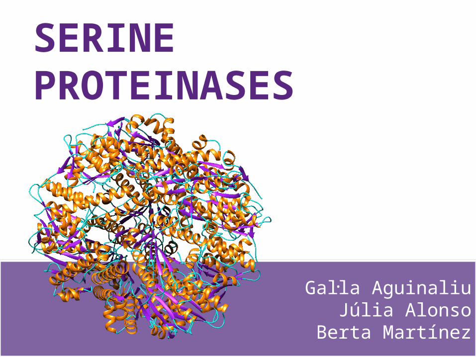
SERINEPROTEINASES
Gal·la AguinaliuJúlia Alonso
Berta Martínez
INDEX
• Introduction• General mechanism of action• Different topologies and foldings• Results• Conclusions• References

INTRODUCTION
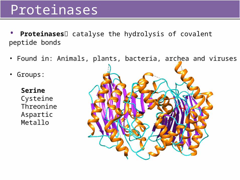
Proteinases
• Proteinases catalyse the hydrolysis of covalent peptide bonds
• Found in: Animals, plants, bacteria, archea and viruses
• Groups:
SerineCysteineThreonine AsparticMetallo
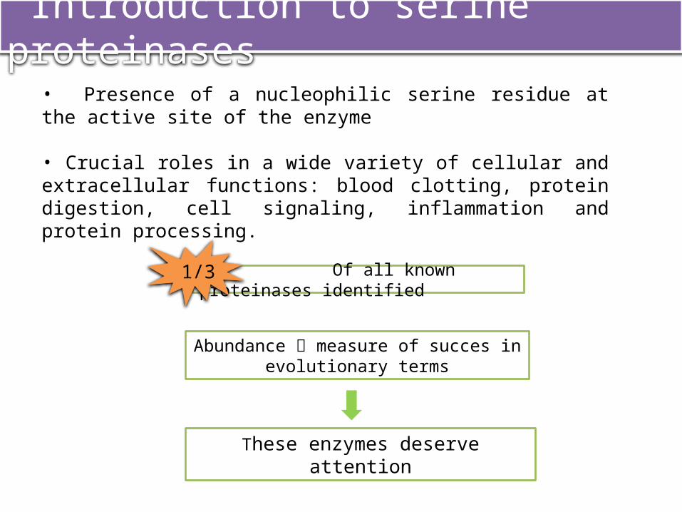
• Presence of a nucleophilic serine residue at the active site of the enzyme
• Crucial roles in a wide variety of cellular and extracellular functions: blood clotting, protein digestion, cell signaling, inflammation and protein processing.
Introduction to serine proteinases
Of all known proteinases identified1/3
Abundance measure of succes in evolutionary terms
These enzymes deserve attention
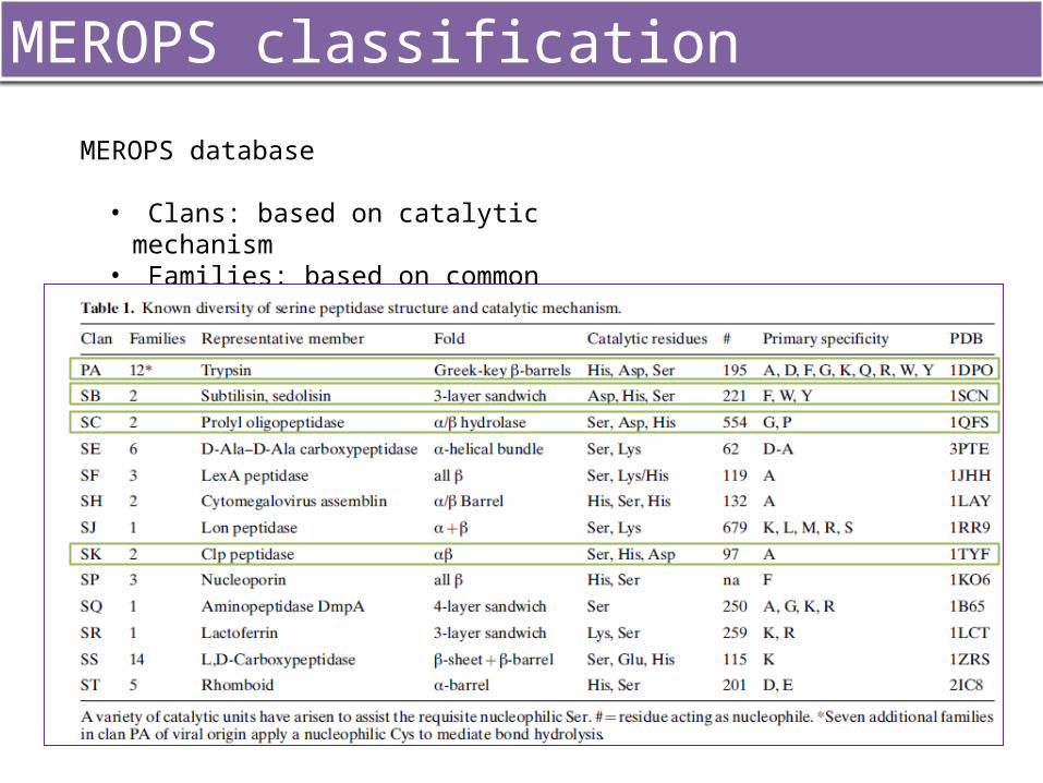
MEROPS classification
MEROPS database
• Clans: based on catalytic mechanism• Families: based on common ancestry
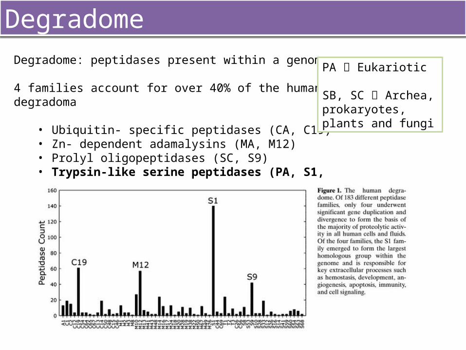
DegradomeDegradome: peptidases present within a genome
4 families account for over 40% of the human degradoma
• Ubiquitin- specific peptidases (CA, C19)• Zn- dependent adamalysins (MA, M12)• Prolyl oligopeptidases (SC, S9)• Trypsin-like serine peptidases (PA, S1, A)
PA Eukariotic
SB, SC Archea, prokaryotes, plants and fungi
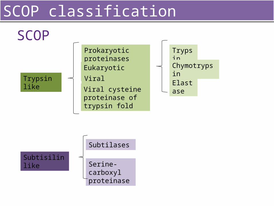
SCOP classification
SCOP
Trypsin like
Prokaryotic proteinasesEukaryotic proteinases
Viral proteinases
Viral cysteine proteinase of trypsin fold
Trypsin
Elastase
Chymotrypsin
Subtisilin like
Subtilases
Serine-carboxyl proteinase
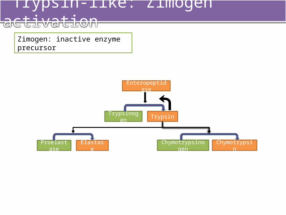
Trypsin-like: Zimogen activation
Enteropeptidase
Trypsinogen Trypsin
Proelastase Elastase Chymotrypsinogen Chymotrypsin
Zimogen: inactive enzyme precursor
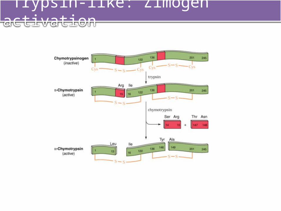
Trypsin-like: Zimogen activation
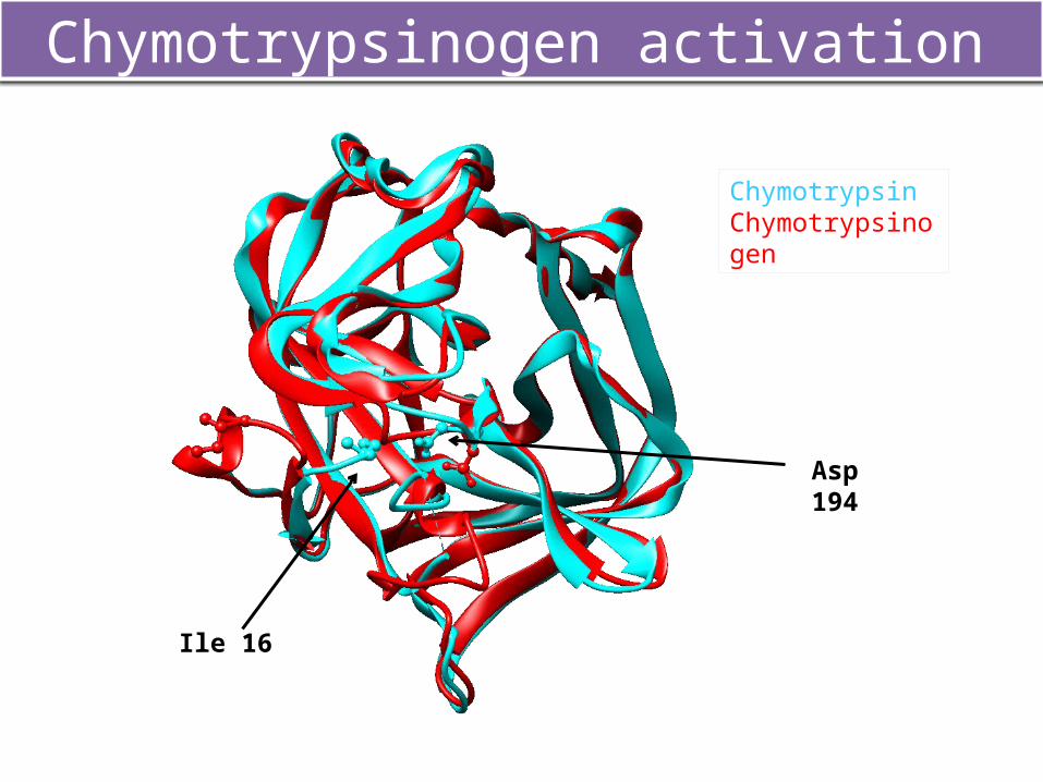
Chymotrypsinogen activation
ChymotrypsinChymotrypsinogen
Ile 16
Asp 194
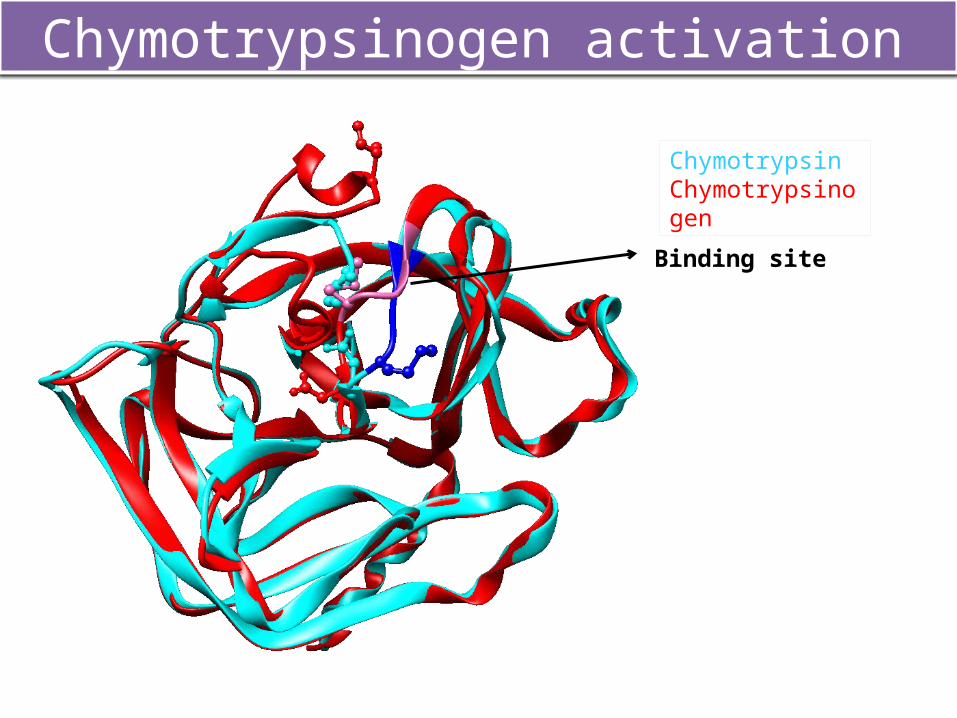
Chymotrypsinogen activation
ChymotrypsinChymotrypsinogen
Binding site
MECHANISM OF
ACTION
Four important structural features required for the catalitic action of SP:
1. Catalytic triad2. The oxanyon hole3. Polypeptide binding site4. Specificity pockets
Chemical mechanism of serine proteinases
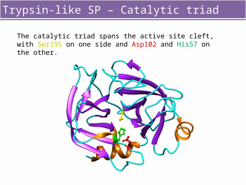
The catalytic triad spans the active site cleft, with Ser195 on one side and Asp102 and His57 on the other.
Trypsin-like SP – Catalytic triad
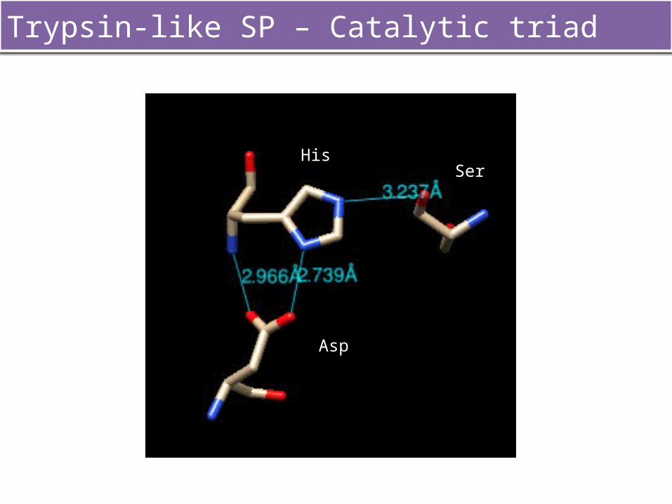
Trypsin-like SP – Catalytic triad
Asp2
Ser2His2
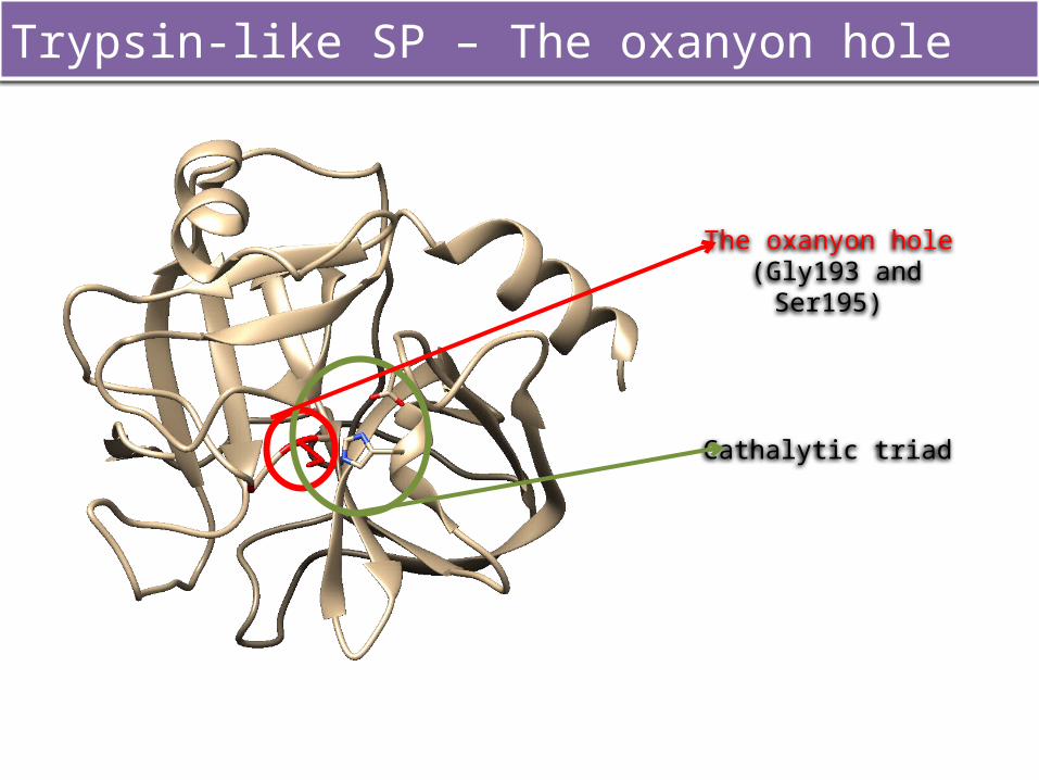
The oxanyon hole (Gly193 and Ser195)
Cathalytic triad
Trypsin-like SP – The oxanyon hole
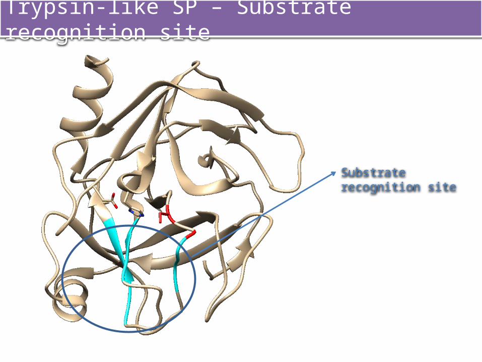
Trypsin-like SP – Substrate recognition site
Substrate recognition site
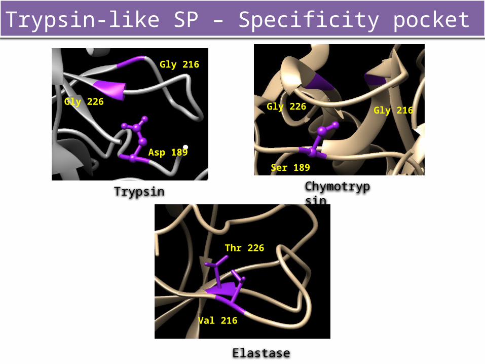
Trypsin-like SP – Specificity pocket
Trypsin Chymotrypsin
Elastase
Gly 226
Gly 216
Asp 189
Ser 189
Gly 216Gly 226
Thr 226
Val 216
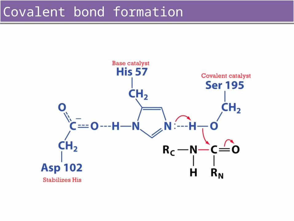
Covalent bond formation
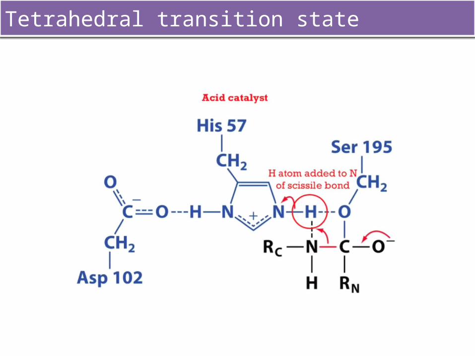
Tetrahedral transition state
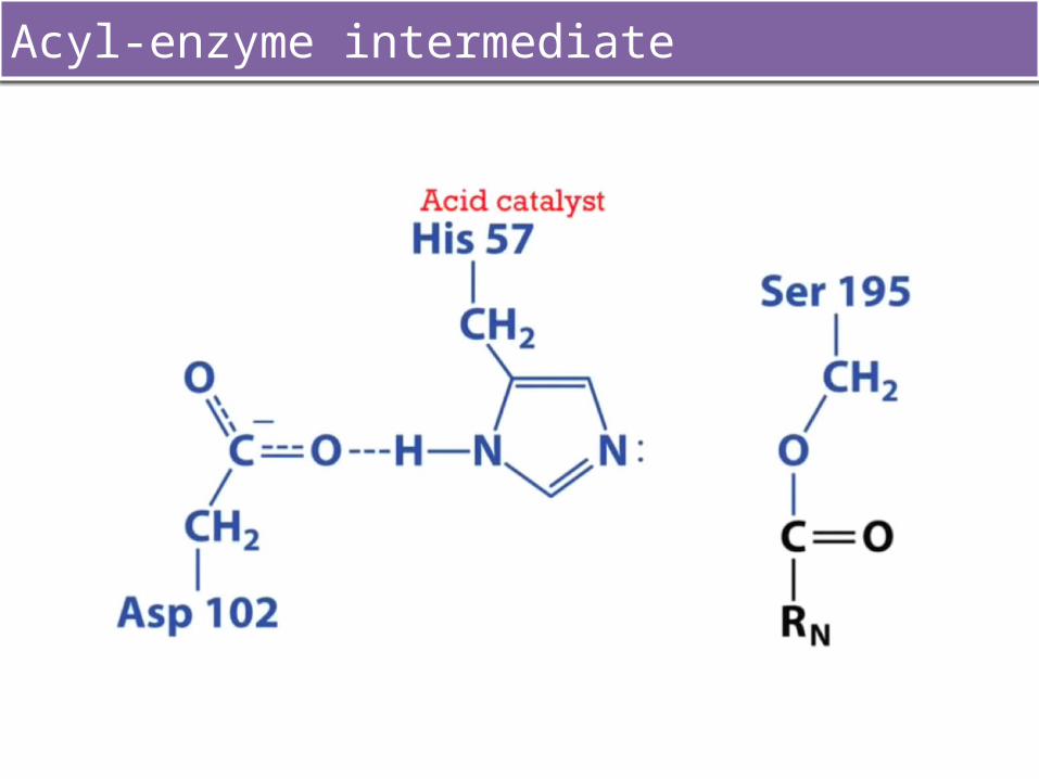
Acyl-enzyme intermediate
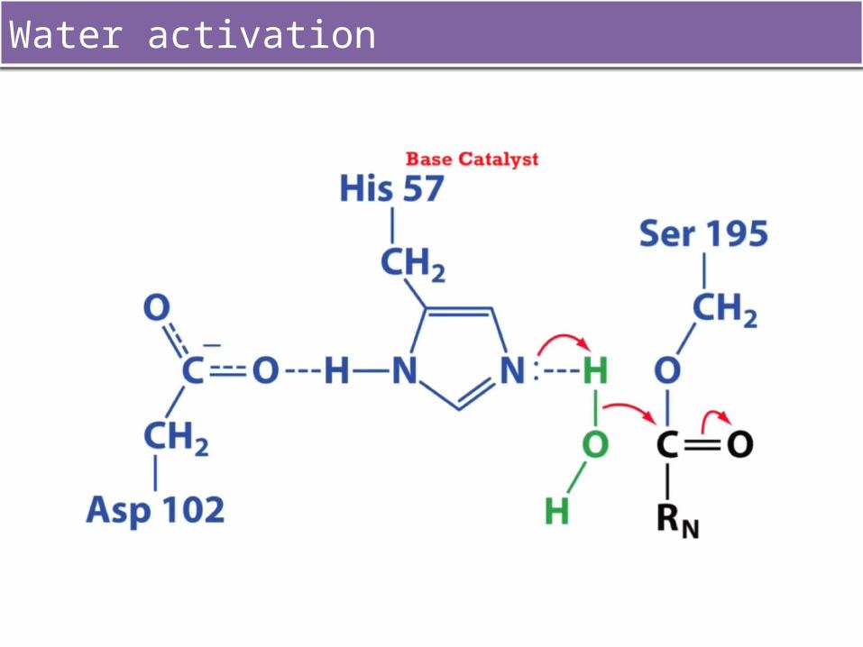
Water activation
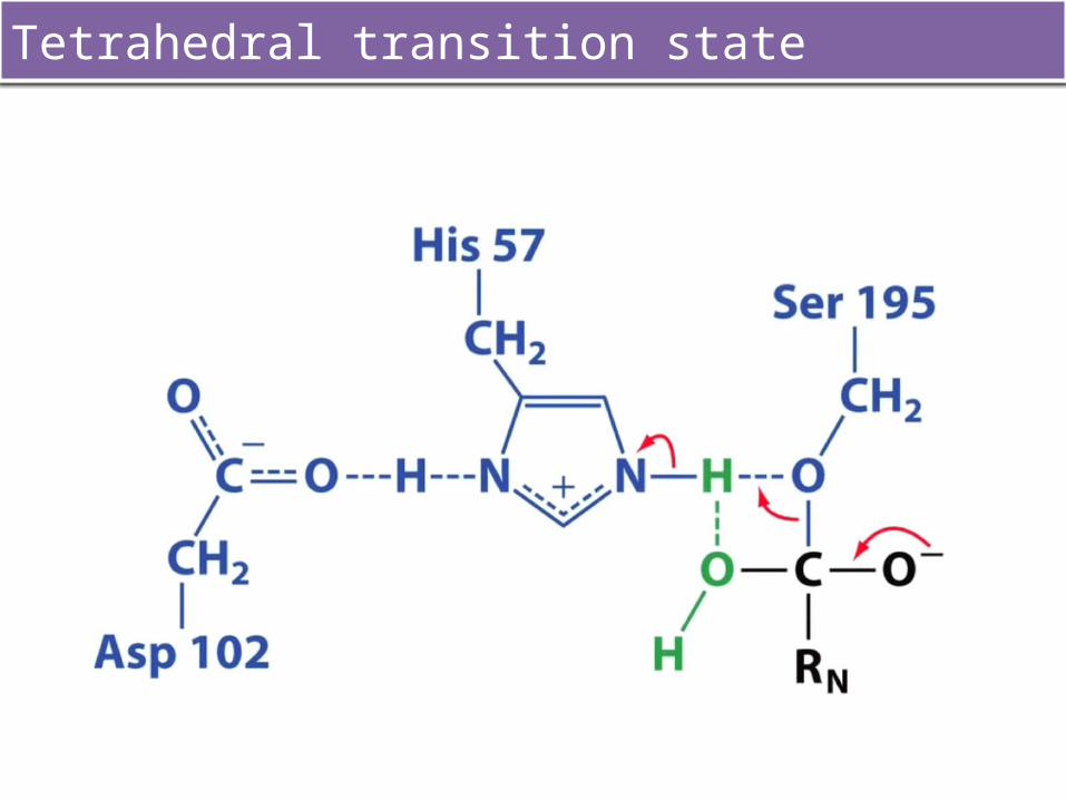
Tetrahedral transition state
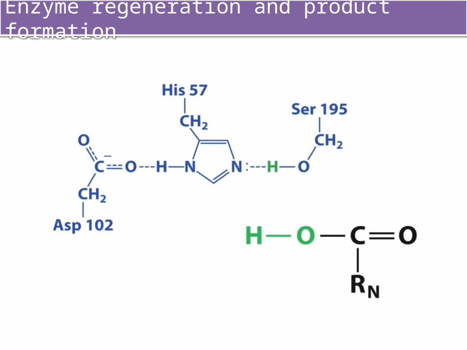
Enzyme regeneration and product formation
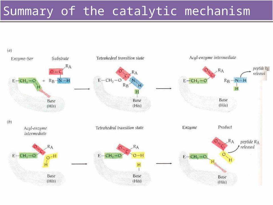
Summary of the catalytic mechanism
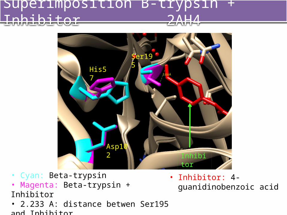
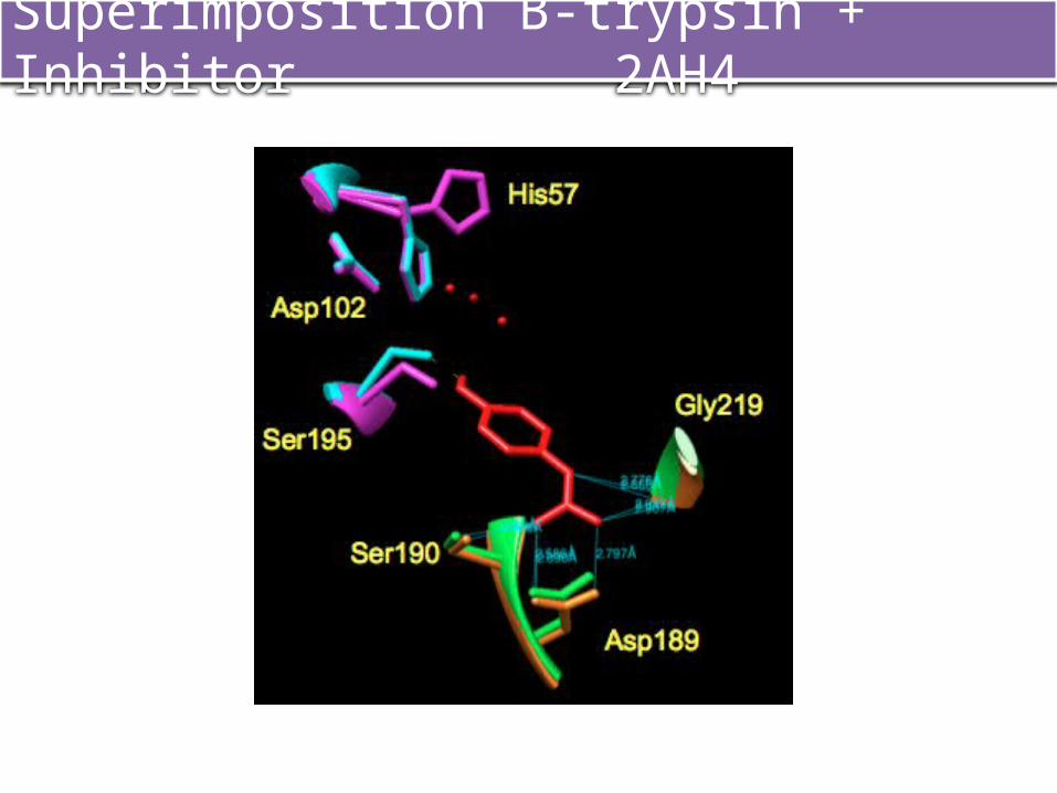
Superimposition B-trypsin + Inhibitor 2AH4
InhibitorAsp102
His57
Ser195
• Inhibitor: 4-guanidinobenzoic acid
• Cyan: Beta-trypsin• Magenta: Beta-trypsin + Inhibitor• 2.233 A: distance betwen Ser195 and Inhibitor
Superimposition B-trypsin + Inhibitor 2AH4
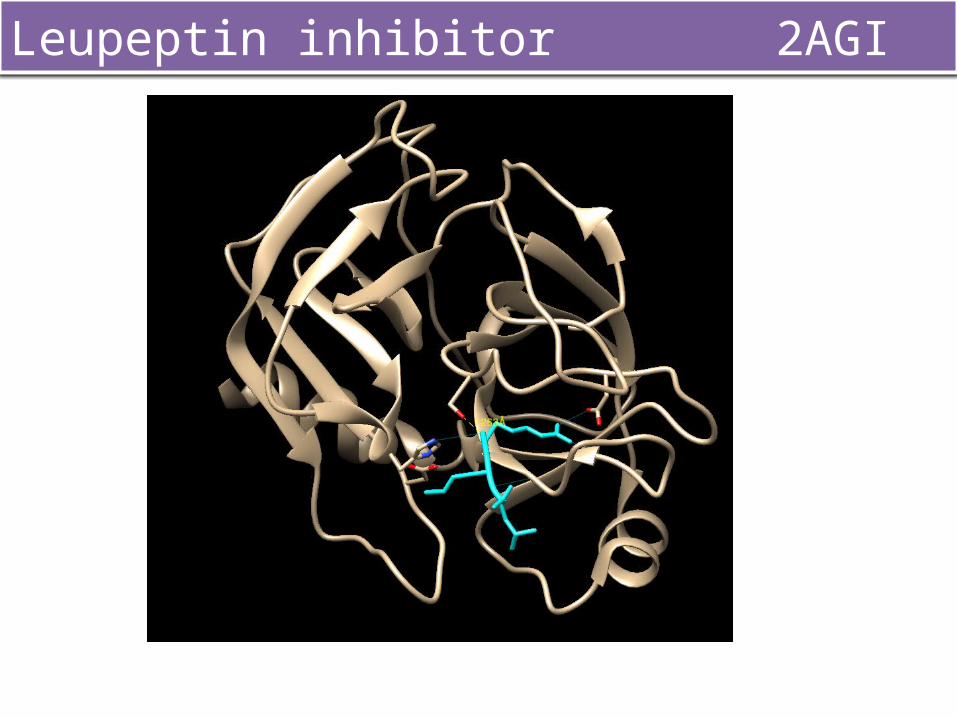
Leupeptin inhibitor 2AGI
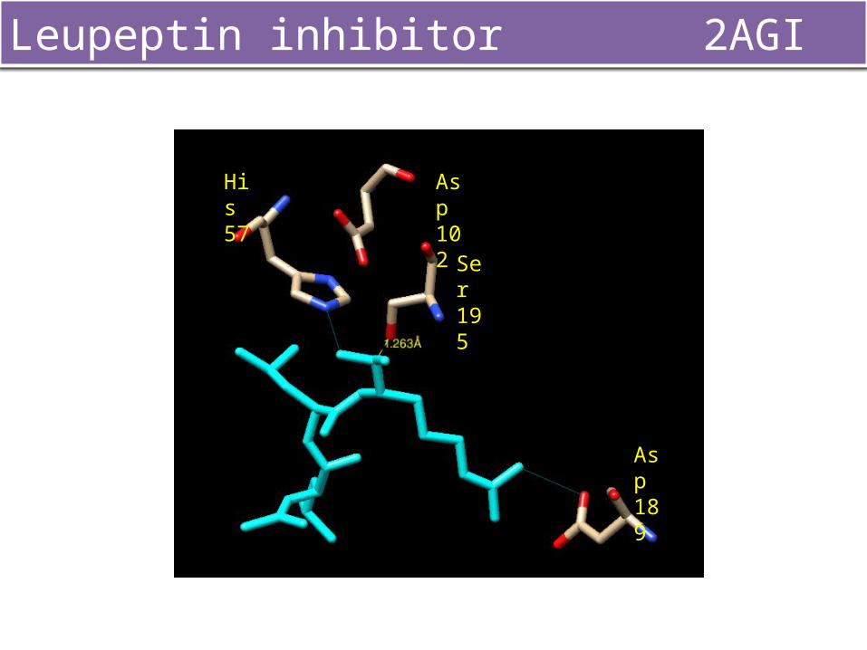
Leupeptin inhibitor 2AGI
His 57
Ser 195
Asp 102
Asp 189

TOPOLOGY and FOLDINGS
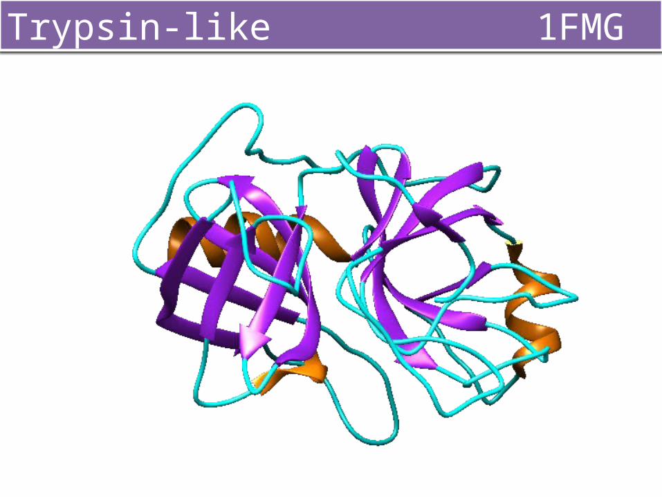
Trypsin-like 1FMG
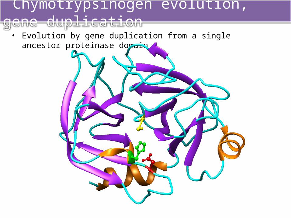
• Evolution by gene duplication from a single ancestor proteinase domain
Chymotrypsinogen evolution, gene duplication
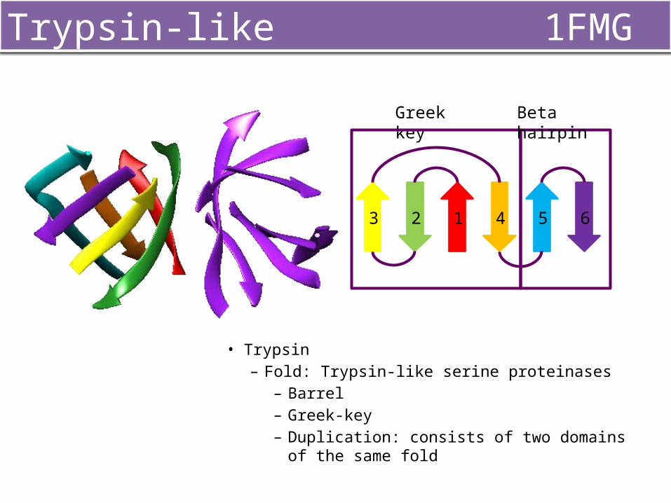
Trypsin-like 1FMG
• Trypsin– Fold: Trypsin-like serine proteinases
– Barrel– Greek-key – Duplication: consists of two domains of the same fold
Greek key Beta hairpin
123 4 5 6
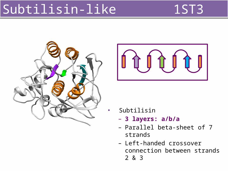
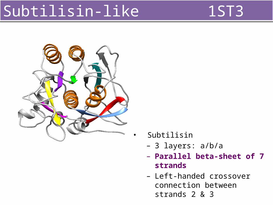
• Subtilisin– 3 layers: a/b/a– Parallel beta-sheet of 7 strands – Left-handed crossover connection
between strands 2 & 3
Subtilisin-like 1ST3
• Subtilisin– 3 layers: a/b/a– Parallel beta-sheet of 7 strands – Left-handed crossover connection
between strands 2 & 3
Subtilisin-like 1ST3
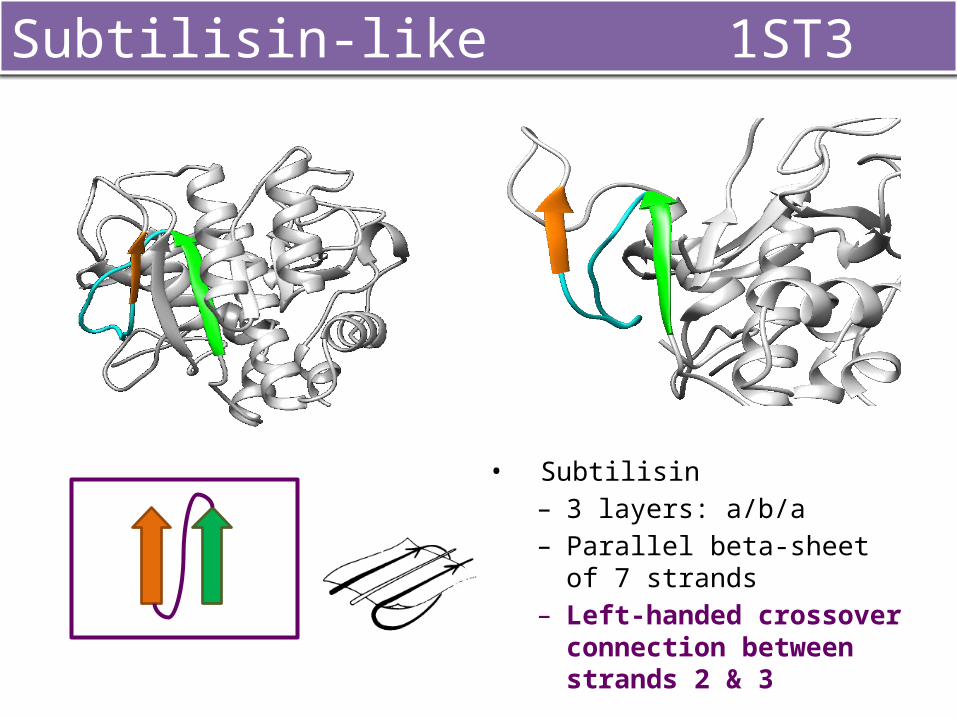
• Subtilisin– 3 layers: a/b/a– Parallel beta-sheet of 7 strands – Left-handed crossover
connection between strands 2 & 3
Subtilisin-like 1ST3
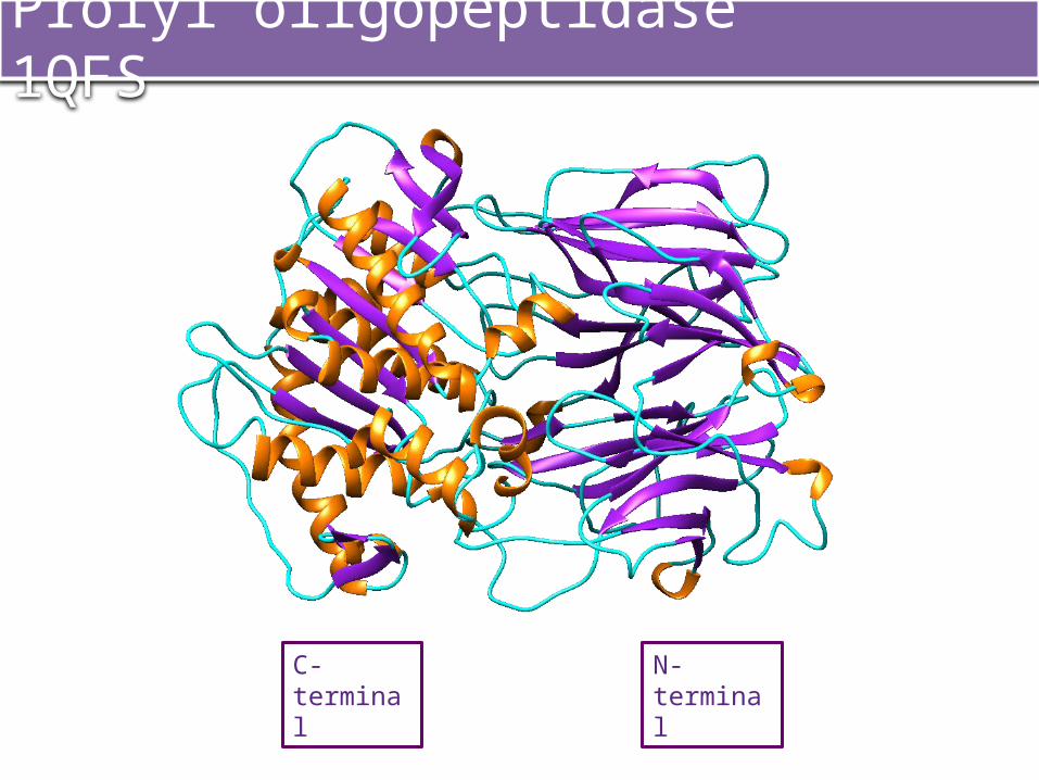
C-terminal N-terminal
Prolyl oligopeptidase 1QFS
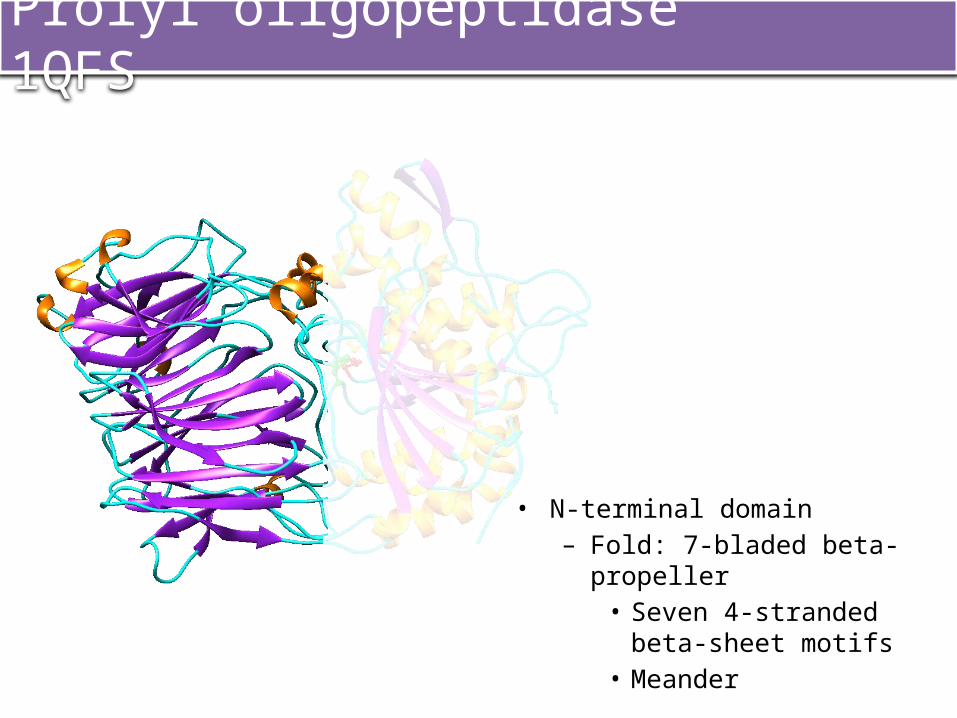
• N-terminal domain– Fold: 7-bladed beta-propeller
• Seven 4-stranded beta-sheet motifs
• Meander
Prolyl oligopeptidase 1QFS
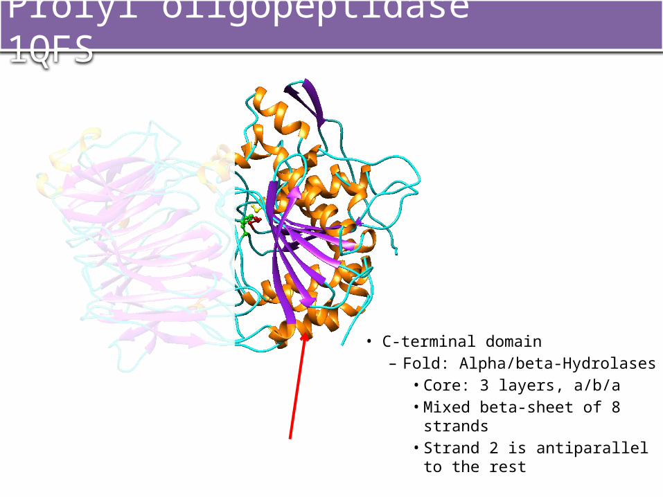
• C-terminal domain– Fold: Alpha/beta-Hydrolases
• Core: 3 layers, a/b/a• Mixed beta-sheet of 8 strands• Strand 2 is antiparallel to the
rest
Prolyl oligopeptidase 1QFS
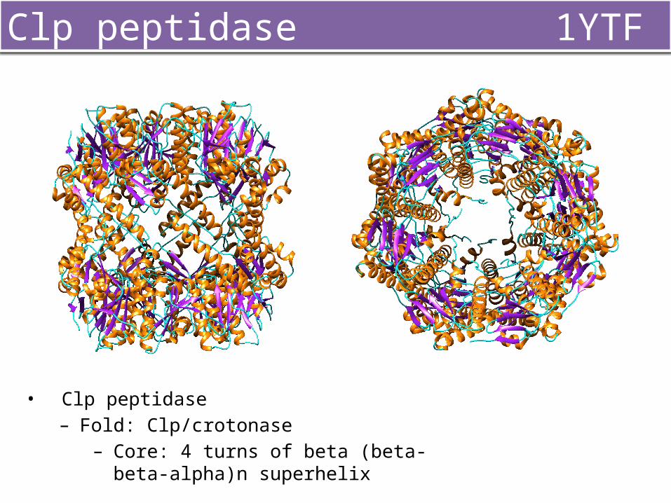
Clp peptidase 1YTF
• Clp peptidase– Fold: Clp/crotonase
– Core: 4 turns of beta (beta-beta-alpha)n superhelix

RESULTS
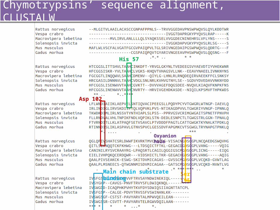
Chymotrypsins’ sequence alignment, CLUSTALW
Ser 195
Asp 102
His 57
Oxyanion hole
Main chain substrate binding
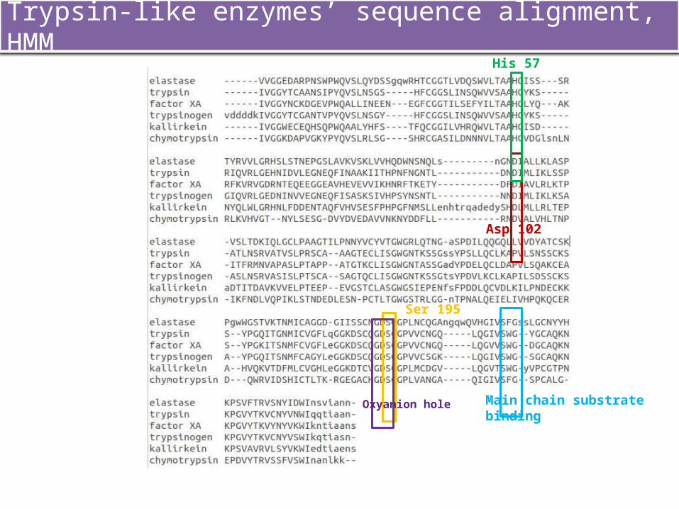
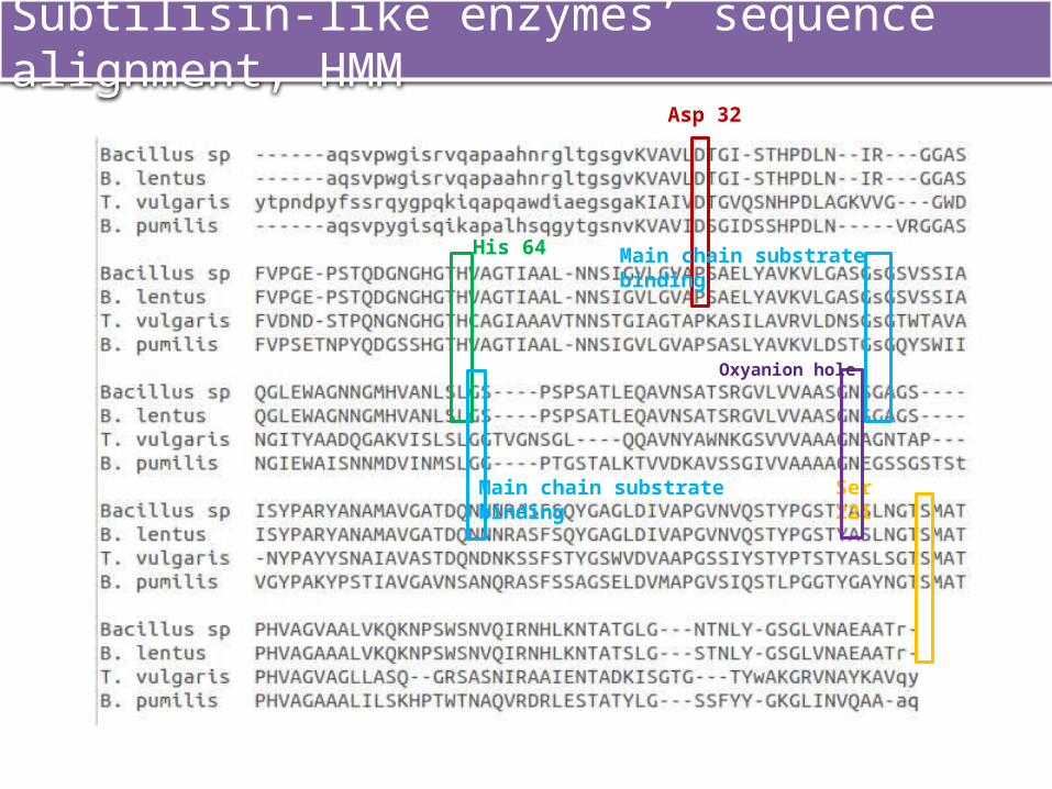
Trypsin-like enzymes’ sequence alignment, HMM
Ser 195
Asp 102
His 57
Oxyanion hole
Main chain substrate binding
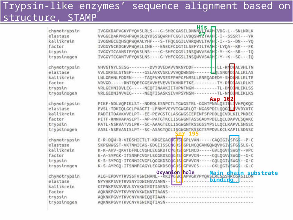
Trypsin-like enzymes’ sequence alignment based on structure, STAMP
Ser 195
Asp 102
His 57
Oxyanion hole
Main chain substrate binding
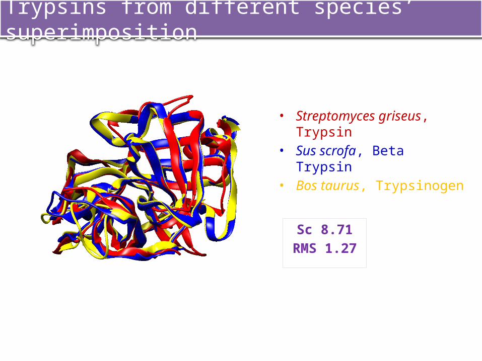
Trypsins from different species’ superimposition
Sc 8.71RMS 1.27
• Streptomyces griseus, Trypsin• Sus scrofa, Beta Trypsin• Bos taurus, Trypsinogen
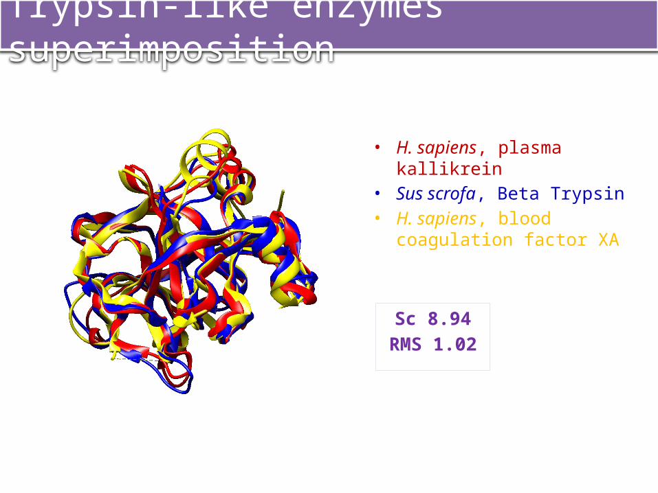
Trypsin-like enzymes’ superimposition
Sc 8.94RMS 1.02
• H. sapiens, plasma kallikrein• Sus scrofa, Beta Trypsin• H. sapiens, blood coagulation
factor XA

• Divergent evolution
Trypsin-like enzymes’ superimposition
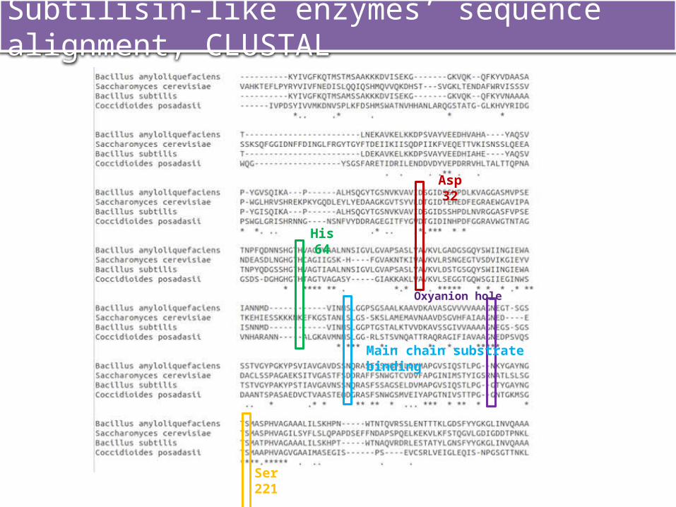
Subtilisin-like enzymes’ sequence alignment, CLUSTAL
Ser 221
Asp 32
His 64
Oxyanion hole
Main chain substrate binding
Subtilisin-like enzymes’ sequence alignment, HMM
Ser 221
Asp 32
His 64
Oxyanion hole
Main chain substrate binding
Main chain substrate binding
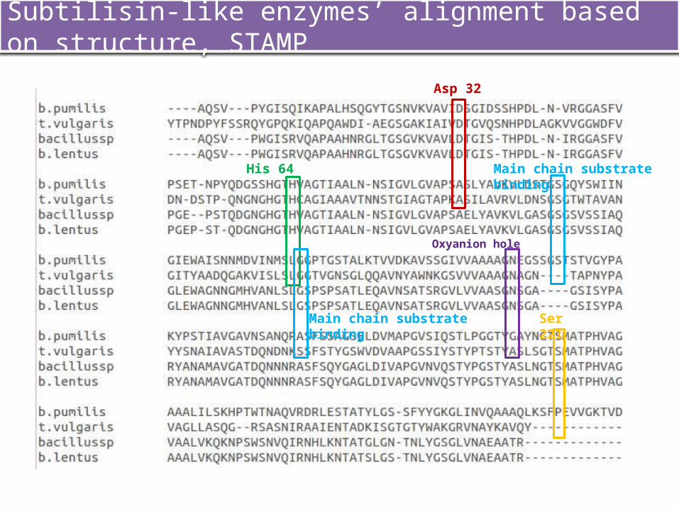
Subtilisin-like enzymes’ alignment based on structure, STAMP
Ser 221
Asp 32
His 64
Oxyanion hole
Main chain substrate binding
Main chain substrate binding
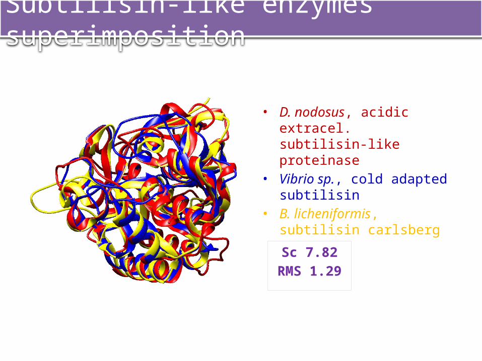
Subtilisin-like enzymes’ superimposition
• D. nodosus, acidic extracel. subtilisin-like proteinase
• Vibrio sp., cold adapted subtilisin
• B. licheniformis, subtilisin carlsberg
Sc 7.82RMS 1.29
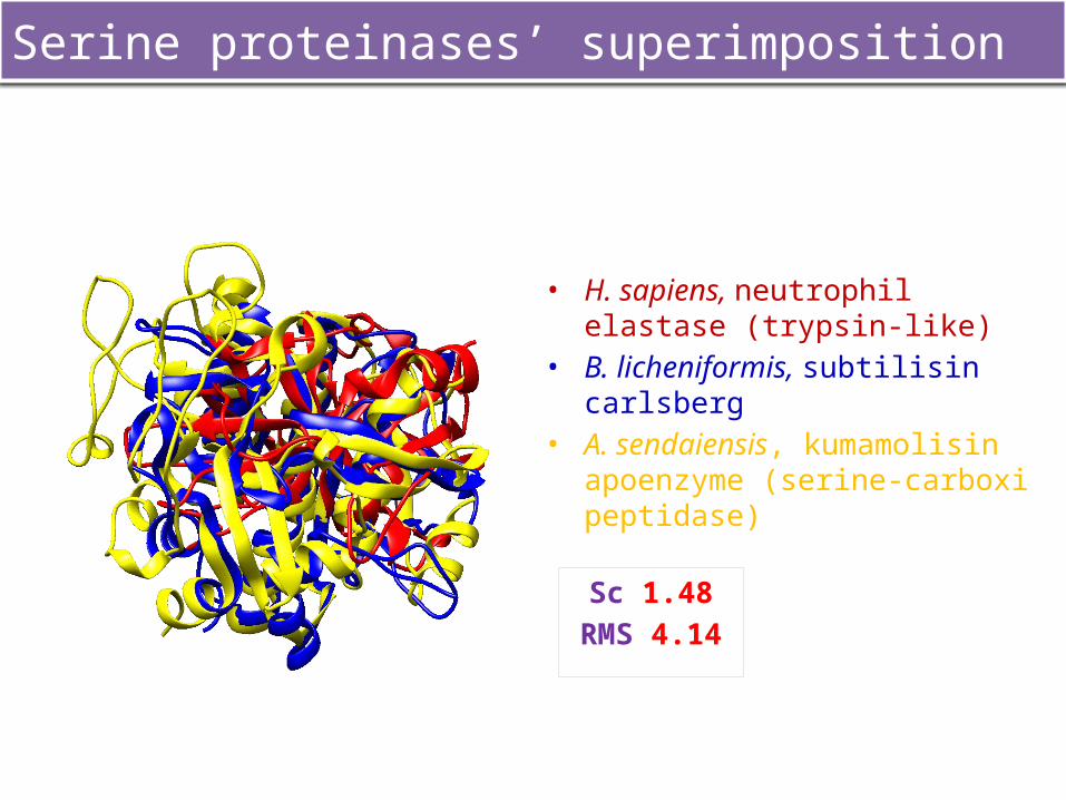
Serine proteinases’ superimposition
• H. sapiens, neutrophil elastase (trypsin-like)
• B. licheniformis, subtilisin carlsberg• A. sendaiensis, kumamolisin
apoenzyme (serine-carboxi peptidase)
Sc 1.48RMS 4.14
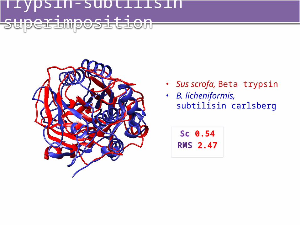
Trypsin-subtilisin superimposition
• Sus scrofa, Beta trypsin• B. licheniformis, subtilisin
carlsberg
Sc 0.54RMS 2.47
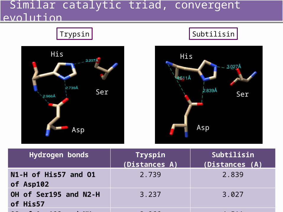
Similar catalytic triad, convergent evolution
SubtilisinTrypsin
Hydrogen bonds Tryspin (Distances A) Subtilisin (Distances (A)
N1-H of His57 and O1 of Asp102 2.739 2.839
OH of Ser195 and N2-H of His57 3.237 3.027
O2 of Asp102 and NHs His57 2.966 4.511
Asp2
Ser2
Asp2
Ser2
His2 His2

CONCLUSIONS
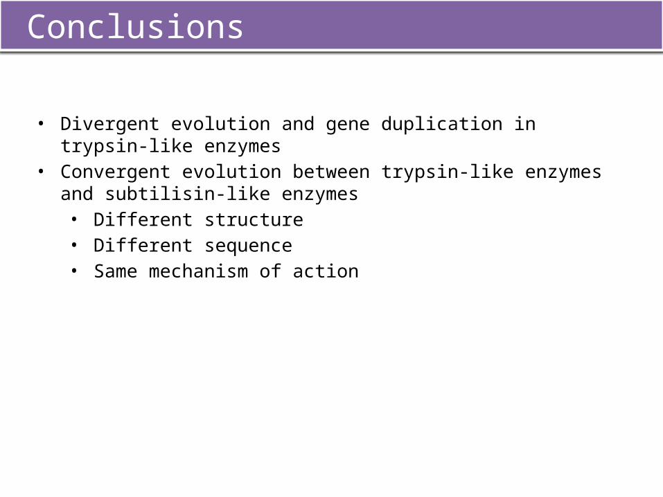
Conclusions
• Divergent evolution and gene duplication in trypsin-like enzymes• Convergent evolution between trypsin-like enzymes and subtilisin-like
enzymes • Different structure• Different sequence• Same mechanism of action

PROGRAMMES USED
• ClustalW• HMM• STAMP• XAM• Chimera• Rasmol
Programmes used
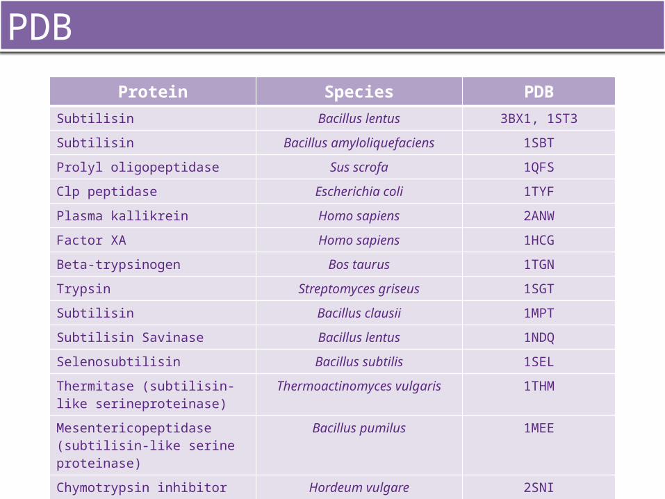
PDBProtein Species PDB
Subtilisin Bacillus lentus 3BX1, 1ST3Subtilisin Bacillus amyloliquefaciens 1SBTProlyl oligopeptidase Sus scrofa 1QFSClp peptidase Escherichia coli 1TYFPlasma kallikrein Homo sapiens 2ANWFactor XA Homo sapiens 1HCGBeta-trypsinogen Bos taurus 1TGNTrypsin Streptomyces griseus 1SGTSubtilisin Bacillus clausii 1MPTSubtilisin Savinase Bacillus lentus 1NDQSelenosubtilisin Bacillus subtilis 1SELThermitase (subtilisin-like serineproteinase)
Thermoactinomyces vulgaris 1THM
Mesentericopeptidase (subtilisin-like serine proteinase)
Bacillus pumilus 1MEE
Chymotrypsin inhibitor CI-2 Hordeum vulgare 2SNI
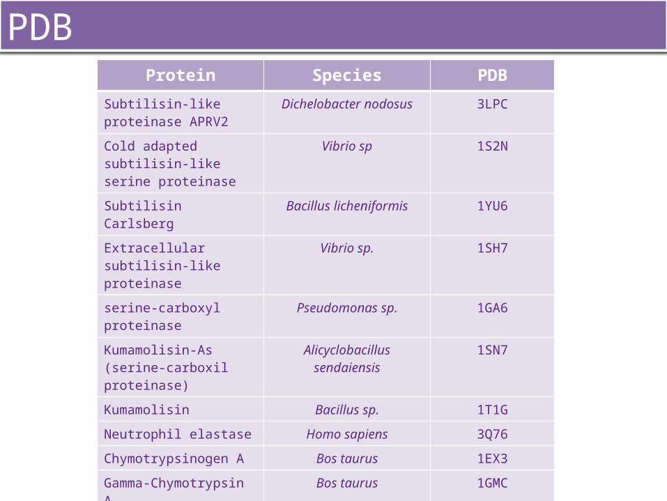
Protein Species PDB
Subtilisin-like proteinase APRV2
Dichelobacter nodosus 3LPC
Cold adapted subtilisin-like serine proteinase
Vibrio sp 1S2N
Subtilisin Carlsberg Bacillus licheniformis 1YU6
Extracellular subtilisin-like proteinase
Vibrio sp. 1SH7
serine-carboxyl proteinase Pseudomonas sp. 1GA6
Kumamolisin-As (serine-carboxil proteinase)
Alicyclobacillus sendaiensis 1SN7
Kumamolisin Bacillus sp. 1T1G
Neutrophil elastase Homo sapiens 3Q76
Chymotrypsinogen A Bos taurus 1EX3
Gamma-Chymotrypsin A Bos taurus 1GMC
Cationic trypsin Bos taurus 4I8G
Beta- Trypsin Sus scrofa 1FMG
Elastase Sus scrofa 1C1M
Chymotrypsin Bos taurus 1GMC, 2CHA
PDB

REFERENCES
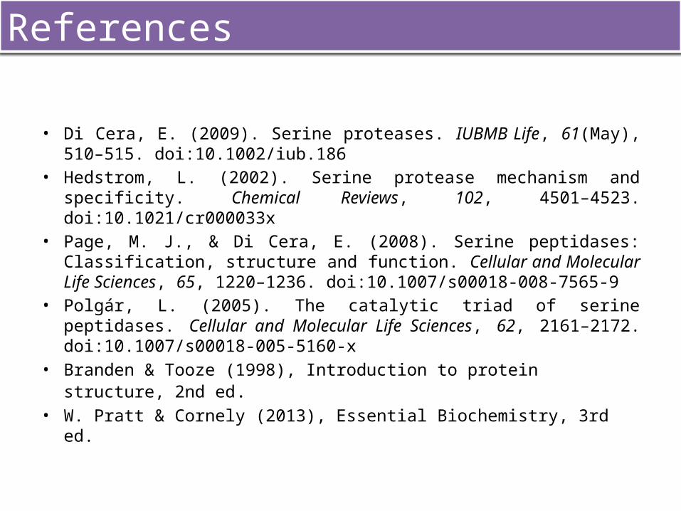
• Di Cera, E. (2009). Serine proteases. IUBMB Life, 61(May), 510–515. doi:10.1002/iub.186
• Hedstrom, L. (2002). Serine protease mechanism and specificity. Chemical Reviews, 102, 4501–4523. doi:10.1021/cr000033x
• Page, M. J., & Di Cera, E. (2008). Serine peptidases: Classification, structure and function. Cellular and Molecular Life Sciences, 65, 1220–1236. doi:10.1007/s00018-008-7565-9
• Polgár, L. (2005). The catalytic triad of serine peptidases. Cellular and Molecular Life Sciences, 62, 2161–2172. doi:10.1007/s00018-005-5160-x
• Branden & Tooze (1998), Introduction to protein structure, 2nd ed.• W. Pratt & Cornely (2013), Essential Biochemistry, 3rd ed.
References

1. Which of these residues are part of the catalytic thriad in serine proteinases?a) Asp-Ser-Hisb) Asp-Thr-Hisc) Ser-Asp-Thrd) Ser-Gly-Hise) Asp-His-Thr
2. Proteinases are found in:a) Animalsb) Bacteria and plantsc) Archaea and virusesd) All of the answers above are incorrecte) a, b and c are correct
3. Regarding trypsin-like and subtilisin-like enzymes, they both have similar:f) Structureg) Sequenceh) Catalytic thriadi) Functionj) A, b, c and d are true
4. A superimposition with STAMP of different chymotrypsins from different species…a) could probably have a SC value lower than 5.5b) could probably have a SC value lower than 2c) could probably have a SC value between 5.5-9.8d) will probably have a RMSD value higher than 2e) will probably have a RMSD value higher than 5.5
5. Which different proteinases groups do exist?a) Serine and cysteinb) Serine, cystein, threonin and glycinec) Cystein, serine, threonin, aspartic and metallod) Cystein, metallo, serine, glycine and histidinee) All of the answers are incorrect
PEM
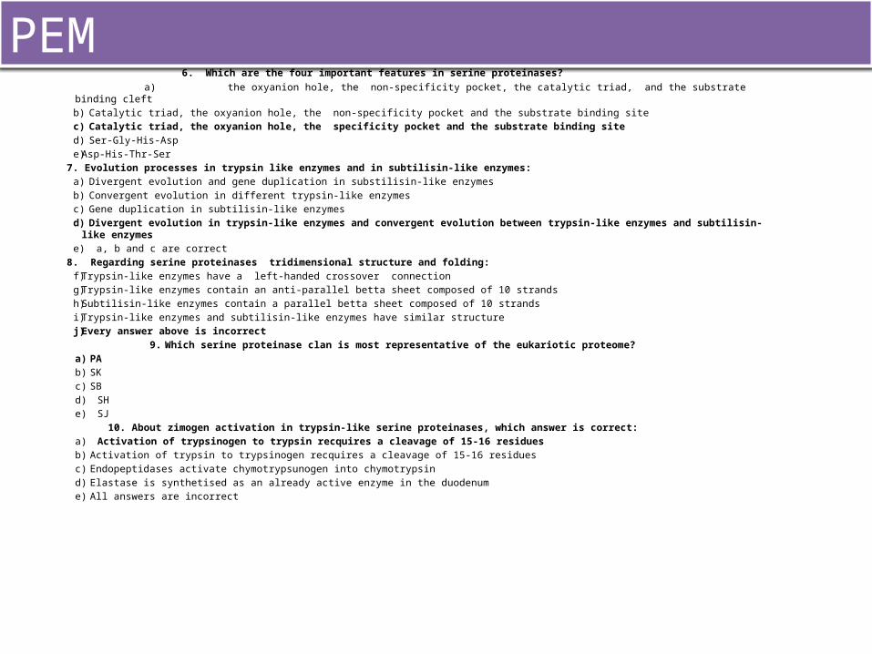
6. Which are the four important features in serine proteinases? a) the oxyanion hole, the non-specificity pocket, the catalytic triad, and the substrate binding cleft
b) Catalytic triad, the oxyanion hole, the non-specificity pocket and the substrate binding sitec) Catalytic triad, the oxyanion hole, the specificity pocket and the substrate binding sited) Ser-Gly-His-Aspe) Asp-His-Thr-Ser
7. Evolution processes in trypsin like enzymes and in subtilisin-like enzymes:a) Divergent evolution and gene duplication in substilisin-like enzymesb) Convergent evolution in different trypsin-like enzymesc) Gene duplication in subtilisin-like enzymesd) Divergent evolution in trypsin-like enzymes and convergent evolution between trypsin-like enzymes and subtilisin-like enzymes e) a, b and c are correct
8. Regarding serine proteinases tridimensional structure and folding:f) Trypsin-like enzymes have a left-handed crossover connectiong) Trypsin-like enzymes contain an anti-parallel betta sheet composed of 10 strandsh) Subtilisin-like enzymes contain a parallel betta sheet composed of 10 strandsi) Trypsin-like enzymes and subtilisin-like enzymes have similar structurej) Every answer above is incorrect
9. Which serine proteinase clan is most representative of the eukariotic proteome?a) PAb) SKc) SBd) SHe) SJ
10. About zimogen activation in trypsin-like serine proteinases, which answer is correct:a) Activation of trypsinogen to trypsin recquires a cleavage of 15-16 residuesb) Activation of trypsin to trypsinogen recquires a cleavage of 15-16 residuesc) Endopeptidases activate chymotrypsunogen into chymotrypsind) Elastase is synthetised as an already active enzyme in the duodenume) All answers are incorrect
PEM