Embed Size (px)
Citation preview
Serine 254 Enhances an Induced Fit Mechanism in Murine5-Aminolevulinate Synthase*□S
Received for publication, September 15, 2009, and in revised form, November 13, 2009 Published, JBC Papers in Press, November 16, 2009, DOI 10.1074/jbc.M109.066548
Thomas Lendrihas‡, Gregory A. Hunter‡, and Gloria C. Ferreira‡§¶1
From the Departments of ‡Molecular Medicine and §Chemistry and the ¶H. Lee Moffitt Cancer Center and Research Institute,University of South Florida, Tampa, Florida 33612
5-Aminolevulinate synthase (EC2.3.1.37) (ALAS), a pyridoxal5�-phosphate (PLP)-dependent enzyme, catalyzes the initialstep of heme biosynthesis in animals, fungi, and some bacteria.Condensation of glycine and succinyl coenzyme A produces5-aminolevulinate, coenzymeA, andcarbondioxide.X-ray crys-tal structures ofRhodobacter capsulatusALAS reveal that a con-served active site serine moves to within hydrogen bonding dis-tance of the phenolic oxygen of the PLP cofactor in the closedsubstrate-bound enzyme conformation and within 3–4 A of thethioester sulfur atom of bound succinyl-CoA. To evaluate therole(s) of this residue in enzymatic activity, the equivalent serinein murine erythroid ALAS was substituted with alanine or threo-nine. Although both the Km
SCoA and kcat values of the S254Avariant increased, by 25- and 2-fold, respectively, the S254Tsubstitution decreased kcat without altering Km
SCoA. Further-more, in relation to wild-type ALAS, the catalytic efficiency ofS254A toward glycine improved�3-fold, whereas that of S254Tdiminished �3-fold. Circular dichroism spectroscopy revealedthat removal of the side chain hydroxyl group in the S254A var-iant altered the microenvironment of the PLP cofactor and hin-dered succinyl-CoA binding. Transient kinetic analyses of thevariant-catalyzed reactions and protein fluorescence quenchingupon 5-aminolevulinate binding demonstrated that the proteinconformational transition step associated with product releasewas predominantly affected. We propose the following: 1) Ser-254 is critical for formation of a competent catalytic complex bycoupling succinyl-CoAbinding to enzyme conformational equi-libria, and 2) the role of the active site serine should be extendedto the entire �-oxoamine synthase family of PLP-dependentenzymes.
5-Aminolevulinate synthase (EC 2.3.1.37; ALAS)2 is ahomodimeric PLP-dependent enzyme that catalyzes the firstand key regulatory step of the heme biosynthetic pathway innon-plant eukaryotes and the �-subclass of purple bacteria,involving the condensation of glycine and succinyl-CoA to pro-duce CoA, carbon dioxide, and ALA (1). Animal genomes
encode two highly conserved but differentially expressedALASgenes, one of which is expressed in all tissues (ALAS1), and theother of which is erythroid-specific (ALAS2) (2). In humans,mutations in theALAS2 gene can result in X-linked sideroblas-tic anemia (3), a hypochromic and microcytic anemia charac-terized by iron accumulation within erythroblast mitochondria(4). Approximately one-third of X-linked sideroblastic anemiapatients are pyridoxine-responsive, and in these patients theALAS mutations are commonly observed in the PLP-bindingsite (5, 6).The ALAS chemical mechanism (Scheme 1) is complex and
involves a high degree of stereoelectronic control, with individ-ual steps, including the following: (i) binding of glycine; (ii)transaldimination with the active site lysine (Lys-313, murineALAS2 numbering) to yield an external aldimine; (iii) abstrac-tion of the pro-R proton of glycine; (iv) condensation withsuccinyl-CoA; (v)CoArelease togeneratean�-amino-�-ketoadi-pate intermediate; (vi) decarboxylation resulting in an enol-quinonoid rapid equilibrium; (vii) protonation of the enol togive an aldimine-bound molecule of ALA; and (viii) ultimatelyrelease of the product (ALA) (7). This mechanistic complexityis manifested structurally as an enzyme with an unusually highdegree of sequence conservation, as exemplified by the observa-tion that the catalytic cores of human ALAS2 and RhodobactercapsulatusALAS are 49% identical and 70% similar (8).PLP-dependent enzymes are classified according to struc-
tural and mechanistic similarities (9). ALAS is evolutionarilyrelated to transaminases and is grouped within class II of thefold type I PLP-dependent enzyme superfamily, for whichthe prototypical enzyme is generally considered to be aspartateaminotransferase (10–12). ALAS is most closely related to thethree other members of the �-oxoamine synthase subfamily,each of which also catalyzes a reaction involving a small aminoacid, a CoA ester, and a 1,3-aminoketone (13, 14).Aspartate aminotransferase exists in two predominant con-
formations, “open” and “closed” (10–12), and this property hasbeen hypothesized to be a general feature of all PLP-dependentenzymes (15, 16). Catalysis occurs in the closed conformation,consistent with the induced fit hypothesis, where electrostaticand hydrophobic interactions between the substrates, cofactor,and amino acids comprising the active site provide the ener-getic impetus to stabilize a catalytically optimal conformation(17).Prior to solution of an ALAS crystal structure, kinetic data
led to the proposal that the ALAS catalytic cycle is dominatedby a rate-limiting conformational change associated withrelease of ALA (7, 18–20). The crystal structures of R. capsula-
* This work was supported, in whole or in part, by National Institutes of HealthGrant DK63191 (to G. C. F.).
□S The on-line version of this article (available at http://www.jbc.org) containssupplemental “Experimental Procedures” and an additional reference.
1 To whom correspondence should be addressed: Dept. of Molecular Medi-cine, College of Medicine, MDC 7, University of South Florida, Tampa, FL33612-4799. Tel.: 813-974-5797; Fax: 813-974-0504; E-mail: [email protected].
2 The abbreviations used are: ALAS, 5-aminolevulinate synthase; ALA, 5-ami-nolevulinate; PLP, pyridoxal 5�-phosphate.
THE JOURNAL OF BIOLOGICAL CHEMISTRY VOL. 285, NO. 5, pp. 3351–3359, January 29, 2010© 2010 by The American Society for Biochemistry and Molecular Biology, Inc. Printed in the U.S.A.
JANUARY 29, 2010 • VOLUME 285 • NUMBER 5 JOURNAL OF BIOLOGICAL CHEMISTRY 3351
by guest on March 23, 2018
http://ww
w.jbc.org/
Dow
nloaded from

tus ALAS in holoenzymic and substrate-bound forms con-firmed that the enzyme adopts open and closed conformations,respectively, and allowed discrimination of the specific regionsand residues of the enzyme involved in protein dynamics (8).Although the structure in general collapses slightly around thebound substrates, a more conformationally mobile loop ofamino acids located between two �-sheets at the subunit inter-
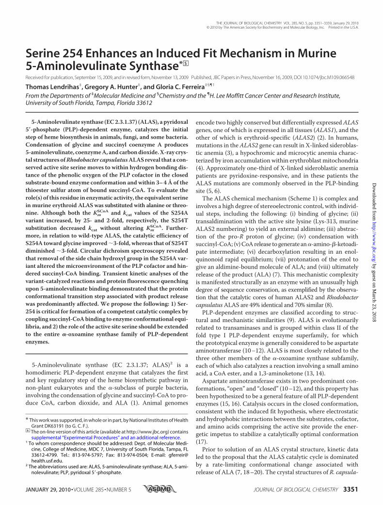
face closes directly over the active site channel, which extends�20Ådown into the core of the enzyme to the PLP-binding site(Fig. 1) (8). A conserved threonine at the apex of the mobileloop forms a strong hydrogen bond (�2.5 Å) with the carbox-ylate tail of succinyl-CoA in the substrate-bound structureand appears to simultaneously provide molecular recogni-tion for succinyl-CoA while helping to lock this substrate into
optimal stereoelectronic positionfor catalysis (21). Coincident withthese changes, the side chain ofSer-189 (R. capsulatus numbering)migrates from noncovalently asso-ciating with the peptide macroskel-eton to within hydrogen bondingdistance of the PLP phenolic oxy-gen, as well as the sulfur atom ofsuccinyl-CoA (8, 22, 23). Theseinteractions suggest that this serineresidue may be an important deter-minant in conformer equilibriumand catalysis by providing orienta-tional binding energy between theenzyme, cofactor, and substrate,exclusively, in the closed Michaeliscomplex conformation. The con-servation of this residue in ALASand the other �-oxoamine syn-thases further suggests an impor-tant functionality that may be gen-eral to these enzymes (Fig. 2). Here,we present experiments aimed atprobing the role of this serine in
FIGURE 1. Structural models for murine erythroid ALAS based on the R. capsulatus crystal structures.A, Michaelis complex modeled by alignment of open holoenzyme and closed glycine- and succinyl-CoA-boundmonomeric structures. Serine 254 is hidden by the succinyl-CoA ester in this view from the perspective of theadjacent subunit, which has been removed. The active site loop is shown in a yellow cartoon for the open andclosed conformations, and all other structural features are for the closed conformation. B, serine 254 in theopen conformation. C, serine 254 in the closed conformation with succinyl-CoA bound.
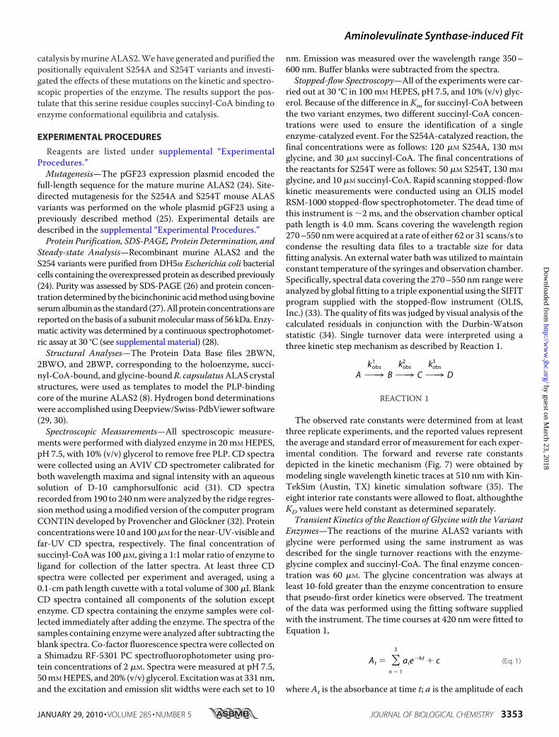
FIGURE 2. Multiple sequence alignment of phylogenetically diverse members of the �-oxoamine synthase family in the region of murine ALAS2serine 254. The amino acid sequences were retrieved from public data bases (NCBI) and aligned using ClustalW (42). The conserved serine residue isboxed and in boldface. The amino acid numbering (i.e. 254) refers to that of murine erythroid ALAS (mALAS2). Represented proteins are as follows: M. mus. AL2,Mus musculus erythroid ALAS (156255176); H. sap. AL2, Homo sapiens erythroid ALAS (28586); H. sap. AL1, H. sapiens housekeeping ALAS (40316939); S. cer. ALA,Saccharomyces cerevisiae ALAS (151942209); R. cap. ALA, R. capsulatus ALAS (974202); A. nig. AON, Aspergillus niger AONS (61696868); A. tha. AON, Arabidopsisthaliana AONS (42573269); M. mar. AON, Methanococcus maripaludis AONS (1599054); E. col. AON, E. coli AONS (85674759); H. sap. KBL, H. sapiens KBL (3342906);C. kor. KBL, Candidatus korarchaeum cryptofilum (17017433); E. col. KBL, E. coli KBL (169753078); H. sap. SPT, H. sapiens SPT (4758668); A. tha. SPT, A. thaliana SPT(17221603); S. cer. SPT, S. cerevisiae SPT (706828), E. col. SPT, E. coli SPT (170517920). AONS, 8-amino-7-oxononanoate synthase; SPT, serine palmitoyltransferase;KBL, 2-amino-3-oxobutyrate CoA ligase.
Aminolevulinate Synthase-induced Fit
3352 JOURNAL OF BIOLOGICAL CHEMISTRY VOLUME 285 • NUMBER 5 • JANUARY 29, 2010
by guest on March 23, 2018
http://ww
w.jbc.org/
Dow
nloaded from
catalysis bymurineALAS2.Wehave generated and purified thepositionally equivalent S254A and S254T variants and investi-gated the effects of these mutations on the kinetic and spectro-scopic properties of the enzyme. The results support the pos-tulate that this serine residue couples succinyl-CoA binding toenzyme conformational equilibria and catalysis.
EXPERIMENTAL PROCEDURES
Reagents are listed under supplemental “ExperimentalProcedures.”Mutagenesis—The pGF23 expression plasmid encoded the
full-length sequence for the mature murine ALAS2 (24). Site-directed mutagenesis for the S254A and S254T mouse ALASvariants was performed on the whole plasmid pGF23 using apreviously described method (25). Experimental details aredescribed in the supplemental “Experimental Procedures.”Protein Purification, SDS-PAGE, Protein Determination, and
Steady-state Analysis—Recombinant murine ALAS2 and theS254 variants were purified from DH5� Escherichia coli bacterialcells containing the overexpressed protein as described previously(24). Purity was assessed by SDS-PAGE (26) and protein concen-trationdeterminedby thebicinchoninic acidmethodusingbovineserumalbuminas the standard (27).All proteinconcentrationsarereportedon thebasis of a subunitmolecularmass of 56 kDa. Enzy-matic activity was determined by a continuous spectrophotomet-ric assay at 30 °C (see supplemental material) (28).Structural Analyses—The Protein Data Base files 2BWN,
2BWO, and 2BWP, corresponding to the holoenzyme, succi-nyl-CoA-bound, and glycine-boundR. capsulatusALAS crystalstructures, were used as templates to model the PLP-bindingcore of the murine ALAS2 (8). Hydrogen bond determinationswere accomplished usingDeepview/Swiss-PdbViewer software(29, 30).Spectroscopic Measurements—All spectroscopic measure-
ments were performed with dialyzed enzyme in 20 mMHEPES,pH 7.5, with 10% (v/v) glycerol to remove free PLP. CD spectrawere collected using an AVIV CD spectrometer calibrated forboth wavelength maxima and signal intensity with an aqueoussolution of D-10 camphorsulfonic acid (31). CD spectrarecorded from190 to 240 nmwere analyzed by the ridge regres-sionmethod using amodified version of the computer programCONTIN developed by Provencher and Glockner (32). Proteinconcentrationswere 10 and 100�M for the near-UV-visible andfar-UV CD spectra, respectively. The final concentration ofsuccinyl-CoAwas 100�M, giving a 1:1molar ratio of enzyme toligand for collection of the latter spectra. At least three CDspectra were collected per experiment and averaged, using a0.1-cm path length cuvette with a total volume of 300 �l. BlankCD spectra contained all components of the solution exceptenzyme. CD spectra containing the enzyme samples were col-lected immediately after adding the enzyme. The spectra of thesamples containing enzymewere analyzed after subtracting theblank spectra. Co-factor fluorescence spectra were collected ona Shimadzu RF-5301 PC spectrofluorophotometer using pro-tein concentrations of 2 �M. Spectra were measured at pH 7.5,50mMHEPES, and 20% (v/v) glycerol. Excitationwas at 331nm,and the excitation and emission slit widths were each set to 10
nm. Emission was measured over the wavelength range 350–600 nm. Buffer blanks were subtracted from the spectra.Stopped-flow Spectroscopy—All of the experiments were car-
ried out at 30 °C in 100 mMHEPES, pH 7.5, and 10% (v/v) glyc-erol. Because of the difference in Km for succinyl-CoA betweenthe two variant enzymes, two different succinyl-CoA concen-trations were used to ensure the identification of a singleenzyme-catalyzed event. For the S254A-catalyzed reaction, thefinal concentrations were as follows: 120 �M S254A, 130 mM
glycine, and 30 �M succinyl-CoA. The final concentrations ofthe reactants for S254T were as follows: 50 �M S254T, 130 mM
glycine, and 10�M succinyl-CoA. Rapid scanning stopped-flowkinetic measurements were conducted using an OLIS modelRSM-1000 stopped-flow spectrophotometer. The dead time ofthis instrument is �2 ms, and the observation chamber opticalpath length is 4.0 mm. Scans covering the wavelength region270–550 nmwere acquired at a rate of either 62 or 31 scans/s tocondense the resulting data files to a tractable size for datafitting analysis. An external water bath was utilized tomaintainconstant temperature of the syringes and observation chamber.Specifically, spectral data covering the 270–550 nm range wereanalyzed by global fitting to a triple exponential using the SIFITprogram supplied with the stopped-flow instrument (OLIS,Inc.) (33). The quality of fits was judged by visual analysis of thecalculated residuals in conjunction with the Durbin-Watsonstatistic (34). Single turnover data were interpreted using athree kinetic step mechanism as described by Reaction 1.
AO¡kobs
1
BO¡kobs
2
CO¡kobs
3
D
REACTION 1
The observed rate constants were determined from at leastthree replicate experiments, and the reported values representthe average and standard error of measurement for each exper-imental condition. The forward and reverse rate constantsdepicted in the kinetic mechanism (Fig. 7) were obtained bymodeling single wavelength kinetic traces at 510 nm with Kin-TekSim (Austin, TX) kinetic simulation software (35). Theeight interior rate constants were allowed to float, althoughtheKD values were held constant as determined separately.Transient Kinetics of the Reaction of Glycine with the Variant
Enzymes—The reactions of the murine ALAS2 variants withglycine were performed using the same instrument as wasdescribed for the single turnover reactions with the enzyme-glycine complex and succinyl-CoA. The final enzyme concen-tration was 60 �M. The glycine concentration was always atleast 10-fold greater than the enzyme concentration to ensurethat pseudo-first order kinetics were observed. The treatmentof the data was performed using the fitting software suppliedwith the instrument. The time courses at 420 nm were fitted toEquation 1,
At � �n � 1
3
aie�kit � c (Eq. 1)
where At is the absorbance at time t; a is the amplitude of each
Aminolevulinate Synthase-induced Fit
JANUARY 29, 2010 • VOLUME 285 • NUMBER 5 JOURNAL OF BIOLOGICAL CHEMISTRY 3353
by guest on March 23, 2018
http://ww
w.jbc.org/
Dow
nloaded from

phase; k is the observed rate constant for each phase, and c is thefinal absorbance. The quality associated with the fit was deter-mined by the calculated residuals and from theDurbin-Watsonratio (34). The observed rate constants were plotted againstincreasing concentrations of glycine, and the resulting datawere fitted to a two-step reaction process represented by Reac-tion 2. Data fitting to Equation 2 used the nonlinear regressionanalysis program SigmaPlot10 (Systat, San Jose, CA),
AO¡kobs
1
BO¡kobs
2
C
REACTION 2
kobs �k1�S�
KD � �S�� k�1 (Eq. 2)
where S is the concentration of substrate; k1 and k�1 are theforward and reverse rate constants; KD is the dissociation con-stant, and kobs, the observed rate constant.Intrinsic Protein Fluorescence Quenching—The presteady-
state kinetics of the product binding reaction of ALAS and thetwo serine variants were examined bymeasuring changes in theintrinsic protein fluorescence intensity. An OLIS RSM-1000Frapid mixing spectrofluorimeter, equipped with a high inten-sity xenon arc lamp, was used to monitor the reaction. Theenzyme and ligand in 20 mM HEPES, pH 7.5, and 10% glycerolwere maintained at 30 °C in separate syringes prior to theirmixing in the reaction chamber. The intrinsic protein fluores-cence, as measured with 5 �M enzyme, was evaluated in thepresence of increasing concentrations of the product ALA. Theexcitation wavelength and the slit width were 280 and 5 mm,respectively. The emitted light was filtered using a cutoff filter(WG 320; 80% transmittance at 320 nm (Edmund Optics, Bar-rington,NJ)). Typically, 500 time pointswere collected for vary-ing lengths of time, and three or more experiments were aver-aged. Each averaged data setwas then fitted to Equation 3, usingthe Global fitting software provided with the instrument.
�Fobs�t� � A1e�kobst � A0 (Eq. 3)
where Fobs(t) is the observed fluorescence change (in arbitrary
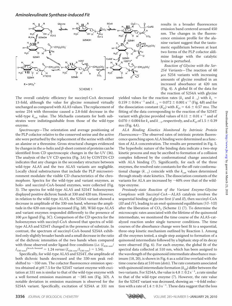
FIGURE 3. Circular dichroism and fluorescence emission spectra of ALAS and the Ser-254 variants. Spectra of wild-type ALAS (O), S254A (���) and S254T(- - -). A and B, ligand-free holoenzymes; C, in the presence of 100 �M succinyl-CoA; D, upon excitation of the cofactor at 331 nm.
TABLE 1Summary of steady-state kinetic parameters
Enzyme KmGly Km
SCoA kcat kcat/KmGly kcat/Km
SCoA
mM �M s�1 mM�1�s�1 �M�1 �s�1
Wild type 25 4 1.3 0.9 0.14 0.02 5.6 10�3 1.1 10�1
S254A 18 2 32 7 0.27 0.01 1.5 10�2 8.4 10�3
S254T 27 3 1.2 0.3 0.050 0.004 1.9 10�3 4.2 10�2
Aminolevulinate Synthase-induced Fit
3354 JOURNAL OF BIOLOGICAL CHEMISTRY VOLUME 285 • NUMBER 5 • JANUARY 29, 2010
by guest on March 23, 2018
http://ww
w.jbc.org/
Dow
nloaded from

units) at time t; kobs is the observed first order rate constant; A1is the pre-exponential factor, andA0 is the offset. The observedrate constants were then plotted against ligand concentration,and the data were fitted to Equation 4 by nonlinear regression.The rates of dissociation (koff) and association (kon) as well asthe ligand binding constants (KD) were calculated from theasymptotic maximal observed rate, the ordinate intercept, andthe ligand concentration (x) in Equation 4.
f�x� � kon �koffx
KD � x(Eq. 4)
RESULTS
Kinetic Characterization of the Ser-254 Variants—Thesteady-state kinetic parameters of the Ser-254 variants weredetermined, and the results are summarized in Table 1. Themutation of serine 254 to alanine resulted in a kcat �2-foldhigher than that of the wild-type ALAS value. The Km value forsuccinyl-CoA was increased �25-fold relative to ALAS,although theKm value for glycine was not significantly affected.
FIGURE 4. Reaction of the Ser-254 variants (60 �M) with increasing con-centrations of glycine. Data were fitted to Reaction 2 for a two-exponentialprocess, yielding equilibrium and rate constants for S254T (KD � 1.5 0.4 mM,k1 � 0.11 0.01 s�1, and k�1 � 0.070 0.004 s�1) (A) and S254A (KD � 6.6 0.6 mM, k1 � 0.159 0.04 s�1, and k�1 � 0.072 0.001 s�1) (B). The fitted rateconstants for wild-type ALAS yielded an overall binding KD of 8.0 0.1 mM
(18).
FIGURE 5. Reaction of wild-type ALAS and the Ser-254 variants (5 �M)with ALA. The observed rate constants were calculated by fitting thedecrease in intrinsic protein fluorescence over time to Equation 2 for a singleexponential process. A, resolved equilibrium and rate constants for wild-typeALAS are as follows: KD � 500 16 �M, k1 � 0.120 0.015 s�1, and k�1 �0.140 0.05 s�1. B, for the S254A variant, the constants are as follows: KD �855 66 �M, k1 � 0.235 0.006 s�1, and k�1 � 0.29 0.02 s�1. C, for theS254T variant, the constants are as follows: KD � 832 49 �M, k1 � 0.19 0.09s�1, and k�1 � 0.057 0.007 s�1.
Aminolevulinate Synthase-induced Fit
JANUARY 29, 2010 • VOLUME 285 • NUMBER 5 JOURNAL OF BIOLOGICAL CHEMISTRY 3355
by guest on March 23, 2018
http://ww
w.jbc.org/
Dow
nloaded from
The overall catalytic efficiency for succinyl-CoA decreased13-fold, although the value for glycine remained virtuallyunchanged as comparedwithALAS values. The replacement ofserine 254 with threonine caused a 2.8-fold decrease in thewild-type kcat value. The Michaelis constants for both sub-strates were indistinguishable from those of the wild-typeenzyme.Spectroscopy—The orientation and average positioning of
the PLP cofactor relative to the conserved serine and the activesite were perturbed by the replacement of the serine with eitheran alanine or a threonine. Gross structural changes evidencedby changes in the�-helix and�-sheet content of proteins can beidentified from CD spectroscopic changes in the far-UV (36).The analysis of the UV CD spectra (Fig. 3A) by CONTIN-CDindicates that any changes in the secondary structure betweenwild-type ALAS and the two ALAS variants are negligible.Locally chiral substructures that include the PLP microenvi-ronment modulate the visible CD characteristics of the chro-mophore. Spectra for the wild-type and variant enzymes, asholo- and succinyl-CoA-bound enzymes, were collected (Fig.3). The spectra for wild-type ALAS and S254T holoenzymesdisplayed positive dichroic bands at 330 and 420 nm. However,in relation to the wild-type ALAS, the S254A variant showed adecrease in amplitude of the 330-nm band, whereas the ampli-tude in the 420-nm region increased (Fig. 3B).Wild-type ALASand variant enzymes responded differently to the presence of100 �M ligand (Fig. 3C). Comparison of the CD spectra for theholoenzymes with succinyl-CoA showed that spectra of wild-type ALAS and S254T changed in the presence of substrate. Incontrast, the spectrum of succinyl-CoA-bound S254A exhib-ited only slightly broader bands and a small decrease in the ratioof the dichroic intensities of the two bands when comparedwith those observed under ligand-free conditions (i.e. (I426 nm/I330 nm)ScoA-bound � 1.24 versus (I426 nm/I330 nm) � 1.54).
Specifically, for wild-type ALAS and S254T, the amplitude ofboth dichroic bands decreased and the 330-nm peak red-shifted to �350 nm. The cofactor fluorescence emission spec-tra obtained at pH 7.5 for the S254T variant enzyme with exci-tation at 331 nm is similar to that of the wild-type enzyme witha well formed emission maximum at �385 nm (Fig. 3D). Anotable deviation in emission maximum is observed for theS254A variant. Specifically, excitation of S254A at 331 nm
results in a broader fluorescenceemission band centered around 450nm. The changes in the fluores-cence emission profile for the ala-nine variant suggest that the tauto-meric equilibrium between at leasttwo forms of the PLP cofactor aldi-mine linkage with the catalyticlysine is perturbed.Reaction of Glycine with the Ser-
254 Variants—The reaction of 60�M S254 variants with increasingamounts of glycine resulted in anincreased absorbance at 420 nm(Fig. 4). A global fit of the data forthe reaction of S254A with glycine
yielded values for the reaction rates (k1 and k�1) with k1 �0.159 0.04 s�1 and k�1 � 0.072 0.001 s�1 (Fig. 4B) and forthe dissociation constant (KD) with KD � 6.6 0.57 mM. Thefitting of the data corresponding to the reaction of the S254Tvariant with glycine provided values of 0.11 0.01 s�1 and of0.070 0.004 for k1 and k�1, respectively, and aKD of 1.5 0.39mM (Fig. 4A).ALA Binding Kinetics Monitored by Intrinsic Protein
Fluorescence—The observed rates of intrinsic protein fluores-cence quenching uponALAbindingwere determined as a func-tion of ALA concentration. The results are presented in Fig. 5.The hyperbolic nature of the binding data indicate a two-stepkinetic process and may be ascribed to formation of a collisioncomplex followed by the conformational change associatedwith ALA binding (7). Significantly, for each of the threeenzymes, the resolved rate constants for the off-rate conforma-tional change (k�1) coincide with the kcat values determinedthrough steady-state kinetics. The dissociation constants of thevariants for ALA are increased by �60% over that of the wild-type enzyme.Presteady-state Reaction of the Variant Enzyme-Glycine
Complexes with Succinyl-CoA—ALAS catalysis involves thesequential binding of glycine first (I and II), then succinyl-CoA(III and IV), leading to an enol-quinonoid equilibrium (VI–VII)after the liberation of CO2 (Scheme 1) (7). To determine themicroscopic rates associated with the lifetime of the quinonoidintermediate, we monitored the time course of the ALAS-cat-alyzed reaction under single turnover conditions. The timecourses of the absorbance change were best fit to a sequential,three-step kinetic mechanism outlined by Reaction 1. Amongall the enzymes tested, a single step assigned to formation of aquinonoid intermediate followed by a biphasic step of its decaywere observed (Fig. 6). For each enzyme, the global fit of thespectral data collected at 510 nm, which has been assigned tothewavelength of the quinonoid intermediate absorbancemax-imum (18, 20), is shown in Fig. 6 as a solid line overlaid with thetime course data at 510 nm (dots). The rate constants associatedwith quinonoid intermediate formation (kQf) differ between thetwo variants. For S254A, the value is 4.8 0.2 s�1, a rate similarto that of the wild-type enzyme (7). However, the rate valuefor the S254T variant was decreased, showing an �4-fold reduc-tion with a rate of 1.4 0.3 s�1. These data suggest that the loss
SCHEME 1
Aminolevulinate Synthase-induced Fit
3356 JOURNAL OF BIOLOGICAL CHEMISTRY VOLUME 285 • NUMBER 5 • JANUARY 29, 2010
by guest on March 23, 2018
http://ww
w.jbc.org/
Dow
nloaded from
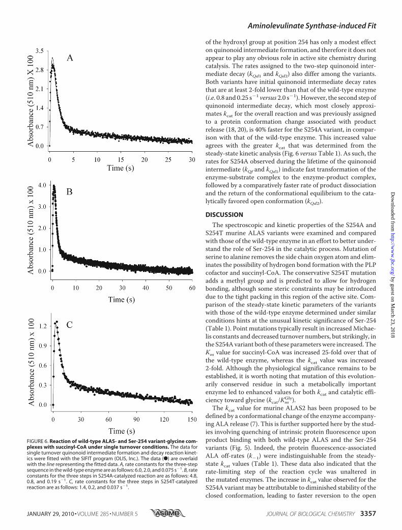
of the hydroxyl group at position 254 has only a modest effecton quinonoid intermediate formation, and therefore it does notappear to play any obvious role in active site chemistry duringcatalysis. The rates assigned to the two-step quinonoid inter-mediate decay (kQd1 and kQd2) also differ among the variants.Both variants have initial quinonoid intermediate decay ratesthat are at least 2-fold lower than that of the wild-type enzyme(i.e. 0.8 and 0.25 s�1 versus 2.0 s�1). However, the second step ofquinonoid intermediate decay, which most closely approxi-mates kcat for the overall reaction and was previously assignedto a protein conformation change associated with productrelease (18, 20), is 40% faster for the S254A variant, in compar-ison with that of the wild-type enzyme. This increased valueagrees with the greater kcat that was determined from thesteady-state kinetic analysis (Fig. 6 versusTable 1). As such, therates for S254A observed during the lifetime of the quinonoidintermediate (kQf and kQd1) indicate fast transformation of theenzyme-substrate complex to the enzyme-product complex,followed by a comparatively faster rate of product dissociationand the return of the conformational equilibrium to the cata-lytically favored open conformation (kQd2).
DISCUSSION
The spectroscopic and kinetic properties of the S254A andS254T murine ALAS variants were examined and comparedwith those of the wild-type enzyme in an effort to better under-stand the role of Ser-254 in the catalytic process. Mutation ofserine to alanine removes the side chain oxygen atom and elim-inates the possibility of hydrogen bond formation with the PLPcofactor and succinyl-CoA. The conservative S254T mutationadds a methyl group and is predicted to allow for hydrogenbonding, although some steric constraints may be introduceddue to the tight packing in this region of the active site. Com-parison of the steady-state kinetic parameters of the variantswith those of the wild-type enzyme determined under similarconditions hints at the unusual kinetic significance of Ser-254(Table 1). Point mutations typically result in increasedMichae-lis constants and decreased turnover numbers, but strikingly, inthe S254Avariant both of these parameterswere increased. TheKm value for succinyl-CoA was increased 25-fold over that ofthe wild-type enzyme, whereas the kcat value was increased2-fold. Although the physiological significance remains to beestablished, it is worth noting that mutation of this evolution-arily conserved residue in such a metabolically importantenzyme led to enhanced values for both kcat and catalytic effi-ciency toward glycine (kcat/Km
Gly).The kcat value for murine ALAS2 has been proposed to be
defined by a conformational change of the enzyme accompany-ing ALA release (7). This is further supported here by the stud-ies involving quenching of intrinsic protein fluorescence uponproduct binding with both wild-type ALAS and the Ser-254variants (Fig. 5). Indeed, the protein fluorescence-associatedALA off-rates (k�1) were indistinguishable from the steady-state kcat values (Table 1). These data also indicated that therate-limiting step of the reaction cycle was unaltered inthe mutated enzymes. The increase in kcat value observed for theS254A variantmay be attributable to diminished stability of theclosed conformation, leading to faster reversion to the open
FIGURE 6. Reaction of wild-type ALAS- and Ser-254 variant-glycine com-plexes with succinyl-CoA under single turnover conditions. The data forsingle turnover quinonoid intermediate formation and decay reaction kinet-ics were fitted with the SIFIT program (OLIS, Inc.). The data (F) are overlaidwith the line representing the fitted data. A, rate constants for the three-stepsequence in the wild-type enzyme are as follows: 6.0, 2.0, and 0.075 s�1. B, rateconstants for the three steps in S254A-catalyzed reaction are as follows: 4.8,0.8, and 0.19 s�1. C, rate constants for the three steps in S254T-catalyzedreaction are as follows: 1.4, 0.2, and 0.037 s�1.
Aminolevulinate Synthase-induced Fit
JANUARY 29, 2010 • VOLUME 285 • NUMBER 5 JOURNAL OF BIOLOGICAL CHEMISTRY 3357
by guest on March 23, 2018
http://ww
w.jbc.org/
Dow
nloaded from
conformation and product release. In this interpretation, thelarge concomitant increase in theKm value for succinyl-CoA inthe S254A variantwould result not only from the direct effect ofloss of hydrogen bonding to the substrate but also from the lessdirect effect of impaired enzyme conformational change as trig-gered by the substrate, which is believed to be required foroptimal binding (7, 18). The 3-fold decrease in the kcat valuewithout any effect on the Km value for succinyl-CoA resultingfrom the more conservative S254T mutation would then ariseprimarily from enhanced stability of the closed conformation.Bothmutations substantially diminished the catalytic efficiencywith succinyl-CoA (kcat/Km
SCoA) but in different ways, andalthough the effects of these very conservativemutations on thesteady-state kinetic parameters may appear relatively modest,they are presumably sufficient to have metabolically harmfuleffects in vivo, given the strong evolutionary selection for serineat this position.Significant effects on the cofactor microenvironment of
ALAS were induced with the S254A mutation, as determinedusing fluorescence and CD spectroscopies. The differences inthe fluorescence spectra reflected alterations in cofactor tauto-meric equilibria, whichmust have occurred upon loss of hydro-gen bonding interactions between the phenolic oxygen of PLPand the side chain of Ser-254 (Fig. 3D). We previously assignedthe 330- and 410-nmabsorption species to a substituted aldam-ine and a ketoenamine, respectively (37). The assignment of the330-nm absorption species to a substituted aldamine, and notthe enolimine form of the Schiff base, was due to the intenseemission fluorescence of this species at 385 nm and not at thecharacteristic 510-nm emission wavelength of an enolimine(37, 38). The diminished substituted aldamine tautomer emis-sion at 386 nm for the S254A variant is consistent with loss ofhydrogen bonding at the cofactor phenolic oxygen. Anincreased proportion of the cofactor is probably in the ketoe-namine tautomer, which we previously ascribed to the activeform of the coenzyme (37), as the 410-nm absorption speciesappears to be more prominent in the S254A variant than in thewild-type ALAS and S254T variant (data not shown). CD spec-troscopic examination of the S254A and S254T variants in thefar-UV region confirmed that the mutations did not signifi-cantly alter the overall secondary structure of the enzymes (Fig.
3A). Any alterations in the confor-mational equilibria of the active siteloop, which adopts an extendedconformation and is thus not CDactive, were not apparent in thesespectra, but the visible CD spectraof the S254A variant do diverge sub-stantially from those of the wild-type and the S254T variant. TheS254A holoenzyme maximum at435 nm was blue-shifted toward�420 nm, and the ratio of the meanresidual ellipticity at this wave-length to the one at 330 nm wasincreased (Fig. 3B). These elliptici-ties arise fromCotton effects associ-atedwith the ketoenamine (435 nm)
and substituted aldamine (330 nm) tautomers of the PLP Schiffbase and are indicative of the microenvironment surroundingthis linkage between the cofactor and the active site lysine (39–41). Succinyl-CoA binding to the wild-type and S254T variantsinduced decreases in asymmetry of the cofactor, whereas theS254A variant was relatively unchanged under similar condi-tions (Fig. 3C). A logical interpretation is that the decrease inasymmetry observed for thewild-type and S254T variants arosefrom partial conversion of the internal aldimine to free PLPaldehyde bound at the active site, as was observed in three offour R. capsulatus crystal structure active sites upon succinyl-CoA binding (8). In the crystal structures these events wereaccompanied by transition to a closed conformation, fromwhich it might be concluded that the S254A variant retainedthe internal aldimine in the presence of succinyl-CoA, and itmay not have been induced to adopt a closed conformationupon binding of this substrate.Monitoring of the formation and decay of the quinonoid
reaction intermediate under single turnover conditions indi-cated that the two Ser-254 variants follow a kinetic mechanismsimilar to that of wild-type ALAS (Fig. 6, A and B). A rapid stepof ALA-bound quinonoid intermediate formation upon decar-boxylation is followed by two successively slower decay steps,associated with protonation of the ALA-quinonoid intermedi-ate and ALA release, respectively (7). The quinonoid interme-diate formation rate decreased 4-fold for the S254T variant-catalyzed reaction, which could be explained by a change in theflow of electrons from the site of bond scission throughout thecofactor, precipitated by a shift in the conformational equilib-rium toward the closed state. In the ALAS crystal structure,PLP is noted to change position by 15° when substrate is bound(8). Changes to the stereoelectronic relationship between thecofactor and the �-carbon bonds of the external aldimine caninfluence the chemical mechanism dramatically (7). Therefore,it is possible that the hydrogen bond between serine 254 and thephenolic oxygen of PLP may be an influential part of maintain-ing the angle of PLP during not only formation and decay of thequinonoid intermediate but also during the complete reactioncycle. The increase observed in the second step of quinonoidintermediate decay in the S254A variant is also consistent with
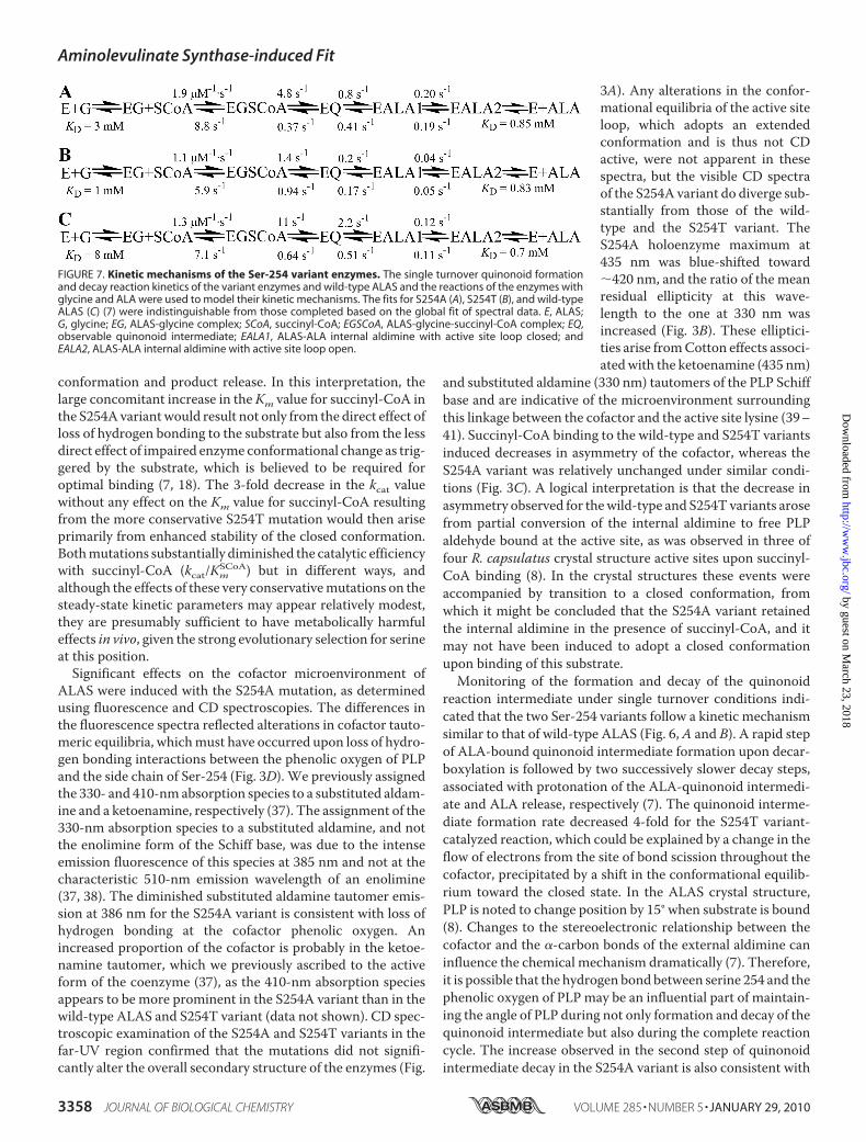
FIGURE 7. Kinetic mechanisms of the Ser-254 variant enzymes. The single turnover quinonoid formationand decay reaction kinetics of the variant enzymes and wild-type ALAS and the reactions of the enzymes withglycine and ALA were used to model their kinetic mechanisms. The fits for S254A (A), S254T (B), and wild-typeALAS (C) (7) were indistinguishable from those completed based on the global fit of spectral data. E, ALAS;G, glycine; EG, ALAS-glycine complex; SCoA, succinyl-CoA; EGSCoA, ALAS-glycine-succinyl-CoA complex; EQ,observable quinonoid intermediate; EALA1, ALAS-ALA internal aldimine with active site loop closed; andEALA2, ALAS-ALA internal aldimine with active site loop open.
Aminolevulinate Synthase-induced Fit
3358 JOURNAL OF BIOLOGICAL CHEMISTRY VOLUME 285 • NUMBER 5 • JANUARY 29, 2010
by guest on March 23, 2018
http://ww
w.jbc.org/
Dow
nloaded from

the increases in kcat and the ALA off-rate determined fromenzyme fluorescence quenching.By utilizing the microscopic parameters obtained from the
transient kinetics for the reactions catalyzed by the variantenzymes under single turnover conditions, coupled with therate constants and dissociation constants determined for thereactions between the product or substrate with the enzymes,we were able to model their kinetic mechanisms as shown inFig. 7. The simulations of the kinetic pathways revealed that theS254T mutation significantly retards the kinetic mechanism inrelation to that of thewild-typeALAS. Conversely, themodeledpathway for S254A highlights the increases observed in thekinetics associated with the variant. Overall, the mechanisticdata for both variants support the hypothesis that the interac-tion between Ser-254 and the O3� of PLP is a limiting factor inenforcing an induced fit mechanism by coupling substrate rec-ognition to conformational equilibria. However, how the struc-tural differences between the variant enzymes with ligandbound accomplish the conformational transition awaits three-dimensional structural and protein dynamics information.
REFERENCES1. Akhtar, M., Abboud, M.M., Barnard, G., Jordan, P., and Zaman, Z. (1976)
Philos. Trans. R. Soc. Lond. B Biol. Sci. 273, 117–1362. May, B. K., Dogra, S. C., Sadlon, T. J., Bhasker, C. R., Cox, T. C., and
Bottomley, S. S. (1995) Prog. Nucleic Acid Res. Mol. Biol. 51, 1–513. May, A., and Bishop, D. F. (1998) Haematologica 83, 56–704. Bottomley, S. S. (2006) Curr. Hematol. Rep. 5, 41–495. Shoolingin-Jordan, P.M., Al-Daihan, S., Alexeev,D., Baxter, R. L., Bottom-
ley, S. S., Kahari, I. D., Roy, I., Sarwar,M., Sawyer, L., andWang, S. F. (2003)Biochim. Biophys. Acta 1647, 361–366
6. Bottomley, S. S. (2004) in Wintrobe’s Clinical Hematology (Greer, J., Fo-errster, J., Lukens, J. N., Rodgers, G.M., Paraskevas, R., andGlader, B., eds)pp. 1012–1033, Lippincott, Williams &Wilkins, Philadelphia
7. Hunter, G. A., Zhang, J., and Ferreira, G. C. (2007) J. Biol. Chem. 282,23025–23035
8. Astner, I., Schulze, J. O., van den Heuvel, J., Jahn, D., Schubert, W. D., andHeinz, D. W. (2005) EMBO J. 24, 3166–3177
9. Eliot, A. C., and Kirsch, J. F. (2004) Annu. Rev. Biochem. 73, 383–41510. Jager, J.,Moser,M., Sauder, U., and Jansonius, J. N. (1994) J.Mol. Biol. 239,
285–30511. Picot, D., Sandmeier, E., Thaller, C., Vincent, M. G., Christen, P., and
Jansonius, J. N. (1991) Eur. J. Biochem. 196, 329–34112. McPhalen, C. A., Vincent, M. G., Picot, D., Jansonius, J. N., Lesk, A. M.,
and Chothia, C. (1992) J. Mol. Biol. 227, 197–21313. Christen, P., and Mehta, P. K. (2001) Chem. Rec. 1, 436–44714. Alexander, F.W., Sandmeier, E., Mehta, P. K., and Christen, P. (1994) Eur.
J. Biochem. 219, 953–960
15. Jansonius, J. N., Eichele, G., Ford, G. C., Kirsch, J. F., Picot, D., Thaller, C.,Vincent, M. G., Gehring, H., and Christen, P. (1984) Biochem. Soc. Trans.12, 424–427
16. Jansonius, J. N., Eichele, G., Ford, G. C., Kirsch, J. F., Picot, D., Thaller, C.,Vincent, M. G., Gehring, H., and Christen, P. (1984) Prog. Clin. Biol. Res.144, 195–203
17. Koshland, D. E., Jr., and Neet, K. E. (1968) Annu. Rev. Biochem. 37,359–410
18. Hunter, G. A., and Ferreira, G. C. (1999) J. Biol. Chem. 274, 12222–1222819. Ferreira, G. C., and Zhang, J. S. (2002) Cell. Mol. Biol. 48, 827–83320. Zhang, J., and Ferreira, G. C. (2002) J. Biol. Chem. 277, 44660–4466921. Lendrihas, T., Zhang, J., Hunter, G. A., and Ferreira, G. C. (2009) Protein
Sci. 18, 1847–185922. Gregoret, L. M., Rader, S. D., Fletterick, R. J., and Cohen, F. E. (1991)
Proteins 9, 99–10723. Rajagopal, S., and Vishveshwara, S. (2005) FEBS J. 272, 1819–183224. Ferreira, G. C., and Dailey, H. A. (1993) J. Biol. Chem. 268, 584–59025. Miyazaki, K., and Takenouchi, M. (2002) BioTechniques 33, 1033–1034,
1036–103826. Laemmli, U. K. (1970) Nature 227, 680–68527. Smith, P. K., Krohn, R. I., Hermanson, G. T., Mallia, A. K., Gartner, F. H.,
Provenzano, M. D., Fujimoto, E. K., Goeke, N. M., Olson, B. J., and Klenk,D. C. (1985) Anal. Biochem. 150, 76–85
28. Hunter, G. A., and Ferreira, G. C. (1995) Anal. Biochem. 226, 221–22429. Schwede, T., Kopp, J., Guex, N., and Peitsch, M. C. (2003) Nucleic Acids
Res. 31, 3381–338530. Guex, N., and Peitsch, M. C. (1997) Electrophoresis 18, 2714–272331. Chen, G. C., and Yang, J. T. (1977) Anal. Lett. 10, 1195–120732. Provencher, S. W., and Glockner, J. (1981) Biochemistry 20, 33–3733. Tsai,M. D.,Weintraub, H. J., Byrn, S. R., Chang, C., and Floss, H. G. (1978)
Biochemistry 17, 3183–318834. Durbin, J., and Watson, G. S. (1950) Biometrika 37, 409–42835. Barshop, B. A., Wrenn, R. F., and Frieden, C. (1983) Anal. Biochem. 130,
134–14536. Kelly, S. M., and Price, N. C. (2000) Curr. Protein Pept. Sci. 1, 349–38437. Zhang, J., Cheltsov, A. V., and Ferreira, G. C. (2005) Protein Sci. 14,
1190–120038. Ikushiro, H., Hayashi, H., and Kagamiyama, H. (2004) Biochemistry 43,
1082–109239. Futaki, S., Ueno, H., Martinez del Pozo, A., Pospischil, M. A., Manning,
J. M., Ringe, D., Stoddard, B., Tanizawa, K., Yoshimura, T., and Soda, K.(1990) J. Biol. Chem. 265, 22306–22312
40. Kallen, R. G., Korpela, T., Martell, A. E., Matsushima, Y., Metzler, C. M.,Metzler, D. E., Morozov, Y. V., Ralston, I. M., Savin, F. A., Torchinsky,Y. M., and Ueno, H. (1985) in Transaminases (Christen, P., and Metzler,D. E., eds) pp. 37–108, John Wiley & Sons, Inc., New York
41. Cellini, B., Bertoldi, M., Montioli, R., and Borri Voltattorni, C. (2005) Bio-chemistry 44, 13970–13980
42. Thompson, J. D., Higgins, D.G., andGibson, T. J. (1994)Nucleic Acids Res.22, 4673–4680
Aminolevulinate Synthase-induced Fit
JANUARY 29, 2010 • VOLUME 285 • NUMBER 5 JOURNAL OF BIOLOGICAL CHEMISTRY 3359
by guest on March 23, 2018
http://ww
w.jbc.org/
Dow
nloaded from
Thomas Lendrihas, Gregory A. Hunter and Gloria C. FerreiraSynthase
Serine 254 Enhances an Induced Fit Mechanism in Murine 5-Aminolevulinate
doi: 10.1074/jbc.M109.066548 originally published online November 16, 20092010, 285:3351-3359.J. Biol. Chem.
10.1074/jbc.M109.066548Access the most updated version of this article at doi:
Alerts:
When a correction for this article is posted•
When this article is cited•
to choose from all of JBC's e-mail alertsClick here
Supplemental material:
http://www.jbc.org/content/suppl/2009/11/16/M109.066548.DC1
http://www.jbc.org/content/285/5/3351.full.html#ref-list-1
This article cites 40 references, 9 of which can be accessed free at
by guest on March 23, 2018
http://ww
w.jbc.org/
Dow
nloaded from