Embed Size (px)
Citation preview
Copyright 2006 Psychonomic Society, Inc. 246
Journal2006, ?? (?), ???-???
The brain has a remarkable ability to adapt flexibly to new demands. Functional plasticity in sensory and motor regions occurs in response to short-term training, on the order of minutes or hours, or in response to long-term training, over the course of weeks or years (Karni et al., 1995; Merzenich et al., 1983; Sanes & Donoghue, 2000). There is evidence that the time course of training has a critical influence on behavioral and neural aspects of learning (Korman, Raz, Flash, & Karni, 2003; Tracy et al., 2001; Ungerleider, Doyon, & Karni, 2002). In particular, “fast” and “slow” phases of skill learning have been asso-ciated with distinct psychophysical and neural character-istics. Fast changes often occur with rapid improvements in performance and may be accompanied by automati-zation and habituation-like decreases in activity (Karni et al., 1998), perhaps due to changes in synaptic strength (Kolb & Whishaw, 1998). Slow changes, on the other hand, occur with more gradual performance improvements and are associated with reorganization of functional topog-raphy (Kleim et al., 2004), enlargement of the cortical map (Pascual-Leone, 2001), and morphological changes to brain structure (Münte, Altenmüller, & Jäncke, 2002). Thus, although these two types of learning are associated with different neural events, it is likely that both result in
improved neural efficiency. Previous neuroimaging stud-ies tracing the time course of practice-related changes across a number of different types of tasks (Karni et al., 1998; Kelly & Garavan, 2005) suggest that fast learning, linked to repetitive task performance, is associated with decreases in activity within a fixed network of regions (Floyer-Lea & Matthews, 2004), whereas slow learning, linked to strategy development, is associated with recruit-ment of additional regions as the skill is learned (Olesen, Westerberg, & Klingberg, 2004).
Studies of musicians are especially well suited to ad-dress questions about slow learning, in particular, since musical expertise engages a network of primary and multimodal regions that may be differently influenced by mechanisms of plasticity. In primary and secondary motor regions, reduced activation has been reported for pianists in comparison with nonpianists during self-paced finger tapping (Jäncke, Shah, & Peters, 2000) or exter-nally paced tapping (Haslinger et al., 2004) and during performance of motor sequences learned prior to scanning that are 5 items (Hund-Georgiadis & von Cramon, 1999; Meister et al., 2005) or 12 items in length (Krings et al., 2000). Reduced activation for pianists relative to nonpia-nists was attributed to pianists’ greater efficiency of move-ments within their expanded motor networks.
The interpretation of these data with respect to musical expertise is limited, however, by small sample sizes and the use of tasks that place minimal demands on sequence-learning processes. The functional networks activated dur-ing the performance of these tasks may be different from those that have been modified through the development of piano expertise. We hypothesize that reduced activa-tion observed for pianists relative to controls may have been due to the use of tasks that may not have utilized the capacity of the optimized motor system.
We thank Roshan Cools, Brian Hawthorne, Ben Inglis, John Jinks-Ollinger, Jarrod Millman, and Felice Sun for technical assistance and helpful suggestions. This research was supported by a predoctoral fel-lowship from the National Science Foundation, grants from the National Institutes of Health (NS 40813 and MH63901), and a grant from the Dana Foundation. Correspondence concerning this article should be ad-dressed to S. Landau, Helen Wills Neuroscience Institute, 132 Barker Hall, University of California, Berkeley, CA 94720-3190 (e-mail: [email protected]).
Sequence learning in pianists and nonpianists: An fMRI study of motor expertise
SUSAN M. LANDAU and MARK D’ESPOSITOUniversity of California, Berkeley, California
Previous studies of motor learning have proposed a distinction between fast and slow learning, but these mechanisms have rarely been examined simultaneously. We examined the influence of long-term motor expertise (slow learning) while pianists and nonpianists performed alternating epochs of sequenced and random keypresses in response to visual cues (fast learning) during functional neuro-imaging. All of the participants demonstrated learning of the sequence as demonstrated by decreasing reaction times (RTs) on sequence trials relative to random trials throughout the session. Pianists also demonstrated faster RTs and superior sequence acquisition in comparison with nonpianists. Within-session decreases in bilateral sensorimotor and parietal activation were observed for both groups. Additionally, there was more extensive activation throughout the session for pianists in comparison with nonpianists across a network of primarily right-lateralized prefrontal, sensorimotor, and parietal regions. These findings provide evidence that different neural systems subserve slow and fast phases of learning.
Cognitive, Affective, & Behavioral Neuroscience2006, 6 (3), 246-259
PIANO EXPERTISE AND SEQUENCE LEARNING 247
Pianists are often described as a model population in which to study the acquisition, control, and execution of abstract motor sequences (Palmer, 1997; Parsons, Sergent, Hodges, & Fox, 2005; Peretz & Zatorre, 2005; Sergent, Zuck, Terriah, & MacDonald, 1992). Models of hierarchi-cal motor sequence acquisition (Lashley, 1951; Rhodes, Bullock, Verwey, Averbeck, & Page, 2004) have proposed that units in a sequence may be combined (chunked) in order to facilitate learning, a skill that is critical for pia-nists. With increased working memory (WM) demand, a hypothesized function of the prefrontal cortex is to medi-ate strategic functions such as chunking multiple units for verbal (Savage et al., 2001) and for spatial (Bor, Duncan, Wiseman, & Owen, 2003; Sakai et al., 1998) information.
Consistent with this, motor learning studies suggest that complex sequence-learning tasks recruit multimodal regions (Catalan, Honda, Weeks, Cohen, & Hallett, 1998) in addition to primary motor regions. Thus, brain regions known to be involved in integrative association are likely critically involved in long-term functional plasticity but have been examined in only a few studies (Bangert et al., 2006; Stewart et al., 2003). Stewart et al. reported in-creases in activation of bilateral superior parietal regions after 15 weeks of piano training that included learning musical notation. That study reported bilateral superior parietal cortex activity during music reading and play-ing for participants who had received training, which was attributed to the acquisition of a code for translation of spatiomotor information. This expansion of activation in superior parietal regions is consistent with other studies (of nonmusicians) during visuospatial skill learning over a period of days or weeks (Olesen et al., 2004; Poldrack & Gabrieli, 2001). The brain’s capacity for functional plas-ticity in these regions may, however, expand further over a long period of time. Bangert et al. (2006) found increased activation across a number of multimodal regions for pia-nists in comparison with nonpianists during arbitrary key-presses. This is initial evidence that a long-term training period of years recruits a network of task-specific associa-tive regions and results in expansions of this network, per-haps via cortical reorganization (Gaser & Schlaug, 2003; Pascual-Leone, 2001).
In this experiment, we compared pianists and non-pianist controls during functional neuroimaging while they performed a motor sequence learning task in order to examine (1) the regional specificity and (2) the time course of functional plasticity. The motor sequence learn-ing task was designed to engage a number of processes that pianists engage during music performance—namely, learning complex sequential relationships between spatial positions, bimanual motor coordination, and fine motor precision (Münte et al., 2002). Our within-subjects fac-tor (practice effects across the session) was designed to identify activation related to short-term fast learning over the course of the scanning session, whereas our between-subjects factor (pianists vs. nonpianists) was designed to identify activation differences related to the slow develop-ment of expertise.
We addressed limitations of previous studies of motor learning in several ways. First, in order to make the sequence-learning task sufficiently challenging for pia-nists but still possible to perform for both subject groups, we used a version of the serial reaction time task (SRT; Nissen & Bullemer, 1987) containing a probabilistic finite state grammar (Berns, Cohen, & Mintun, 1997; Reber, 1967). The performance of this task differs from piano playing in a number of ways, such as in its lack of tempo-ral variability and auditory feedback, and the probabilistic structure of keypresses. Our task was, however, designed to engage processes related to learning of sequential rela-tionships. During music performance, any given note on the piano may be followed by many different combina-tions of notes, and a pianist must learn to flexibly switch between these different sequential associations (Palmer, 1997). As a pianist becomes familiar with more extensive repertoire, he or she learns to associate certain sequences of notes with greater frequency than others, thus acquiring knowledge about probabilistic relationships between dif-ferent spatial positions. Second, responses were externally cued rather than self-paced, in order to control for over-all motor stimulation between groups. Third, pianists and nonpianists had to attend to the stimuli equally, since the sequence was probabilistic and therefore never entirely predictable even after substantial learning. The use of a probabilistic, rather than a deterministic, sequence also meant that the task could not be automatized. Finally, in order to reduce possible differences in effort and strat-egy use between pianists and nonpianists, the participants were not told ahead of time about the presence of regulari-ties in the stimuli.
With respect to regional specificity, we predicted that experts with years of motor training would recruit a dif-ferent functional network during sequence learning than would novices, and that this network would include as-sociative regions as well as the motor regions that are typically engaged by sequence learning (Grafton, Hazel-tine, & Ivry, 1995). With respect to the time course of functional plasticity, we predicted that practice would influence functional activation such that within-session practice effects would be characterized by habituation-like decreases within a primarily motor network, whereas between-subjects (pianist vs. nonpianist) effects would reflect increases in functional activation in associative regions for pianists, resulting from the underlying corti-cal reorganization. Because fast (within-session) and slow (between-subjects) learning are likely associated with dif-ferent underlying mechanisms of plasticity, their activa-tion profiles should be distinct, reflecting different forms of neural efficiency.
METHOD
ParticipantsNine right-handed, experienced pianists (mean age 21.8) and 8
nonpianists (mean age 20.6) were recruited from the population of undergraduate students at the University of California at Berke-ley. Pianists selected as participants were recruited through the UC
248 LANDAU AND D’ESPOSITO
Berkeley music department on the basis of the following criteria: (1) piano experience of at least 8 years, and (2) currently playing the piano for a minimum of 3 h per week. Pianists had an average of 10.7 years of piano experience and played for an average of 7.7 h per week. Nonpianists had no experience with the piano or other musical instruments. All of the participants gave written, informed consent prior to participation in the study. The participants were screened for medical, neurological, and psychiatric illnesses, and for substance abuse and use of prescription medications.
Behavioral TaskThe participants performed a modified version of the SRT task
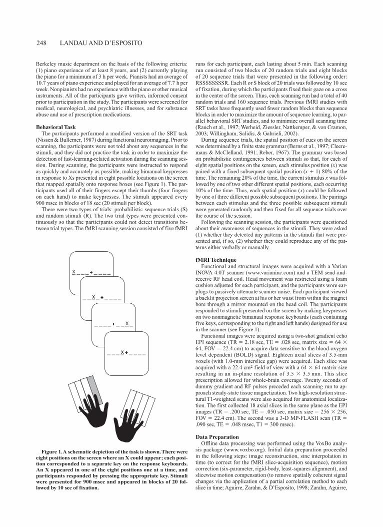
(Nissen & Bullemer, 1987) during functional neuroimaging. Prior to scanning, the participants were not told about any sequences in the stimuli, and they did not practice the task in order to maximize the detection of fast-learning-related activation during the scanning ses-sion. During scanning, the participants were instructed to respond as quickly and accurately as possible, making bimanual keypresses in response to Xs presented in eight possible locations on the screen that mapped spatially onto response boxes (see Figure 1). The par-ticipants used all of their fingers except their thumbs (four fingers on each hand) to make keypresses. The stimuli appeared every 900 msec in blocks of 18 sec (20 stimuli per block).
There were two types of trials: probabilistic sequence trials (S) and random stimuli (R). The two trial types were presented con-tinuously so that the participants could not detect transitions be-tween trial types. The fMRI scanning session consisted of five fMRI
runs for each participant, each lasting about 5 min. Each scanning run consisted of two blocks of 20 random trials and eight blocks of 20 sequence trials that were presented in the following order: RSSSSSSSSR. Each R or S block of 20 trials was followed by 10 sec of fixation, during which the participants fixed their gaze on a cross in the center of the screen. Thus, each scanning run had a total of 40 random trials and 160 sequence trials. Previous fMRI studies with SRT tasks have frequently used fewer random blocks than sequence blocks in order to maximize the amount of sequence learning, to par-allel behavioral SRT studies, and to minimize overall scanning time (Rauch et al., 1997; Werheid, Ziessler, Nattkemper, & von Cramon, 2003; Willingham, Salidis, & Gabrieli, 2002).
During sequence trials, the spatial position of cues on the screen was determined by a finite state grammar (Berns et al., 1997; Cleere-mans & McClelland, 1991; Reber, 1967). The grammar was based on probabilistic contingencies between stimuli so that, for each of eight spatial positions on the screen, each stimulus position (x) was paired with a fixed subsequent spatial position (x 1) 80% of the time. The remaining 20% of the time, the current stimulus x was fol-lowed by one of two other different spatial positions, each occurring 10% of the time. Thus, each spatial position (x) could be followed by one of three different possible subsequent positions. The pairings between each stimulus and the three possible subsequent stimuli were generated randomly and then fixed for all sequence trials over the course of the session.
Following the scanning session, the participants were questioned about their awareness of sequences in the stimuli. They were asked (1) whether they detected any patterns in the stimuli that were pre-sented and, if so, (2) whether they could reproduce any of the pat-terns either verbally or manually.
fMRI TechniqueFunctional and structural images were acquired with a Varian
INOVA 4.0T scanner (www.varianinc.com) and a TEM send-and- receive RF head coil. Head movement was restricted using a foam cushion adjusted for each participant, and the participants wore ear-plugs to passively attenuate scanner noise. Each participant viewed a backlit projection screen at his or her waist from within the magnet bore through a mirror mounted on the head coil. The participants responded to stimuli presented on the screen by making keypresses on two nonmagnetic bimanual response keyboards (each containing five keys, corresponding to the right and left hands) designed for use in the scanner (see Figure 1).
Functional images were acquired using a two-shot gradient echo EPI sequence (TR 2.18 sec, TE .028 sec, matrix size 64 64, FOV 22.4 cm) to acquire data sensitive to the blood oxygen level dependent (BOLD) signal. Eighteen axial slices of 3.5-mm voxels (with 1.0-mm interslice gap) were acquired. Each slice was acquired with a 22.4 cm2 field of view with a 64 64 matrix size resulting in an in-plane resolution of 3.5 3.5 mm. This slice prescription allowed for whole-brain coverage. Twenty seconds of dummy gradient and RF pulses preceded each scanning run to ap-proach steady-state tissue magnetization. Two high-resolution struc-tural T1-weighted scans were also acquired for anatomical localiza-tion. The first collected 18 axial slices in the same plane as the EPI images (TR .200 sec, TE .050 sec, matrix size 256 256, FOV 22.4 cm). The second was a 3-D MP-FLASH scan (TR .090 sec, TE .048 msec, T1 300 msec).
Data PreparationOffline data processing was performed using the VoxBo analy-
sis package (www.voxbo.org). Initial data preparation proceeded in the following steps: image reconstruction, sinc interpolation in time (to correct for the fMRI slice-acquisition sequence), motion correction (six-parameter, rigid-body, least-squares alignment), and slicewise motion compensation (to remove spatially coherent signal changes via the application of a partial correlation method to each slice in time; Aguirre, Zarahn, & D’Esposito, 1998; Zarahn, Aguirre,
+_ __ _ _ __ _X
+_ __ _ _ __ _
+_ __ _ _ __ _
+_ __ _ _ __ _
X
X
X
Figure 1. A schematic depiction of the task is shown. There were eight positions on the screen where an X could appear; each posi-tion corresponded to a separate key on the response keyboards. An X appeared in one of the eight positions one at a time, and participants responded by pressing the appropriate key. Stimuli were presented for 900 msec and appeared in blocks of 20 fol-lowed by 10 sec of fixation.
PIANO EXPERTISE AND SEQUENCE LEARNING 249
& D’Esposito, 1997). Images were then smoothed with a 7-mm FWHM kernel and masked using a whole-brain mask to remove ex-traneous signal caused by ghosting.
Statistical AnalysisSince fMRI data are temporally autocorrelated under the null hy-
pothesis (Zarahn et al., 1997), statistical analyses were conducted within the framework of the modified general linear model (GLM) for serially correlated error terms (Worsley & Friston, 1995). A time-domain representation of the expected 1/f power structure (Zarahn et al., 1997) and a notch filter that removed frequencies above 0.25 Hz and below 0.01 Hz (i.e., the portions of highest power in the noise spectrum) were placed in the convolution matrix (Wors-ley & Friston, 1995).
A blocked design was used. The GLM describes fMRI signal change as a series of amplitude-scaled and time-shifted covari-ates or regressors. Within that model, each type of block (random, sequence) was convolved by a canonical hemodynamic response function. Fixation blocks served as a baseline. A different set of co-variates was used to model random and sequence blocks separately for each run in the scanning session in order to examine incremental changes in signal. Two trial types with five runs of each gave a total of 10 covariates of interest. An additional nuisance covariate was included to model an intercept.
Random Effects AnalysesEach participant’s brain was normalized to the Montreal Neuro-
logical Institute (MNI) reference brain using SPM99. Spatial nor-malization was performed as a two-step procedure: First, a structural image acquired to overlay the EPI images was coregistered to the high-resolution MP-FLASH anatomical structural image. Then this structural image was spatially normalized. The two resulting trans-formations were combined into a single transformation and used to spatially normalize the EPI images directly. Normalized whole-brain maps for the specific conditions of interest were calculated for each participant. Primary analyses (identification of ROIs for corresponding ANOVAs) were conducted at a statistical threshold of p .01, cluster-corrected for multiple comparisons (Cao, 1999; Worsley et al., 1996). Secondary analyses were conducted at p .05, cluster-corrected. For the early versus late comparison, we used a more liberal threshold of p .001, uncorrected, in order to il-lustrate changes in the extent of activation from early to late (when cluster sizes may have been reduced late in practice). One-tailed t tests were conducted for contrasts comparing activation with fixa-tion baseline, and two-tailed t tests were conducted for sequence versus random contrasts or pianist versus nonpianist contrasts.
Primary AnalysesA. All-participants mapwise analysis. We identified regions
that were active during each task condition (random and sequence) across all participants (N 17). The union of these maps (all random- related activation and all sequence-related activation) was used to construct the group-level functionally defined regions of interest (ROIs; see below) and to plot changes in signal across runs.
B. Functionally defined ROI analysis. Functionally defined ROIs were defined on the basis of the activations we observed in our mapwise analysis of all task-active regions (Item A, above). We constructed a map of task-active regions for all of the participants (N 17) to use as the basis of our functionally defined ROIs for subsequent analyses of dynamic changes in these regions.
Using these ROIs, we extracted mean parameter estimates from each participant’s normalized functional data. Functional activity from our mapwise analysis was subdivided within anatomical re-gions on the basis of findings of previous experiments of motor se-quence learning (Catalan et al., 1998; Grafton et al., 1995; Honda et al., 1998; Müller, Kleinhans, Pierce, Kemmotsu, & Courchesne, 2002; Rauch et al., 1997; Willingham et al., 2002): bilateral primary
motor cortex (M1), dorsolateral premotor cortex (dPMC), ventrolat-eral premotor cortex (vPMC), caudate and putamen, primary motor cortex, supplementary motor area (SMA), pre-SMA, superior pari-etal lobule, and inferior parietal lobule. In addition to activations in these regions, we also found activations in the bilateral inferior fron-tal gyrus and the thalamus, as well as the right superior and inferior temporal gyrus, which we included in our analyses.
ROIs were constructed containing the local maximum and the suprathreshold voxels within the anatomical regions named above. We extracted mean parameter estimates from each ROI for each trial type (random, sequence) and for each scanning run (10 total) for each participant’s normalized activation maps. Parameter estimates were averaged across participant groups to give an estimate of per-centage of signal change for each group on each of the five scanning runs and each trial type, for every ROI.
We used these parameter estimates to evaluate practice and group effects. For each ROI, we conducted an ANOVA at p .05 with one between-subjects factor, group (pianists, nonpianists), and two within-subjects factors, trial type (sequence, random) and run (1–5). Based on our hypotheses, we were interested in regions showing a main effect of group, a main effect of trial type, a linear effect of run (significant linear increase or decrease from Run 1 to 5), or interac-tions between these factors.
Secondary AnalysesA. Sequence versus random contrast. We were interested in
identifying other regions that were active for sequence or random keypresses, but may have been below statistical significance when both groups were averaged together. To identify these regions, we conducted a sequence versus random contrast for each group.
B. Early and late mapwise analysis. To identify differences between groups in the extent of activation early and late, we identi-fied activation in each group associated with sequence early [1st run], sequence late [5th run], random early [1st run], and random late [5th run].
C. Group contrast. A two-tailed between-groups t test was per-formed in order to identify regions of activation that differed be-tween groups for random and sequence conditions. Each cluster of activation included a peak, which was the local maxima identified by the t test and contiguous voxels that reached an uncorrected p .005 threshold.
RESULTS
Behavioral DataTable 1 shows the mean RTs in each group for random
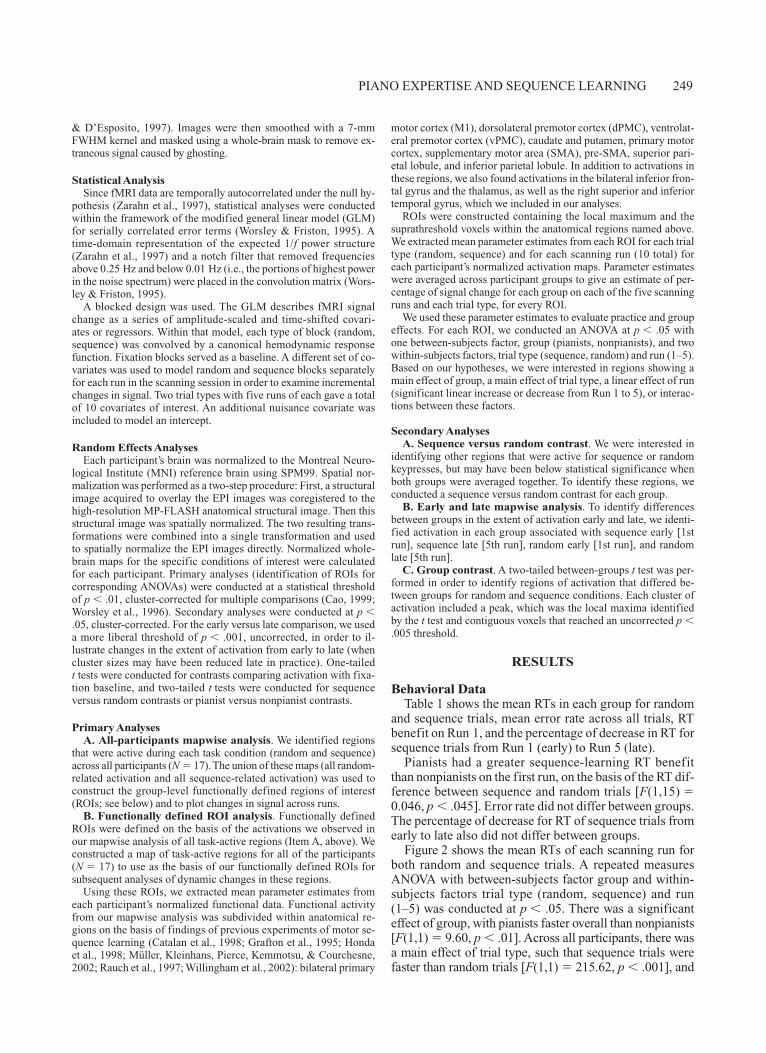
and sequence trials, mean error rate across all trials, RT benefit on Run 1, and the percentage of decrease in RT for sequence trials from Run 1 (early) to Run 5 (late).
Pianists had a greater sequence-learning RT benefit than nonpianists on the first run, on the basis of the RT dif-ference between sequence and random trials [F(1,15) 0.046, p .045]. Error rate did not differ between groups. The percentage of decrease for RT of sequence trials from early to late also did not differ between groups.
Figure 2 shows the mean RTs of each scanning run for both random and sequence trials. A repeated measures ANOVA with between-subjects factor group and within-subjects factors trial type (random, sequence) and run (1–5) was conducted at p .05. There was a significant effect of group, with pianists faster overall than nonpianists [F(1,1) 9.60, p .01]. Across all participants, there was a main effect of trial type, such that sequence trials were faster than random trials [F(1,1) 215.62, p .001], and
250 LANDAU AND D’ESPOSITO
a significant linear effect of run, with RTs decreasing from Runs 1 to 5 [F(1,1) 26.79, p .001]. The effect of de-creasing RTs across runs interacted with trial type [F(1,1) 32.94, p .001] so that sequence trials decreased dispro-portionately in comparison with random trials.
During the postscanning interview, the participants were asked (1) whether they were aware of any sequen-tial regularities during any part of the session, and, if so, (2) whether they could reproduce (manually or verbally) any parts of the sequence. All 17 participants (pianists and nonpianists) reported that they had at least minimal aware-ness of sequential regularities in the stimuli (i.e., they said that they knew they were not completely random). When prompted to reproduce any parts of the sequence, 4 participants named stimulus positions they noticed were often paired (e.g., “the second and fourth fingers on my right hand alternated sometimes”). The remaining 13 par-ticipants could not reproduce any parts of the sequence and reported that they hadn’t been paying attention and/or hadn’t been making an effort to learn the sequence. No participants reported knowledge of the presence of alter-nating blocks of sequenced and random stimuli, or of the probabilistic nature of the grammar.
Fast Learning: Within-Session Changes in Activation
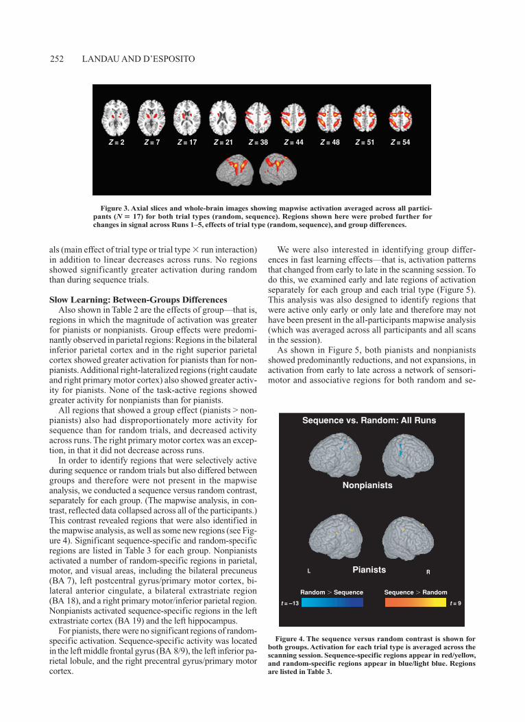
We identified task-active regions by combining voxel-wise maps of the main effect of random trials and the main effect of sequence trials. The task-active regions are listed in Table 2 and shown in Figure 3, and included a net-work of bilateral inferior frontal, premotor, sensorimotor, and parietal regions, as well as bilateral basal ganglia, thalamus, and right temporal regions. We subsequently probed these regions for effects of trial type (random, sequence) and linear changes across runs by conducting planned ANOVAs on mean parameter estimates. No re-gions showed linear increases across runs. Table 2 lists each region according to whether it showed a significant linear decrease across runs, and whether it showed ad-ditional main effects or interactions of trial type (random, sequence) and/or group (pianists, nonpianists).
Several key findings are apparent from this analysis. First, the majority of ROIs showed linear decreases across runs. A second striking finding is that all subdivisions of the precentral gyrus (BA 6), including dorsal and ventral premotor cortex, pre-SMA, and SMA, showed signifi-cantly greater activation for sequence than for random tri-
Table 1 Mean Reaction Times (RTs, in Milliseconds) and Error Rate Measures
With Standard Deviations for Each Group
Random Trials
Sequence Trials
RT Benefit for Sequence Trials: Run 1
OnlyPercent Error
Rate
Percent Decrease in
Sequence RT From Run 1
to Run 5
Group RT SD RT SD RTB SD PE SD PD SD
Nonpianists (n 8) 559 27 478 20 41.49 36.02 14.64 9.42 12.15 7.00Pianists (n 9) 505 42 423 53 75.53 21.73 9.94 6.65 10.35 7.46
Note—RT benefit (mean random RT minus mean sequence RT) for the first scanning run only is shown. Mean RTs of all runs for each group are also shown in Figure 2. The difference be-tween pianists and nonpianists was significant for both random and sequence trials. The group difference in RT benefit on Run 1 was also significant. There was no group difference in error rate or percent decrease on sequence trials. RTB, reaction time benefit; PE, percent error; PD, percent decrease.
A. Nonpianists
RandomSequence
B. Experienced Pianists
Rea
ctio
n T
ime
(mse
c)
350
400
450
500
550
600
Rea
ctio
n T
ime
(mse
c)
350
400
450
500
550
600
Run0 1 2 3 4 5
Run0 1 2 3 4 5
Figure 2. Changes in RT across all five runs are shown separately for random trials and sequence trials and for (A) nonpianists and (B) experienced pianists.
PIANO EXPERTISE AND SEQUENCE LEARNING 251
Tab
le 2
L
ocal
Max
ima
of t
he S
tati
stic
al P
aram
etri
c M
aps
for
the
Tas
k-A
ctiv
e R
egio
ns (
Show
n in
Fig
ure
3)
Eff
ect
ofTa
lair
ach
Coo
rd.
No.
tB
rain
Reg
ion
R
/L
Run
?
Oth
er E
ffec
t?
x
y
z
BA
V
oxel
s
Val
ue
Infe
rior
fron
tal g
yrus
R68
108
4412
3 6
.19
Puta
men
L20
414
188
6.6
1M
iddl
e te
mpo
ral g
yrus
R44
5810
3787
5.3
1In
feri
or fr
onta
l gyr
usL
•60
826
4470
5.9
1C
auda
teL
•22
418
144
6.3
0T
hala
mus
L•
1614
1925
6 6
.26
Prim
ary
mot
or c
orte
xL
•46
2446
459
6.1
7Pr
esup
plem
enta
ry m
otor
are
aL
•M
E tr
ial t
ype
(Seq
> R
and)
86
546
239
7.9
4Su
pple
men
tary
mot
or a
rea
L•
ME
tria
l typ
e (S
eq >
Ran
d)6
258
686
7.2
5V
entr
al p
rem
otor
cor
tex
L•
ME
tria
l typ
e (S
eq >
Ran
d)28
250
682
412
.01
Dor
sal p
rem
otor
cor
tex
R•
ME
tria
l typ
e (S
eq >
Ran
d)32
252
687
012
.98
Supp
lem
enta
ry m
otor
are
aR
•M
E tr
ial t
ype
(Seq
> R
and)
22
586
15 5
.21
Supe
rior
tem
pora
l gyr
usR
ME
tria
l typ
e (S
eq >
Ran
d)62
3824
40/2
237
4.7
1V
entr
al p
rem
otor
cor
tex
R•
ME
tria
l typ
e (S
eq >
Ran
d)32
250
645
212
.43
Puta
men
RM
E tr
ial t
ype
(Seq
> R
and)
260
268
5.1
6D
orsa
l pre
mot
or c
orte
xL
•In
t. tr
ial t
ype
run
(Seq
> R
and)
282
526
813
12.0
4Su
peri
or p
arie
tal l
obul
eL
•In
t. tr
ial t
ype
run
(Seq
> R
and)
2464
607
612
7.7
3Pr
esup
plem
enta
ry m
otor
are
aR
•In
t. tr
ial t
ype
run
(Seq
> R
and)
26
566
129
6.0
8T
hala
mus
RIn
t. tr
ial t
ype
run
(Seq
> R
and)
1216
1023
6 5
.62
Infe
rior
par
ieta
l lob
ule
L•
ME
Gro
up (P
> N
P), I
nt. t
rial
type
r
un (S
eq >
Ran
d)36
3850
401,
335
14.1
5C
auda
teR
ME
Gro
up (P
> N
P), I
nt. t
rial
type
r
un (S
eq >
Ran
d)20
214
160
5.3
8Su
peri
or p
arie
tal l
obul
eR
•M
E G
roup
(P >
NP)
, Int
. tri
al ty
pe
run
(Seq
> R
and)
2656
507
630
6.5
8In
feri
or p
arie
tal l
obul
eR
•M
E G
roup
(P >
NP)
, Int
. tri
al ty
pe
run
(Seq
> R
and)
4344
4840
449
8.6
0Pr
imar
y m
otor
cor
tex
R
In
t. tr
ial t
ype
gro
up (P
> N
P fo
r Seq
tria
ls o
nly)
48
24
46
4
75
4.9
3
Not
e—E
ach
loca
l max
imum
is li
sted
acc
ordi
ng to
whe
ther
it s
how
ed a
sig
nifi
cant
line
ar d
ecre
ase
acro
ss r
uns
and
whe
ther
it s
how
ed o
ther
sig
nifi
cant
eff
ects
of
tria
l typ
e an
d/or
gro
up. L
ater
ality
, coo
rdin
ates
, BA
of
supr
athr
esho
ld v
oxel
s in
clud
ed in
RO
I (s
ee M
etho
d), c
lust
er s
ize
of R
OI,
and
t va
lue
are
also
list
ed. C
oor-
dina
tes
corr
espo
nd to
thos
e fr
om th
e M
NI r
efer
ence
bra
in te
mpl
ate.
ME
, mai
n ef
fect
; Int
., In
tera
ctio
n; S
eq, s
eque
nce;
Ran
d, ra
ndom
; P, p
iani
sts;
NP,
non
pian
ists
; L
, lef
t; R
, rig
ht; B
A, B
rodm
ann’
s ar
ea.
252 LANDAU AND D’ESPOSITO
als (main effect of trial type or trial type run interaction) in addition to linear decreases across runs. No regions showed significantly greater activation during random than during sequence trials.
Slow Learning: Between-Groups DifferencesAlso shown in Table 2 are the effects of group—that is,
regions in which the magnitude of activation was greater for pianists or nonpianists. Group effects were predomi-nantly observed in parietal regions: Regions in the bilateral inferior parietal cortex and in the right superior parietal cortex showed greater activation for pianists than for non-pianists. Additional right-lateralized regions (right caudate and right primary motor cortex) also showed greater activ-ity for pianists. None of the task-active regions showed greater activity for nonpianists than for pianists.
All regions that showed a group effect (pianists > non-pianists) also had disproportionately more activity for sequence than for random trials, and decreased activity across runs. The right primary motor cortex was an excep-tion, in that it did not decrease across runs.
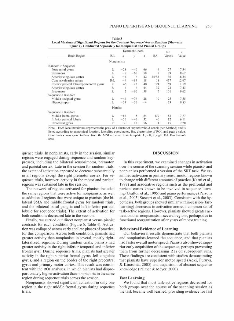
In order to identify regions that were selectively active during sequence or random trials but also differed between groups and therefore were not present in the mapwise analysis, we conducted a sequence versus random contrast, separately for each group. (The mapwise analysis, in con-trast, reflected data collapsed across all of the participants.) This contrast revealed regions that were also identified in the mapwise analysis, as well as some new regions (see Fig-ure 4). Significant sequence-specific and random-specific regions are listed in Table 3 for each group. Nonpianists activated a number of random-specific regions in parietal, motor, and visual areas, including the bilateral precuneus (BA 7), left postcentral gyrus/primary motor cortex, bi-lateral anterior cingulate, a bilateral extrastriate region (BA 18), and a right primary motor/inferior parietal region. Nonpianists activated sequence-specific regions in the left extrastriate cortex (BA 19) and the left hippocampus.
For pianists, there were no significant regions of random-specific activation. Sequence-specific activity was located in the left middle frontal gyrus (BA 8/9), the left inferior pa-rietal lobule, and the right precentral gyrus/primary motor cortex.
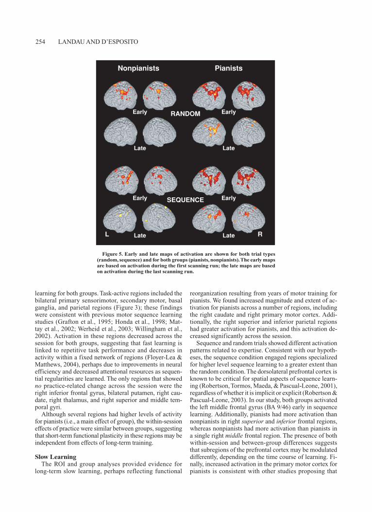
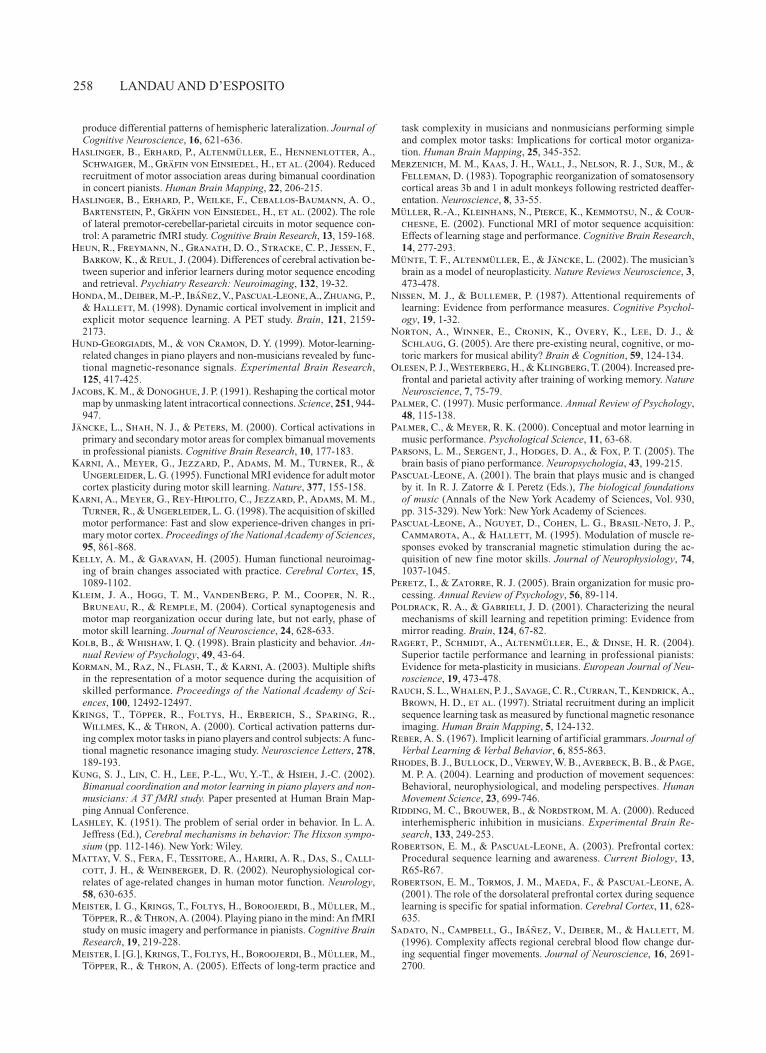
We were also interested in identifying group differ-ences in fast learning effects—that is, activation patterns that changed from early to late in the scanning session. To do this, we examined early and late regions of activation separately for each group and each trial type (Figure 5). This analysis was also designed to identify regions that were active only early or only late and therefore may not have been present in the all-participants mapwise analysis (which was averaged across all participants and all scans in the session).
As shown in Figure 5, both pianists and nonpianists showed predominantly reductions, and not expansions, in activation from early to late across a network of sensori-motor and associative regions for both random and se-
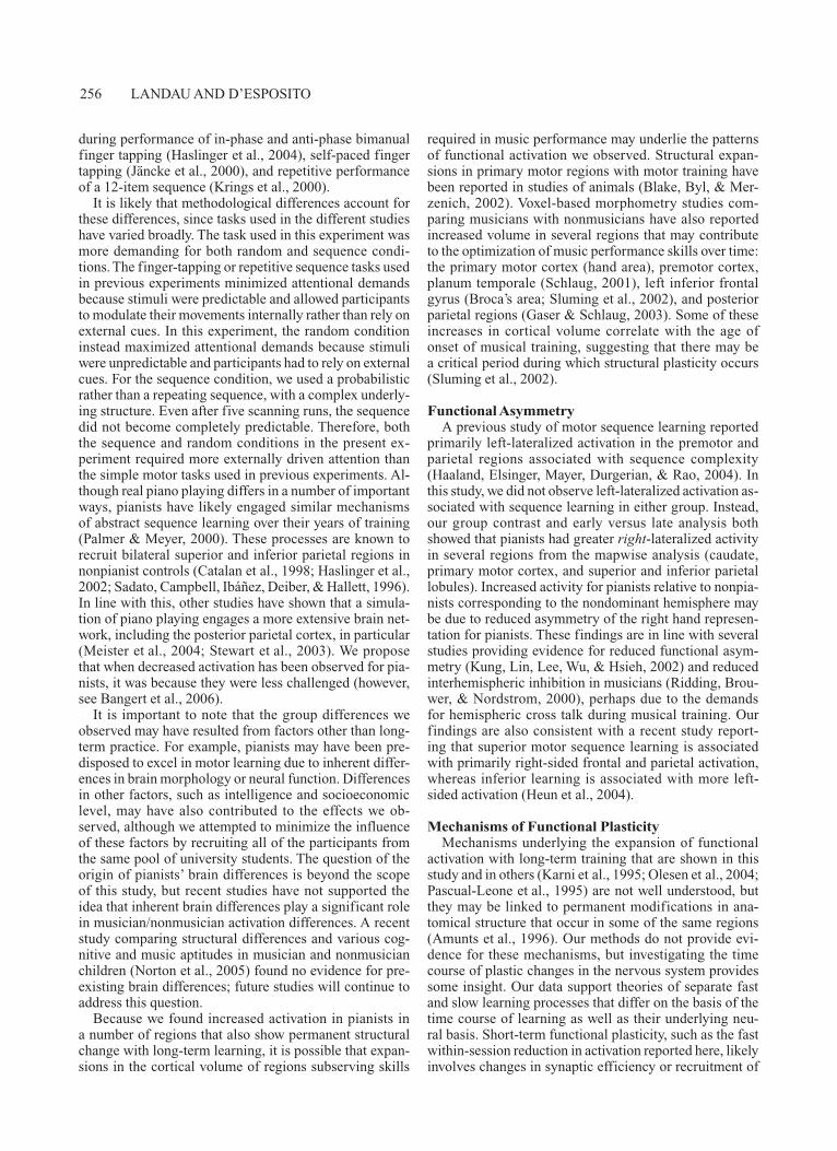
Figure 3. Axial slices and whole-brain images showing mapwise activation averaged across all partici-pants (N 17) for both trial types (random, sequence). Regions shown here were probed further for changes in signal across Runs 1–5, effects of trial type (random, sequence), and group differences.
Z = 2 Z = 7 Z = 17 Z = 21 Z = 38 Z = 44 Z = 48 Z = 51 Z = 54
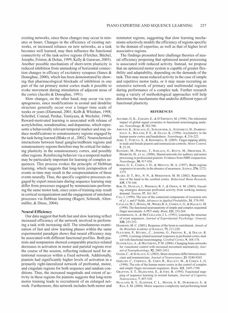
Figure 4. The sequence versus random contrast is shown for both groups. Activation for each trial type is averaged across the scanning session. Sequence-specific regions appear in red/yellow, and random-specific regions appear in blue/light blue. Regions are listed in Table 3.
Nonpianists
Pianists
Sequence vs. Random: All Runs
Random Sequence Sequence Random
t = –13 t = 9
RL
PIANO EXPERTISE AND SEQUENCE LEARNING 253
quence trials. In nonpianists, early in the session, similar regions were engaged during sequence and random key-presses, including the bilateral sensorimotor, premotor, and parietal cortex. Late in the session for random trials, the extent of activation appeared to decrease substantially in all regions except the right premotor cortex. For se-quence trials, however, activity in the motor and parietal regions was sustained late in the session.
The network of regions activated for pianists included the same regions that were active for nonpianists, as well as additional regions that were unique to pianists (the bi-lateral SMA and middle frontal gyrus for random trials, and the bilateral basal ganglia and left inferior parietal lobule for sequence trials). The extent of activation for both conditions decreased late in the session.
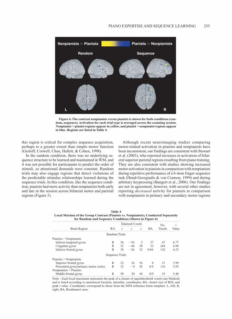
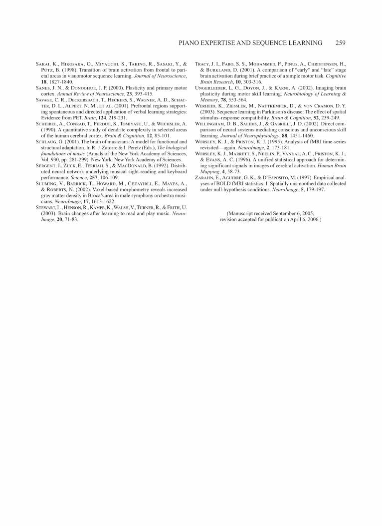
Finally, we carried out direct nonpianist versus pianist contrasts for each condition (Figure 6, Table 4). Activa-tion was collapsed across early and late phases of practice, for this comparison. Across both conditions, pianists had greater activity than nonpianists in several, mostly right-lateralized, regions. During random trials, pianists had greater activity in the right inferior temporal and inferior frontal gyri. During sequence trials, pianists had greater activity in the right superior frontal gyrus, left cingulate gyrus, and a region on the border of the right precentral gyrus and primary motor cortex. This result was consis-tent with the ROI analyses, in which pianists had dispro-portionately higher activation than nonpianists in the same region during sequence trials across the session.
Nonpianists showed significant activation in only one region in the right middle frontal gyrus during sequence trials.
DISCUSSION
In this experiment, we examined changes in activation over the course of the scanning session while pianists and nonpianists performed a version of the SRT task. We ex-amined activation in primary sensorimotor regions known to change with different amounts of practice (Karni et al., 1998) and associative regions such as the prefrontal and parietal cortex known to be involved in sequence learn-ing (Grafton et al., 1995) and piano performance (Parsons et al., 2005; Stewart et al., 2003). Consistent with the hy-potheses, both groups showed similar within-session (fast-learning) decreases in activation across a common set of task-active regions. However, pianists showed greater ac-tivation than nonpianists in several regions, perhaps due to functional reorganization after years of motor training.
Behavioral Evidence of LearningOur behavioral results demonstrate that both pianists
and nonpianists learned the sequence, and that pianists had faster overall motor speed. Pianists also showed supe-rior early acquisition of the sequence, perhaps preventing them from further decreasing RTs on subsequent runs. These findings are consistent with studies demonstrating that pianists have superior motor speed (Aoki, Furuya, & Kinoshita, 2005) and acquisition of abstract sequence knowledge (Palmer & Meyer, 2000).
Fast LearningWe found that most task-active regions decreased for
both groups over the course of the scanning session as the task became well learned, providing evidence for fast
Table 3 Local Maxima of Significant Regions for the Contrast Sequence Versus Random (Shown in
Figure 4), Conducted Separately for Nonpianist and Pianist Groups
Talairach Coord. No. tBrain Region R/L x y z BA Voxels Value
NonpianistsRandom > Sequence Postcentral gyrus L 28 40 66 4 27 7.34 Precuneus L 2 60 58 7 89 8.62 Anterior cingulate cortex L 4 6 42 24/32 36 8.34 Cuneus/calcarine sulcus R/L 4 84 18 18 437 12.67 Inferior parietal lobule/postcentral gyrus R 46 22 40 3/4 169 11.59 Anterior cingulate cortex R 4 6 44 32 22 7.43 Precuneus R 2 60 58 7 101 9.62Sequence > Random Middle occipital gyrus L 44 76 28 19 25 7.55 Hippocampus L 34 36 4 33 9.85
PianistsSequence > Random Middle frontal gyrus L 56 8 54 8/9 53 7.77 Inferior parietal lobule L 56 46 52 40 12 6.11 Precentral gyrus R 30 18 56 4 15 7.20
Note—Each local maximum represents the peak of a cluster of suprathreshold voxels (see Method) and is listed according to anatomical location, laterality, coordinates, BA, cluster size of ROI, and peak t value. Coordinates correspond to those from the MNI reference brain template. L, left; R, right; BA, Brodmann’s area.
254 LANDAU AND D’ESPOSITO
learning for both groups. Task-active regions included the bilateral primary sensorimotor, secondary motor, basal ganglia, and parietal regions (Figure 3); these findings were consistent with previous motor sequence learning studies (Grafton et al., 1995; Honda et al., 1998; Mat-tay et al., 2002; Werheid et al., 2003; Willingham et al., 2002). Activation in these regions decreased across the session for both groups, suggesting that fast learning is linked to repetitive task performance and decreases in activity within a fixed network of regions (Floyer-Lea & Matthews, 2004), perhaps due to improvements in neural efficiency and decreased attentional resources as sequen-tial regularities are learned. The only regions that showed no practice-related change across the session were the right inferior frontal gyrus, bilateral putamen, right cau-date, right thalamus, and right superior and middle tem-poral gyri.
Although several regions had higher levels of activity for pianists (i.e., a main effect of group), the within-session effects of practice were similar between groups, suggesting that short-term functional plasticity in these regions may be independent from effects of long-term training.
Slow LearningThe ROI and group analyses provided evidence for
long-term slow learning, perhaps reflecting functional
reorganization resulting from years of motor training for pianists. We found increased magnitude and extent of ac-tivation for pianists across a number of regions, including the right caudate and right primary motor cortex. Addi-tionally, the right superior and inferior parietal regions had greater activation for pianists, and this activation de-creased significantly across the session.
Sequence and random trials showed different activation patterns related to expertise. Consistent with our hypoth-eses, the sequence condition engaged regions specialized for higher level sequence learning to a greater extent than the random condition. The dorsolateral prefrontal cortex is known to be critical for spatial aspects of sequence learn-ing (Robertson, Tormos, Maeda, & Pascual-Leone, 2001), regardless of whether it is implicit or explicit (Robertson & Pascual-Leone, 2003). In our study, both groups activated the left middle frontal gyrus (BA 9/46) early in sequence learning. Additionally, pianists had more activation than nonpianists in right superior and inferior frontal regions, whereas nonpianists had more activation than pianists in a single right middle frontal region. The presence of both within-session and between-group differences suggests that subregions of the prefrontal cortex may be modulated differently, depending on the time course of learning. Fi-nally, increased activation in the primary motor cortex for pianists is consistent with other studies proposing that
Figure 5. Early and late maps of activation are shown for both trial types (random, sequence) and for both groups (pianists, nonpianists). The early maps are based on activation during the first scanning run; the late maps are based on activation during the last scanning run.
PianistsNonpianists
Early Early
Early Early
Late Late
Late Late
RANDOM
SEQUENCE
RL
PIANO EXPERTISE AND SEQUENCE LEARNING 255
this region is critical for complex sequence acquisition, perhaps to a greater extent than simple motor function (Gerloff, Corwell, Chen, Hallett, & Cohen, 1998).
In the random condition, there was no underlying se-quence structure to be learned and maintained in WM, and it was not possible for participants to predict the order of stimuli, so attentional demands were constant. Random trials may also engage regions that detect violations of the predictable stimulus relationships learned during the sequence trials. In this condition, like the sequence condi-tion, pianists had more activity than nonpianists both early and late in the session across bilateral motor and parietal regions (Figure 5).
Although recent neuroimaging studies comparing motor-related activation in pianists and nonpianists have been inconsistent, our findings are consistent with Stewart et al. (2003), who reported increases in activation of bilat-eral superior parietal regions resulting from piano training. They are also consistent with studies showing increased motor activation in pianists in comparison with nonpianists during repetitive performance of a 6-item finger sequence task (Hund-Georgiadis & von Cramon, 1999) and during arbitrary keypressing (Bangert et al., 2006). Our findings are not in agreement, however, with several other studies reporting decreased activity for pianists in comparison with nonpianists in primary and secondary motor regions
Figure 6. The contrast nonpianists versus pianists is shown for both conditions (ran-dom, sequence). Activation for each trial type is averaged across the scanning session. Nonpianist > pianist regions appear in yellow, and pianist > nonpianist regions appear in blue. Regions are listed in Table 4.
Table 4 Local Maxima of the Group Contrast (Pianists vs. Nonpianists), Conducted Separately
for Random and Sequence Conditions (Shown in Figure 6)
Talairach Coord. No. tBrain Region R/L x y z BA Voxels Value
Random TrialsPianists > Nonpianists Inferior temporal gyrus R 56 56 2 37 67 6.77 Cingulate gyrus R 32 44 30 32 264 6.98 Inferior frontal gyrus R 38 26 32 9/44 142 6.23
Sequence TrialsPianists > Nonpianists Superior frontal gyrus R 22 26 56 8 51 5.99 Precentral gyrus/primary motor cortex R 32 4 42 6/4 116 5.95Nonpianists > Pianists Middle frontal gyrus R 50 30 44 8/9 53 5.48
Note—Each local maximum represents the peak of a cluster of suprathreshold voxels (see Method) and is listed according to anatomical location, laterality, coordinates, BA, cluster size of ROI, and peak t value. Coordinates correspond to those from the MNI reference brain template. L, left; R, right; BA, Brodmann’s area.
RL
Pianists NonpianistsNonpianists Pianists
Random Sequence
256 LANDAU AND D’ESPOSITO
during performance of in-phase and anti-phase bimanual finger tapping (Haslinger et al., 2004), self-paced finger tapping (Jäncke et al., 2000), and repetitive performance of a 12-item sequence (Krings et al., 2000).
It is likely that methodological differences account for these differences, since tasks used in the different studies have varied broadly. The task used in this experiment was more demanding for both random and sequence condi-tions. The finger-tapping or repetitive sequence tasks used in previous experiments minimized attentional demands because stimuli were predictable and allowed participants to modulate their movements internally rather than rely on external cues. In this experiment, the random condition instead maximized attentional demands because stimuli were unpredictable and participants had to rely on external cues. For the sequence condition, we used a probabilistic rather than a repeating sequence, with a complex underly-ing structure. Even after five scanning runs, the sequence did not become completely predictable. Therefore, both the sequence and random conditions in the present ex-periment required more externally driven attention than the simple motor tasks used in previous experiments. Al-though real piano playing differs in a number of important ways, pianists have likely engaged similar mechanisms of abstract sequence learning over their years of training (Palmer & Meyer, 2000). These processes are known to recruit bilateral superior and inferior parietal regions in nonpianist controls (Catalan et al., 1998; Haslinger et al., 2002; Sadato, Campbell, Ibáñez, Deiber, & Hallett, 1996). In line with this, other studies have shown that a simula-tion of piano playing engages a more extensive brain net-work, including the posterior parietal cortex, in particular (Meister et al., 2004; Stewart et al., 2003). We propose that when decreased activation has been observed for pia-nists, it was because they were less challenged (however, see Bangert et al., 2006).
It is important to note that the group differences we observed may have resulted from factors other than long-term practice. For example, pianists may have been pre-disposed to excel in motor learning due to inherent differ-ences in brain morphology or neural function. Differences in other factors, such as intelligence and socioeconomic level, may have also contributed to the effects we ob-served, although we attempted to minimize the influence of these factors by recruiting all of the participants from the same pool of university students. The question of the origin of pianists’ brain differences is beyond the scope of this study, but recent studies have not supported the idea that inherent brain differences play a significant role in musician/nonmusician activation differences. A recent study comparing structural differences and various cog-nitive and music aptitudes in musician and nonmusician children (Norton et al., 2005) found no evidence for pre-existing brain differences; future studies will continue to address this question.
Because we found increased activation in pianists in a number of regions that also show permanent structural change with long-term learning, it is possible that expan-sions in the cortical volume of regions subserving skills
required in music performance may underlie the patterns of functional activation we observed. Structural expan-sions in primary motor regions with motor training have been reported in studies of animals (Blake, Byl, & Mer-zenich, 2002). Voxel-based morphometry studies com-paring musicians with nonmusicians have also reported increased volume in several regions that may contribute to the optimization of music performance skills over time: the primary motor cortex (hand area), premotor cortex, planum temporale (Schlaug, 2001), left inferior frontal gyrus (Broca’s area; Sluming et al., 2002), and posterior parietal regions (Gaser & Schlaug, 2003). Some of these increases in cortical volume correlate with the age of onset of musical training, suggesting that there may be a critical period during which structural plasticity occurs (Sluming et al., 2002).
Functional AsymmetryA previous study of motor sequence learning reported
primarily left-lateralized activation in the premotor and parietal regions associated with sequence complexity (Haaland, Elsinger, Mayer, Durgerian, & Rao, 2004). In this study, we did not observe left-lateralized activation as-sociated with sequence learning in either group. Instead, our group contrast and early versus late analysis both showed that pianists had greater right-lateralized activity in several regions from the mapwise analysis (caudate, primary motor cortex, and superior and inferior parietal lobules). Increased activity for pianists relative to nonpia-nists corresponding to the nondominant hemisphere may be due to reduced asymmetry of the right hand represen-tation for pianists. These findings are in line with several studies providing evidence for reduced functional asym-metry (Kung, Lin, Lee, Wu, & Hsieh, 2002) and reduced interhemispheric inhibition in musicians (Ridding, Brou-wer, & Nordstrom, 2000), perhaps due to the demands for hemispheric cross talk during musical training. Our findings are also consistent with a recent study report-ing that superior motor sequence learning is associated with primarily right-sided frontal and parietal activation, whereas inferior learning is associated with more left-sided activation (Heun et al., 2004).
Mechanisms of Functional PlasticityMechanisms underlying the expansion of functional
activation with long-term training that are shown in this study and in others (Karni et al., 1995; Olesen et al., 2004; Pascual-Leone et al., 1995) are not well understood, but they may be linked to permanent modifications in ana-tomical structure that occur in some of the same regions (Amunts et al., 1996). Our methods do not provide evi-dence for these mechanisms, but investigating the time course of plastic changes in the nervous system provides some insight. Our data support theories of separate fast and slow learning processes that differ on the basis of the time course of learning as well as their underlying neu-ral basis. Short-term functional plasticity, such as the fast within-session reduction in activation reported here, likely involves changes in synaptic efficiency or recruitment of
PIANO EXPERTISE AND SEQUENCE LEARNING 257
existing networks, since these changes may occur in min-utes or hours. Changes in the efficiency of existing net-works, or increased reliance on new networks, as a task becomes well learned, may then influence the functional connectivity of the task-active regions (Fletcher, Büchel, Josephs, Friston, & Dolan, 1999; Kelly & Garavan, 2005). Another possible mechanism of short-term plasticity is reduced inhibition from unmasking of horizontal connec-tion changes in efficacy of excitatory synapses (Sanes & Donoghue, 2000), which has been demonstrated by show-ing that pharmacological blockade of inhibition in one part of the rat primary motor cortex made it possible to evoke movement during stimulation of adjacent areas of the cortex (Jacobs & Donoghue, 1991).
Slow changes, on the other hand, may occur via syn-aptogenesis, since modifications in axonal and dendritic structure generally occur over a longer time scale of weeks or years (Diamond, 2001; Kolb & Whishaw, 1998; Scheibel, Conrad, Perdue, Tomiyasu, & Wechsler, 1990). Reward-motivated learning is associated with release of acetylcholine, noradrenaline, and dopamine, which repre-sents a behaviorally relevant temporal marker and may in-duce modifications to somatosensory regions engaged by the task being learned (Blake et al., 2002). Neurochemical interactions between basal ganglia/midbrain regions and somatosensory regions therefore may be critical for induc-ing plasticity in the somatosensory cortex, and possibly other regions. Reinforcing behavior via a temporal marker may be particularly important for learning of complex se-quences. This process evokes the principle of Hebbian learning, which suggests that long-term juxtaposition of events in time may result in the corepresentation of these events neurally. Thus, the specific cognitive processes en-gaged by expert musicians during sequence learning may differ from processes engaged by nonmusicians perform-ing the same motor task, since years of training may result in cortical reorganization and the engagement of unrelated processes via Hebbian learning (Ragert, Schmidt, Alten-müller, & Dinse, 2004).
Neural EfficiencyOur data suggest that both fast and slow learning reflect
increased efficiency of the network involved in perform-ing a task with increasing skill. The simultaneous exami-nation of fast and slow learning phases within the same experimental paradigm shows that neural efficiency may be associated with different functional profiles. Both pia-nists and nonpianists showed comparable practice-related decreases in activation in motor and parietal regions over the course of the session, reflecting reduced need for at-tentional resources within a fixed network. Additionally, pianists had significantly higher levels of activation in a primarily right-lateralized network of prefrontal, motor, and cingulate regions for both sequence and random con-ditions. Thus, the increased magnitude and extent of ac-tivity in these regions for pianists suggests that long-term motor training leads to recruitment of an enlarged net-work. Furthermore, this network includes both motor and
nonmotor regions, suggesting that slow learning mecha-nisms selectively modify the efficiency of regions specific to the domain of expertise, as well as that of higher level associative regions.
The findings presented here challenge theories of neu-ral efficiency proposing that optimized neural processing is associated with reduced activity. Instead, we propose that an optimized motor system is capable of greater flex-ibility and adaptability, depending on the demands of the task. This may mean reduced activity in the case of simple and repetitive motor tasks, or it may mean recruiting an extensive network of primary and multimodal regions during performance of a complex task. Further research using a variety of methodological approaches will help determine the mechanisms that underlie different types of functional plasticity.
REFERENCES
Aguirre, G. K., Zarahn, E., & D’Esposito, M. (1998). The inferential impact of global signal covariates in functional neuroimaging analy-ses. NeuroImage, 8, 302-306.
Amunts, K., Schlaug, G., Schleicher, A., Steinmetz, H., Dabring-haus, A., Roland, P. E., & Zilles, K. (1996). Asymmetry in the human motor cortex and handedness. NeuroImage, 4, 216-222.
Aoki, T., Furuya, S., & Kinoshita, H. (2005). Finger-tapping ability in male and female pianists and nonmusician controls. Motor Control, 9, 23-39.
Bangert, M., Peschel, T., Schlaug, G., Rotte, M., Drescher, D., Hinrichs, H., et al. (2006). Shared networks for auditory and motor processing in professional pianists: Evidence from fMRI conjunction. NeuroImage, 30, 917-926.
Berns, G. S., Cohen, J. D., & Mintun, M. A. (1997). Brain regions responsive to novelty in the absence of awareness. Science, 276, 1272-1275.
Blake, D. T., Byl, N. N., & Merzenich, M. M. (2002). Representa-tion of the hand in the cerebral cortex. Behavioral Brain Research, 135, 179-184.
Bor, D., Duncan, J., Wiseman, R. J., & Owen, A. M. (2003). Encod-ing strategies dissociate prefrontal activity from working memory demand. Neuron, 37, 361-367.
Cao, J. (1999). The size of the connected components of excursion sets of 2, t, and F fields. Advances in Applied Probability, 31, 579-595.
Catalan, M. J., Honda, M., Weeks, R. A., Cohen, L. G., & Hallett, M. (1998). The functional neuroanatomy of simple and complex sequential finger movements: A PET study. Brain, 121, 253-264.
Cleeremans, A., & McClelland, J. L. (1991). Learning the structure of event sequences. Journal of Experimental Psychology: General, 120, 235-253.
Diamond, M. C. (2001). Response of the brain to enrichment. Annals of the Brazilian Academy of Sciences, 73, 211-220.
Fletcher, P., Büchel, C., Josephs, O., Friston, K., & Dolan, R. (1999). Learning-related neuronal responses in prefrontal cortex stud-ied with functional neuroimaging. Cerebral Cortex, 9, 168-178.
Floyer-Lea, A., & Matthews, P. M. (2004). Changing brain networks for visuomotor control with increased movement automaticity. Jour-nal of Neurophysiology, 92, 2405-2412.
Gaser, C., & Schlaug, G. (2003). Brain structures differ between musi-cians and nonmusicians. Journal of Neuroscience, 23, 9240-9245.
Gerloff, C., Corwell, B., Chen, R., Hallett, M., & Cohen, L. G. (1998). The role of the human motor cortex in the control of complex and simple finger movement sequences. Brain, 121, 1695-1709.
Grafton, S. T., Hazeltine, E., & Ivry, R. (1995). Functional map-ping of sequence learning in normal humans. Journal of Cognitive Neuroscience, 7, 497-510.
Haaland, K. Y., Elsinger, C. L., Mayer, A. R., Durgerian, S., & Rao, S. M. (2004). Motor sequence complexity and performing hand
258 LANDAU AND D’ESPOSITO
produce differential patterns of hemispheric lateralization. Journal of Cognitive Neuroscience, 16, 621-636.
Haslinger, B., Erhard, P., Altenmüller, E., Hennenlotter, A., Schwaiger, M., Gräfin von Einsiedel, H., et al. (2004). Reduced recruitment of motor association areas during bimanual coordination in concert pianists. Human Brain Mapping, 22, 206-215.
Haslinger, B., Erhard, P., Weilke, F., Ceballos-Baumann, A. O., Bartenstein, P., Gräfin von Einsiedel, H., et al. (2002). The role of lateral premotor-cerebellar-parietal circuits in motor sequence con-trol: A parametric fMRI study. Cognitive Brain Research, 13, 159-168.
Heun, R., Freymann, N., Granath, D. O., Stracke, C. P., Jessen, F., Barkow, K., & Reul, J. (2004). Differences of cerebral activation be-tween superior and inferior learners during motor sequence encoding and retrieval. Psychiatry Research: Neuroimaging, 132, 19-32.
Honda, M., Deiber, M.-P., Ibáñez, V., Pascual-Leone, A., Zhuang, P., & Hallett, M. (1998). Dynamic cortical involvement in implicit and explicit motor sequence learning. A PET study. Brain, 121, 2159-2173.
Hund-Georgiadis, M., & von Cramon, D. Y. (1999). Motor-learning- related changes in piano players and non-musicians revealed by func-tional magnetic-resonance signals. Experimental Brain Research, 125, 417-425.
Jacobs, K. M., & Donoghue, J. P. (1991). Reshaping the cortical motor map by unmasking latent intracortical connections. Science, 251, 944-947.
Jäncke, L., Shah, N. J., & Peters, M. (2000). Cortical activations in primary and secondary motor areas for complex bimanual movements in professional pianists. Cognitive Brain Research, 10, 177-183.
Karni, A., Meyer, G., Jezzard, P., Adams, M. M., Turner, R., & Ungerleider, L. G. (1995). Functional MRI evidence for adult motor cortex plasticity during motor skill learning. Nature, 377, 155-158.
Karni, A., Meyer, G., Rey-Hipolito, C., Jezzard, P., Adams, M. M., Turner, R., & Ungerleider, L. G. (1998). The acquisition of skilled motor performance: Fast and slow experience-driven changes in pri-mary motor cortex. Proceedings of the National Academy of Sciences, 95, 861-868.
Kelly, A. M., & Garavan, H. (2005). Human functional neuroimag-ing of brain changes associated with practice. Cerebral Cortex, 15, 1089-1102.
Kleim, J. A., Hogg, T. M., VandenBerg, P. M., Cooper, N. R., Bruneau, R., & Remple, M. (2004). Cortical synaptogenesis and motor map reorganization occur during late, but not early, phase of motor skill learning. Journal of Neuroscience, 24, 628-633.
Kolb, B., & Whishaw, I. Q. (1998). Brain plasticity and behavior. An-nual Review of Psychology, 49, 43-64.
Korman, M., Raz, N., Flash, T., & Karni, A. (2003). Multiple shifts in the representation of a motor sequence during the acquisition of skilled performance. Proceedings of the National Academy of Sci-ences, 100, 12492-12497.
Krings, T., Töpper, R., Foltys, H., Erberich, S., Sparing, R., Willmes, K., & Thron, A. (2000). Cortical activation patterns dur-ing complex motor tasks in piano players and control subjects: A func-tional magnetic resonance imaging study. Neuroscience Letters, 278, 189-193.
Kung, S. J., Lin, C. H., Lee, P.-L., Wu, Y.-T., & Hsieh, J.-C. (2002). Bimanual coordination and motor learning in piano players and non-musicians: A 3T fMRI study. Paper presented at Human Brain Map-ping Annual Conference.
Lashley, K. (1951). The problem of serial order in behavior. In L. A. Jeffress (Ed.), Cerebral mechanisms in behavior: The Hixson sympo-sium (pp. 112-146). New York: Wiley.
Mattay, V. S., Fera, F., Tessitore, A., Hariri, A. R., Das, S., Calli-cott, J. H., & Weinberger, D. R. (2002). Neurophysiological cor-relates of age-related changes in human motor function. Neurology, 58, 630-635.
Meister, I. G., Krings, T., Foltys, H., Boroojerdi, B., Müller, M., Töpper, R., & Thron, A. (2004). Playing piano in the mind: An fMRI study on music imagery and performance in pianists. Cognitive Brain Research, 19, 219-228.
Meister, I. [G.], Krings, T., Foltys, H., Boroojerdi, B., Müller, M., Töpper, R., & Thron, A. (2005). Effects of long-term practice and
task complexity in musicians and nonmusicians performing simple and complex motor tasks: Implications for cortical motor organiza-tion. Human Brain Mapping, 25, 345-352.
Merzenich, M. M., Kaas, J. H., Wall, J., Nelson, R. J., Sur, M., & Felleman, D. (1983). Topographic reorganization of somatosensory cortical areas 3b and 1 in adult monkeys following restricted deaffer-entation. Neuroscience, 8, 33-55.
Müller, R.-A., Kleinhans, N., Pierce, K., Kemmotsu, N., & Cour-chesne, E. (2002). Functional MRI of motor sequence acquisition: Effects of learning stage and performance. Cognitive Brain Research, 14, 277-293.
Münte, T. F., Altenmüller, E., & Jäncke, L. (2002). The musician’s brain as a model of neuroplasticity. Nature Reviews Neuroscience, 3, 473-478.
Nissen, M. J., & Bullemer, P. (1987). Attentional requirements of learning: Evidence from performance measures. Cognitive Psychol-ogy, 19, 1-32.
Norton, A., Winner, E., Cronin, K., Overy, K., Lee, D. J., & Schlaug, G. (2005). Are there pre-existing neural, cognitive, or mo-toric markers for musical ability? Brain & Cognition, 59, 124-134.
Olesen, P. J., Westerberg, H., & Klingberg, T. (2004). Increased pre-frontal and parietal activity after training of working memory. Nature Neuroscience, 7, 75-79.
Palmer, C. (1997). Music performance. Annual Review of Psychology, 48, 115-138.
Palmer, C., & Meyer, R. K. (2000). Conceptual and motor learning in music performance. Psychological Science, 11, 63-68.
Parsons, L. M., Sergent, J., Hodges, D. A., & Fox, P. T. (2005). The brain basis of piano performance. Neuropsychologia, 43, 199-215.
Pascual-Leone, A. (2001). The brain that plays music and is changed by it. In R. J. Zatorre & I. Peretz (Eds.), The biological foundations of music (Annals of the New York Academy of Sciences, Vol. 930, pp. 315-329). New York: New York Academy of Sciences.
Pascual-Leone, A., Nguyet, D., Cohen, L. G., Brasil-Neto, J. P., Cammarota, A., & Hallett, M. (1995). Modulation of muscle re-sponses evoked by transcranial magnetic stimulation during the ac-quisition of new fine motor skills. Journal of Neurophysiology, 74, 1037-1045.
Peretz, I., & Zatorre, R. J. (2005). Brain organization for music pro-cessing. Annual Review of Psychology, 56, 89-114.
Poldrack, R. A., & Gabrieli, J. D. (2001). Characterizing the neural mechanisms of skill learning and repetition priming: Evidence from mirror reading. Brain, 124, 67-82.
Ragert, P., Schmidt, A., Altenmüller, E., & Dinse, H. R. (2004). Superior tactile performance and learning in professional pianists: Evidence for meta-plasticity in musicians. European Journal of Neu-roscience, 19, 473-478.
Rauch, S. L., Whalen, P. J., Savage, C. R., Curran, T., Kendrick, A., Brown, H. D., et al. (1997). Striatal recruitment during an implicit sequence learning task as measured by functional magnetic resonance imaging. Human Brain Mapping, 5, 124-132.
Reber, A. S. (1967). Implicit learning of artificial grammars. Journal of Verbal Learning & Verbal Behavior, 6, 855-863.
Rhodes, B. J., Bullock, D., Verwey, W. B., Averbeck, B. B., & Page, M. P. A. (2004). Learning and production of movement sequences: Behavioral, neurophysiological, and modeling perspectives. Human Movement Science, 23, 699-746.
Ridding, M. C., Brouwer, B., & Nordstrom, M. A. (2000). Reduced interhemispheric inhibition in musicians. Experimental Brain Re-search, 133, 249-253.
Robertson, E. M., & Pascual-Leone, A. (2003). Prefrontal cortex: Procedural sequence learning and awareness. Current Biology, 13, R65-R67.
Robertson, E. M., Tormos, J. M., Maeda, F., & Pascual-Leone, A. (2001). The role of the dorsolateral prefrontal cortex during sequence learning is specific for spatial information. Cerebral Cortex, 11, 628-635.
Sadato, N., Campbell, G., Ibáñez, V., Deiber, M., & Hallett, M. (1996). Complexity affects regional cerebral blood flow change dur-ing sequential finger movements. Journal of Neuroscience, 16, 2691-2700.
PIANO EXPERTISE AND SEQUENCE LEARNING 259
Sakai, K., Hikosaka, O., Miyauchi, S., Takino, R., Sasaki, Y., & Pütz, B. (1998). Transition of brain activation from frontal to pari-etal areas in visuomotor sequence learning. Journal of Neuroscience, 18, 1827-1840.
Sanes, J. N., & Donoghue, J. P. (2000). Plasticity and primary motor cortex. Annual Review of Neuroscience, 23, 393-415.
Savage, C. R., Deckersbach, T., Heckers, S., Wagner, A. D., Schac-ter, D. L., Alpert, N. M., et al. (2001). Prefrontal regions support-ing spontaneous and directed application of verbal learning strategies: Evidence from PET. Brain, 124, 219-231.
Scheibel, A., Conrad, T., Perdue, S., Tomiyasu, U., & Wechsler, A. (1990). A quantitative study of dendrite complexity in selected areas of the human cerebral cortex. Brain & Cognition, 12, 85-101.
Schlaug, G. (2001). The brain of musicians: A model for functional and structural adaptation. In R. J. Zatorre & I. Peretz (Eds.), The biological foundations of music (Annals of the New York Academy of Sciences, Vol. 930, pp. 281-299). New York: New York Academy of Sciences.
Sergent, J., Zuck, E., Terriah, S., & MacDonald, B. (1992). Distrib-uted neural network underlying musical sight-reading and keyboard performance. Science, 257, 106-109.
Sluming, V., Barrick, T., Howard, M., Cezayirli, E., Mayes, A., & Roberts, N. (2002). Voxel-based morphometry reveals increased gray matter density in Broca’s area in male symphony orchestra musi-cians. NeuroImage, 17, 1613-1622.
Stewart, L., Henson, R., Kampe, K., Walsh, V., Turner, R., & Frith, U. (2003). Brain changes after learning to read and play music. Neuro- Image, 20, 71-83.
Tracy, J. I., Faro, S. S., Mohammed, F., Pinus, A., Christensen, H., & Burkland, D. (2001). A comparison of “early” and “late” stage brain activation during brief practice of a simple motor task. Cognitive Brain Research, 10, 303-316.
Ungerleider, L. G., Doyon, J., & Karni, A. (2002). Imaging brain plasticity during motor skill learning. Neurobiology of Learning & Memory, 78, 553-564.
Werheid, K., Ziessler, M., Nattkemper, D., & von Cramon, D. Y. (2003). Sequence learning in Parkinson’s disease: The effect of spatial stimulus–response compatibility. Brain & Cognition, 52, 239-249.
Willingham, D. B., Salidis, J., & Gabrieli, J. D. (2002). Direct com-parison of neural systems mediating conscious and unconscious skill learning. Journal of Neurophysiology, 88, 1451-1460.
Worsley, K. J., & Friston, K. J. (1995). Analysis of fMRI time-series revisited—again. NeuroImage, 2, 173-181.
Worsley, K. J., Marrett, S., Neelin, P., Vandal, A. C., Friston, K. J., & Evans, A. C. (1996). A unified statistical approach for determin-ing significant signals in images of cerebral activation. Human Brain Mapping, 4, 58-73.
Zarahn, E., Aguirre, G. K., & D’Esposito, M. (1997). Empirical anal-yses of BOLD fMRI statistics: I. Spatially unsmoothed data collected under null-hypothesis conditions. NeuroImage, 5, 179-197.
(Manuscript received September 6, 2005; revision accepted for publication April 6, 2006.)