Embed Size (px)
Citation preview

September 2021 Volume 4, Issue 1
www.pmf.ni.ac.rs/chemianaissensis
ISSN 2620-1895
University of Niš, Faculty of Sciences and Mathematics

ISSN 2620-1895
Volume 4, Issue 1
September 2021
Category: M54
https://kobson.nb.rs/upload/documents/MNTR/Kategoriza
cija_casopisa/2019/MNTR2019_hemija.pdf
Publisher:
Faculty of Sciences and Mathematics, University of Niš
Editor-in-Chief:
Dr Vesna Stankov Jovanović, Department of Chemistry, Faculty of Sciences and Mathematics,
University of Niš, Republic of Serbia
Deputy Editor:
Dr Biljana Arsić, Department of Chemistry, Faculty of Sciences and Mathematics, University
of Niš, Republic of Serbia
Editors:
Analytical Chemistry
Dr Aleksandra Pavlović, Department of Chemistry, Faculty of Sciences and Mathematics,
University of Niš, Republic of Serbia
Physical Chemistry
Dr Snežana Tošić, Department of Chemistry, Faculty of Sciences and Mathematics, University
of Niš, Republic of Serbia
Organic Chemistry and Biochemistry
Dr Aleksandra Đorđević, Department of Chemistry, Faculty of Sciences and Mathematics,
University of Niš, Republic of Serbia
Inorganic Chemistry
Dr Dragan Đorđević, Department of Chemistry, Faculty of Sciences and Mathematics,
University of Niš, Republic of Serbia

Chemical Engineering
Dr Marjan Ranđelović, Department of Chemistry, Faculty of Sciences and Mathematics,
University of Niš, Republic of Serbia
Chemical Education
Dr Vesna Stankov Jovanović, Department of Chemistry, Faculty of Sciences and Mathematics,
University of Niš, Republic of Serbia
Language Editors:
1. Dr Selena Stanković, Department of French Language and Literature, Faculty of Philosophy,
University of Niš, Republic of Serbia (French)
2. Jovana Golubović (French)
3. Dr Nikoleta Momčilović, Department of German Language, Faculty of Philosophy,
University of Niš, Republic of Serbia (German)
4. Katarina Stamenković, Department of German Language, Faculty of Philosophy, University
of Niš, Republic of Serbia (German)
5. Dr Nadežda Jović, Department of Serbian language and literature, Faculty of Philosophy,
University of Niš, Republic of Serbia (Serbian)
6. Jelena Stošić, Department of Serbian language and literature, Faculty of Philosophy,
University of Niš, Republic of Serbia (Serbian)
PR Manager:
Dr Radomir Ljupković, Department of Chemistry, Faculty of Sciences and Mathematics,
University of Niš, Republic of Serbia
Technical Secretary:
1. Dr Jelena Nikolić, Department of Chemistry, Faculty of Sciences and Mathematics,
University of Niš, Republic of Serbia
2. Milica Nikolić, Department of Chemistry, Faculty of Sciences and Mathematics, University
of Niš, Republic of Serbia
IT Support:
Predrag Nikolić, Haed of the Informational&Computional Center of Faculty of Science and
Mathematics, University of Niš, Republic of Serbia
Cover design:
Dr Vesna Stankov Jovanović

Chemia Naissensis, Vol 4, Issue 1
CONTENT
Review article
Danijela Kostic, Nenad Krstic and Marina Blagojevic
Alternative Periodic System of the Elements 1
Alternativni periodni sistem elemenata 14
Tableau périodique alternatif des éléments 15
Альтернативные периодические системы элементов 16
Alternative Periodensysteme der Elemente 17
Research articles
Jelena M. Purenović, Milovan M. Purenović and Marjan S. Ranđelović
The Influence of Metal Microelements, Colloids and Organic Phase on Physical-chemical
Properties and Processes in Peloids 18
Uticaj metalnih mikroelemenata, koloida i organske faze na fizičko-hemijska svojstva i
procese u peloidima 36
L’influence des microéléments métalliques, des colloïdes et de la phase organique sur les
propriétés et les processus physico-chimiques des péloïdes 37
Влияние металлических микроэлементов, коллоидов и органической фазы на
физико-химические свойства и процессы в пелоидах 38
Der Einfluss von metallischen Mikroelementen, Kolloiden und der organischen Phase auf
physikalisch-chemische Eigenschaften und Prozesse in Peloiden 39
Emilija Pecev-Marinković, Ana Miletić, Aleksandra Pavlović and Vidoslav
Dekić
Development and application of kinetic-spectrophotometric method for analysis of
diflubenzuron in soil samples using SPE followed by HPLC method 40
Razvoj i primena kinetičko-spektrofotometrijske metode za analizu diflubenzurona u
uzorcima zemljišta primenom SPE praćene HPLC metodom 62

Chemia Naissensis, Vol 4, Issue 1
Développement et application d’une méthode cinétique-spectrophotométrique pour
l’analyse du diflubenzuron dans des échantillons de sol à l’aide de la SPE suivie de la
méthode HPLC 63
Разработка и применение кинетико-
спектрофотометрического метода анализа дифлубензурона в образцах почвы с ис
пользованием ТФЭ с последующей ВЭЖХ 64
Entwicklung und Anwendung einer kinetisch-spektrophotometrischen Methode zur
Analyse von Diflubenzuron in Bodenproben mittels SPE gefolgt von HPLC-Methode 65
Todosijević Anka, Jančić Minić Aleksandra, Mihailović Vladimir and
Srećković Nikola
Synthesis, characterization, and antimicrobial activity of novel 2-ferrocenyl-1,3-
thiazolidin-4-thiones 66
Sinteza, karakterizacija i antimikrobna aktivnost novih 2-ferocenil-1,3-tiazolidin-4-tiona
85
Synthèse, caractérisation et activité antimicrobienne de nouvelles 2-ferrocényl-1,3-
thiazolidine-4-thiones 86
Синтез, характеристика и антимикробная активность новых 2-ферроценил-1,3-
тиазолидин-4-тионов 87
Synthese, Charakterisierung und antimikrobielle Aktivität von neuartigen 2-Ferrocenyl-
1,3-Thiazolidin-4-thionen 88
Necati MENEK, Serpil ZEYREKLİ and Yeliz KARAMAN
Investigation of Electrochemical Behavior of Mordant Dye (C.I. 17135) at Glassy Carbon
and Silver Electrodes 89
Istraživanje elektrohemijskog ponašanja Mordant boje (C.I. 17135) na staklenim
ugljeničnim i srebrnim elektrodama 105
Enquête sur le comportement électrochimique du colorant Mordant (C.I. 17135) aux
électrodes de carbone vitreux et d’argent 106
Исследование электрохимического поведения красителя протравы (C.I. 17135) на
стеклоуглеродных и серебряных электродах 107
Untersuchung zum elektrochemischen Verhalten von Mordant-Farbstoff (C.I. 17135) an
Glaskohlenstoff- und Silberelektroden 108

Chemia Naissensis, Vol 4, Issue 1, REVIEW ARTICLE, 1-13
1
Alternative Periodic System of the Elements
Danijela Kostic1 *, Nenad Krstic1, Marina Blagojevic1
1- University of Niš, Faculty of Sciences and Mathematics, Department of Chemistry, Višegradska 33,
18000 Niš, Serbia
ABSTRACT
For more than 150 years since the discovery of the periodic table of elements, there has been a need
for its constant supplementation and improvement. As a result, today there are over 700 different periodic
systems that aim to present the position of the elements more simply, effectively in the periodic table,
their interrelationships and the possibility of building different compounds. In addition to two-
dimensional, both three- and four-dimensional systems of elements have appeared. All of them, in
addition to their great didactic significance, also have great scientific significance and represent
guidelines for scientists in various multidisciplinary research. The exploration of new elements of both
a more perfect and a comprehensive periodic table continues.
Keywords: Periodic system, modern forms of the Periodic system, alternative forms of the Periodic
systems
Danijela Kostic*: [email protected]
Nenad Krstic: [email protected]
Marina Blagojevic: [email protected]

Chemia Naissensis, Vol 4, Issue 1, REVIEW ARTICLE, 1-13
2
Introduction
The periodic elements’ table is a tabular arrangement of chemical elements, organized on the basis
of their atomic numbers (number of protons in the nucleus), electronic configuration and repetitive
chemical properties. The elements are arranged in ascending order of atomic numbers, which is usually
indicated by a chemical symbol in each field. It ranges from element 1 (hydrogen H) in the upper left
corner to the newly approved element 118 (oganesson Og) in the lower right corner. The standard form
of the table consists of a network of elements arranged in 18 columns and 7 rows, with two rows of
elements below that table - lanthanides and actinoids (Figure 1). The rows of the table are called periods,
and the columns are called groups, and some of the columns have special names such as halogen
elements or noble gases. The table can be divided into four rectangular blocks: s-block to the left, p-
block to the right, d-block in the middle and f-block below it (Mazur, 1974).
Figure 1. Periodic Table of the Elements
https://www.thoughtco.com/how-to-use-a-periodic-table-608807
Mendeleev's Periodic Table has historically expanded and improved with the discovery or
synthesis of new elements and the development of new theoretical models.
When the four most recent additions to the table (synthetic elements nihonium, moscovium,
tennessine and oganesson) were formally recognized in 2016, the remaining gaps were finally filled.
All elements from atomic numbers 1 to 118 have been discovered or synthesized. On December 30,
2015, the International Union of Pure and Applied Chemistry (IUPAC, 2015) confirmed the
completeness of the first seven rows of the Periodic Table (Figure 2).

Chemia Naissensis, Vol 4, Issue 1, REVIEW ARTICLE, 1-13
3
Figure 2. The discovery of chemical elements mapped to significant dates in the development of
the periodic table
https://commons.wikimedia.org/wiki/File:Discovery_of_chemical_elements-en.svg
Elements with ordinal numbers up to 81 are stable elements found on Earth and they build most
of the objects in the Universe. The next 13 elements are radioactive, but they are also on our planet.
Although their half-life is very long, a million or even a billion years, they are rare on Earth, with the
origin from meteorites and samples from the Moon. There are 24 more radioactive elements, which are
created artificially, in special, laboratory conditions. Unlike naturally occurring radioactive elements,
the half-life of this group of elements is much shorter.
The consequence of rapid decay is that these elements cannot be found in nature and are therefore
synthesized in laboratories or nuclear reactors. Due to the rapid decay, detecting or determining their
properties after production is a real challenge. The first element to be added as a synthetic was
neptunium in 1940. (Siborg, 1946)
Ancient times
Until 1789 (Lavoisier)
Until 1869 (Mendeleev)
Until 1923 (Demming)
Until 1945 (Seaborg)
Until 2000
Until 1923

Chemia Naissensis, Vol 4, Issue 1, REVIEW ARTICLE, 1-13
4
Alternative periodic systems
The periodic table remained essentially indisputable even after numerous discoveries in the world
of science. Of course, there were some changes in the table, although they were relatively small and, in
some cases, almost "cosmetic".
One might get the impression that Mendeleev's masterpiece has finally been completed, but the
search for element 119 - which would be the first in a new order - is already underway in some
laboratories in Japan.
The number of possible elements is not known. A somewhat recent estimate is that the Periodic
Table could end shortly after crossing the ‘island of stability’, which is believed to occur around element
126, because the expansion of periodic and nuclide systems is limited by proton and neutron drop lines.
Other significant proposals regarding the end of the Periodic Table include a break in element 128
proposed by John Emsley, a break in element 137 proposed by Richard Feynman, and a break in element
155 proposed by Albert Kazan.
It is not known whether the newly discovered elements will follow the trend of the current
Periodic Table as the 8th period or whether additional adjustments and corrections will be necessary.
There are currently several competing theoretical models for determining the position of elements with
an atomic number less than or equal to 172 (Fricke et al., 1971)
It would be understandable to think that this would be the end of research. However, this is not
the case. A simple internet search will reveal different variants of the periodic table.
Many researchers have created hundreds of variations in search of the perfect periodic table.
There are short versions, long versions, circular versions, spiral versions, three and even four-
dimensional versions. Many of them are certainly simply different ways of conveying the same
information, but there are still disagreements about where some elements should be located. Alternative
periodic systems are tabular representations of chemical elements that differ significantly from their
organization or traditional layout in the Periodic Table. Many such systems have been invented so far,
often for didactic reasons, because not all correlations between chemical elements can be effectively
represented by a standard Periodic Table.
There are literally hundreds of variations (see Mark Leach's database), and spirals and 3D versions
are particularly popular, such as "tongue behind cheek" (Figure 3) or the London Underground (Figure
4). Paul Giguere's 3-D periodic table consists of 4 billboards with the elements written on the front and
the back (Giguère, 1965)

Chemia Naissensis, Vol 4, Issue 1, REVIEW ARTICLE, 1-13
5
Figure 3. 3D 'Mendeleev flower' version of the table
https://theconversation.com/the-periodic-table-is-150-but-it-could-have-looked-very-diffe
rent-106899
Figure 4. The Mark Lorch's underground map of the elements
https://theconversation.com/the-periodic-table-is-150-but-it-could-have-looked-very-different-
106899
Alternative periodic systems are most often developed to emphasize the different chemical and
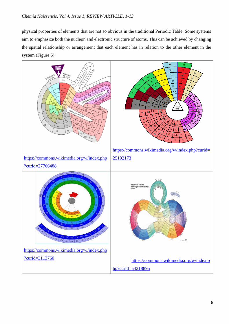
Chemia Naissensis, Vol 4, Issue 1, REVIEW ARTICLE, 1-13
6
physical properties of elements that are not so obvious in the traditional Periodic Table. Some systems
aim to emphasize both the nucleon and electronic structure of atoms. This can be achieved by changing
the spatial relationship or arrangement that each element has in relation to the other element in the
system (Figure 5).
https://commons.wikimedia.org/w/index.php
?curid=27766488
https://commons.wikimedia.org/w/index.php?curid=
25192173
https://commons.wikimedia.org/w/index.php
?curid=3113760
https://commons.wikimedia.org/w/index.p
hp?curid=54218895

Chemia Naissensis, Vol 4, Issue 1, REVIEW ARTICLE, 1-13
7
https://www.chemistryworld.com/news/does-the-periodic-table-make-ore-
sense-upside-down/3010360.article
Figure 5. Alternative Periodic Table of the Elements
Other systems have emphasized the isolation of chemical elements throughout history.
Timmothy Stowe created the physicist's periodic table. This table is a three-dimensional and the
three axes represent the principal quantum number, orbital quantum number, and orbital magnetic
quantum number. Helium is again a group 2 element (Bradley, 2011).
In 1984 an idea of a “structure map” was explored by Pettifor, who suggested that a well-
structured chemical space could be derived by changing the sequence of the elements in the periodic
table. He proposed a chemical scale that determines the “distance” between the elements on an one-
dimensional axis and a Mendeleev number (MN): an integer showing the position of an element in the
sequence. Pettifor claimed that binary compounds with the same structure type occupy the same region
in a two-dimensional map plotted using the MNs (the Pettifor’s map). He evaluated the chemical scale
by presenting a map clearly separating 34 different structure types of 574 binary AB compounds
(Pettifor, 1984).
Later, Pettifor showed that the MN approach also works for other AxBy compounds. Although
Pettifor derived the chemical scale and Mendeleev number empirically and based his assessment on
only several hundred binary compounds, his study provided a phenomenally successful ordering of the
elements confirmed in many later works. In this work, we denote Pettifor’s MN as MNP. We expect
that a nonempirical method of finding the MNs would perform even better (Pettifor, 1986).

Chemia Naissensis, Vol 4, Issue 1, REVIEW ARTICLE, 1-13
8
Villars et al. proposed a different enumeration of the elements (called periodic number, PN),
emphasizing the role of valence electrons. The atomic number (AN) of the elements together with their
‘periodic number’ (PN) were found to form an efficient pair for the discussion of metallurgical and
structural problems. The periodic number PN represents a different enumeration of the elements,
emphasizing the role of the valence electrons. In contrast to the atomic number, PN depends in detail on
the underlying Periodic Table of the elements. As a first result we describe the elemental-property
parameters ‘atomic size SZa’ and ‘atomic reactivity REa’, derived from fits to various experimental and
theoretical data sets. We argue that all elemental-property parameter patterns are derived from AN and PN.
AN and PN represent fundamental elemental-property parameters independent from each other. Any
pattern, which shows well-defined functional behavior within each group number GN, as well as within
each main quantum number QN, can be included. On the example of compound formers/non-formers in
binary, ternary and quaternary chemical systems we demonstrate that a quantitative link exists between
material properties and AN, PN (or simple functions of both) of the constituent element (Villars, 2008).
Glawe et al. proposed another sequence of elements (modified MN, in this work, we represent
this as MNm) based on their similarity, defining elements A and B to be similar if they crystallize in
the same structure type when combined with other elements of the periodic table. We believe that our
proposed 'modified Pettifor scale' can be of use not only for the representation of structure maps, but
also as a tool for both theorists and experimentalists to study possible chemical substitutions in the quest
for new materials with tailored properties (Glawe, 2016).
In a correctly defined chemical space, closely located materials should have similar properties.
The most promising materials will then be clustered in one or a few “islands” in this space. To predict
new materials, it could be sufficient to explore these as lands instead of the entire chemical space. The
fewer these islands are, the easier it would be to locate and explore them for promising materials. A
chemical space containing many small islands is less amenable for the prediction of materials than the
one with fewer big islands. Therefore, for evaluating each chemical space, it is useful to have these
islands and calculate the number of (similar) materials they cover. For doing this, we used the idea of
the clustering algorithm proposed by Rodriguez and Laio and applied it to clustering regions of the
chemical space based on their similarity (Rodriguez and Laio, 2014).
The precise placement of certain elements depends on which features we want to emphasize. The
last attempt to arrange the elements in this way was recently published in the journal Physical Chemistry
by scientists Zahed Allahyari and Artem R. Oganov. Their approach, building on the earlier work of
others, is to assign to each element what is called a Mendelian number (MN). There are several ways

Chemia Naissensis, Vol 4, Issue 1, REVIEW ARTICLE, 1-13
9
to derive such numbers, but the latest study uses a combination of two basic quantities that can be
measured directly: the atomic radius of the element and electronegativity (Festschrift, 2020).
In a well-ordered sequence of elements, the atoms with similar properties are close to each other.
Therefore, in the two-dimensional chemical space based on such a sequence, the properties of
neighboring binary systems should exhibit a close relation. On this premise, we evaluate different MNs:
atomic number (AN), Villars' periodic number (PN), Pettifor's Mendeleev number (MNP), modified
Mendeleev number (MNm), and Mendeleev numbers obtained in this work-the universal sequence of
elements (USE).
Only 1591 binary and 80 unary systems are studied in the database which is about half of the total
binary and unary systems that can be created from the combination of 80 elements; in total, 3240
systems can be created.
The number of clusters (i.e., islands) that cover all binary systems in the chemical spaces of the
MNs is a good quantitative evaluation of these MNs. The lower the number of clusters is, the better-
clustered the chemical space is.
Having a well-defined sequence of the elements (Mendeleev numbers, or MNs), where similar
elements take neighboring places, one can produce an organized map of properties for binary or more
complex systems that leads to the prediction of new materials by having information on their
neighboring systems. A simple, physically meaningful, and universal way to order the elements was
defined. MN (USE), in addition to a few previously known MNs such as atomic number (AN), Villars'
periodic number (PN), Pettifor's Mendeleev number (MNP), and modified Mendeleev number (MNm),
using provided data on binary systems from different databases, such as ICSD and COD, was examined.
Two dimensional maps of the hardness, magnetization, enthalpy of formation, and atomization energy
were plotted using the provided data in the space of MNs, and it turned out that most of these sequences
(except for AN) indeed work well for clustering materials with similar properties. The evaluation of the
MNs showed the overall best clustering rate of the chemical spaces produced by USE for target spaces,
i.e., hardness, magnetization, and enthalpy of formation. Also, unlike other MNs, USE can be defined
at any arbitrary pressure, which is a step forward for the prediction of materials under pressure. Physical
meaning of the Mendeleev number (previously defined empirically): it is a collapsed one-number
representation of the important atomic properties (such as atomic radius, electronegativity,
polarizability, and valence).
If we want to develop replacement materials that omit the use of certain elements, insights from
the arrangement of elements according to their MN can prove useful in that search. The importance of
the prevalence of elements used in the production of new materials is presented by the example of the

Chemia Naissensis, Vol 4, Issue 1, REVIEW ARTICLE, 1-13
10
periodic table in the figure below. This table not only illustrates the relative representation of the
elements (the larger the framework for each element, the more it has), but also highlights the potential
supply issue, relevant to technologies that have become ubiquitous and essential in our daily lives.
Figure 6. Periodic table showing the relative abundance of elements
https://theconversation.com/periodic-table-new-version-warns-of-elements-that-are-end
angered-110377
As part of the celebrations, the European Chemical Society published a completely new version
of the periodic table (Figure 6).
Each area of the new system is marked with a color that indicates its distribution. In most cases,
the elements are not lost, but as we use them, they fall apart and are much easier to recycle. Red indicates
that the elements will be much less available in 100 years or less. The orange and yellow surfaces on
the new periodic table predict problems caused by the increased use of these elements. Green means
that a large amount is available. The four elements - tin (Sn), tantalum (Ta), tungsten (W) and gold (Au)
- are colored black because they often originate from “conflicting” minerals; that is, from mines where
wars are fought over their ownership. Their color can have a more ethical meaning because it is a
reminder that producers must carefully monitor their origin to make sure that people are not harmed to
provide the minerals in question.
The three main ways to preserve some elements that are already at a minimum are: replace them
Chemia Naissensis, Vol 4, Issue 1, REVIEW ARTICLE, 1-13
11
with others, recycle them, or simply reduce their use. Huge efforts are being made to find alternative
materials. If we do not take these issues more seriously, many of the items and technologies we take for
granted today may be relics of old age after a few generations - or only available to wealthier people.
But as the new version of the periodic table underscores, we must do everything in our power to preserve
and recycle the first 90 precious elements that make up our wonderfully diverse world (Norman, 2020).
Is the resistance of chemists to changes related to the line boundaries of the standard Periodic
Table so great that they cannot accept other solutions, even when other tables offer a better
representation of basic chemical principles? Maybe it is just pragmatism. One cynical critic suggested
that the compressed version was favored because it fits well on a standard sheet of paper. Aesthetics are
important, but they always take the last place in relation to clarity. Ideally, the data in the table should
be visible and obvious.
One of the many virtues of the Periodic Table is that it brings simplification and coherence to the
world of chemistry. Since the Periodic Table is by definition based on recurring trends, any such table
can be used to obtain relationships between the properties of elements or to predict the properties of
others. Instead of knowing the properties of all 118 currently known elements, it is enough to gain
knowledge about the typical properties of about 10 of them. Therefore, the Periodic Table of the
Elements, whether in standard form or in some other variant, provides a useful framework for the
analysis of chemical behavior and is widely used in chemistry and other sciences.
Conclusion
Scientific theories are changing. The periodic table contains 118 elements and all rows and
columns are filled in. Is it complete and perfect? Laboratories around the world are synthesizing new,
even more difficult elements. Synthesis or the discovery of new weights raises the question of how much
the 150-year-old Periodic Table of the Elements can be modified to meet any new extensions. After so
many years since the creation of the Periodic Table, we can conclude that it is not only a basic educational
tool but is useful for researchers looking for new materials. New Periodic Table models should not serve
as replacements for previous views. Alternative periodic tables are developed often to highlight or
emphasize different chemical or physical properties of the elements which are not so obvious in
traditional periodic tables.
Acknowledgement
Authors want to thank Dr Biljana Arsić, Department of Chemistry, Faculty of Sciences and Mathematics,
University of Nis, Republic of Serbia for language corrections.

Chemia Naissensis, Vol 4, Issue 1, REVIEW ARTICLE, 1-13
12
Conflict-of-Interest Statement
None.
References
Bradley, D., (2011) At Last, A Definitive Periodic Table?, ChemistryViews.org.,
doi: 10.1002/chemv.201000107
Festschrift, A. B., Allahyari, Z., Oganov, A. R. (2020), Nonempirical Definition of the Mendeleev
Numbers: The Journal of Physical Chemistry virtual special issue, doi:10.1021/acs.jpcc.0c07857
Fricke, B., Greiner, W., Waber, J. T. (1971). The continuation of the periodic table up to Z = 172. The
chemistry of superheavy elements". Theoretica Chimica Acta. 21 (3), 235– 260.
Giguère, P. (1965) "The 'new look' for the periodic system". Chemistry in Canada vol. 18 (12), 36–39
Glawe, H., Sanna, A., Gros, E., Marques M. A. L., (2016) The optimal one-dimensional periodic table:
a modified Pettifor chemical scale from data mining, New Journal of Physic. 18 093011
IUPAC, Discovery and Assignment of Elements with Atomic Numbers 113, 115, 117 and 118,
December 30, 2015
Mazurs, E. G. (1974) Graphical Representations of the Periodic System During One Hundred Years.
University of Alabama Press, Alabama
Norman, N., (2020) Periodic table: scientists propose new way of ordering the elements
Pettifor, D. G., (1984) A Chemical Scale for Crystal-Structure Maps. Solid State Commun. 1984, 51,
31−34.
Pettifor, D. G., (1986), The Structures of Binary Compounds. I. Phenomenological Structure Maps.
Journal of Physic C: Solid State Physic. 19, 285−313.
Rodriguez, A., Laio, A., (2014) Clustering by fast search and find of density peaks, Science 344, 1492-
1496
Seaborg, G., (1946), Seaborg Announces Fissionable Neptunium, Chemical Engeenering News, 24, 20,
2764-2746
Villars, P., Daams, J., Shikata, Y., Rajan, S., Iwat, A., (2008) A new approach to describe elemental-
property parameters, Chemistry Metal Alloys 1, 1-23
https://www.thoughtco.com/how-to-use-a-periodic-table-608807
https://commons.wikimedia.org/wiki/File:Discovery_of_chemical_elements-en.svg
https://theconversation.com/the-periodic-table-is-150-but-it-could-have-looked-very-diffe
rent-106899

Chemia Naissensis, Vol 4, Issue 1, REVIEW ARTICLE, 1-13
13
https://theconversation.com/the-periodic-table-is-150-but-it-could-have-looked-very-different-106899
https://commons.wikimedia.org/w/index.php?curid=27766488
https://commons.wikimedia.org/w/index.php?curid=25192173
https://commons.wikimedia.org/w/index.php?curid=3113760
https://commons.wikimedia.org/w/index.php?curid=54218895
https://www.chemistryworld.com/news/does-the-periodic-table-make-more-
sense-upside-down/3010360.article

Chemia Naissensis, Vol 4, Issue 1, REVIEW ARTICLE, 14
14
Alternativni periodni sistem elemenata
Danijela Kostic1*, Nenad Krstic1, Marina Blagojevic1
1- Univerzitet u Nišu, Prirodno-matematički fakultet, Departman za hemiju, Višegradska 33, 18000 Niš,
Srbija
SAŽETAK
Više od 150 godina, od otkrica periodnog sistema elemenata, postojala je potreba za njegovim
stalnim dopunjavanjem i poboljšavanjem. Kao rezultat toga, danas postoji preko 700 različitih
periodnih sistema koji imaju za cilj da jednostavnije i efikasnije predstave položaj elemenata u
periodnom sistemu, njihove međusobne odnose i mogucnost građenja različitih jedinjenja. Pored
dvodimenzionalnih, pojavili su se i trodimenzionalni i četvorodimenzionalni sistemi elemenata.
Svi oni, pored velikog didaktičkog značaja, imaju i veliki naučni značaj i predstavljaju smernice
naučnicima u različitim multidisciplinarnim istraživanjima. Istraživanja savršenijih i
sveobuhvatnijih periodnih sistema se nastavljaju.
Ključne reči: periodni sistem, savremeni oblik periodnog sistema, alternativni oblik periodnog
sistema

Chemia Naissensis, Vol 4, Issue 1, REVIEW ARTICLE, 15
15
Tableau périodique alternatif des éléments
Danijela Kostic1*, Nenad Krstic1, Marina Blagojevic1
1- Université de Niš, Faculté des sciences naturelles et des mathématiques, Département de chimie,
Višegradska 33, 18000 Niš, Serbie
RÉSUMÉ
Depuis plus de cent cinquante ans, depuis la découverte du tableau périodique des éléments, il
a été nécessaire de le compléter et de l’améliorer constamment. En conséquence, il existe
aujourd’hui plus de sept cents tableaux périodiques différents qui visent à présenter plus
simplement et plus efficacement la position des éléments dans le tableau périodique, leurs
interrelations, ainsi que la possibilité de construire les différents composés. En plus des
tableaux bidimensionnels, sont apparus aussi des tableaux des éléments tridimensionnels et
quadridimensionnels. Tous ces tableaux, outre leur grande importance didactique, possèdent
également une grande valeur scientifique et représentent des lignes directrices pour les
scientifiques dans les diverses recherches multidisciplinaires. Les explorations des tableaux
périodiques plus parfaits et plus complets se poursuivent.
Mots-clés: tableau périodique, forme moderne du tableau périodique, forme alternative du
tableau périodique.

Chemia Naissensis, Vol 4, Issue 1, REVIEW ARTICLE, 16
16
Альтернативные периодические системы элементов
Даниела Костич1*, Ненад Крстич1, Марина Благоевич1
1- Университет в Нише, Естественно-математический факультет, Кафедра химии,
Вишеградска 33, 18000 Ниш, Сербия
АННОТАЦИЯ
Более 150 лет с момента открытия периодической таблицы элементов существовала
потребность в ее постоянном дополнении и улучшении. В результате этого, сегодня
существует более 700 различных периодических систем, которые стремятся более
просто и эффективно представить положение элементов в периодической таблице, их
взаимосвязи и возможность построения различных соединений. Помимо двухмерных,
появились как трехмерные, так и четырехмерные системы элементов. Все они, помимо
большого дидактического значения, также имеют большое научное значение и
представляют собой руководящие принципы для ученых в различных
междисциплинарных исследованиях. Продолжается поиск более совершенных и
всеобъемлющих периодических систем.
Ключевые слова: периодическая система, современная форма периодической системы,
альтернативная форма периодической системы.

Chemia Naissensis, Vol 4, Issue 1, REVIEW ARTICLE, 17
17
Alternative Periodensysteme der Elemente
Danijela Kostic1*, Nenad Krstic1, Marina Blagojevic1
1- Universität in Niš, Fakultät für Naturwissenschaften und Mathematik, Lehrstuhl für Chemie,
Višegradska 33, 18000 Niš, Serbien
ABSTRACT
Seit mehr als 150 Jahren, seit der Entdeckung des Periodensystems der Elemente, besteht die
Notwendigkeit, es ständig zu ergänzen und zu verbessern. Infolgedessen gibt es heute über 700
verschiedene Periodensysteme, die das Ziel haben, die Position der Elemente im
Periodensystem, ihre Beziehungen untereinander und die Möglichkeit der Bildung
verschiedener Verbindungen einfacher und effektiver darzustellen. Neben zweidimensionalen
sind auch drei- und vierdimensionale Periodensysteme erschienen. Sie alle haben neben ihrer
großen didaktischen auch eine große wissenschaftliche Bedeutung und stellen Leitlinien für
Wissenschaftler in verschiedenen multidisziplinären Forschungen dar. Die Forschung nach
vollkommeneren und umfassenderen Periodensystemen wird fortgesetzt.
Schlüsselwörter: Periodensystem, moderne Form des Periodensystems, alternative Form des
Periodensystems

Chemia Naissensis, Vol 4, Issue 1, RESEARCH ARTICLE, 18-35
18
The Influence of Metal Microelements, Colloids and Organic Phase on
Physical-chemical Properties and Processes in Peloids
Jelena M. Purenović1*, Milovan M. Purenović2, Marjan S. Ranđelović2
1-University of Kragujevac, Faculty of Technical Sciences, Svetog Save 65, Čačak, Serbia
2-University of Niš, Faculty of Sciences and Mathematics, Department of Chemistry, Višegradska 33,
Niš, Serbia
ABSTRACT
The main emphasis in this study was on the modification of peloid characteristics through
maturation processes, physical-chemical analysis of salty geothermal water and intact
geomaterial, content of toxic heavy metals, radionuclides, and microorganisms in matured
peloid, and physical-chemical processes that occur in a highly heterogeneous and
microheterogeneous system solid-water. Main processes were considered to be mass transfer,
colloidal processes, adsorption and surface compounding by macro- and micronutrients from
salty mineral water with surface groups of intact geomaterial. This study indicated that
inorganic and organic components of peloid could be in the form of colloids, suspended
macro- and microparticles, ions and molecules. Colloidal silica had special importance in
peloids. Due to low maximum solubility of silica, there were a number of processes in which
coagulated and floculated particles were created during maturation, especially in the presence
of metal cations (e.g., Fe3+ and Al3+) and colloidal metal hydroxides which noticeably reduced
the solubility of silica. Single charged alkali metal cations caused coagulation of colloidal
silica occupying bridging positions between negatively charged colloidal particles. Colloidal
silica in peloid together with other micro- and macro phases, and with the help of numerous
microelements, comes in interaction, building a complex surface and occluded compounds. In
the multiphase system, very complex organic and inorganic compunds are formed, which are
important for therapeutic purposes.
Keywords: Peloid, Macro/micro elements, Thermo mineral water, Colloidal particles.
Jelena M. Purenović: [email protected]
Milovan M. Purenović: [email protected]
Marjan S. Ranđelović: [email protected]

Chemia Naissensis, Vol 4, Issue 1, RESEARCH ARTICLE, 18-35
19
Introduction
The term “peloid” usually refers to the natural healing mud, which is a multi-component
macro- and micro-heterogeneous system consisting of mineral water, colloidal clay minerals,
organic matter, organic-mineral complexes, etc. According to The International Society of
Medical Hydrology, peloid can be defined as a natural product consisting of a mixture of sea,
salt lakes, or mineral-medicinal water (liquid phase), with organic and inorganic material
(solid phase) produced by biological action (humus) and geological action (clay minerals).
Peloids are used in “pelotherapy” for local or generalized recovering from rheumatism,
arthritis, and bone-muscle traumatic damages. The main factors which determine peloid
characteristics and its suitability for therapy are: composition, granulometry, geochemistry of
mineral water, low-cooling rate, high ion exchange capacity, good adhesivity, ease of
handling and pleasant sensation when applied to the skin (Veniale et al., 2004). Today it is
known that peloids accelerate blood circulation and metabolism, affect the activity of some
endocrine glands, and soothe tension and pain, primarily due to the thermal effect. They also
have a beneficial effect on chronic rheumatic diseases, and are used for cosmetic purposes, for
example, to remove cellulite. Moreover, some studies have confirmed that peloids possess
antimicrobial, antiviral, antineoplastic and anti-inflammatory effects (Suárez et al., 2011).
Primary or secondary mixing of clayey (geo)materials with salty thermo-mineral waters,
accompanied by organic materials produced by the metabolic activity of micro-organisms
growing during the so-called “maturation” process, is a very common procedure for
improving and stabilizing the therapeutic properties of the final product-peloid (Sanchez et
al., 2002; Veniale et al., 2007). It is established that the maturation process improves some
physical properties of the clay minerals, such as heat retention capacity, rheology, and
adhesion. Some studies report that maturation leads to a decrease in grain size, while both
mineralogy and chemistry are almost unchanged (Summa and Tateo, 1998).
However, for practical reasons, some studies suggest use of extemporaneous peloids,
whose preparation procedure assumes a contact between both phases (clayey material and
salty thermo-mineral waters) for around 48 h, instead of several months or up to two years in
the case of usual maturation process (Carretero et al., 2006; Gámiz et al., 2009). Organic
substances as well as macro- and micronutrient elements present in water are taken up by the
peloid during interaction of phases and can be released during application to the human body
(Carretero et al., 2010; Gámiz et al., 2009).

Chemia Naissensis, Vol 4, Issue 1, RESEARCH ARTICLE, 18-35
20
Geothermal water from Bujanovačka Spa (south of Serbia) is highly mineralized,
hyperthermal (43°C), sodium, hydrogen carbonate, fluoride, sulfate, and carbonic-acid rich
(http://bujanovackabanja.org/) . Uniqueness of this spa is in the specific combination of three
natural factors: therapeutic thermal waters, peloids and carbon dioxide having a purity of
about 98%. During pelotherapy, mud exhibits three effects: mechanical, thermal, and
pharmacological. Peloids can be used raw or after passing maturation process, but also as a
mixture of mud and paraffin and for preparation of cosmetics (http://bujanovackabanja.org/) .
Experimental
This paper focused on: i) detailed physical-chemical and elemental analysis of salty
geothermal water and virgin geomaterial used for peloid maturation process; ii) content of
toxic heavy metals, radionuclides, and microorganisms in matured peloid, and iii) processes
that took place in a highly heterogeneous and microheterogeneous system solid-water. To
date, the latter aspect was very little studied and/or considered in detail.
Virgin clay (native clayey geomaterial for peloid preparation) and thermo-mineral
water were the basic ingredients used for the peloid preparation through “maturation”
treatment. It was clayey mineral-rich geomaterial of volcanic origin, which was mixed in open
air with salty thermo-mineral water, undergoing a maturation process. Already prepared
peloid and thermo-mineral water were analyzed without further modifications. All used
chemicals were of analytical grade.
Bearing in mind the subject and purpose of this work, program and research
methodology included the following:
Physical and physical-chemical properties of geothermal mineral water (pH,
electrical conductivity, free CO2, alkalinity, metal cations, anions, etc.) were
analyzed by the accredited laboratories using the standard methods. These
important parameters and factors influenced the ratio of ionic and colloidal states of
different ingredients. Details regarding the used analytical methods were provided
in Table 1. These analyses were courteously performed at the Institute of Public
Health of Serbia “Dr Milan Jovanović Batut”, Belgrade, using adequate analytical
methods.

Chemia Naissensis, Vol 4, Issue 1, RESEARCH ARTICLE, 18-35
21
Determination of the chemical composition of mineral water for the dumping of
peloids, the content of fluoride, chloride, nitrite and nitrate, phosphate, sulfate, Na,
K, Mg, Ca, Fe, Mn, Cu, Zn, Pb, Cr, Cd, Co, Ba, Si, Ni, As, Hg, Se, Sb, Al and Sn;
Content of the weak electrolyte metasilicicacid (H2SiO3) and metaboric (HBO2)
acid.
Determining the content of radionuclides in appropriate samples.
EDXRF (Energy Dispersive X-ray Fluorescence) spectrometry technique using
radioisotopes 241Am and 109Cd as the excitation sources was employed for
determination of chemical compositions of two different samples (184/9V and
183/8V), including macro- and microelements, in salty thermo-mineral water and
the virgin clay for peloid preparation. The EDXRF spectra were collected using a
modified method EPA 6200. These analyses were courteously performed at the
“Vinča” Institute of Nuclear Sciences, Belgrade.
Macro- and microelements in the virgin clay were determined following the
microwave digestion with HNO3 and H2O2. Heavy metals in the samples of the
matured peloid were analyzed after their extraction by boiling in the 0.1 M HCl
during 15 min (method: AEL M01-05).
Content of relevant radionuclides in the samples was determined using high-
resolution gamma-ray spectroscopy with HPGe detector, according to ISO
10703:1997 and ASTMC1402-98.
These analyses were courteously performed at the Institute of Chemistry,
Technology and Metallurgy – ICTM using adequate analytical methods.
All samples for testing, indicated by numbers, were taken in accordance with the
standards and were taken on the spot by expert of accredited laboratory, therefore,
under objective conditions.
Results and Discussion
The results of EDXRF spectrometry with the use of radioactive isotopes 109Cd and 241Am, for samples 184 / 9V and sample 183 / 8V
The qualitative analysis of thermo-mineral water (sample 184 / 9V) by non-destructive
EDXRF spectrometry (Figure 1) showed the presence of the following elements: S, Cl, K, Ca,
Fe, Ni, Cu, Pb, Br, Rb, Sr, Ag, Sn, Sb, Cs and Ba. The size of the peaks indicated that K and

Chemia Naissensis, Vol 4, Issue 1, RESEARCH ARTICLE, 18-35
22
Ca were macroelements, and, besides them, the most common were S and Cl. Other detected
elements were present in a smaller proportion, or in trace quantities. Marked small peak,
observed in Figure 1a, energetically corresponded to Hg (L line 9.9 keV) or germanium (K
line 9.87 keV). Hence, in the absence of other/additional lines, it could not be unambiguously
specified which one occurred in water. Moreover, there was a possibility that both of them
were simultaneously present or the peak could be the result of some other weaker peaks
summation.
a) b)
Figure 1. EDXRF spectra of salty geothermal water acquired by using a) 109Cd and b) 241Am
as the excitation sources.
As Figure 2 revealed, the elements detected in the EDXRF spectra of virgin clay (sample
183 / 8V) were as follows: Si, K, Ca, Ti, Mn, Fe, Ni, Zn, Pb, Rb, Sr, Y, Zr, Ag, Sn, Ba and
Ce. Moreover, by considering the size of peaks in the spectra, it could be concluded that the
most common elements were Si, K, Ca and Fe, while other elements were present in smaller
proportion, or in trace amounts. On the other hand, the peaks of Fe, Ni, and Pb (for 109Cd) and
Ag, Pb, Sn, and Ba (for 241Am) could be aroused as a result of the influence of background
radiation. Therefore, on the basis of EDXRF results, the possible presence of these elements
in the sample could not be excluded.

Chemia Naissensis, Vol 4, Issue 1, RESEARCH ARTICLE, 18-35
23
a) b)
Figure 2. EDXRF spectra of virgin clayey geomaterial acquired by using a) 109Cd and b)
241Am as the excitation sources.
The results of sensory, physical, and physical-chemical analysis of water for the sinking
of the curative mud, for samples with the identification mark 184 / 9V and sample 183 /
8V
Physical-chemical parameters and quantitative analysis of cations and anions of
thermomineral water and virgin clay were summarized in Tables 1 and 2, respectively.
Table 1. Physical and physical-chemical analysis of thermomineral water (sample 184/9V).
No Analyzed parameter Unit Result Analytical method
1. Smell - no /
2. pH - 7.55 SRPS H.Z1.111.:1987
3. Electrical conductivity µS/cm 4500 EPA 120.1:1982
4. Dry residue at 105ºC mg/dm3 3180 Handbook1) P-IV-7
5. Hydrogen sulphide (H2S) mg/dm3 - /
6. Free CO2 mg/dm3 55.3 /
7. KMnO4 consumption mg/dm3 0.69 SRPS EN ISO 8467:2007
8. Alkalinity (p-alkalinity) meq/dm3 0 SRPS EN ISO 9963-1:2007
9. Alkalinity (m-alkalinity) meq/dm3 50.2 SRPS EN ISO 9963-1:2007
10. Fluoride (F-) mg/dm3 < 1.0 EN ISO 10304-1:1995

Chemia Naissensis, Vol 4, Issue 1, RESEARCH ARTICLE, 18-35
24
11. Chloride (Cl-) mg/dm3 55.2 SRPS-ISO 9297:1997
SRPS-ISO 9297:2007
12. Nitrite (NO2-) mg/dm3 0.016 APHA-Method 4500-NO2
-
13. Nitrate (NO3-) mg/dm3 5.8 EN ISO 10304-1:1995
14. Phosphate (PO43-) mg/dm3 0.046 Handbook1) P-V-16/A
15. Sulphate (SO42-) mg/dm3 200 EN ISO 10304-1:1995
16. Sodium (Na+) mg/dm3 726 APHA-Method 3111-B, 1999
17. Potassium (K+) mg/dm3 49 APHA-Method 3111-B, 1999
18. Magnesium (Mg2+) mg/dm3 8.02 SRPS H.Z1.181:1985
19. Calcium (Ca2+) mg/dm3 41.6 SRPS H.Z1.181:1985
20 Strontium mg/dm3 1.5 /
21. Total hardness as CaCO3 mg/dm3 156 AEL M02-01
22. Iron (Fe) mg/dm3 0.36 APHA-Method 311-B, 1999
23. Manganese (Mn) mg/dm3 0.05 APHA-Method 311-B, 1999
24. Copper (Cu) mg/dm3 < 0.03 APHA-Method 311-B, 1999
25. Zinc (Zn) mg/dm3 0.018 APHA-Method 311-B, 1999
26. Lead (Pb) mg/dm3 < 0.19 APHA-Method 311-B, 1999
27. Chromium (Cr) mg/dm3 < 0.13 EPA-Method 218.1, 1978
28. Cadmium (Cd) mg/dm3 < 0.014 APHA-Method 311-B, 1999
29. Cobalt (Co) mg/dm3 < 0.05 APHA-Method 311-B, 1999
30. Barium (Ba) mg/dm3 < 1.0 EPA-Method 208.1, 1974
31. Nickel (Ni) mg/dm3 < 0.06 APHA-Method 311-B, 1999
32. Silicon (Si) mg/dm3 57.5 Handbook P-V-40/A
33. Arsenic (As) µg/dm3 72 SRPS ISO 11969:2002

Chemia Naissensis, Vol 4, Issue 1, RESEARCH ARTICLE, 18-35
25
34. Mercury (Hg) µg/dm3 < 1.0 BS EN 1483:2007
35. Selenium (Se) µg/dm3 < 0.3 APHA-Method 3114-B, 1999
36. Antimony (Sb) µg/dm3 2.5 AEL M-02
37. Aluminum (Al) mg/dm3 < 1.5 /
38. Tin (Sn) µg/dm3 < 0.5 AEL M02-03
30. Metasilicic acid mg/dm3 118.00 /
31. Metaboric acid mg/dm3 17.00 /
32. Total dissolved solids mg/dm3 4983.60 /
Peloids acted as ionic mediators on human skin. The pathway of incorporation of
exchangeable ions and other compounds existing in peloids in the human body, applied for
therapeutic and cosmetic purposes, was the absorption through skin (dermal absorption). For
this reason, concentration of toxic heavy metals and other harmful components in peloids
should be at the level that does not cause damage to human health (Gerencsér et al., 2010;
Vreca and Dolenec, 2005). To date, only the maximum acceptable concentrations established
for soils and cosmetics could be used as guidelines for healing mud and peloids. For example,
in December 2008, Health Canada released Draft Guidance on Heavy Metal Impurities in
Cosmetics, which outlined recommended impurity limits in cosmetic products for lead (10
ppm), arsenic (3 ppm), cadmium (3 ppm), mercury (3 ppm), and antimony (5 ppm)
(https://www.canada.ca/en/health-canada/consumer-product-safety/reports-
producations/industry-professionals/guidance-heavy-metal-impurities-cosmetics.html).
In Europe there was no specific legislation and regulation regarding maximum acceptable
concentrations of potential toxic elements in peloids. The European Cosmetics Directive
(76/768) and the new Cosmetics Products Regulation (1223/2009) allowed "the non-intended
presence of small quantities of a prohibited substance, stemming from impurities of natural or
synthetic ingredients, the manufacturing process, storage, migration from packaging, which
was technically unavoidable in good manufacturing practice, provided that such presence did
not cause damage to human health when the product was applied under normal or reasonably
foreseeable conditions of use" (European Union, 2009).

Chemia Naissensis, Vol 4, Issue 1, RESEARCH ARTICLE, 18-35
26
Table 2. Chemical analysis of raw/virgin clayey geomaterial for peloid formulation (sample
183/8V).
No Analyzed parameter Unit Result
Guideline
value1
Microwave digestion with HNO3 and H2O2
1 Lead, Pb mg/kg 69.1 < 30.0
2 Cadmium, Cd mg/kg 2.20 < 5.0
3 Chromium, Cr mg/kg 49.8 < 50.0
4 Arsenic, As mg/kg 9.2 < 5.0
5 Mercury, Hg mg/kg 2.98 < 10.0
6 Nickel, Ni mg/kg 42.5 < 50.0
7 Manganese, Mn mg/kg 562 -
8 Cobalt, Co mg/kg 13.7 -
9 Zinc, Zn mg/kg 184.5 -
10 Sodium, Na mg/kg 22900 -
11 Potassium, K mg/kg 2770 -
12 Magnesium, Mg mg/kg 4500 -
13 Calcium, Ca mg/kg 347.5 -
14 Iron, Fe mg/kg 2100 -
15 Copper, Cu mg/kg 59.2 -
16 Tin, Sn mg/kg < 0.02 -
17 Selenium, Se mg/kg < 0.02 -
18 Antimony, Sb mg/kg 1.35 -
1) Mark * refers to the non-accredited method

Chemia Naissensis, Vol 4, Issue 1, RESEARCH ARTICLE, 18-35
27
As Tables 1 and 2 described the amount of heavy metal cations in the matured peloid mud
was lower with respect to virgin. When geothermal water was mixed with the virgin clayey
geomaterial, content of heavy metals decreased due to ion exchange, hydrolysis, partial
dissolution of minerals and other phenomena that will be discussed in detail. The importance
of ion exchange mainly refers to major elements, such as alkaline or alkaline earth metal
cations (Ca2+, Mg2+, K+ and Na+), between geothermal water and the clay. Due to their high
concentrations in geothermal water, major elements could replace potentially toxic cations in
virgin geomaterial, resulting in the final peloid with significantly lower content of heavy
metals.
The results of chemical analysis of thermal mineral water, referring to the characteristic
cations, anions, and weak electrolytes
The water temperature at the source was 42 °C, its pH was 7.2, specific mass 1.002, and
belongs to category of sodium hydrocarbonate water, fluoride, sulfide and carbonic acid
hyperthermia. Dry residue at 180°C was 3.2, and the total mineralization 4.996 g/dm3 in its
composition was given in Table 3.
Table 3. The results of chemical analysis of thermal mineral water, referring to the
characteristic cations, anions and weak electrolytes.
Cations
Content
(mg/dm3)
Sodium 1132.00
Potassium 66.00
Calcium 42.00
Magnesium 11.00
Strontium 1.50
Iron 0.70

Chemia Naissensis, Vol 4, Issue 1, RESEARCH ARTICLE, 18-35
28
Anions
Hydrocarbonate 3355.00
Chloride 56.00
Fluoride 0.70
Sulphate 183.70
Weak electrolytes
Metasilicic acid 118.00
Metaboric acid 17.00
The sum of all soluble ingredients 4983.60
The results of radiological tests
Radioactive phases could produce ionising radiations that could be dangerous when
exceeding certain levels. Results of radiological study of thermomineral water, virgin clay and
peloid samples were comparatively shown in Table 4. The content of radionuclides in the
samples was below the limits of radioactive contamination, established for medicinal and
cosmetics products based on natural raw materials.
Table 4. The results of radiological examination of mineral water.
Sample
40K
(Bq/kg)
232Th
(Bq/kg)
226Ra
(Bq/kg)
137Cs
(Bq/kg)
238U
(Bq/kg)
235U
(Bq/kg)
Mineral
water
1.4±0.1 < 0.2 < 1.1 < 0.1 < 0.8 < 0.4
Virgin
geomate
rial
276±8 22±1 20±2 < 0.1 12±4 < 0.6
Peloid 388±11 29±1 27±2 < 0.1 18±4 < 0.9

Chemia Naissensis, Vol 4, Issue 1, RESEARCH ARTICLE, 18-35
29
Diagnostic criteria established on the basis of the analysis of obtained results
By the analysis of the research results, and bearing in mind the review of literature and
theoretical knowledge in the field of colloids and colloidal state, as an important prerequisite
for the necessary physical-chemical and medicinal properties of the curative mud, the
following diagnostic criteria were achieved:
Medicinal waters, by definition, belonged to the category of sodium hydrocarbonate,
fluoride, sulfate and carbonic acid hypothermia, with a temperature of 42ºC;
Thermomineral waters had a very high value of dry residue of 3.2 g/dm3, at 180°C and
high mineralization value in an amount of 4.983 g/dm3;
According to the content of cations, anions and weak electrolytes (Table 3) and total
chemical composition of mineral water for peloid sinking (Table 1), it could be seen
that water contained predominantly sodium and potassium, followed by calcium and
magnesium, as macrocomponents;
Concerning the anions present, the water was rich with HCO3- ions (3355 mg/dm3),
which was mainly related to the alkali and alkaline earth metals;
The presence of Cl- and SO42- ions was noted, and F- ion in very small quantities of 70
g/dm3;
Metasilicic and metaboric acid were found in high quantity, far above the solubility
limit;
Thermal water contained very small quantities of aluminum (Al) 1500 mg/dm3,
arsenic (As) 72 mg/dm3 and silicon (Si) 57.5 mg/dm3;
Wet mud contained a number of heavy metals and others; some in traces (Cd, As, Hg,
Ni, Sn, and Se), others in microquantities (Pb, Cr, Mn, Co, Sb, and Zn), as well as
macrocomponents (Na, K, Mg, Ca and Fe);
EDXRF spectra of geothermal water for the peloid preparation (Figure 1) revealed the
following elements: S, Cl, K, Ca, Fe, Ni, Cu, Pb, Br, Rb, Sr, Ag, Sn, Sb, Cs and Ba;
Based on the size of the peaks of EDXRF spectra for geothermal water, it was
concluded that the most common elements were K, Ca, Ni, Cu, Sr, Cl and S, while the
other detected elements were present in a smaller share or trace;
In EDXRF spectrum of geothermal water a small peak was detected, which
corresponded to the Hg (L line 9.9 keV) or germanium (K line 9.87 keV), which in

Chemia Naissensis, Vol 4, Issue 1, RESEARCH ARTICLE, 18-35
30
this case, due to the lack of other lines, could not be determined with certainty (Figure
1);
In the EDXRF spectra related to crude mud, which did not undergo the process of
ripening, the following elements were detected: Si, K, Ca, Ti, Mn, Fe, Ni, Zn, Pb, Rb,
Sr, Y, Zr, Ag, Sn, Ba and Ce (Figure 2);
Due to the size of the peaks on EDXRF spectra for raw peloid, it could be concluded
that in this sample the most frequent elements were Si, K, Ca and Fe, while the other
elements were present in a smaller proportion, i.e., in traces, as microelements;
On the other hand, peaks Ni, Pb, Ag, Sn and Ba in the spectra of raw peloid occurred
because of the influence of background from the instrument, so the possible presence
of these elements in the sample could not be ruled out.
Discussion of possible chemical interactions and processes in heterogeneous systems of
peloid
Previous research on peloids primarly focused on their curative and ion-exchange
characteristics, paying no attention to other physical-chemical processes and colloidal
phenomena in the peloid, and even ignoring a number of microcomponents which had
considerable influence on the consistency of peloids. Physico-chemical processes that most
likely occurred in peloid during the maturation process were as follows:
Water retention, i.e., retention on the external surface of solid particles and within the
interlayer space of swelling clay minerals as well as hydration and dehydration of the
surface groups on solid surfaces;
Adsorption and absorption of ions and molecules on solid surfaces and within pores;
Coagulation - floculation processes;
Ion exchange process between mineral water and aluminosilicates;
Reactions between adsorbates and adsorbents in which surface complexes were
formed;
Polymerization of silica aid monomers;
Biochemical decomposition of organic phase and its mineralization;
Generation of metabolites of some colonizing organisms;
Creating inorganic and organic sols and gels;
Processes of mass exchange;
Formation of crystallization centers and solid phases of new deposits;

Chemia Naissensis, Vol 4, Issue 1, RESEARCH ARTICLE, 18-35
31
Oxido-reduction processes.
The peloid was extremely macro and micro heterogeneous solid-water system which
contained soluble inorganic and organic components in the form of colloids, suspended
particles, ions and molecules. Dominant constituents of the peloid were clay minerals,
coagulated inorganic and organic sols, complexes of heavy metals with organic ligands,
chelates, aqua complexes, hydroxide gels and various metal species (Al, Mg, Ca, Fe); a
number of suspended solid particles of silicates, carbonates, sulphates and silica; soluble ions
of many cations and anions; chemical compounds in the form of inorganic and organic
molecules and polymeric forms of silica. The structures of the most probable colloidal
micelles in peloid could be shown as follows.
{m[(SiO2)]nSiO32- · 2(n-x)Na+} 2xNa+ (I)
{m[M2(SiO3)p]nSiO32- · (2/p)(n-x)Mp+} (2/p)xMp+ (II)
where Mp+ was general symbol for the cations (e.g., Fe3+, Al3+, Ca2+, Mg2+, etc.). Cations
adsorbed on the surface of colloidal silica species acted as cristalization centers for
carbonates, sulphates of calcium and other deposits that could be formed in water.
This complex system, with dominant interfacial mass exchange, was out of equilibrium
and unstable. For this reason, colloids tended to be destabilized through coagulation-
floculation process, forming precipitate or gelatinous state. Due to low maximum solubility of
silica, there were a number of processes in which coagulated and floculated particles were
created, especially in the presence of metal cations (e.g., Fe3+ and Al3+) and colloidal
hydroxides which noticeably reduced the solubility of silica. Research in this work focused
precisely on the impact of colloidal silica on a number of phenomena and processes in
peloids. Depending on the crystallographic modification of crystalline SiO2, solubility in
water was in the range of from 70 to 120 mg/dm3. Thus, for example, at extremely low
concentrations of Fe and Al, the solubility of silica was drastically reduced.
For instance, the addition of Fe3+ ions in a solution of Si(OH)4 significantly delayed the
polymerization process.
(-SiOH)m + Fe3+ → (-SiOH)m-n(-SiO)nFe3-n + nH+ (III)
2Si(OH)4 + 2Al3+ + H2O → Al2Si2O5(OH)4 + 6H+ (IV)
Within the peloid, interaction of monomer Si(OH)4 with metal hydroxides or oxides could not
be avoided. This interaction may be depicted by the following equation:

Chemia Naissensis, Vol 4, Issue 1, RESEARCH ARTICLE, 18-35
32
(V)
Deposition of colloidal silica was influenced by floculating ions and it was significantly
diminished if metal cations were previously complexed with the chelate ligands.
The presence of Cl- ions in geothermal water could significantly stabilize colloidal state,
acting as an anticoagulant. On the other hand, the F- ion had a major impact on the creation
and structure of numerous suspended particles and easily transformed ortosilicic acid into
H2SiF6 in accordance with the following mechanism:
Si(OH)4 + 6HF → 2H+ + SiF6- + 4H2O (VI)
Since it was not possible to avoid the presence of the organic phase in the structure of
peloids, and this was not desirable, its presence ensured the creation of molecular solution and
organic sols, and in interactions with other micro and macro components chelate a number of
organic complexes could be formed, as well as silica-organic compounds.
Water used in the preparation of peloid had a high concentration of sodium, which was
available not only for the above reaction, but also for the formation of new phases, such as
trona (Na3H(CO3)2 ∙ 2H2O), seen in Figure 3. Previous processes which lead to formation of
solid deposits caused a considerable change in the chemical composition of geothermal water
(a liquid phase in peloid).

Chemia Naissensis, Vol 4, Issue 1, RESEARCH ARTICLE, 18-35
33
Figure 3. XRD pattern of salty geothermal water.
The main clay minerals in peloid were smectites and minerals based on metal oxides and
hydroxides. Smectite was a very good absorber of water, whereby it swelled ensuring high
water retention capacity (Cara et al., 2001). Noteworthy, smectite possessed high content of
variable exchangeable cations, which could largely vary in hydration degree (Sparks, 2002),
thus influencing moisture and water diffusion. A multicomponent mixture in the form of pulp,
saturated with H2S from geothermal waters, upon the occurrence of redox processes of
ripening (biochemical processes), could create peloid of strongly mineralized structure, which
had good healing and plastic qualities, important for its implementation.
Conclusion
Peloids were considered as highly complex and heterogeneous systems, composed of
minerals, amorphous inorganic materials, and organic matter. Therapeutic and curative
characteristics of peloid were realized through thermal effects and ion exchange properties in
contact with human skin. Due to the potential human toxicity or radiation risks during
pelotherapy, this study has involved detailed physical-chemical and radiological analysis of
mineral water, virgin geomaterial and final peloid from Bujanovačka Spa. All tested
parameters have fulfilled the requirements, according to Serbian regulations.
High mineralized water was extraordinarily suitable for the preparation of peloids.
Presence of silicic acid, as well as Al3+, Fe3+, F- and trace elements, were reflected in the
extraordinary process within the peloid at phase boundaries solid-water. Moreover, clay
minerals and silicic acid had a key role in processes of hydration and dehydration. Polymer
form of silicic acid significantly affected the formation of solid phase and deposits in the form
of layers or clusters within the peloids and mediated mass exchange processes between human
body and peloids.
The presence of metal cations and hydroxides, anions, clayey material, and organics in
ionic, molecular, or colloidal state gave rise to many processes and interactions between all
mentioned constituents, forming chelates, organosols, organic complexes with metal cations
and polymeric forms of various inorganic and organic compounds.
Peloids should be active, but stable colloidal systems, so, in that sense, a harmonious
correlation between the added macro and micro elements and the process of creating peloids
was established in this paper.

Chemia Naissensis, Vol 4, Issue 1, RESEARCH ARTICLE, 18-35
34
Acknowledgement
This research is a part of investigations performed within the scope of the project 451-03-
9/2021-14/200132. The authors gratefully acknowledge the financial support of Serbian
Ministry of Education, Science and Technological Development.
Conflict-of-Interest Statement
None.
References
Cara, S., Carcangiu, G., Padalino, G., Palomba, M., & Tamanini, M. (2000). The bentonites in
pelotherapy: Thermal properties of clay pastes from Sardinia (Italy). Applied Clay Science,
16, 125–132.
Carretero, M. I., Gomes, C. S. F., & Tateo, F. (2006). Clays and human health. In F. Bergaya,
B. K. G. Theng, , G. Lagaly, (Eds.), The Handbook of Clay Science, Developments in Clay
Science, (vol. 1, pp. 717-741). Amsterdam: Elsevier.
Carretero, M. I., Pozo, M., Martín-Rubí, J. A., Pozo, E., & Maraver, F. (2010). Mobility of
elements in interaction between artificial sweat and peloids used in Spanish spas. Applied
Clay Science, 48, 506–515.
Cosmetics Products Regulation (1223/2009): Official Journal of the European Union
Regulation (EC) No 1223/2009 of the European parliament and of the council of 30
November 2009 on cosmetic products (recast) (Text with EEA relevance).
Gámiz, E., Martín-García, J. M., Fernández-González, M. V., Delgado, G., & Delgado, R.
(2009). Influence of water type and maturation time on the properties of kaolinite-saponite
peloids. Applied Clay Science, 46, 117–123.
Gerencsér, G., Murányi, E., Szendi, K., & Varga, C. (2010). Ecotoxicological studies on
Hungarian peloids (medicinal muds). Applied Clay Science, 50, 47–50.
http://bujanovackabanja.org/
http://www.hc-sc.gc.ca/cps-spc/pubs/indust/heavy_metals-metaux_lourds/index-eng.php
Sanchez, C. J, Parras, J., & Carretero, M. I. (2002). The effect of maturation upon the
mineralogical and physicochemical properties of illitic-smectitic clays for pelotherapy. Clay
Minerals, 37, 457–463.
Chemia Naissensis, Vol 4, Issue 1, RESEARCH ARTICLE, 18-35
35
Sparks, D. (2002). Environmental soil chemistry. (2nd ed.). Academic Press.
Suárez, M., González, P., Dominguez, R., Bravo, A., Melián, C., Pérez, M., Herrera, I.,
Blanco, D., Hernández, R., & Fagundo, J. R. (2011). Identification of organic compounds in
San Diego de los Banos Peloid (Pinar del rio, Cuba). The Journal of Alternative and
Complementary Medicine, 17(2), 155-165.
Summa, V., & Tateo, F. (1998). The use of pelitic raw materials in thermal centres:
Mineralogy, geochemistry, grain size and leaching tests. Examples from the Lucania area
(southern Italy). Applied Clay Science, 12, 403–417.
Veniale, F., Barberis, E., Carcangiu, G., Morandi, N., Setti, M., Tamanini M., & Tessier D.
(2004). Formulation of muds for pelotherapy: effects of “maturation” by different mineral
waters. Applied Clay Science, 25, 135– 148.
Veniale, F., Bettero, A., Jobstraibizer, P. G, & Setti, M. (2007). Thermal muds: Perspectives
of innovations. Applied Clay Science, 36, 141–147.
Vreca, P., & Dolenec, T. (2005). Geochemical estimation of copper contamination in the
healing mud from Makirina Bay, central Adriatic. Environment International, 31, 53– 61.

Chemia Naissensis, Vol 4, Issue 1, RESEARCH ARTICLE, 36
36
Uticaj metalnih mikroelemenata, koloida i organske faze na fizičko-
hemijska svojstva i procese u peloidima
Jelena M. Purenovic1*, Milovan M. Purenovic2, Marjan S. Ranđelovic2
1- Univerzitet u Kragujevcu, Fakultet tehničkih nauka, Svetog Save 65, Čačak, Srbija
2- Univerzitet u Nišu, Prirodno-matematički fakultet, Odeljenje za hemiju, Višegradska 33, Niš, Srbija
SAŽETAK
Glavni akcenat u ovom proučavanju bio je na modifikovanju peloidnih karakteristika kroz
procese sazrevanja, fizičko-hemijskoj analizi slane geotermalne vode i netaknutog
geomaterijala, kao i na sadržaju toksičnih teških metala, radionukleida i mikroorganizama u
zrelim peloidima i fizičko-hemijskim procesima koji se javljaju u visoko heterogenom i
mikroheterogenom sistemu čvrsto-voda. Smatra se da su glavni procesi: prenos mase, koloidni
procesi, adsorpcija i usložavanje površine makro i mikro elementima iz slane mineralne vode
sa površinskim grupama izvornog geomaterijala. Ova proučavanja su pokazala da neorganske
i organske komponente peloida mogu biti u obliku koloida, suspendovanih makro i mikro
čestica, jona i molekula. Koloidni silicijum-dioksid imao je poseban značaj u peloidima. Zbog
niske maksimalne rastvorljivosti silicijum-dioksida, postoji više procesa u kojima su tokom
zrenja nastale koagulisane i flokulisane čestice, posebno u prisustvu metalnih katjona (npr. Fe3+
i Al3+) i koloidnih hidroksida metala koji su primetno smanjili rastvorljivost silicijum-dioksida.
Jednonaelektrisani katjoni alkalnih metala prouzrokovali su koagulaciju koloidnog silicijum-
dioksida zauzimajuci mostne položaje između negativno naelektrisanih koloidnih čestica.
Koliodni silicijum dioksid u peloidima zajedno sa drugim mikro i makro fazama, a uz pomoc
brojnih mikroelemenata, stupa u interakcije, gradeci složena površinska i okludovana
jedinjenja. U višefaznom sistemu stvaraju se veoma složena jedinjenja organskog i
neorganskog porekla, koja imaju značaj u terapeutske svrhe.
Ključne reči: Peloid, makro/mikro elementi, termomineralna voda, koloidne čestice

Chemia Naissensis, Vol 4, Issue 1, RESEARCH ARTICLE, 37
37
L’influence des microéléments métalliques, des colloïdes et de la phase
organique sur les propriétés et les processus physico-chimiques des péloïdes
Jelena M. Purenović1*, Milovan M. Purenović2, Marjan S. Ranđelović2
1- Université de Kragujevac, Faculté des sciences techniques, Svetog Save 65, Čačak, Serbie
2- Université de Niš, Faculté des sciences naturelles et des mathématiques, Département de
chimie, Višegradska 33, Niš, Serbie
RÉSUMÉ
Dans cette étude, l’accent principal était sur la modification des caractéristiques péloïdes à
travers les processus de maturation, sur l’analyse physico-chimique de l’eau géothermique
salée et du géomatériau intact, sur la teneur en métaux lourds toxiques, radionucléides et
microorganismes dans les péloïdes mûris, ainsi que sur les processus physico-chimiques qui se
produisent dans un système solide-eau hautement hétérogène et microhétérogène. Les
processus qui sont considérés comme principaux sont les suivants : le transfert de la masse, les
processus colloïdaux, l’adsorption et la composition en surface par des macro et
micronutriments à partir d’eau minérale salée avec des groupes de surface du géomatériau
intact. Cette recherche a montré que les composants inorganiques et organiques du péloïde
peuvent apparaître sous forme de colloïdes, de macro et microparticules en suspension, d’ions
et de molécules. La silice colloïdale avait une importance particulière dans les péloïdes. En
raison de la faible solubilité maximale de la silice, il existe plusieurs processus dans lesquels
ont été créées des particules coagulées et floculées durant la maturation, en particulier en
présence des cations métalliques (p. ex. Fe3+ et Al3+) et des hydroxydes métalliques colloïdaux
qui ont sensiblement réduit la solubilité de la silice. Des cations de métaux alcalins à charge
unique ont provoqué la coagulation de la silice colloïdale en occupant des positions de pontage
entre les particules colloïdales chargées négativement. Dans les péloïdes, la silice colloïdale
entre en interaction avec d’autres micro et macrophases à l’aide de nombreux microéléments
en créant ainsi une surface complexe et des composés occlus. Au sein du système
multiphasique, se forment des composés organiques et inorganiques très complexes, qui sont
importants à des fins thérapeutiques.
Mots-clés : péloïde, macro/microéléments, eau thermominérale, particules colloïdales.

Chemia Naissensis, Vol 4, Issue 1, RESEARCH ARTICLE, 38
38
Влияние металлических микроэлементов, коллоидов и органической
фазы на физико-химические свойства и процессы в пелоидах
Елена М. Пуренович1*, Милован М. Пуренович2, Марьян С. Ранжелович2
1- Университет в Крагуевце, Факультет технических наук, Светог Саве 65, Чачак, Сербия
2- Университет в Нише, Естественно-математический факультет, Кафедра химии,
Вишеградска 33, 18000 Ниш, Сербия
АННОТАЦИЯ
Основное внимание в этом исследовании уделялось модификации характеристик
пелоидов посредством процессов созревания, физико-химическому анализу соленой
геотермальной воды и неповрежденного геоматериала, содержанию токсичных тяжелых
металлов, радионуклидов и микроорганизмов в зрелых пелоидах, а также физико-
химическим процессам, которые происходят в сильно неоднородной и
микрогетерогенной системе твердое тело-вода. Основными процессами считались
массоперенос, коллоидные процессы, адсорбция и компаундирование поверхности
макро- и микроэлементами из соленой минеральной воды с поверхностными группами
неповрежденного геоматериала. Это исследование показало, что неорганические и
органические компоненты пелоида могут быть в форме коллоидов, взвешенных макро-
и микрочастиц, ионов и молекул. Коллоидный кремнезем имел особое значение в
пелоидах. Из-за низкой максимальной растворимости кремнезема был ряд процессов, в
которых во время созревания образовывались коагулированные и флокулированные
частицы, особенно в присутствии катионов металлов (например, Fe3+ и Al3+) и
коллоидных гидроксидов металлов, которые заметно снижали растворимость
кремнезема. Однозарядные катионы щелочных металлов вызывали коагуляцию
коллоидного кремнезема, занимающего мостиковые позиции между отрицательно
заряженными коллоидными частицами. Коллоидный кремнезем в пелоиде вместе с
другими микро- и макрофазами и с помощью многочисленных микроэлементов
вступает во взаимодействие, создавая сложную поверхность и поглощая соединения. В
многофазной системе образуются очень сложные органические и неорганические
соединения, которые важны для терапевтических целей.
Ключевые слова: пелоид, макро / микроэлементы, термоминеральная вода, коллоидные
частицы.

Chemia Naissensis, Vol 4, Issue 1, RESEARCH ARTICLE, 39
39
Der Einfluss von metallischen Mikroelementen, Kolloiden und der
organischen Phase auf physikalisch-chemische Eigenschaften und Prozesse
in Peloiden
Jelena M. Purenović1*, Milovan M. Purenović2, Marjan S. Ranđelović2
1- Universität in Kragujevac, Fakultät für Technische Wissenschaften, Svetog Save 65, Čačak,
Serbien
2- Universität in Niš, Fakultät für Naturwissenschaften und Mathematik, Lehrstuhl für Chemie,
Višegradska 33, Niš, Serbien
ABSTRACT
Der Schwerpunkt dieser Studie lag auf der Veränderung der Eigenschaften der Peloide durch
Reifungsprozesse, auf der physikalisch-chemischen Analyse des geothermischen Salzwassers
und des intakten Geomaterials, auf dem Gehalt an toxischen Schwermetallen, Radionukliden
und Mikroorganismen im gereiften Peloid, sowie auf auftretenden physikalisch-chemischen
Prozessen in einem sehr heterogenen und mikroheterogenen Fest-Wasser-System. Als
Hauptprozesse werden Massentransfer, kolloidale Prozesse, Adsorption und zunehmende
Oberflächenkomplexität durch Makro- und Mikroelemente aus Salzmineralwasser mit
Oberflächengruppen des intakten Geomaterials angesehen. Diese Studie zeigte, dass
anorganische und organische Bestandteile des Peloids in Form von Kolloiden, suspendierten
Makro- und Mikropartikeln, Ionen und Molekülen vorliegen können. Eine besondere
Bedeutung hatte kolloidales Siliziumdioxid bei Peloiden. Aufgrund der geringen maximalen
Löslichkeit des Siliziumdioxids gab es eine Reihe von Prozessen, bei denen koagulierte und
ausgeflockte Partikel während der Reifung entstanden, insbesondere in Anwesenheit von
Metallkationen (z. B. Fe3+ und Al3+) und kolloidalen Metallhydroxiden, die die Löslichkeit des
Siliziumdioxids merklich verringerten. Einfach geladene Alkalimetallkationen verursachten
die Koagulation des kolloidalen Siliciumdioxids, die Brückenpositionen zwischen den negativ
geladenen kolloidalen Partikeln besetzten. Kolloidales Siliziumdioxid im Peloid kommt in
Wechselwirkung mit anderen Mikro- und Makrophasen, und mittels zahlreicher
Mikroelemente, und bildet komplexe Oberflächen- und okkludierte Verbindungen. Im
Mehrphasensystem entstehen sehr komplexe organische und anorganische Verbindungen, die
für therapeutische Zwecke wichtig sind.
Schlüsselwörter: Peloid, Makro-/Mikroelemente, Thermomineralwasser, kolloidale Partikel.

Chemia Naissensis, Vol 4, Issue 1, RESEARCH ARTICLE, 40-61
40
Development and application of kinetic-spectrophotometric method for
analysis of diflubenzuron in soil samples using SPE followed by HPLC
method
Emilija Pecev-Marinković1*, Ana Miletić1, Aleksandra Pavlović1, Vidoslav Dekić2
1-Faculty of Sciences and Mathematics, Department of Chemistry, Višegradska 33, 18000 Niš, Republic
of Serbia
2-Faculty of Sciences and Mathematics, Department of Chemistry, Ivo Lole Ribara 29, Kosovska
Mitrovica, Republic of Serbia
ABSTRACT
The purpose of this paper is to present a new sensitive and simple kinetic-spectrophotometric
method for the determination of the insecticide diflubenzuron (DFB). The kinetic method is
based on the inhibition effect of DFB on the oxidation of sulfanilic acid (SA) by potassium
periodate in acetate buffer in the presence of Fe(III) ion as a catalyst and 1,10-phenantroline. The
reaction was monitored spectrophotometrically by measuring the increase in absorbance with
time of the reaction product at 368 nm. Diflubenzuron was determined with linear calibration
graph in the interval from 0.0374 to 0.374 μg/cm3 and from 0.374 to 26.18 μg/cm3. The detection
limit and quantification limit of the method with 3σ criteria were 0.0039 μg/cm3 and 0.0131
μg/cm3, respectively. The relative standard deviations for five replicate determinations of 0.0374,
0.188 and 0.374 μg/cm3 DFB were 2.24, 2.11 and 1.10%, respectively. The method was
successfully applied to determine DFB residues in soil samples. Solid-phase extraction (SPE)
was used for extraction of DFB from soil and samples with Chromabond® (Macherey-Nagel)
C18 cartridges. The HPLC method was used as a comparative method to verify the results. The
results obtained by two different methods showed good agreement.
Keywords: Diflubenzuron, Kinetic method, HPLC method, Solid-phase extraction, Soil samples
Emilija Pecev-Marinković: [email protected]
Ana Miletić: [email protected]
Aleksandra Pavlović: [email protected]
Vidoslav Dekić: [email protected]
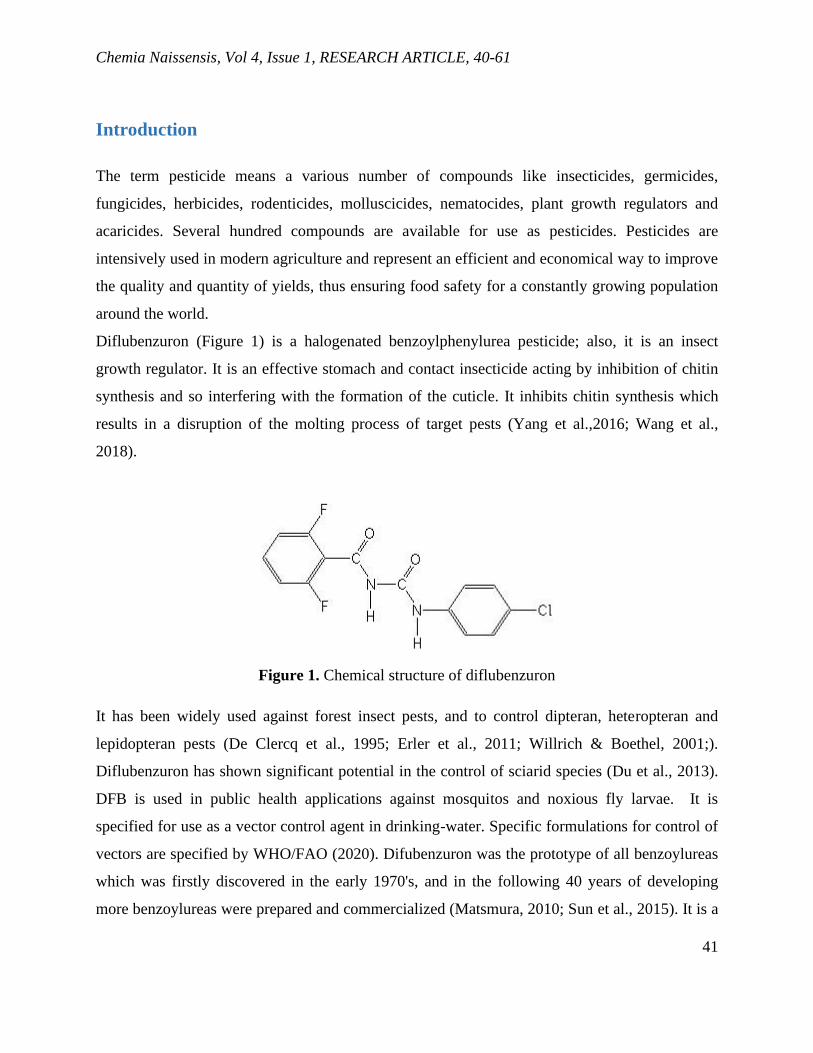
Chemia Naissensis, Vol 4, Issue 1, RESEARCH ARTICLE, 40-61
41
Introduction
The term pesticide means a various number of compounds like insecticides, germicides,
fungicides, herbicides, rodenticides, molluscicides, nematocides, plant growth regulators and
acaricides. Several hundred compounds are available for use as pesticides. Pesticides are
intensively used in modern agriculture and represent an efficient and economical way to improve
the quality and quantity of yields, thus ensuring food safety for a constantly growing population
around the world.
Diflubenzuron (Figure 1) is a halogenated benzoylphenylurea pesticide; also, it is an insect
growth regulator. It is an effective stomach and contact insecticide acting by inhibition of chitin
synthesis and so interfering with the formation of the cuticle. It inhibits chitin synthesis which
results in a disruption of the molting process of target pests (Yang et al.,2016; Wang et al.,
2018).
Figure 1. Chemical structure of diflubenzuron
It has been widely used against forest insect pests, and to control dipteran, heteropteran and
lepidopteran pests (De Clercq et al., 1995; Erler et al., 2011; Willrich & Boethel, 2001;).
Diflubenzuron has shown significant potential in the control of sciarid species (Du et al., 2013).
DFB is used in public health applications against mosquitos and noxious fly larvae. It is
specified for use as a vector control agent in drinking-water. Specific formulations for control of
vectors are specified by WHO/FAO (2020). Difubenzuron was the prototype of all benzoylureas
which was firstly discovered in the early 1970's, and in the following 40 years of developing
more benzoylureas were prepared and commercialized (Matsmura, 2010; Sun et al., 2015). It is a

Chemia Naissensis, Vol 4, Issue 1, RESEARCH ARTICLE, 40-61
42
direct acting insecticide normally applied directly to plants or water. It is rapidly adsorbed in soil
and particles and is immobile in soil. It also rapidly adsorbs to sediments and the sides of vessels
and pipes, but it may also partition into the surface film because of its low water solubility.
Pesticide analysis is very important because of widespread human exposure to these chemicals.
However, with widespread use and accumulation of pesticides over time, the residues of
benzoylureas can contaminate water, soil, and food. The pesticides can cause damage effects on
human health, such as carcinogenic and allergies, because of long-term toxicity and chronic
exposure to these compounds (Olsvik et al., 2013; Wang et al., 2016). The benzoylurea
pesticides are not very toxic for numerous marine species such as fish and algae, but due to their
specific mode of action they are likely to have adverse effects on non-target species such as the
crustaceans and amphipods in the marine environment (Klima, 2011; Macken et al., 2015).
Because of these reasons it is important to develop a simple, fast, and sensitive technique for the
determination of DFB in different samples.
Numerous analytical methods have been reported for the determination of DFB in various
matrices. Many papers reported determination of DFB by HPLC using different detectors
(Ambike and Argekar, 2017; Amelin et al., 2013; Huang et al., 2011; Kim et al., 2013; Ouyang
et al., 2015; Tfouni et al., 2013; Yang et al., 2015). GC-MS method is frequently used technique
for the analysis and the determination of a different group of pesticides in water samples and in
river waters (Chen, 2014; Łozowicka, 2017; Shi et al., 2014; Tian, 2020; Zhang et al., 2014).
Some authors reported an ultra-performance liquid chromatography coupled with tandem mass
spectrometry (UPLC-MS/MS) for the determination of DFB (Carneiro et al., 2013; Chen et al.,
2013; Pengqiang, 2013; Wang et al., 2013; Wang et al., 2014). DFB was determined previously
using ultra high-performance liquid chromatography coupled to triple quadrupole tandem mass
spectrometry (UHPLC–QqQ-MS/MS) (Martínez-Domínguez et al., 2015). There are various
studies on the determination of DFB using Liquid Chromatography-Tandem Mass Spectrometry
(LC–MS/MS) (Choi et al., 2015; Irungu et al., 2016; Kim et al., 2015; Lee et al., 2011; Macken
et al., 2015; Tran et al., 2012; Zainudin et al., 2015).
Authors from Senegal and France reported a Direct Laser-Photo-Induced Fluorescence (DL-PIF)
method for the determination of DFB in river and sea water (Diaw et al., 2013).

Chemia Naissensis, Vol 4, Issue 1, RESEARCH ARTICLE, 40-61
43
Sample preparation plays an important role in the pesticide residues analysis. To date, a large
number of methods have been developed and reported for the analysis of benzoylureas in
different samples (water, juice, fruit, food, soil samples) such as dispersive liquid–liquid
microextraction (Wang et al., 2017), solid-phase extraction (SPE) (Huang et al., 2011), solid-
phase microextraction (SPME) (Mei et al., 2015), dispersive solid-phase extraction (DSPE)
(Martin Pozo et al., 2019) and magnetic dispersive solid-phase extraction (MDSPE) (Huang et
al., 2019a). QuEChERS methods are considered to be a quick, easy, cheap, effective, robust, and
safe sample preparation methods. These methods have been used for sample preparation of
benzoylurea before LC-MS method (Guimarães de Oliveira et al., 2019; Huang et al., 2019b) or
HPLC-MS/MS method (Garsia Melo et al., 2020). Some authors developed multiple monolithic
fiber-solid-phase microextraction (MMF-SPME) of samples followed by high performance
liquid chromatography with diode array detection (Mei et al., 2014).
The aim of this study was to develop a simple, fast, and sensitive kinetic-spectrophotometric
method for the determination of pesticide DFB. Finally, it was proposed the method for the
determination of DFB in soil samples after their preparation based on SPE. Kinetic methods of
chemical analysis are popular methods for rapid determination of organic species. These
methods have some advantages like high sensitivity, low detection limit, good selectivity, rapid
analysis and using of inexpensive instrument such as spectrophotometer. In the previous work
our laboratory developed and validated two kinetic methods for DFB determination. The first
work reported DFB determination in the range 0.31 – 3.10 μg/cm3 and the application in
mushroom samples (Grahovac et al., 2010). The second kinetic method reported DFB
determination in the interval from 0.102 to 3.40 μg/cm3. This method was applied for the
determination of DFB in water and baby food samples (Pecev-Marinković et al., 2018).
In this study we reported a kinetic - spectrophotometric method for DFB determination based on
its inhibitory effect on the oxidation of sulfanilic acid by potassium periodate in acetate buffer in
the presence of Fe(III) ion as a catalyst and 1,10-phenantroline, which was monitored at 368 nm.
The method is simple, sensitive, rapid, precise, and accurate. The limit of detection (LOD) is
0.0039 μg/cm3.
Chemia Naissensis, Vol 4, Issue 1, RESEARCH ARTICLE, 40-61
44
Experimental
Reagents and chemicals
All chemicals used were analytical reagent grade. Pesticide standard diflubenzuron DFB with a
certified purity of 99% was obtained from Dr. Ehrenstorfer (Augsburg, Germany). Standard
stock solutions containing 100 μg/cm3 of DFB were prepared by dissolving the required amounts
of the standards in methanol: water (50/50, v/v). They were stored in a refrigerator at 4°C.
A Sulfanilic Acid (SA) solution (4×10⁻² mol/dm3) was prepared by dissolving a 0.3463 g of SA
(Merck) in water in the volumetric flask (50 cm3).
The initial 1.5×10⁻² mol/dm3 solution of potassium periodate was prepared by dissolving 0.075 g
KIO4 (Merck) in 50 cm3 of water.
A solution of Fe (III) 1.0×10⁻³ mol/dm3 was prepared by dissolving FeCl3×6H2O (Merck) in 0.1
mol/dm3 HCl.
A solution of 1,10-phenantroline was prepared by dissolving exact amounts in water.
The acetate buffer pH 4.7 was obtained by mixing solutions of CH3COOH (1 mol/dm3) and
NaOH (1 mol/dm3). Analytical-reagent grade solvents, methanol (MeOH), acetone ((CH3)2CO),
dichlormethane (DCM) and cyclohexane (CHX) were obtained from J. T. Baker (UK). High
purity distilled water obtained from Micro Med water purification system TKA
Wasseraufbereitungssysteme GmbH was used for solutions preparation.
Soil samples were collected in the period April-May 2011 from different locations (Knez Selo
(village), Grdelica (village), Svrljig (town), Nis (town)).
Apparatus
A Perkin-Elmer Lambda UV/Vis spectrophotometer with 10-cm quartz cell pairs was used for
recording the absorbance at 368 nm. A water bath thermostat (n-BIOTEK, INC, model NB-301)
was employed to control the reaction temperature. A stopwatch was used to record the reaction
time.
Chromatographic analyses were performed with an Agilent Technologies, Series 1200 liquid
chromatograph, equipped with an Agilent photodiode array detector (DAD), Model 1200 with
RFID tracking technology for flow cells and a UV lamp, an automatic injector and Chem Station
software. The analytical column was an Agilent – Eclipse XDBC-18 C18 column (150×4.6 mm).

Chemia Naissensis, Vol 4, Issue 1, RESEARCH ARTICLE, 40-61
45
A model BÜCHI R-200/205 rotary vacuum evaporator including bath B-490 with a vacuum
pump was used to evaporate the extracts.
A solid phase extraction system (J. T. Baker Model SPE-12, UK) with a vacuum pump was used
for solid phase extraction of samples. SPE with Chromabond® HR-P cartridges (sorbent mass
200 mg, Macherey Nagel, Germany) were used for extraction of DFB.
Hanna pH-meter instrument was used for checking the pH measurements.
The solutions were thermostated at 25 ± 0.1 C º before the beginning of the reaction.
In addition, high precision volume micropipettes (Lab Mate+) of 50, 500 and 1000 μL were used
for handling or pipetting the solutions.
General procedure
The reaction was performed in a special glass four-compartment reaction vessel-mixer with
lapped flap. The reaction was carried out in the following way: in reaction-mixture vessel with
four compartments, the solution of SA was placed in one compartment, KIO4 in the second,
buffer solution in the third, Fe (III), o-phenantroline and DFB solution were added in the fourth
compartment and water was added to the total volume of 10 cm3. The mixer-vessel was kept for
10 min at temperature of 25±0.1ºC.
The solutions were mixed and homogenized by shaking, and then transferred into 10 cm constant
temperature cell of spectrophotometer. The absorption at 368 nm was read over a period of 6
min. The rate of the reactions at different concentrations of reactants was obtained by measuring
the slope of the linear part of kinetic curve to the absorbance – time plot. The calibration graphs
were obtained by tangent method under the optimum conditions.
Soil sample preparation
10 g of soil samples were measured and then prepared by the addition of appropriate amount of
standard stock solution DFB (2 μg/cm3) and solution of methanol was added until the solvent
completely covered the soil particles. The prepared samples were stayed for 1 day. After that, the
prepared soil sample was transferred to a separatory funnel and mechanically shaken with
acetone/water mixture (80:20, v/v), then it was centrifuged 3 times for 10 min at 3000 rpm. The
separated supernatant was transferred into the separating funnel and extracted using DCM:CHX
(1:1) using 3 portions of 100 cm3 solution for 10 min under mechanical shaking. Extracted
Chemia Naissensis, Vol 4, Issue 1, RESEARCH ARTICLE, 40-61
46
solution was put into SPE cartridge which was firstly conditioned. The solution passed through
the cartridge under manual positive pressure at flow rate of 1 ml/min. The sample was eluted
with 3×1 cm3 methanol and then extract was collected and evaporated at 60 °C in a rotary
vacuum evaporator till dryness. The residue was dissolved with methanol:water (80:20, v/v),
transferred into volumetric flask (25 cm3), and divided into two parts. One part of the solution
was filtered through a 0.45-μm microporous nylon membrane (Sigma – Aldrich, USA), then it
was transferred into vials for HPLC analysis. For kinetic determination 10 cm3 of this solution
was taken and evaporated at temperature of 60 °C till dryness. The residue was dissolved in
methanol and made up with water in 10-cm3 volumetric flask and used for kinetic determination.
SPE with Chromabond® HR-P cartridge was used for the extraction of DFB. Each sample
solution was poured into a Chromabond HR-P C18 cartridge which had been conditioned with 3
ml acetone and 2 ml ethanol.
Comparative method
HPLC method was performed with an Agilent Technologies Model 1200 instrument with UV
detector, fitted with C18 (Zorbax 5 μm, 250 mm x 4.6 mm) analytical column, operating at 25 °C.
The mobile phase was methanol-water (80:20, v/v), delivered at a flow rate 1 cm3/min. The
eluate was monitored at wavelength of 254 nm. Injected volume was 10 μl, and the flow rate of
the mobile phase was 1 cm3/min.
Kinetic Procedure
To obtain good mechanical and thermal stability, the instruments were run for 10 min before the
first measurement. The reaction was carried out in the following way. In reaction-mixture vessel
with four compartments, the solution of SA was placed in one compartment, KIO4 in the second,
acetate buffer in the third, Fe(III), o-phenantroline and DFB solution were added in the fourth
compartment and water was added to the total volume of 10 cm3. The vessel was thermostated at
25.0 ± 0.1 ºC.
The content was mixed well and then immediately transferred to the spectrophotometric cell with
a path length of 10 cm. The change in absorbance was recorded at 368 nm as a function of time
every 30 s over a period of 6 min. The rate of the reaction at different concentrations of each of
the reactants was obtained by measuring the slope of the linear part of the kinetic curves of the

Chemia Naissensis, Vol 4, Issue 1, RESEARCH ARTICLE, 40-61
47
absorbance-time plot (from Beer’s law A=εlc, dA/dt=εl(dc/dt), slope = dA/dt, rate=dc/dt
dc/dt=(dA/dt)/εl). The calibration graph was constructed by plotting the slope of the linear part
of the kinetic curve versus concentration of the DFB (cDFB, μg/cm3).
Results and Discussion
Kinetic studies
The tangent method was used for processing the kinetic data. The rate of the reaction was
obtained by measuring the slope of the linear part of the kinetic curves of the absorbance-time
plot (slope=dA/dt). In order to determine the lowest possible determinable concentration of DFB,
working conditions had to be optimized. Therefore, the dependence of the rate of reactions on
the concentration of each of the reactants was determined. In Figure 2 the influence of pH on the
initial rate in the presence and absence of DFB is shown. The effect of pH on the rate of both
reactions, catalytic and inhibited, was studied in the interval pH from 4.0 to 5.0. Reaction rate is
increased with increasing pH from 4.0-4.7 for catalytic reaction, and for inhibited reaction is
increased from pH 4.0 to 4.5. For further work a pH of 4.7 was used. Catalytic reaction is – 0.75
order in the interval pH 4.0 – 4.70, and the inhibited reaction is –1.2 in the mentioned pH
interval.

Chemia Naissensis, Vol 4, Issue 1, RESEARCH ARTICLE, 40-61
48
Figure 2. Dependence of the reaction rate on the pH for the catalyzed (1) and inhibited (2)
reaction. Initial concentrations: c(SA) = 4.0×10⁻³ mol/dm3; c(KIO4) = 18.0×10⁻⁴ mol/dm3;
c(Fe(III)) = 3.0×10⁻⁸ mol/dm3; c(phen) = 6.0×10⁻⁵ mol/dm3; c(DFB) = 26.18 μg/cm3; t =
25.0±0.1°C.
Figure 3. Dependence of the reaction rate on the KIO4 concentration for the catalyzed (1) and
inhibited (2) reaction. Initial concentrations: pH = 4.7; c(SA) = 4.0×10⁻³ mol/dm3; c(Fe(III)) =
3.0×10⁻⁸ mol/dm3; c(phen) = 6.0×10⁻⁵ mol/dm3; c(DFB) = 26.18 μg/cm3; t = 25.0±0.1°C.

Chemia Naissensis, Vol 4, Issue 1, RESEARCH ARTICLE, 40-61
49
The effect of the concentration of KIO4 on the rates is shown in Figure 3. The influence of KIO4
was studied in the range 6.0×10⁻⁴ – 22.5×10⁻⁴ mol/dm3. The reaction rate of both reactions is
increased with increasing KIO4 concentration. A KIO4 concentration of 18.0×10⁻⁴ mol/dm3 was
selected for the further work. Catalytic reaction is the first order in the interval KIO4 6.0×10⁻⁴ –
18.0×10⁻⁴ mol/dm3, and inhibited reaction is the first order through the whole investigated
interval.
The effect of the concentration of SA was studied (Figure 4) in the interval of 1.0×10⁻³ – 6.0×10-
3 mol/dm3.
Figure 4. Dependence of the reaction rate on the SA concentration for the catalyzed (1) and
inhibited (2) reaction. Initial concentrations: pH = 4.7; c(KIO4) = 18.0×10⁻⁴ mol/dm3; c (Fe (III))
= 3.0×10⁻⁸ mol/dm3; c(phen) = 6.0×10⁻⁵ mol/dm3; c(DFB) = 26.18 μg/cm3; t = 25.0±0.1°C.
The rate of the catalyzed and inhibited reaction is increased with increasing SA concentration
through the whole investigated interval. For further work a concentration of 4.8×10⁻³ mol/dm3
was selected. Both reactions are the first order in the whole investigated interval.
Influence of the o-phenantroline concentration on reaction rates is shown in Figure 5. It is
examined in the interval 2.0×10⁻⁵ – 8.0×10⁻⁵ mol/dm3. The rate of the catalyzed and inhibited
reaction was increased with increasing o-phenantroline concentration from 2.0×10⁻⁵ to 6.0×10⁻⁵
mol/dm3. For further work a concentration of 6.0×10⁻⁵ mol/dm3 was selected.

Chemia Naissensis, Vol 4, Issue 1, RESEARCH ARTICLE, 40-61
50
Figure 5. Dependence of the reaction rate on the o-phenantroline concentration for the catalyzed
(1) and inhibited (2) reaction. Initial concentrations: pH = 4.7; c(KIO4) = 18.0×10⁻⁴ mol/dm3;
c(SA) = 4.8×10⁻³ mol/dm3; c (Fe (III)) = 3.0×10⁻⁸ mol/dm3; c(DFB) = 26.18 μg/cm3; t =
25.0±0.1°C.
Figure 6. Dependence of the reaction rate on the Fe(III) concentration for the catalyzed (1) and
inhibited (2) reaction. Initial concentrations: pH = 4.7; c(KIO4) = 18.0×10⁻⁴ mol/dm3; c(SA) =
4.8×10⁻³ mol/dm3; c(phen) = 6.0×10⁻⁵ mol/dm3; c(DFB) = 26.18 μg/cm3; t = 25.0±0.1°C.

Chemia Naissensis, Vol 4, Issue 1, RESEARCH ARTICLE, 40-61
51
The correlation between the slope and the Fe (III) concentration is given in Figure 6. The
influence of the concentration of Fe (III) ion on the reaction rates of catalyzed and inhibited
reactions was examined in the range 0.5×10⁻⁸– 4.0×10⁻⁸ mol/dm3. A concentration of 3.0×10⁻⁸
mol/dm3 in the final solution was used throughout the experiments.
Under the optimum reaction conditions: pH=4.7; c(KIO4) =18.0×10⁻⁴ mol/dm3; c(SA)=4.8×10⁻³
mol/dm3; c(o-phenantroline)=6.0×10⁻⁵ mol/dm3; c(Fe(III))=3.0x10⁻⁸ mol/dm3; t=25.0±0.1°C, the
DFB concentration was varied from 0.374 to 26.18 μg/cm3 and from 0.0374 to 0.374 μg/cm3.
Figure 7 shows the calibration curve at the temperature of 25°C, which can be used for the
determination of the DFB concentration in the interval from 0.0374 to 0.374 μg/cm3. The least
squares equation (y = bx + a, where b and a are the slope and intercept, respectively) for the
calibration graphs and correlation coefficient, r (Miller, 1991) for the determination of DFB in
the concentration range 0.0374 to 0.374 μg/cm3 and 0.374 – 26.18 μg/cm3 under the optimal
reaction conditions, mentioned above, were calculated:
Slope×102 = – 0.1057×cDFB + 7.89 r = – 0.9979 (1)
Slope×102 = – 0.00066×cDFB + 3.80 r = – 0.9927 (2)
where slope is the slope of the linear part of the kinetic curve of the absorbance-time plot (Slope
= dA/dt = εl (dc/dt)) and cDFB is the DFB concentration expressed in μg/cm3.
The following kinetic equations for the catalyzed and inhibited reaction were deduced based on
the obtained graphic correlations:
(3)
(4)
,where k1 and k2 are constant proportional to the rate constant of the catalyzed and inhibited
reaction, respectively.
The equations are valid for the following concentrations: acetate buffer pH 4.0 – 4.7 for catalytic
reaction and from 4.5 to 5.0 for inhibited reaction; c(KIO4) = 6.0×10⁻⁴ – 18.0×10⁻⁴ mol/dm3;
c(SA) = 1.0×10⁻³– 6.0×10⁻³ mol/dm3; c(o-phen) = 5.0×10⁻⁵ – 8.0×10⁻⁵ mol/dm3; c(Fe(III)) =
0.5×10⁻⁸ – 4.0×10⁻⁸ mol/dm3 and c(DFB) = 0.0374 – 26.8 μg/cm3.

Chemia Naissensis, Vol 4, Issue 1, RESEARCH ARTICLE, 40-61
52
Figure 7. Dependence of the reaction rate on the DFB concentration in the interval 0.0374-0.374
μg/cm3. Initial concentrations: pH = 4.7; c(KIO4) = 18.0×10⁻⁴ mol/dm3; c(SA) = 4.8×10⁻³
mol/dm3; c(o-phen) = 6.0×10⁻⁵ mol/dm3; c (Fe (III)) = 3.0×10⁻⁸ mol/dm3; t = 25.0±0.1°C.
The Limit of Detection (LOD) and Quantification (LOQ) were evaluated using the following
equations (Bendito and Silva, 1988; Motolla, 1988; Prichard and Barwick, 2007): LOD = 3.3S0/b
and LOQ = 10S0/b, where S0 is the standard deviation of the calibration line and b is the slope.
They were found to be 0.0039 μg/cm3 and 0.0131 μg/cm3, respectively.
The precision and accuracy of the system were studied by performing the experiment 5 times for
different concentration of DFB. The results of accuracy and precision of the recommended
procedure are presented in Table 1.
Table 1. Accuracy and precision of DFB determination
Added
(μg/cm3)
Determineda)
x ±SD
(μg/cm3)
n
RSD
(%)
G
(%)
100−
x
(%)b)
Recovery
(%)
0.0374 0.0366±0.0008
5
2.22 2.78 -2.14 97.86
0.188 0.193±0.004 2.11 2.63 2.66 102.65
0.374 0.382±0.005 1.30 1.35 2.13 102.10 a) Mean and standard deviation of five determinations at the 95 % confidence level; n- number of
replicates; RSD - relative standard deviation; G- relative error; b) accuracy of the method

Chemia Naissensis, Vol 4, Issue 1, RESEARCH ARTICLE, 40-61
53
Interference studies
To assess the selectivity of the method, the interference due to several cations and anions was
studied in detail. Different amounts of ionic species were added to the DFB solution. Table 2
gives the tolerance limits (expressed as v/v ratios), for the species studied in the determination of
3.74 μg/cm3 of DFB. The maximum tolerated level was taken as that causing a difference in the
rate of the inhibited reaction not larger than 5%. It may be seen that Cu2+ to DFB interferes with
reaction. The other investigated ions have practically no influence on the determination of DFB
by this method. And it means that selectivity of the method is good.
Table 2. Effect of the foreign species on the determination of 3.74 μg/cm3 of DFB
Foreign species Tolerance level (cInterferent/cDFB)
Li+, Na+, K+, Mg2+, , , F– , 103 5-10
Mg2+, Ca2+, SO42-, As3+, 102 5-10
10 <5
Ba2+, , , Mn2+, Ni2+, Zn2+Al3+ 1 <5
Co2+, Fe2+ 0.1 5-10
Cu2+ interfere
Applicability of the proposed method
The proposed method was applied to the determination of DFB in soil samples using the direct
calibration curve. The results were compared with parallel HPLC method using a point
hypothesis test (Hartman et al., 1995; Skoog et al., 1996). Detection of DFB under the optimum
conditions (methanol-water (80:20, v/v)) delivered at a flowrate of 1 mL/min, and detection at a
constant wavelength of 254 nm gave satisfactory results for the sensitivity of all spiked samples.
They were treated as described in the Experimental section. As can be seen from Table 3, the
results obtained for this method are in accordance with the parallel HPLC method. Therefore, the
proposed method could be used for the determination of DFB in real samples after SPE. Table 3
shows that F and t values at 95% confidence level are less than the theoretical ones, confirming
no significant differences between the performance of the proposed and HPLC method.

Chemia Naissensis, Vol 4, Issue 1, RESEARCH ARTICLE, 40-61
54
Table 3. Determination of diflubenzuron in soil samples by kinetic and HPLC method
Soil
sample
Added
DFB
(μg/cm3)
Found by kinetic
methoda
(μg/cm3)
RSDa
(%)
Recoverya
(%)
Found by
HPLCa
(μg/cm3)
Recoverya
(%)
F
valueb
t
valueb
S1 0.10 0.093±0.003 3.20 -7.0 93.00 0.094±0.002 94.00 1.12 0.63
S2 0.20 0.195±0.01 5.10 -2.5 97.50 0.193±0.01 96.50 2.56 1.33
S3 0.26 0.249±0.006 2.40 -4.23 95.70 0.25±0.002 96.15 2.45 1.05
S4 0.18 0.17±0.003 1.76 -5.55 94.40 0.178±0.002 98.80 1.86 0.74
S5 0.08 0.085±0.001 1.17 6.25 106.25 0.087±0.001 108.75 1.09 0.25
S6 0.05 0.047±0.007 6.40 -6.0 94.00 0.049±0.001 98.00 1.17 0.74
S7 0.60 0.56±0.02 3.57 -6.66 95.00 0.58±0.03 96.66 2.86 1.25
S8 0.90 0.86±0.05 5.80 -4.44 95.50 0.85±0.05 94.44 2.92 2.14
S9 1.70 1.73±0.03 1.73 1.76 101.00 1.72±0.02 101.17 1.03 0.71
S10 3.60 3.57±0.2 5.60 -0.83 99.16 3.58±0.1 99.45 1.78 0.65
S11 7.00 7.05±0.3 4.25 0.71 100.00 7.02±0.3 100.30 2.59 1.05
S12 10.20 10.13±0.5 4.93 -0.68 99.30 10.19±0.5 99.90 1.37 0.96 a Data are based on the average obtained from five determinations.
b Theoretical F-value (ν1=4, ν2=4) and t-value (ν=8) at 95 % confidence level are 6.39 and 2.306, respectively

Chemia Naissensis, Vol 4, Issue 1, RESEARCH ARTICLE, 40-61
55
Conclusion
The new kinetic-spectrophotometric method for the determination of diflubenzuron proposed in
this paper is simple, rapid, inexpensive, and thus, it is very appropriate for routine quality control
analysis of DFB in real samples. Spectrophotometry is the technique of choice even today due to
its inherent simplicity. It is frequently used in the laboratories of the developing countries to
overcome a variety of analytical problems. For the most laboratories for kinetic evaluations,
spectrophotometer is available, and it is not an expensive apparatus. Advantage of the proposed
method is simplicity owing to the elimination of some experimental steps such as extraction, and
derivatization prior to absorbance measurements. The simple and not expensive chemicals are
used. The procedure is easy to execute and requires less sample handling than some other
methods currently described in the literature. Statistical comparison of the results with parallel
HPLC method showed good agreement and indicates no significant difference in accuracy and
precision. Reliable recovery data were found at various concentrations, after spiking samples,
and good limits of quantification were attained. Therefore, the proposed method could be used
for the determination of DFB in soil samples after extraction.
Acknowledgment
This research was supported by the Serbian Ministry of Education, Science and Technological
Development (Agreement number 451-03-9/2021-14/200124). The authors are grateful for the
financial support provided by the Ministry.
Conflict-of-Interest Statement
None.

Chemia Naissensis, Vol 4, Issue 1, RESEARCH ARTICLE, 40-61
56
References
Amelin, V. G., Lavrukhin, D. K., & Tret’yakov, A. V. (2013). Dispersive liquid-liquid
microextraction for the determination of herbicides of urea derivatives family in natural waters
by HPLC. Journal of Analytical Chemistry, 68, 822-830.
Ambike, V., & Argekar, A. (2017). Stability indicating HPLC method for the determination of
diflubenzuron insecticide. Science Journal of Energy Engineering, 5(6), 152-157.
Bendito, D. P., & Silva, M. (1988.). Kinetic Methods in Analytical Chemistry. Ellis Horwood:
Chichester.
Carneiro, R. P., Oliveira, F. A. S., Madureira, F. D., Silva, G., de Souza W. R., & Pereira Lopes,
R. (2013). Development and method validation for determination of 128 pesticides in bananas by
modified QuEChERS and UHPLC-MS/MS analysis. Food Control, 33, 413-423.
Chen, B., Jin, B., Jiang, R., Xie, L., Lin, Y., Feng, W., & Ouyang, G. (2014). Screening and
quantification of 304 pesticides and related organic pollutants in surface water using dispersive
Liquid–Liquid Microextraction Coupled with Gas Chromatography–Mass Spectrometry.
Analytical Methods, 6, 1743–1752.
Chen, X., Zhao, K., Ge, B., & Chen, Q. (2013). Simultaneous determination of 44 pesticides in
tobacco by UPLC/MS/MS and a modified QuEChERS procedure. Journal of AOAC
International, 96, 422-431.
Choi, S., Kim, S., Shin, J.Y., Kim, M, & Kim, J.-H. (2015). Development and verification for
analysis of pesticides in eggs and egg products using QuEChERS and LC-MS/MS. Food
Chemistry, 173, 1236-1242.
Diaw, P. A., Maroto, A., Mbaye, O. M. A., Gaye-Seye, M. D., Stephan, L., Coly, A.,
Deschamps, L., Tine, A., Aaron, J. J., & Giamarchi, P. (2013). Determination of phenylurea
pesticides by direct laser photo-induced fluorescence. Talanta, 116, 569–574.
De Clercq, P., De Cock, A., Tirry, L., Viñuela, E., & Degheele, D. (1995). Toxicity of
diflubenzuron and pyriproxyfen to the predatory bug Podisus maculiventris. Entomologia
Experimentalis et Applicata, 74, 17–22.
Du, P., Liu, X., Gu, X., Dong, F., Xu, J., Kong, Z., Wu, Y., Zhu, Y., Li, Y., & Zheng, Y. (2013).
Rapid residue analysis of pyriproxyfen, avermectins and diflubenzuron in mushrooms by ultra-

Chemia Naissensis, Vol 4, Issue 1, RESEARCH ARTICLE, 40-61
57
performance liquid chromatography coupled with tandem mass spectrometry. Analytical
Methods, 5, 6741-6747.
Erler, F., Polat, E., Demir, H., Catal, M., & Tuna, G. (2011). Control of mushroom sciarid fly
Lycoriella ingenua populations with insect growth regulators applied by soil drench. Journal of
Economic Entomology, 104(3), 839-844.
Graça Melo, M., Carqueijo, A., Freitas, A., Barbosa, J., & Sanches Silva, A. (2019). Modified
QuEChERS extraction and HPLC-MS/MS for simultaneous determination of 155 pesticide
residues in rice (Oryza sativa L.). Foods 9, 18. doi:10.3390/foods9010018.
Grahovac, Z. M., Mitić, S. S., Pecev, E. T., & Pavlović, A. N. (2010). Determination of the
insecticide diflubenzuron in mushrooms by kinetic method and high-performance liquid
chromatographic method. Journal of Environmental Science and Health, Part B: Pesticides, Food
Contaminants, and Agricultural Wastes, 45, 783-789.
Guimarães de Oliveira, L., Ramkumar, A., Moloney, M., Scherer Kurz, M. H., Ferreira
Gonçalves, F., Prestes, O. D., & Danaher, M. (2019). Vibrational extraction QuEChERS for
analysis of antiparasitic agents in fish by liquid chromatography coupled with tandem mass
spectrometry. Analytical and Bioanalytical Chemistry, 411, 6913-6929.
Huang, X., Qiao, K., Li, L., Liu, G., Xu, X., Lu, R., Gao, H., & Xu, D. (2019a). Preparation of a
magnetic graphene/polydopamine nanocomposite for magnetic dispersive solid-phase extraction
of benzoylurea insecticides in environmental water samples. Scientific Reports, 9:8919. DOI:
10.1038/s41598-019-45186-z
Huang, Y., Zhou, Q., Xie, G., Liu, H., & Lin, H. (2011). Titanium dioxide nanotubes for solid
phase extraction of benzoylurea insecticides in environmental water samples, and determination
by high performance liquid chromatography with UV detection. Microchimica Acta, 172, 109–
115.
Hartmann, C., Smeyers-Verbeke, J., Penninckx, W., Vander Heyden, Y., Vankeerberghen, P., &
Massart, D. L. (1995). Reappraisal of hypothesis testing the method validation: detection of
systematic error by comparing the means of two methods or of two laboratories. Analytical
Chemistry, 67, 4491-4499.

Chemia Naissensis, Vol 4, Issue 1, RESEARCH ARTICLE, 40-61
58
Huang, Y., Shi, T., Luo, X., Xiong, H., Min, F., Chen, Y., Nie, S., & Xie, M. (2019b).
Determination of multi-pesticide residues in green tea with a modified QuEChERS protocol
coupled to HPLC-MS/MS. Food Chemistry, 275, 255-264.
Irungu, J., Raina, S., & Torto, B. (2016). Determination of pesticide residues in honey: a
preliminary study from two of Africa’s largest honey producers. International Journal of Food
Contamination, 3, 1-14.
Choi, S., Kim, S., Shin, J. Y., Kim, M., & Kim, J.-H. (2015). Development and verification for
analysis of pesticides in eggs and egg products using QuEChERS and LC–MS/MS. Food
Chemistry, 173, 1236–1242.
Kim, J.-H., Seo, J.-S., Moon, J.-K., & Kim, J.-H. (2013). Multi-residue method development of 8
benzoylurea insecticides in mandarin and apple using high performance liquid chromatography
and liquid chromatography-tandem mass spectrometry. Journal of the Korean Society for
Applied Biological Chemistry, 56, 47-54.
Klima of Forensnings Direktoratet (Klif). (2011). Environmental screening of veterinary
medicines used in aquaculture – diflubenzuron and teflubenzuron. TA 2773. Utført av NIVA
(Norwegian Institute for Water Research).
Łozowicka, B., Rutkowska, E., & Jankowska, M. (2017). Influence of QuEChERS modifications
on recovery and matrix effect during the multi-residue pesticide analysis in soil by GC/MS/MS
and GC/ECD/NPD. Environmental Science and Pollution Research, 24, 7124–7138.
Lee, S. W., Choi, J.-H., Cho, S.-K., Yu, H.-A., Abd El-Aty, A.M., & Shim, J.-H. (2011).
Development of a new QuEChERS method based on dry ice for the determination of 168
pesticides in paprika using tandem mass spectrometry. Journal of Chromatography A, 1218,
4366–4377.
Matsumura, F. (2010). Studies on the action mechanism of benzoylurea insecticides to inhibit the
process of chitin synthesis in insects: A review on the status of research activities in the past, the
present and the future prospects. Pesticide Biochemistry and Physiology, 97, 133–139.
Macken, A., Lillicrap, A., & Langford, K. (2015). Benzoylurea pesticides used as veterinary
medicines in aquaculture: risks and developmental effects on non-target crustaceans.
Environmental Toxicology and Chemistry, 34(7), 1533-1542.

Chemia Naissensis, Vol 4, Issue 1, RESEARCH ARTICLE, 40-61
59
Martínez-Domínguez, G., Nieto-García, A. J., Romero-González, R., & Frenich, A. G. (2015).
Application of QuEChERS based method for the determination of pesticides in nutraceutical
products (Camellia sinensis) by liquid chromatography coupled to triple quadrupole tandem
mass spectrometry. Food Chemistry, 177, 182–190.
Martín-Pozo, L., de Alarcón-Gómez, B., Rodríguez-Gómez, R., García-Córcoles, M. T., Çipa,
M., & Zafra-Gómez, A. (2019). Analytical methods for the determination of emerging
contaminants in sewage sludge samples. A review. Talanta, 192, 508-533.
Mei, M., Huang X., Liao, K., & Yuan, D. (2015). Sensitive monitoring of benzoylurea
insecticides in water and juice samples treated with multiple monolithic fiber solid-phase
microextraction and liquid chromatographic analysis. Analytica Chimica Acta, 860, 29–36.
Mei, M., Huang, X., & Yuan, D. (2014). Multiple monolithic fiber solid-phase microextraction:
A new extraction approach for aqueous samples. Journal of Chromatography A, 1345, 29-36.
Miller, J. N. (1991). Basic statistical methods for analytical chemistry. Part 2. Calibration and
regression methods. A review. Analyst, 116, 3-14.
Mottola, H. A. (1988). Kinetic Aspects of Analytical Chemistry. Wiley: New York.
Ouyang, H., He, Y., & Lin, L. (2015). Determination of four kinds of pesticide residues in edible
mushrooms by HPLC. Journal of Southern Agriculture, 46, 437-440.
Olsvik, P. A., Samuelsen, O. B., Erdal, A., Holmelid, B., & Lunestad, B. T. (2013).
Toxicological assessment of the anti-salmon lice drug difubenzuron on Atlantic cod Gadus
morhua. Diseases of Aquatic Organisms, 105, 27–43.
Pecev-Marinković, E. T., Grahovac, Z. M., Pavlović, A. N., Tošić, S. B., Rašić Mišić, I. D.,
Mitić, M. N., Miletić, A. S., & Sejmanović, D. M. (2018). Development of a kinetic
spectrophotometric method for insecticide diflubenzuron determination in water and baby food
samples. Hemijska Industrija, 72(5), 305–314.
Prichard, E., & Barwick, V. (2007). Quality Assurance in Analytical Chemistry. John Wiley &
Sons: Teddington.
Shi, X., Tang, Z., Sun, A., Zhou, L., Zhao, J., Li, D., Chen, J., & Pan, D. (2014). Simultaneous
analysis of polychlorinated biphenyls and organochlorine pesticides in seawater samples by
membrane–assisted solvent extraction combined with Gas Chromatography–Electron Capture

Chemia Naissensis, Vol 4, Issue 1, RESEARCH ARTICLE, 40-61
60
Detector and Gas Chromatography–Tandem Mass Spectrometry. Journal of Chromatography B,
972, 58–64.
Sun, R., Liu, C., Zhang, H., & Wang, Q. (2015). Benzoylurea chitin synthesis inhibitors. Journal
of Agricultural and Food Chemistry, 63, 6847–6865.
Skoog, D. A., West, D. M., & Holler, F. J. (1996). Fundamentals of Analytical Chemistry.
Saunders College Publishing: Philadelphia.
Tian, F., Qiao, C., Luo, J., Guo, L., Pang, T., Pang, R., Li, J., Wang, C., Wang, R., & Xie, H.
(2020). Method development and validation of ten pyrethroid insecticides in edible mushrooms
by modifed QuEChERS and gas chromatography-tandem mass spectrometry. Scientific Reports.
10:7042. https://doi.org/10.1038/s41598-020-64056-7
Tfouni, S.A.V., Furlani, R.P.Z., Carreiro, L.B, Loredo, I.S.D., Gomes, A.G, Alves, L.A, Mata,
R.S.S., Fonseca, A.M.D., & Rocha, R.M.S. (2013). Determination of diflubenzuron residues in
milk and cattle tissues. Arquivo Brasileiro de Medicina Veterinaria e Zootecnia, 65, 301-307.
Tran, K., Eide, D., Nickols, S. M., Cromer, M. R., Sabaa-Srur, A., & Smith, R. E. (2012).
Finding of pesticides in fashionable fruit juices by LC–MS/MS and GC–MS/MS. Food
Chemistry, 134, 2398–2405.
Wang, J., Jiao, C., Li, M., Wang, X., Wang, C., Wu, Q., & Wang, Z. (2018). Porphyrin based
porous organic polymer modified with Fe3O4 nanoparticles as an efficient adsorbent for the
enrichment of benzoylurea insecticides. Microchimica Acta, 185, 36.
Wang, X., Yang, C., Huang, M., Wang, M., Zha, Y., Lin, L., & Zeng, S. (2014). Determination
of diflubenzuron residues in vegetables by UPLC-MS/MS. Advanced Materials Research, 852,
266-269.
Willrich, M. M., & Boethel, D. J. (2001). Effects of diflubenzuron on Pseudoplusia includens
(Lepidoptera: Noctuidae) and its parasitoid Copidosoma floridanum (Hymenoptera: Encyrtidae).
Environmental Entomology, 30, 794-797.
WHO/FAO (2020). Fao specifications and evaluations for agricultural pesticides, Food and
Agriculture Organisation of the United Nations.
Wang, H., Hu, L., Li, W., Lu, R., Zhang, S., Zhou, W., & Gao, H. (2016). A rapid and simple
pretreatment method for benzoylurea insecticides in honey samples using in-syringe dispersive

Chemia Naissensis, Vol 4, Issue 1, RESEARCH ARTICLE, 40-61
61
liquid–liquid microextraction based on the direct solidification of ionic liquids. Journal of
Chromatography A, 1471, 60–67.
Wang, H., Hu, L., Li, W., Yang, X., Lu, R., Zhang, S., Zhou, W., Gao, H., & Li, J. (2017). In-
syringe dispersive liquid-liquid microextraction based on the solidification of ionic liquids for
the determination of benzoylurea insecticides in water and tea beverage samples. Talanta, 162,
625–633.
Wang, S., Zhang, S., Dong, C., Wang, G., Guo, J., & Sun, W. (2013). Simultaneous
determination of four herbicide and pesticide residues in Chinese green tea using QuEChERS
purification procedure and UPLC-MS/MS analysis. Advanced Materials Research, 634-638,
1586-1590.
Yang, X., Qiao, K., Ye, Y., Yang, M., Li, J., Gao, H., Zhang, S., Zhou, W., & Lu, R. (2016).
Facile synthesis of multifunctional attapulgite/Fe3O4/polyaniline nanocomposites for magnetic
dispersive solid phase extraction of benzoylurea insecticides in environmental water samples.
Analytica Chimica Acta, 934, 114–121.
Yang, M., Xi, X., Yang, X., Bai, L., Lu, R., Zhou, W., Zhang, S. & Gao, H. (2015).
Determination of benzoylurea insecticides in environmental water and honey samples using
ionic-liquid-mingled air-assisted liquid–liquid microextraction based on solidification of floating
organic droplets. RCS Advances, 32(5), 25572-25580.
Zhang, Z., Lefebvre, T., Kerr, C., & Osprey, M. (2014). Simultaneous extraction and
determination of various pesticides in environmental waters. Journal of Separation Science, 37,
3699–3705.
Zainudin, B. H., Salleh, S., Mohamed, R., Choy Yap, K., & Muhamad, H. (2015). Development,
validation, and determination of multiclass pesticide residues in cocoa beans using gas
chromatography and liquid chromatography tandem mass spectrometry. Food Chemistry, 172,
585–595.

Chemia Naissensis, Vol 4, Issue 1, RESEARCH ARTICLE, 62
62
Razvoj i primena kinetičko-spektrofotometrijske metode za analizu
diflubenzurona u uzorcima zemljišta primenom SPE pracene HPLC
metodom
Emilija Pecev-Marinkovic1*, Ana Miletic1, Aleksandra Pavlovic1, Vidoslav Dekic2
1- Univerzitet u Nišu, Prirodno-matematički fakultet, Departman za hemiju, Višegradska 33, 18000
Niš, Republika Srbija
2- Univerzitet u Prištini, Prirodno-matematički fakultet, Departman za hemiju, Ivo Lole Ribara 29,
Kosovska Mitrovica, Republika Srbija
SAŽETAK
U radu je predstavljena nova osetljiva i jednostavna kinetičko-spektrofotometrijska metoda za
određivanje insekticida diflubenzurona (DFB). Kinetička metoda je bazirana na inhibitornom
efektu DFB na oksidaciju sulfanilne kiseline (engl. SA) kalijum-perjodatom u prisustvu Fe(III)
jona kao katalizatora i 1,10-fenantrolina. Reakcija je praćena spektrofotometrijski merenjem
apsorbance proizvoda reakcije sa vremenom na 368 nm. Diflubenzuron je određen pomoću
linearnog kalibracionog grafika u intervalu od 0,0374 do 0,374 μg/cm3 i od 0,374 do 26,18
μg/cm3. Limit detekcije i limit kvantifikacije metode sa 3σ-kriterijumima su bili 0,0039 μg/cm3
i 0,0131 μg/cm3, redom. Relativne standardne devijacije za pet replikatnih određivanja od
0.0374, 0.188 i 0,374 μg/cm3 DFB su bile 2,24, 2,11 i 1,10%, redom. Metoda je uspešno
primenjena za određivanje DFB ostataka u uzorcima zemljišta. Tvrdofazna ekstrakcija (engl.
SPE) je korišćena za ekstrakciju DFB iz zemljišta sa Chromabond® (Macherey-Nagel) C18-
kertridžima. HPLC metoda korišćena je kao komparativna metoda za verifikaciju rezultata.
Rezultati dobijeni korišćenjem dve različite metode pokazali su dobro slaganje.
Ključne reči: diflubenzuron, kinetička metoda, HPLC metoda, čvrsto-fazna ekstrakcija,
uzorci zemljišta

Chemia Naissensis, Vol 4, Issue 1, RESEARCH ARTICLE, 63
63
Développement et application d’une méthode cinétique-
spectrophotométrique pour l’analyse du diflubenzuron dans des
échantillons de sol à l’aide de la SPE suivie de la méthode HPLC
Emilija Pecev-Marinković1*, Ana Miletić1, Aleksandra Pavlović1, Vidoslav Dekić2
1- Université de Niš, Faculté des sciences naturelles et des mathématiques, Département de chimie,
Višegradska 33, 18000 Niš, République de Serbie
2- Université de Priština, Faculté des sciences naturelles et des mathématiques, Département de chimie,
Ivo Lole Ribara 29, Kosovska Mitrovica, République de Serbie
RÉSUMÉ
Le but de cet article est de présenter une nouvelle méthode cinétique-spectrophotométrique
sensible et simple pour la détermination de l’insecticide diflubenzuron (DFB). La méthode
cinétique est basée sur l’effet inhibiteur du DFB sur l’oxydation de l’acide sulfanilique (en
anglais SA) par le periodate de potassium dans un tampon acétate en présence de l’ion Fe(III)
comme catalyseur et du 1,10-phénantroline. La réaction a été contrôlée par spectrophotométrie
en mesurant l’augmentation de l’absorbance du produit de la réaction avec le temps à 368 nm.
Le diflubenzuron a été déterminé au moyen du graphique d’étalonnage linéaire dans l’intervalle
de 0,0374 à 0,374 μg/cm3 et de 0,374 à 26,18 μg/cm3. La limite de la détection et la limite de
la quantification de la méthode avec les critères 3σ étaient respectivement de 0,0039 μg/cm3et
0,0131 μg/cm3. Les écarts relatifs standard pour les cinq déterminations répétées de 0,0374,
0,188 et 0,374 μg/cm3de DFB étaient de 2,24, 2,11 et 1,10%, respectivement. La méthode a été
appliquée avec succès pour déterminer les résidus du DFB dans des échantillons de sol.
L’extraction en phase solide (en anglais SPE) a été utilisée pour l’extraction du DFB du sol et
des échantillons avec des cartouches Chromabond® (Macherey-Nagel) C18. La méthode
HPLC a été utilisée en tant que méthode comparative pour la vérification des résultats. Les
résultats obtenus à l’aide des deux méthodes différentes ont montré une bonne concordance.
Mots-clés : diflubenzuron, méthode cinétique, méthode HPLC, extraction en phase solide,
échantillons de sol.

Chemia Naissensis, Vol 4, Issue 1, RESEARCH ARTICLE, 64
64
Разработка и применение кинетико-спектрофотометрического метода
анализа дифлубензурона в образцах почвы с использованием ТФЭ с
последующей ВЭЖХ.
Эмилия Пецев-Маринкович1*, Ана Милетич1, Александра Павлович1, Видослав
Декич2
1- Университет в Нише, Естественно-математический факультет, Кафедра химии,
Вишеградска 33, 18000 Ниш, Сербия
2- Университет в Приштине, Естественно-математический факультет, Кафедра химии, Иво
Лоле Рибара 29, Косовска Митровица, Республика Сербия
АННОТАЦИЯ
Цель данной статьи - представить новый чувствительный и простой кинетико-
спектрофотометрический метод определения инсектицида дифлубензурона (ДФБ).
Кинетический метод основан на ингибирующем действии ДФБ на окисление
сульфаниловой кислоты (СК) периодатом калия в ацетатном буфере в присутствии иона
Fe(III) в качестве катализатора и 1,10-фенантролина. За реакцией следили
спектрофотометрические измеряя увеличение поглощения продукта реакции при длине
волны 368 nm с течением времени. Дифлубензурон определяли по линейному
градуировочному графику в интервале от 0,0374 до 0,374 μg/cm3 и от 0,374 до 26,18
μg/cm3. Предел обнаружения и предел количественной оценки метода с критерием 3σ
составили 0,0039 μg/cm3 и 0,0131 μg/cm3 соответственно. Относительные стандартные
отклонения для пяти повторных определений 0,0374, 0,188 и 0,374 µg/cm3 ДФБ
составили 2,24, 2,11 и 1,10% соответственно. Метод успешно применен для определения
остатков ДФБ в образцах почвы. Твердофазную экстракцию (ТФЭ) использовали для
экстракции ДФБ из почвы и образцов с картриджами Chromabond® (Macherey-Nagel)
C18. Метод ВЭЖХ использовался в качестве сравнительного метода для проверки
результатов. Результаты, полученные двумя разными методами, показали хорошее
согласие.
Ключевые слова: дифлубензурон, кинетический метод, метод ВЭЖХ, твердофазная
экстракция, образцы почвы.

Chemia Naissensis, Vol 4, Issue 1, RESEARCH ARTICLE, 65
65
Entwicklung und Anwendung einer kinetisch-spektrophotometrischen
Methode zur Analyse von Diflubenzuron in Bodenproben mittels SPE
gefolgt von HPLC-Methode
Emilija Pecev-Marinković1*, Ana Miletić1, Aleksandra Pavlović1, Vidoslav Dekić2
1- Universität in Niš, Fakultät für Naturwissenschaften und Mathematik, Lehrstuhl für Chemie,
Višegradska 33, 18000 Niš, Republik Serbien
2- Universität in Priština, Fakultät für Naturwissenschaften und Mathematik, Lehrstuhl für Chemie,
Ivo Lole Ribara 29, Kosovska Mitrovica, Republik Serbien
ABSTRACT
Ziel dieser Arbeit ist es, eine neue empfindliche und einfache kinetisch-spektrophotometrische
Methode zur Bestimmung des Insektizids Diflubenzuron (DFB) vorzustellen. Die kinetische
Methode basiert auf hemmender Wirkung von DFB auf die Oxidation von Sulfanilsäure (eng.
SA) durch Kaliumperiodat in Acetatpuffer in Gegenwart von Fe(III)-Ionen als Katalysator und
1,10-Phenantrolin. Die Reaktion wurde spektrophotometrisch überwacht, indem die
Absorbtion des Reaktionsprodukts mit der Zeit bei 368 nm gemessen wurde. Diflubenzuron
wurde mit linearer Kalibrierungsgrafik im Intervall von 0,0374 bis 0,374 µg/cm3 und von 0,374
bis 26,18 µg/cm3 bestimmt. Die Nachweis- und Bestimmungsgrenze der Methode mit 3σ-
Kriterien betrug 0,0039 µg/cm3 bzw. 0,0131 µg/cm3, nacheinander. Die relativen
Standardabweichungen für fünf Wiederholungsbestimmungen von 0,0374, 0,188 und 0,374
µg/cm3 DFB betrugen 2,24, 2,11 bzw. 1,10 %, nacheinander. Die Methode wurde erfolgreich
zur Bestimmung von DFB-Rückständen in Bodenproben angewendet. Die
Festphasenextraktion (eng. SPE) wurde zur Extraktion von DFB aus Boden mit Chromabond®
(Macherey-Nagel) C18-Kartuschen verwendet. Als Vergleichsmethode wurde die HPLC-
Methode zur Überprüfung der Ergebnisse verwendet. Die mit zwei verschiedenen Methoden
erhaltenen Ergebnisse zeigten eine gute Übereinstimmung.
Schlüsselwörter: Diflubenzuron, Kinetische Methode, HPLC-Methode, Festphasenextraktion,
Bodenproben
Chemia Naissensis, Vol 4, Issue 1, RESEARCH ARTICLE, 66-84
66
Synthesis, characterization, and antimicrobial activity of novel 2-ferrocenyl-
1,3-thiazolidin-4-thiones
Todosijević Anka1*, Jančić Minić Aleksandra2, Mihailović Vladimir3, Srećković Nikola3
1- University of Niš, Faculty of Agriculture, Kosančićeva 4, 37000 Kruševac, Serbia
2- University of Priština, Faculty of Technical Sciences, Knjaza Miloša 7, 38220 Kosovska Mitrovica,
Serbia
3- University of Kragujevac, Faculty of Science Department of Chemistry, Radoja Domanovića 12, 34000
Kragujevac, Serbia
ABSTRACT
Synthesis of five novel 2-ferrocenyl-1,3-thiazolidin-4-thiones has been achieved in good to
excellent yields through the treatment of 2-ferrocenyl-1,3-thiazolidin-4-ones with Lawesson’s
reagent. The reaction was performed by refluxing the reactants mixture in toluene overnight. All
prepared compounds were characterized by the IR and NMR spectral data. Further, the obtained
products were evaluated for their antibacterial and antifungal activity.
Keywords: Ferrocene, 2-Ferrocenyl-1,3-thiazolidin-4-thiones, 2-Ferrocenyl-1,3-thiazolidin-4-
ones, Characterization, Antimicrobial activity.
Todosijević Anka*: [email protected]
Minić Aleksandra: [email protected] and [email protected]
Mihailović Vladimir: [email protected]
Srećković Nikola: [email protected]

Chemia Naissensis, Vol 4, Issue 1, RESEARCH ARTICLE, 66-84
67
Introduction
Since the middle of the last century, antimicrobial agents have been efficiently used in health
care. However, recent success in the application of antibiotics in infections treatment has been
eroded by the increased resistance of bacteria. Therefore, there is a permanent interest in the
development of novel compounds that exhibit antimicrobial activity (Hawkey, 2008; Cantón,
2009). Ferrocene derivatives have been demonstrated to possess a broad spectrum of biological
activity, including antibacterial (Damljanović et al., 2009; Li et al., 2013), antifungal (Rubbiani,
2016), anti-inflammatory (Shinde et al., 2018), antitumor (Staveren and Metzler-Nolte, 2004;
Fouda, 2007; Hillard, 2010; Biot, 2000) and anti-HIV (Kondapi et al., 2006) activity. In addition,
due to the favorable electronic properties of ferrocene and its easy functionalization, these
substances have been found many applications in materials science, including sensors (Labande
and Astruc, 2000; Labande et al., 2002; Daniel et. al., 2003, Astruc et. al., 2004, Armada et al.,
2006; Ornelas, 2007; Astruc, 2008; Ornelas et al., 2009; Djeda, 2010), catalysts (Astruc, 2008;
Wei et al. 2002, Zhang et al., 2002; Ornelas et al., 2007; Diallo et al., 2007; Ornelas et al.,
2008), electroactive materials (Daniel et al., 2006, Astruc et al., 2008; Ornelas et al., 2008; Wang
et al., 2009; Astruc et al., 2009; Megiatto et al., 2010) and aerospace materials (Ansari et al.,
2018; Neuenfeldt et al., 2011).
On the other hand, the chemistry of thiazolidinone ring is of substantial interest as it
represents is a core structure in numerous synthetic medicaments displaying a wide variety of
biological activities (Makwana and Malani, 2017). The thiazolidinone scaffold is also found in
various natural products, especially thiamines, molecules possessing cardiac and glycemic
benefits such as troglitazone (Ghazzi et al., 1997) and many metabolic products of fungi and
primitive marine animals, including 2-(aminoalkyl)-4-carboxylic acids (Scmidt et al., 1987). A
number of thiazolidinone derivatives have displayed various biological activities such are
antidiabetic (Verma and Thareja, 2016), anticancer (Szychowski et al., 2017), anticonvulsant
(Mishchenko et al., 2020), antimicrobial (Viswajanani et al., 2005), anti-HIV (Rawal et al.,
2005), PAF antagonist (Tanabe et al., 1991), COX inhibitory (Ottana et al., 2002), tumor
necrosis factor-α antagonist (Voss et al., 2003) and antioxidant (Shih and Ke, 2004). Besides,
thiazolidinone derivatives are of interest in a wide range of pharmaceutical, agrochemical,

Chemia Naissensis, Vol 4, Issue 1, RESEARCH ARTICLE, 66-84
68
coordination, medicinal, and organic chemistry applications (Chaves et al., 2014; Corrêa et al.,
2016; Deng et al., 2011).
Scheme 1. Synthesis of N-substituted 2-ferrocenyl-1,3-thiazolidin-4-ones 3 (Pejović et al.,
2014).
Several years ago, our team reported an easy and efficient method for the synthesis of the
novel thiazolidinone derivatives - 2-ferrocenyl-1,3-thiazolidin-4-ones 3 (Scheme 1) (Pejović et
al., 2014). These heterocycles that showed strong anxiolytic activity also demonstrated great
initial results in the synthesis of another ferrocene containing thiazolidinones (Pejović et al.,
2018). Bearing all previously mentioned in mind, we planned to synthesize 2-ferrocenyl-1,3-
thiazolidin-4-thiones using 2-ferrocenyl-1,3-thiazolidin-4-ones 3 as starting material. Thus, in
this research, we put emphasis on the synthesis, assignment of 1H and 13C NMR spectral data of
novel 2-ferrocenyl-1,3-thiazolidin-4-thiones as well as examination of their antimicrobial
activity.
Experimental
Reagents and chemicals
All chemicals were commercially available and used as received, except the solvents, which
were purified by distillation. Chromatographic separations were carried out using silica gel 60
(Merck, 230–400 mesh ASTM), whereas silica gel 60 on Al plates, layer thickness 0.2 mm
(Merck) was used for TLC. Melting points (uncorrected) were determined on a Mel-Temp
capillary melting points apparatus, model 1001. The 1H and 13C NMR spectra of the samples in
CDCl3 were recorded on a Varian Gemini (200 MHz) (1H at 200 MHz, 13C at 50 MHz) NMR

Chemia Naissensis, Vol 4, Issue 1, RESEARCH ARTICLE, 66-84
69
spectrometer. Chemical shifts are expressed in (ppm), relative to residual solvent protons as the
internal standard (CDCl3: 7.26 ppm for 1H and 77 ppm for 13C). IR measurements were carried
out with a Perkin–Elmer FTIR 31725-X spectrophotometer.
2-Ferrocenyl-1,3-thiazolidin-4-ones (3a-e)
2-Ferrocenyl-1,3-thiazolidin-4-ones (3a-e) were synthesized according to the previously
described procedure (Pejović et al., 2014).
General procedure for the synthesis of 2-ferrocenyl-1,3-thiazolidin-4-thiones (5a-e)
A toluene solution of the suitable 2-ferrocenyl-1,3-thiazolidin-4-one (1 mmol), obtained
according to a previously reported procedure (Pejović et al., 2014), was heated under stirring
with Lawesson’s reagent (1 mmol) overnight. The solvent was then evaporated, and the residue
extracted with dichloromethane (three 20 mL portions). After drying overnight (anhydrous
Na2SO4), the solvent was evaporated and the crude product purified by column chromatography
(SiO2, hexane/ethyl acetate = 8: 2).
3-Butyl-2-ferrocenyl-1,3-thiazolidin-4-thione (5a). Orange solid. Yield 88%; m.p. = 119 oC; IR
(neat): νmax = 2952, 1181 cm-1; 1H NMR (200 MHz, CDCl3, ppm): δ 5.88 (br t, 1H), 4.50 – 4.44
(m, 1H), 4.37 – 4.17 (m, 5H), 4.25 (s, 5H), 3.94 – 3.74 (m, 1H), 3.35 – 3.13 (m, 1H), 1.68 – 1.12
(m, 4H), 0.85 (t, J = 7.0 Hz, 3H); 13C NMR (50 MHz, CDCl3, ppm): δ 195.3, 83.6, 71.5, 70.1,
70.0, 69.3, 68.9, 68.1, 47.5, 45.2, 27.7, 20.1, 13.7.
2-Ferrocenyl-3-hexyl-1,3-thiazolidin-4-thione (5b). Orange solid. Yield 90%; m.p. = 92 oC; IR
(neat): νmax = 2928, 1176 cm-1; 1H NMR (200 MHz, CDCl3, ppm): δ 5.88 (br t, 1H), 4.51 – 4.41
(m, 1H), 4.36 – 4.05 (m, 6H), 4.24 (s, 5H), 3.90 – 3.70 (m, 1H), 3.35 – 3.15 (m, 1H), 1.65 – 1.07
(m, 8H), 0.84 (t, J = 6.5 Hz, 3H).; 13C NMR (50 MHz, CDCl3, ppm): δ 195.1, 83.5, 71.4, 70.1,
70.0, 69.2, 68.8, 68.0, 47.7, 45.2, 31.1, 26.4, 25.5, 22.4, 13.9.
2-Ferrocenyl-3-octyl-1,3-thiazolidin-4-thione (5c). Orange solid. Yield 88%; m.p. = 78 oC;
(neat): νmax = 2920, 1203 cm-1; 1H NMR (200 MHz, CDCl3, ppm): δ 5.87 (br t, 1H), 4.49 – 4.31
(m, 1H), 4.35 – 4.14 (m, 6H), 4.25 (s, 5H), 3.92 – 3.66 (m, 1H), 3.37 – 3.13 (m, 1H), 1.74 – 1.04

Chemia Naissensis, Vol 4, Issue 1, RESEARCH ARTICLE, 66-84
70
(m, 12H), 0.87 (t, J = 6.5 Hz, 3H); 13C NMR (50 MHz, CDCl3, ppm): δ 195.3, 83.6, 71.5, 70.1,
70.0, 69.3, 68.8, 68.1, 47.8, 45.2, 31.7, 29.0, 26.8, 25.6, 22.6, 14.1.
2-Ferrocenyl-3-(m-tolyl)-1,3-thiazolidin-4-thione (5d). Orange solid. Yield 65%; m.p. = 78 oC;
IR (neat): νmax = 2956, 1165 cm-1; 1H NMR (200 MHz, CDCl3, ppm): δ 7.35 – 7.29 (m, 2H), 6.93
– 6.83 (m, 2H), 6.16 (br s, 1H), 4.36 – 4.07 (m, 4H), 4.25 (s, 5H), 3.86 (d, J = 10.3 Hz, 1H), 3.45
(d, J = 10.3 Hz, 1H), 2.29 (s, 3H); 13C NMR (50 MHz, CDCl3, ppm): δ 198.4, 139.6, 129.2,
128.6, 127.9, 83.3, 74.5, 70.7, 69.7, 69.1, 68.7, 67.5, 45.7.
2-Ferrocenyl-3-(p-tolyl)-1,3-thiazolidin-4-thione (5e). Yield 60%, brown oil; IR (neat): νmax =
2917, 1168 cm-1; 1H NMR (200 MHz, CDCl3, ppm): δ 7.09 – 6.93 (m, 2H), 6.90 – 6.68 (m, 2H),
6.10 (br s, 1H), 4.55 – 4.36 (m, 4H), 4.23 (s, 5H), 3.84 (d, J = 10.3 Hz, 1H), 3.55 (d, J = 10.3 Hz,
1H), 2.77 (s, 3H); 13C NMR (50 MHz, CDCl3, ppm): δ 195.24, 158.18, 130.07, 129.59, 113.86,
83.38, 71.96, 70.23, 70.19, 69.25, 68.91, 68.11, 49.70, 30.9.
Antimicrobial activity determination
The antimicrobial activity of newly synthesized compounds was tested on four bacterial
(Enterococcus faecalis ATCC 29212, Escherichia coli ATCC 25922, Staphylococcus aureus
ATCC 25923, and Salmonella enteritidis ATCC 13076) and four fungal strains (Candida
albicans ATCC 10231, Fusarium oxysporum FSB 91, Aspergillus brasiliensis ATCC 16404, and
Penicillium canescens FSB 24). The microorganisms were obtained from the Institute of Public
Health, Kragujevac, Serbia. For the determination of the antimicrobial activity of synthesized
compounds, bacterial strains were grown on nutrient agar 24 h before the experiment, C.
albicans was grown on sabouraud dextrose agar for 48 h, while molds were grown on potato
glucose agar for 3–7 days before testing.
The minimal inhibitory concentrations (MICs) of tested compounds, standard antibiotic
chloramphenicol, and antimycotic nystatin against selected bacterial and fungal strains were
determined by the microdilution method in the sterile 96-well microtiter plates (A. Pejović et al.,
2017). The different concentrations of compounds for antimicrobial testing were made in the
sterile 96-well microtiter plates using Mueller–Hinton broth for bacterial strains and sabouraud
dextrose broth for fungal growth. The evaluation of antibacterial and antifungal activities was
Chemia Naissensis, Vol 4, Issue 1, RESEARCH ARTICLE, 66-84
71
performed according to the CLSI recommendations (Clinical and Laboratory Standards Institute,
2012; Clinical and Laboratory Standards Institute, 2008a; Clinical and Laboratory Standards
Institute, 2008b). Indicator resazurin was used for the detection of bacterial growth in microtiter
plates, while the growth of fungi was monitored visually. The lowest concentration (in mg per
mL) of tested compounds that inhibit visible bacterial or fungal growth was considered as
minimal inhibitory concentration (MIC) value.
Results and Discussion
General synthesis of 2-ferrocenyl-1,3-thiazolidin-4-thiones 5a-e
The obtaining of target compounds was conceived as the functionalization of the 2-
ferrocenyl-1,3-thiazolidin-4-ones (Table 1, 3a-e). It should comprise their thionation to afford
the series of appropriate 2-ferrocenyl-1,3-thiazolidin-4-thiones. The thionation reaction of the
thiazolidinone derivatives is commonly carried out in one of two reagents, phosphorus
pentasulfide (P4S10) or Lawesson’s reagent (LR) (Ozturk et al., 2008). In the last few years,
scientists are claimed that Lawesson’s reagent has advantages over P4S10 in terms of
requirements for excess P4S10, longer reaction time and higher temperature (Ozturk et al., 2008).
Therefore, we considered synthesizing a series of novel ferrocene-containing thiazolidinthiones
using Lawesson’s reagent, and all in order to evaluate antimicrobial potential of the desired
products.
The realization of this synthetic plan began with the preparation of the ferrocene-
containing thiazolidinones, 2-ferrocenyl-1,3-thiazolidin-4-ones (Table 1, 3a-e). A few years ago,
we designed and optimized reaction conditions for their synthesis in high yields. The protocol
involved a 25 min ultrasonic irradiation of the reaction mixture consisting of an amine,
ferrocenecarboxaldehyde and thioglycolic acid in the ratio of 1/1/2. Thus, we synthesized the
starting material by using this protocol. After the preparation of starting material, in the test
experiment, the reaction between 3a and Lawesson’s reagent 4 was investigated. This reaction
mixture was refluxed in the toluene during the night. After usual workup and column
chromatography (SiO2/hexane = 8:2, ν/ν), the desired thiazolidinthione 5a was obtained in great

Chemia Naissensis, Vol 4, Issue 1, RESEARCH ARTICLE, 66-84
72
yield (88%, Table 1, entry 1). These results did not require additional screenings, so we accepted
them as the optimal ones.
Table 1. The substrate scope of synthesis of 2-ferrocenyl-1,3-thiazolidin-4-thiones 5a-e
Entry R Product Yields* (%)
1 n-Butyl 5a 88
2 n-Hexyl 5b 90
3 n-Octyl 5c 88
4 m-Tolyl 5d 65
5 p-Tolyl 5e 60 *Isolated yield after column chromatography.
The scope of the reaction was investigated on five known ferrocene-containing
thiazolidinones. The desired products 5a-e have been obtained in moderate to excellent yields
(up to 90%) after the purification by means of column chromatography. Structural
characterization has been confirmed by spectroscopic methods (NMR, IR) for all prepared
compounds.
We have observed that electronic properties, as well as steric hindrance of the
substituents on the N-atom, have an influence on reaction outcomes (See Table 1). Regarding
that, the products with the aliphatic group bonded to the N-atom were obtained in excellent
yields (up to 90%). The hexyl derivatives were isolated in the highest yield (90%, Table 1, entry

Chemia Naissensis, Vol 4, Issue 1, RESEARCH ARTICLE, 66-84
73
2), while the butyl derivative was obtained in a slightly low yield (88%, Table 1, entry 1). These
results can be explained by electron-donating effect of these groups. The octyl derivative
deviates a little bit from this rule (88%, entry 3, Table 1). We believe that the volume of the octyl
group influenced the reaction outcome. In addition, the electronic properties also have an
influence on the reaction outcome since the thiazolidinones with a methyl group on the phenyl
ring leads to moderate yields (60 and 65%, Table 1, entries 5 nad 4).
Several research groups investigated a mechanism of the thionation reaction of carbonyl
compounds with Lawesson’s reagent (Ozturk et al., 2008). Thus, a plausible reaction mechanism
of thionation of ferrocene-containing thiazolidinone 3a with Lawesson’s reagent 4 is illustrated
in Scheme 2. The first step of the mechanism of the reaction involves dissociation of Lawesson’s
reagent 4. After dissociation, the reaction takes place through a two-step mechanism involving
(i) a concerted cycloaddition between dithiophosphine ylide 4' and the thiazolidinone compound
3a to form a four-membered intermediate thiaoxaphosphetane I and (ii) a cycloreversion leading
to the thiocarbonyl derivative 5a and phenyl(thioxo)phosphine oxide 6.
Spectral characterization
All synthesized ferrocene-containing thiazolidin-4-thiones 5a-e were characterized by
standard spectroscopic techniques (IR, 1H-, and 13C NMR). The collected data were in complete
agreement with the proposed structures.
The main common feature of the IR spectra of five new thiazolidin-4-thiones 5a-e is a strong
band in range 1203-1165 cm-1, which is attributed to the C=S stretching vibrations. NMR spectra
utterly confirm the structures of synthesized compounds. Thus, in the 1H NMR spectra of all
thiazolidin-4-thiones 5a-e signals of the cyclopentadienyl rings, protons appear at similar
positions in the spectra (4.25-4.23 ppm for the unsubstituted, and 4.55 - 4.05 ppm for the
substituted rings). The 1H NMR spectra of 5a-e also contain characteristic signals for aliphatic
and aromatic protons. They are located in the expected regions of 1H NMR spectra. In addition,
in 1H NMR spectra of 5a-e, one signal appears at about 6.16-5.87 ppm, which corresponds to the
methine protons of thiazolidinthione moiety. On the other hand, their 13C NMR spectra lack the
carbonyl carbon from the thiazolidinone ring resonances, thus distinguishing themselves from

Chemia Naissensis, Vol 4, Issue 1, RESEARCH ARTICLE, 66-84
74
the corresponding precursors 3a-e. The 1H-, and 13C NMR spectra of 5a are presented in Figure 1
and Figure 2.
Scheme 2. The plausible schematic mechanism for the synthesis of titled compounds 5

Chemia Naissensis, Vol 4, Issue 1, RESEARCH ARTICLE, 66-84
75
Figure 1. 1H NMR (200 MHz, CDCl3) spectrum of 5a
Figure 2. 13C NMR (50 MHz, CDCl3) spectrum of 5a

Chemia Naissensis, Vol 4, Issue 1, RESEARCH ARTICLE, 66-84
76
Figure 3. FTIR spectrum of 5a
Antimicrobial activity
All types of synthesized compounds showed a low to moderate antimicrobial activity against
most of the tested bacterial and fungal species. The most active compounds against E. faecalis
(0.5 mg/mL) were 5a and 5d, while the MIC values for other bacteria were ranged from 1 to 2
mg/mL for all tested compounds, as presented in Table 2. However, 5e did not show
antibacterial activity against S. aureus in applied concentration. The antifungal activity of
synthesized compounds was slightly better than their antibacterial activity according to obtained
MIC values in tests with bacteria and fungi. The most of synthesized compounds inhibited the
growth of tested molds and C. albicans in a concentration of 1 mg/mL. The only exception was
A. brasiliensis, as the most resistant fungi on tested compounds with MIC values of 2 mg/mL for
all compounds. MIC values of synthesized compounds were compared with the well-known

Chemia Naissensis, Vol 4, Issue 1, RESEARCH ARTICLE, 66-84
77
antibiotic (chloramphenicol) and antimycotic (nystatin), whereby results indicated much higher
antibacterial and antifungal activities than synthesized compounds.
Table 2. Antibacterial and antifungal activities of synthesized compounds
MICa (mg/mL) MIC (µg/mL)
Bacteria 5a 5b 5c 5d 5e Chloramphenicol
E. faecalis ATCC 29212 0.5 1 2 0.5 2 0.625
E. coli ATCC 25922 2 2 1 2 2 0.625
S. aureus ATCC 25923 2 2 2 2 ˃2 5
S. enteritidis ATCC 13076 2 2 2 2 2 2.5
Fungi Nystatin
C. albicans ATCC 10259 1 1 1 2 1 2.5
F. oxysporom FSB 91 1 1 1 1 2 1.25
A. brasiliensis ATCC 16404 2 2 2 2 2 1.25
P. canescens FSB 24 1 2 1 1 1 2.5
aMIC- minimum inhibitory concentration.
Conclusion
Five novel 2-ferrocenyl-1,3-thiazolidin-4-thiones were prepared in moderate to excellent
yields (up to 90%) using the standard synthetic protocol. The structures of all the obtained
products were confirmed by the usage of standard spectroscopic methods.
The prepared heterocycles were tested against four bacterial (Enterococcus faecalis,
Escherichia coli, Staphylococcus aureus, and Salmonella enteritidis) and four fungal strains
(Candida albicans, Fusarium oxysporum, Aspergillus brasiliensis, and Penicillium canescens)
using the microdilution method. The results of the experiment indicated that synthesized
heterocycles showed moderate to weak antibacterial activity against less number of examined
microorganisms. Besides, it was found that N-butyl and N-m-tolyl derivatives are the most active
compounds against E. faecalis. Furthermore, antifungal activity of the synthesized ferrocene

Chemia Naissensis, Vol 4, Issue 1, RESEARCH ARTICLE, 66-84
78
derivatives tested on the human pathogen yeasts C. albicans, F. oxysporom, and P. canescens,
showed that the obtained compounds have more pronounced antifungal potential.
Acknowledgment
This work was supported by the Ministry of Education, Science and Technological Development
of the Republic of Serbia (Agreement No. 451-03-68/2020-14/200122).
Conflict-of-Interest Statement
None.
References
Ansari, M. F., Idrees, D., Hassan, Md. I., Ahmad, K., Avecilla, F., Azam, A. (2018). Design,
synthesis and biological evaluation of novel pyridine-thiazolidinone derivatives as anticancer
agents: Targeting human carbonic anhydrase IX. European Journal of Medicinal Chemistry, 144,
544–556.
Armada, M. P. G., Losada, J., Zamora, M., Alonso, B., Cuadrado, I.,. Casado, C. M. (2006).
Electrocatalytical properties of polymethylferrocenyl dendrimers and their applications in
biosensing. Bioelectrochemistry, 69, 65–73.
Astruc, D., Daniel, M. C., Ruiz, J. (2004). Dendrimers and gold nanoparticles as exo-receptors
sensing biologically important anions. Chemical Communication, 2637–2649.
Astruc, D., Ornelas, C., Aranzaes, J. R. (2008). Ferrocenyl-terminated dendrimers: Design for
applications in molecular electronics, molecular recognition and catalysis. Journal of Inorganic
Organometallic Polymers and Materials, 18, 4–17.
Astruc, D., Ornelas, C., Ruiz, J. (2008). Metallocenyl dendrimers and their applications in
molecular electronics, sensing, and catalysis. Accounts of Chemical Research, 41, 841–856.
Astruc, D., Ornelas, C., Ruiz, J. (2009). Dendritic molecular electrochromic batteries based on
redox-robust metallocenes. Chemistry A European Journal, 15, 8936–8944.

Chemia Naissensis, Vol 4, Issue 1, RESEARCH ARTICLE, 66-84
79
Biot, C., Franҫois, N., Maciejewski, L., Brocard, J., Poulain, D. (2000). Synthesis and antifungal
activity of a ferrocene–fluconazole analogue. Bioorganic & Medicinal Chemistry Letters, 10,
839–841.
Cantón, R. (2009). Antibiotic resistance genes from the environment: a perspective through
newly identified antibiotic resistance mechanisms in the clinical setting. Clinical Microbiology
and Infecttion, 15, 20–25.
Chaves, J. D. S., Neumann, F. T., Francisco, T. M., Corrêa, C. C., Lopes, M. T. P., Silva, H.,
Fontes, A. P. S., Almeida, M. V. (2014). Synthesis and cytotoxic activity of gold(I) complexes
containing phosphines and 3-benzyl-1,3-thiazolidine-2-thione or 5-phenyl-1,3,4-oxadiazole-2-
thione as ligands. Inorganica Chimica Acta, 414, 85–90.
Clinical and Laboratory Standards Institute (2008a). Reference method for broth dilution
antifungal susceptibility testing of yeasts (approved standard. document M27-A3). (3rd
ed.).Wayne, PA.
Clinical and Laboratory Standards Institute (2008b). Reference method for broth dilution
antifungal susceptibility testing of filamentous fungi (approved standard. document M38-A2.).
(2nd ed.). Wayne, PA.
Clinical and Laboratory Standards Institute (2012). Methods for dilution antimicrobial
susceptibility tests for bacteria that grow aerobically (approved standard. document M07-A9).
(9th ed.). Wayne, PA.
Corrêa, R. S., Silva, M. M., Graminha, A. E., Meira, C. S., Santos, J. A. F., Moreira, D. R. M.,
Soares, M. B. P., Poelhsitz, G. V., Castellano, E. E., Bloch, C., Cominetti, M. R., Batista, A. A.
(2016). Ruthenium(II) complexes of 1,3-thiazolidine-2-thione: Cytotoxicity against tumor cells
and anti-Trypanosoma cruzi activity enhanced upon combination with benznidazole. Journal of
Inorganic Biochemistry, 156, 153–163.
Daniel, M. C., Ruiz, J., Astruc, D. (2003). Supramolecular H-bonded assemblies of redox-active
metallodendrimers and positive and unusual dendritic effects on the recognition of H2PO4-.
Journal of the American Chemical Society, 125, 1150–1151.

Chemia Naissensis, Vol 4, Issue 1, RESEARCH ARTICLE, 66-84
80
Daniel, M. C., Sakamoto, A., Ruiz, J., Astruc, D., Nishihara, H. (2006). Photoisomerization-
induced Change in the Size of Ferrocenylazobenzene-attached Dendrimers. Chemistry Letters,
35, 38–39.
Damljanović, I., Vukićević, M., Radulović, N., Palić, R., Ellmerer, E., Ratković, Z., Joksović, M.
D., Vukićević, R. D. (2009). Synthesis and antimicrobial activity of some new pyrazole
derivatives containing a ferrocene unit. Bioorganic & Medicinal Chemistry Letters, 19, 1093–
1096.
Deng, X., Chen, N., Wang, Z., Li , X., Hu, H., Xu. J. (2011). A convenient synthesis of
thiazolidin-2-ones from thiazolidine-2-thiones: Antibiotic activity and revisiting the mechanism.
Phosphorus, Sulfur, Silicon Related Elements, 186, 1563–1571.
Diallo, A. K., Ornelas, C., Salmon, L., Aranzaes, J. R., Astruc, D. (2007). "Homeopathic"
catalytic activity and atom‐leaching mechanism in Miyaura–Suzuki reactions under ambient
conditions with precise dendrimer‐stabilized Pd nanoparticles. Angewandte Chemie International
Edition, 46, 8644–8648.
Djeda, R., Rapakousiou, A., Liang, L., Guidolin, N., Ruiz, J., Astruc, D. (2010). Click syntheses
of 1,2,3‐triazolylbiferrocenyl dendrimers and the selective roles of the inner and outer ferrocenyl
groups in the redox recognition of ATP2− and Pd2+. Angewandte Chemie Intenational Edition,
49, 8152–8156.
Fouda, M. F. R., Abd-Elzaher, M. M., Abdelsamaia, R. A., Labib, A. A. (2007). On the
medicinal chemistry of ferrocene. Applied Organometallic Chemistry, 21, 613–625.
Hawkey, P. M. (2008). The growing burden of antimicrobial resistance. Journal of Antimicrobial
Chemotherapy, 62, 1–9.
Hillard, E. A., Vessières A., Jaouen, G. (2010). Ferrocene functionalized endocrine modulators
as anticancer agents. Topics in Organometallic Chemistry, 32, 81–117.
Kondapi, A. K., Satyanarayana, N., Saikrishna, A. D. (2006). A study of the topoisomerase II
activity in HIV-1 replication using the ferrocene derivatives as probes. Archives of Biochemistry
and Biophysics, 450, 123–132.
Labande, A., Astruc, D. (2000). Colloids as redox sensors: recognition of H2PO4- and HSO4
- by
amidoferrocenylalkylthiol–gold nanoparticles. Chemical Communications, 1007–1008.

Chemia Naissensis, Vol 4, Issue 1, RESEARCH ARTICLE, 66-84
81
Labande, A., Ruiz, J., Astruc, D. (2002). Supramolecular gold nanoparticles for the redox
recognition of oxoanions: Syntheses, titrations, stereoelectronic effects, and selectivity. Journal
of the American Chemical Society, 124, 1782–1789.
Li, S., Wang, Z., Wei, Y., Wu, C., Gao, S., Jiang, H., Zhao, X., Yan, H., Wang, X. (2013).
Antimicrobial activity of a ferrocene-substituted carborane derivative targeting multidrug-
resistant infection. Biomaterials, 34, 902–911.
Makwana, H. R., Malani, A. H. (2017). A brief review article: thiazolidines derivatives and their
pharmacological activities Journal of Applied Chemistry, 10, 76–84.
Megiatto, J. D., Li, K., Schuster, D. I., Palkar, A., Herranz, M. A., Echegoyen, L., Abwandner,
S., Miguel, G., Guldi, D. M. (2010). Convergent synthesis and photoinduced processes in multi-
chromophoric rotaxanes. The Journal of Physical Chemistry B, 114, 14408–14419.
Mishchenko, M., Shtrygol, S., Kaminskyy, D., Lesyk, R. (2020). Thiazole-bearing 4-
thiazolidinones as new anticonvulsant agents. Scientia Pharmceutica, 88, 16.
Neuenfeldt, P. D., Drawanz, B. B., Aguiar, A. C. C., Figueiredo, Jr. F., Krettli, A. U., Cunico, W.
(2011). Multicomponent synthesis of new primaquine thiazolidinone derivatives. Synthesis, 23,
3866–3870.
Ornelas, C., Aranzaes, J. R., Cloutet, E., Alves, S., Astruc, D. (2007). Click assembly of
1,2,3‐triazole‐linked dendrimers, including ferrocenyl dendrimers, which sense both oxo anions
and metal cations. Angewandte Chemie International Edition, 46, 872–877.
Ornelas, C., Aranzaes, J. R., Salmon, L., Astruc, D. (2008). "Click" dendrimers: Synthesis, redox
sensing of Pd(OAc)2, and remarkable catalytic hydrogenation activity of precise Pd nanoparticles
stabilized by 1,2,3‐triazole‐containing dendrimers. Chemistry– A European Journal, 14, 50–64.
Ornelas, C., Ruiz, J., Astruc, D. (2009). Dendritic and ion-pairing effects in oxo-anion
recognition by giant alkylferrocenyl dendrimers. Organometallics, 28, 4431–4437.
Ornelas, C., Ruiz, J., Belin, C., Astruc, D. (2009). Giant dendritic molecular electrochrome
batteries with ferrocenyl and pentamethylferrocenyl termini. Journal of the American Chemical
Society, 131, 590–601.

Chemia Naissensis, Vol 4, Issue 1, RESEARCH ARTICLE, 66-84
82
Ornelas, C., Salmon, L., Aranzaes, J. R., Astruc, D. (2007). Catalytically efficient palladium
nanoparticles stabilized by "click" ferrocenyl dendrimers. Chemical Communications, 46, 4946–
4948.
Ottana, R., Mazzon, E., Dugo, L., Monforte, F., Maccari, R., Sautebin, L., De Luca, G., Vigorita,
M. G., Alcaro, S., Ortuso, F., Caputi, A. P., Cuzzocrea, S. (2002). Modeling and biological
evaluation of 3,3'-(1,2-ethanediyl)bis[2-(4-methoxyphenyl)-thiazolidin-4-one], a new synthetic
cyclooxygenase-2 inhibitor. European Journal of Pharmacology, 448, 71–80.
Ozturk, T., Ertas, E., Mert, O. (2007). Use of Lawesson's reagent in organic syntheses. Chemical
Reviews, 107, 5210–5278.
Pejović, A., Denić, M. S., Stevanović, D., Damljanović, I., Vukićević, M., Kostova, K.,
Tavlinova-Kirilova, M., Randjelović, P., Stojanović, N. M., Bogdanović, G. A., Blagojević, P.,
D'hooghe, M., Radulović, N. S., Vukićević, R. D. (2014). Discovery of anxiolytic 2-ferrocenyl-
1,3-thiazolidin-4-ones exerting GABAA receptor interaction via the benzodiazepine-binding site.
European Journal of Medicinal Chemistry, 83, 57–73.
Pejović, A., Damljanović, I., Stevanović, D., Minić, A., Jovanović, J., Mihailović, V., Katanić,
J., Bogdanović, G. A. (2017). Synthesis, characterization and antimicrobial activity of novel
ferrocene containing quinolines: 2-ferrocenyl-4-methoxyquinolines, 1-benzyl-2-ferrocenyl-2,3-
dihydroquinolin-4(1H)-ones and 1-benzyl-2-ferrocenylquinolin-4(1H)-ones. Journal of
Organometallic Chemistry, 846, 6–17.
Pejović, A., Minić, A., Jovanović, J., Pešić, M., Ilić Komatina, D., Damljanović, I., Stevanović,
D., Mihailović, V., Katanić, J., Bogdanović, G. A. (2018). Synthesis, characterization,
antioxidant and antimicrobial activity of novel 5-arylidene-2-ferrocenyl-1,3-thiazolidin-4-ones.
Journal of Organometallic Chemistry, 869, 1–10.
Rawal, R. K., Prabhakar, Y. S., Katti, S. B., Clercq, E. D. (2005). 2-(Aryl)-3-furan-2-ylmethyl-
thiazolidin-4-ones as selective HIV-RT inhibitors. Bioorganic Medicinal Chemistry, 13, 6771–
6776.
Rubbiani, R., Blacque, O., Gasser, G. (2016). Sedaxicenes: potential new antifungal
ferrocene‐based agents? Dalton Transactions, 45, 6619–6626.

Chemia Naissensis, Vol 4, Issue 1, RESEARCH ARTICLE, 66-84
83
Scmidt, U., Utz, R., Lieberknecht, A., Griesser, H., Potzolli, B., Bahr, J., Wagner, K., Fischer, P.
(1987). Amino acids and peptides; Synthesis of biologically active cyclopeptides; Synthesis of
16 isomers of dolastatin 3; Synthesis of the 2-(1-aminoalkyl)-thiazole-4-carboxylic acids.
Synthesis, 233–236.
Shih, M. H., Ke, F. Y. (2004). Syntheses and evaluation of antioxidant activity of sydnonyl
substituted thiazolidinone and thiazoline derivatives. Bioorganic & Medicinal Chemistry, 12,
4633–4643.
Shinde, D. N., Trivedi, R., Krishna, N. V., Giribabu, L., Sridhar, B., Rathod, B. B., Prakasham,
R. S. (2018). Facile synthesis, characterisation and anti‐inflammatory activities of ferrocenyl
ester derivatives of 4‐arylidene‐5‐imidazolinones. Applied Organometallic Chemistry, 32, e4021.
Staveren, D. R., Metzler-Nolte, N. (2004). Bioorganometallic chemistry of ferrocene. Chemical
Reviews, 104, 5931–5986.
Szychowski, K. A., Leja, M. L., Kaminskyy, D. V., Binduga, U. E., Pinyazhko, O. R., Lesyk,
R. B., Gmiński, J. (2017). Study of novel anticancer 4-thiazolidinone derivatives. Chemico-
Biological Interactions, 262, 46–56.
Tanabe, Y., Suzukamo, G., Komuro, Y., Imanishi, N., Morooka, S., Enomoto, M., Kojima, A.,
Sanemitsu, Y., Mizutani, M. (1991). Structure-activity relationship of optically active 2-(3-
pyridyl)thiazolidin-4-ones as a PAF antagonists. Tetrahedron Letters, 32, 379–382.
Verma, S. K., Thareja, S. (2016). Molecular docking assisted 3D-QSAR study of benzylidene-2,4-
thiazolidinedione derivatives as PTP-1B inhibitors for the management of type-2 diabetes mellitus. RSC
Advances, 6, 33857–33867.
Viswajanani, J. S., Ajay, S., Smita, S., Seema, K., Manisha, P., Pragya, B., Tarun, M., Ashok, R.,
Mohan, K. J., Anitha, M. (2005). Synthesis and antimicrobial activity of novel thiazolidinones.
ARKIVOC, 46–59.
Voss, M. E., Carter, P. H., Tebben, A. J., Scherle, P. A., Brown, G. D., Thampson, L. A., Xu, M.,
Lo, Y. C., Yang, G., Liu, R. Q., Strzemenski, P., Everlof, J. G., Trzaskos, J. M., Decicco, C. P.
(2003). Both 5-arylidene-2-thioxodihydropyrimidine-4,6(1H,5H)-diones and 3-thioxo-2,3-
dihydro-1H-imidazo[1,5-a]indol-1-ones are light-dependent tumor necrosis factor-α antagonists.
Bioorganic & Medicinal Chemistry Letters, 13, 533–538.

Chemia Naissensis, Vol 4, Issue 1, RESEARCH ARTICLE, 66-84
84
Wang, A., Ornelas, C., Astruc, D., Hapiot, P. (2009). Electronic communication between
immobilized ferrocenyl-terminated dendrimers. Journal of the American Chemical Society, 131,
6652–6653.
Wei, B., Vajtai, R., Choi, Y. Y., Ajayan, P. M., Zhu, H., Xu, C., Wu, D. (2002). Structural
characterizations of long single-walled carbon nanotube strands. Nano Letters, 2, 1105–1107.
Zhang, X., Cao, A., Wei, B., Li, Y., Wei, J., Xu, C., Wu, D. (2002). Rapid growth of well-
aligned carbon nanotube arrays. Chemical Physics Letters, 362, 285–290.

Chemia Naissensis, Vol 4, Issue 1, RESEARCH ARTICLE, 85
85
Sinteza, karakterizacija i antimikrobna aktivnost novih 2-ferocenil-1,3-
tiazolidin-4-tiona
Todosijević Anka1, Minić Aleksandra2, Mihailović Vladimir3, Srećković Nikola3
1- Univerzitet u Nišu, Poljoprivredni fakultet, Kosančiceva 4, 37000 Kruševac, Srbija
2- Univerzitet u Prištini, Fakultet tehničkih nauka, Knjaza Miloša 7, 38220 Kosovska Mitrovica, Srbija
3- Prirodno-matematički fakultet, Univerzitet u Kragujevcu, Institut za hemiju, Radoja Domanovica 12,
34000 Kragujevac, Srbija
SAŽETAK
Serija novih derivata 3-ferocenil-1,3-tiazolidin-4-tiona sintetisana je polazeći od odgovarajućih 3-
ferocenil-1,3-tiazolidin-4-ona. Sintetisana jedinjenja su u potpunosti okarakterisana
spektroskopskim metodama analize (1H i 13C NMR, IR).
U narednom koraku biološka aktivnost novosintetisanih molekula testirana je protiv četiri soja
bakterija (Enterococcus faecalis, Escherichia coli, Staphylococcus aureus, i Salmonella
enteritidis) i četiri soja gljivica (Candida albicans, Fusarium oxysporum, Aspergillus brasiliensis,
i Penicillium canescens). Na osnovu rezultata dobijenih ovim eksperimentom može se zaključiti
da su sintetisani heterocikli pokazali umerenu do slabu antibakterijsku aktivnost protiv testiranih
sojeva bakterija. Najveću antibakterijsku aktivnost ispoljila su dva ferocenska derivata, N-butil- i
N-tolil-taizolidintioni, protiv E. faecalis. Osim toga, rezultati antimikrobne aktivnosti ukazuju da
novosintetisani ferocenski derivati imaju veći antifungalni potencijal.
Ključne reči: ferocen, 2-ferocenil-1,3-tiazolidin-4-tioni, 2-ferocenil-1,3-tiazolidin-4-oni,
karakterizacija, antimikrobna aktivnost.

Chemia Naissensis, Vol 4, Issue 1, RESEARCH ARTICLE, 86
86
Synthèse, caractérisation et activité antimicrobienne de nouvelles 2-
ferrocényl-1,3-thiazolidine-4-thiones
Todosijević Anka1, Minić Aleksandra2, Mihailović Vladimir3, Srećković Nikola3
1- Université de Niš, Faculté d’agriculture, Kosančićeva 4, 37000 Kruševac, Serbie
2- Université de Priština, Faculté des sciences techniques, Knjaza Miloša 7, 38220 Kosovska Mitrovica,
Serbie
3- Université de Kragujevac, Faculté des sciences, Département de chimie, Radoja Domanovića 12,
34000 Kragujevac, Serbie
RÉSUMÉ
La synthèse des cinq nouvelles 2-ferrocényl-1,3-thiazolidine-4-thiones a été réalisée avec des
rendements bons à excellents grâce au traitement des 2-ferrocényl-1,3-thiazolidine-4-ones avec
le réactif de Lawesson. La réaction a été effectuée en chauffant au reflux le mélange de réactifs
dans du toluène pendant une nuit. Tous les composés préparés ont été caractérisés par les
données spectrales IR et RMN. De plus, les produits obtenus ont été évalués pour leurs activités
antibactérienne et antifongique.
Mots-clés : ferrocène, 2-ferrocényl-1,3-thiazolidin-4-thiones, 2-ferrocényl-1,3-thiazolidin-4-
ones, caractérisation, activité antimicrobienne.

Chemia Naissensis, Vol 4, Issue 1, RESEARCH ARTICLE, 87
87
Синтез, характеристика и антимикробная активность новых 2-
ферроценил-1,3-тиазолидин-4-тионов
Тодосиевич Анка1*, Минич Александра2, Михайлович Владимир3, Сречкович
Никола3
1- Унивеситет в Нише, Факультет сельского хозяйства, Косанчичева 4, 37000 Крушевац, Сербия
2- Университет в Приштине, Факультет технических наук, Княза Милоша 7, 38220 Косовска
Митровица, Сербия
3- Университет в Крагуевце, Естественно-математический факультет, Кафедра химии, Радоя
Домановича 12, 34000 Крагуевац, Сербия
АННОТАЦИЯ
Синтез пяти новых 2-ферроценил-1,3-тиазолидин-4-тионов был достигнут с выходами от
хороших до отличных за счет обработки 2-ферроценил-1,3-тиазолидин-4-онов реактивом
Лавессона. Реакцию проводили путем кипячения смеси реагентов в толуоле в течение ночи.
Все полученные соединения охарактеризованы данными ИК- и ЯМР-спектров. Далее
полученные продукты оценивали на их антибактериальную и противогрибковую
активность.
Ключевые слова: ферроцен, 2-ферроценил-1,3-тиазолидин-4-тионы, 2-ферроценил-1,3-
тиазолидин-4-оны, характеристика, антимикробная активность.

Chemia Naissensis, Vol 4, Issue 1, RESEARCH ARTICLE, 88
88
Synthese, Charakterisierung und antimikrobielle Aktivität von neuartigen
2-Ferrocenyl-1,3-Thiazolidin-4-thionen
Todosijević Anka1, Minić Aleksandra2, Mihailović Vladimir3, Srećković Nikola3
1- Universität in Niš, Landwirtschaftliche Fakultät, Kosančićeva 4, 37000 Kruševac, Serbien
2- Universität in Priština, Fakultät für Technische Wissenschaften, Knjaza Miloša 7, 38220 Kosovska
Mitrovica, Serbien
3- Universität in Kragujevac, Mathematisch-Naturwissenschaftliche Fakultät, Lehrstuhl für Chemie,
Radoja Domanovića 12, 34000 Kragujevac, Serbien
ABSTRACT
Ausgehend von entsprechenden 3-Ferrocenyl-1,3-Thiazolidin-4-on wurde eine Reihe neuer 3-
Ferrocenyl-1,3-Thiazolidin-4-thion-Derivate synthetisiert. Die synthetisierten Verbindungen
wurden durch spektroskopische Analysemethoden (1H und 13C-NMR, IR) vollständig
charakterisiert. Im nächsten Schritt wurde die biologische Aktivität der neu synthetisierten
Moleküle gegen vier Bakterienstämme (Enterococcus faecalis, Escherichia coli,
Staphylococcus aureus und Salmonella enteritidis) und vier Pilzstämme (Candida albicans,
Fusarium oxysporum, Aspergillus brasilensis und Penicillium canescens) getestet. Basierend
auf den Ergebnissen dieses Versuchs wurde festgestellt, dass die synthetisierten Heterocyclen
eine mäßige bis schwache antibakterielle Aktivität gegen die getesteten Bakterienstämme
aufwiesen. Die höchste antibakterielle Aktivität zeigten zwei Ferrocen-Derivate, N-Butyl- und
N-Tolyl-Thiazolidintione, gegen E. faecalis. Darüber hinaus weisen die Ergebnisse der
antimikrobiellen Aktivität darauf hin, dass neu synthetisierte Ferrocenderivate ein höheres
antimykotisches Potential aufweisen.
Schlüsselwörter: Ferrocen, 2-Ferrocenyl-1,3-Thiazolidin-4-thione, 2-Ferrocenyl-1,3-
Thiazolidin-4, Charakterisierung, antimikrobielle Aktivität.

Chemia Naissensis, Vol 4, Issue 1, RESEARCH ARTICLE, 89-104
89
Investigation of Electrochemical Behavior of Mordant Dye (C.I.
17135) at Glassy Carbon and Silver Electrodes
Necati Menek1*, Serpil Zeyrekl 1 and Yeliz Karaman2
1- Ondokuz Mayıs University, Sciences and Arts Faculty, Department of Chemistry, 55139, Kurupelit-
Samsun, Turkey
2- Sinop University, Sciences and Arts Faculty, Department of Chemistry, 57000, Sinop, Turkey
ABSTRACT
In this study, the electrochemical behaviour of Mordant dye (C.I. 17135) was investigated in
Britton-Robinson (BR) buffer (pH 2.0-12.0) media by using different voltammetric techniques:
square wave voltammetry (SWV), cyclic voltammetry (CV), differential pulse voltammetry (DPV)
and direct current voltammetry (DCV). The electrochemical behavior of the dye has been
investigated by using a glassy carbon electrode (GCE) and silver electrode (SE). The brode
peak of the azo dye occurred at SW and DP voltammograms, is due to its adsorption on the
glassy carbon and silver electrode surfaces. Two reduction peaks were observed at pH < 9.5,
and one reduction peak was observed at pH > 9.5 for SWV and DPV techniques at a glassy
carbon electrode. From the voltammetric data electrochemical reaction mechanism of the azo
dye has been suggested at glassy carbon and silver electrodes.
Keywords: Azo compound, Mordant dyes, Voltammetry, Reaction mechanism.
Necati Menek*: [email protected]

Chemia Naissensis, Vol 4, Issue 1, RESEARCH ARTICLE, 89-104
90
Introduction
Azo dyes, which are characterized by the azo functional group (-N=N-), have
widespread applications in lots of areas such as textile processing, paper, food, cosmetics,
medicine, leather, plastics, varnish, automobiles. The azo dyes are called as monoazo, diazo,
triazo, polyazo and azoic dyes, according to the number of azo groups, which is in the same
molecule of the dye.
Mordant dyes compose the largest class of synthetic azo dyes, which are extensively
employed for the coloration of textile fibers, such as wool, silk, polyester, cotton, and nylon
(Yıldız and Boztepe 2002, Pervez et al., 2019). Mordant dyes which contain hydroxyl groups
in the ortho position, have capacity to produce metal-dye complexes, by binding the dye
molecule to the fibers (Abu-El-Wafa et al., 2005, Ali et al., 2019, Ding and Freeman 2017,
Yıldız and Boztepe 2002).
In the literature, there were studies reported about the voltammetric behavior of o-
hydroxy azo dyes such as Amaranth and Allura Red AC on silver solid amalgam electrodes and
solvent Orange 7 and Eriochrome Black T on glassy carbon electrode and carbon paste
electrode, respectively (Chandra et al., 2008, Romanini et al., 2009, Tvorynska et al., 2019).
The purpose of this work is to investigate the voltammetric behavior of Mordant dye (C.I.
17135) at a GCE and a SE. The voltammetric techniques employed for the voltammetric
analysis of the dye are, SWV, DPV, DCV and CV techniques. At the same time, this present
study contributes to previous studies on the voltammetric behavior of azo compounds (Eriksson
and Nyholm, 2001, Lucilha et al., 2010, Menek, 1998, Socha et al., 2007). The structure of the
Mordant dye (C.I. 17135) is given below (Scheme 1). For Mordant dye (C.I. 17135), no studies
have been found so far on glassy carbon and silver electrodes.
Experimental
6-[2-hydroxyl-6-sulfonaphthaleneazo]-3-amino-5-hydroxy-1-naphthalene sulfonic acid
disodium salt (Mordant dye C.I. 17135) was purchased from Aldrich and employed without
further purification. A 10-3 M stock solution of Mordant dye was prepared by dissolving
weighted mass of solid in deionized water. The BR (0.04 M) buffer solution employed as the
supporting electrolyte was prepared by using 0.04 M phosphoric acid, 0.04 M acetic acid and

Chemia Naissensis, Vol 4, Issue 1, RESEARCH ARTICLE, 89-104
91
0.04 M boric acid and fixed to the desired pH with concentrated sodium hydroxide. All
chemicals used for buffer preparation were of p.a. purity.
The Metrohm 757 VA Computrace Electrochemical Analyser was used for the
voltammetric measurements of Mordant dye at room temperature. A three-electrode system was
employed which was consisted of a working electrode (GCE and SE), a Ag/AgCl (saturated
KCl) reference electrode and a platinum wire auxiliary electrode. Voltammetric experiments
were carried out in the voltammetric cell, where nitrogen gas was passed through the solution
for five minutes before the experiments. SWV, DPV, DCV and CV techniques were employed
for the voltammetric analysis of the dye. The experimental parameters for the SWV, DPV, DCV
and CV techniques were pulse amplitude, 50 mV and potential step 4 mV, DPV was recorded
at v = 4 mV/s and pulse time 1 s. For the SWV technique, frequency was 50 Hz and v = 200
mV/s.
Scheme 1. Molecular structure of the Mordant dye (C.I. 17135), (6-[2-hydroxyl-6-
sulfonaphthaleneazo]-3-amino-5-hydroxy-1-naphthalene sulfonic acid disodium salt).
Results and discussion
Glassy carbon electrode
SWV and DPV studies
The SW and DP voltammograms of the Mordant dye in BR (pH 2.0-12.0) buffer are
presented in Figs. 1 and 3. Two reduction peaks were observed at pH < 9.5, and one reduction
peak was observed at pH > 9.5 for SWV and DPV techniques (Fig. 1). The presence of two
peaks at pH < 9.5 and a single reduction peak at pH > 9.5, indicated that two-step electrode
reaction occurred at pH < 9.5 and one-step electrode reaction occurred at pH > 9.5. The
observed shift of the reduction peaks to more negative potentials with increasing pH indicates
that protons are involved in the electrode reaction (Fig.2). For SWV and DPV techniques, the
current values of the first peak in the acidic media, was much greater than the current values of

Chemia Naissensis, Vol 4, Issue 1, RESEARCH ARTICLE, 89-104
92
the second peak in the acidic media. Especially in voltammograms obtained in the basic media,
the current values are quite low for both peaks (Fig. 1). As seen in Fig. 1, the broad peak of the
azo dye occurred is due to its adsorption on the electrode surface (Char et al., 2008, Karaman
and Menek, 2012, Karaman, 2014, Ma and Song, 2008, Mirceski and Lovri, 2004, Sun et al.,
2005). The linear regression equations of azo dye between the peak potentials and pH were
given for the SWV and DPV techniques, respectively, in BR buffer media (Table 1). The data
given in Figures 1, 2 and Table 1, show that the addition of protons occurs in the reduction
reaction.
Figure 1. a) SW b) DP voltammograms of 4.76 x10⁻⁵ M Mordant dye in BR buffer (0.04 M)
pH 4.0, 5.0, 6.0 and 7.0, at GCE.
Figure 2. The change in a) cathodic peak potentials and b) cathodic peak currents versus pH in
BR buffer obtained for 4.76 x10⁻⁵ M Mordant dye using SWV technique at GCE.
a b
a b
1.000 0.500 0.000 -0.500 -1.000
E (V)
-2.00e-6
-3.00e-6
-4.00e-6
-5.00e-6
I (A
)
pH
5.0
7.0
4.0
4.0 5.0
7.0 pH
a b
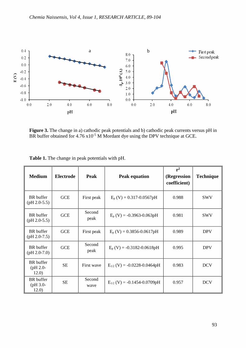
Chemia Naissensis, Vol 4, Issue 1, RESEARCH ARTICLE, 89-104
93
Figure 3. The change in a) cathodic peak potentials and b) cathodic peak currents versus pH in
BR buffer obtained for 4.76 x10⁻⁵ M Mordant dye using the DPV technique at GCE.
Table 1. The change in peak potentials with pH.
Medium Electrode Peak Peak equation
r2
(Regression
coefficient)
Technique
BR buffer
(pH 2.0-5.5) GCE First peak Ep (V) = 0.317-0.0567pH 0.988 SWV
BR buffer
(pH 2.0-5.5) GCE
Second
peak Ep (V) = -0.3963-0.063pH 0.981 SWV
BR buffer
(pH 2.0-7.5) GCE First peak Ep (V) = 0.3856-0.0617pH 0.989 DPV
BR buffer
(pH 2.0-7.0) GCE
Second
peak Ep (V) = -0.3182-0.0618pH 0.995 DPV
BR buffer
(pH 2.0-
12.0)
SE First wave E1/2 (V) = -0.0228-0.0464pH 0.983 DCV
BR buffer
(pH 3.0-
12.0)
SE Second
wave E1/2 (V) = -0.1454-0.0709pH 0.957 DCV
a b

Chemia Naissensis, Vol 4, Issue 1, RESEARCH ARTICLE, 89-104
94
DCV studies
DC voltammograms of the azo dye in BR buffer media were also studied. One broad
reduction wave was observed in the pH range of 2.0 to 12.0. The dependence of the peak
potential on pH is given in Table 1. The limiting current is approximately constant in neutral
and basic media. Electron transfer coefficient (α) was calculated from the Heyrovsky Ilkovic
equation. The change αn versus pH is given in Figure 4. The αn values are below 1.0 at all pH
values.
Figure 4. The change in αn with pH for Mordant dye in BR buffer with a GCE.
CV studies
To investigate the electrochemical behavior of the azo dyes, cyclic voltammetry is an
important technique that give information about the reversibility or irreversibility of the
electrode reactions. For this purpose, the CVvoltammograms obtained in BR buffer (pH 2.0-
12.0) are given in Fig. 5. During the cathodic potential sweep from 1.2 to -1.2 V, two cathodic
peaks (1c, 2c) were seen at 0.097 V and -0.617 V for pH 3.0 in B-R buffer, which were
attributed to the reduction of the azo group to amines in acidic media. On the reverse scan from
-1.2 to 1.2 V, small corresponding anodic peaks (1a, 2a) were observed at 0.319 and 0.692V
for pH 3.0 in BR buffer which were attributed to the oxidation of phenolic hydroxyl groups in
the Mordant dye (Fig. 5) (Hattori et al.,2006, Karaman, 2014). As stated in previous studies,
amines were produced with the reduction of the azo compounds, which have hydroxyl groups
adjacent to an azo group, which was most likely to be reoxidized in the return scan (Yu et al.,
0.0
0.5
1.0
1.5
2.0
0.0 2.0 4.0 6.0 8.0 10.0 12.0
αn
pH

Chemia Naissensis, Vol 4, Issue 1, RESEARCH ARTICLE, 89-104
95
2004). One reduction peak was observed in cathodic scan and one anodic peak was seen in
reverse scan in basic media. Peak currents have a sharp increase and decrease in the pH range
of 2.0-4.0. After pH 4.0, in the absence of the second peak, the first peak current values increase
with increasing pH (Fig. 6).
The peak potential values shifted to more negative values with increasing pH, indicated
that protons are involved in the electrode reaction (Fig. 6).
Figure 5. CV voltammograms of 4.76x10⁻⁵ M Mordant dye in BR (0.04 M) buffer at GCE a)
pH 2.0, 3.0, 4.0, 5.0, and 6.0 b) pH 9.5, 10.0 and 10.5.
Figure 6. The change in a) cathodic peak potentials and b) cathodic peak currents versus pH in
BR buffer obtained for 4.76x10⁻⁵ M Mordant dye using CV technique at GCE, v = 200 mV/s.
First peak
Second peak b a
pH pH
1a 2a
2c
1c

Chemia Naissensis, Vol 4, Issue 1, RESEARCH ARTICLE, 89-104
96
The effect of square root of scan rate (v = 20-1000 mV/s) on the peak current of the dye
at a GCE was examined in BR buffer at different pH values (pH 3.0, 4.5, 6.0 and 7.0) (Fig. 7).
Two reduction peaks were observed at pH 3.0 and one reduction peak was observed at pH 4.5,
6.0 and 7.0. The Ip versus v1/2 relation is not linear, and the Ip/v1/2 slope increases with increasing
scan rate. This result confirms that the azo compound has an adsorption controlled reaction on
the electrode surface (Char et al., 2008, Guaratini et al., 2001).
Figure 7. The change in cathodic peak currents with square root of scan rate at different pH
values at GCE for 4.76 x10⁻⁵ M Mordant dye in BR buffer, v = 20-1000 mV/s.
Small anodic peaks were observed at acidic and neutral pH. However, the exponential
dependence of peak current with the square root of scan rate is evidence of the kinetic
controlling reaction occurring. This indicates that the reaction mechanism is complex.
Silver Electrode
SWV and DPV studies
The SW and DP voltammograms of the Mordant dye in BR (pH 2.0-12.0) buffer are
presented in Figure 8. Two reduction peaks were seen at pH 6.0, 7.0, 9.5 and 11.0 for SWV
technique and two reduction peaks were observed at pH 3.0, 4.0, 5.0, 6.0, 7.0, 8.0 and 9.0 for
DPV technique (Fig. 8). The presence of two peaks indicated that two-step electrode reaction
occurred at the electrode surface. As seen in Fig. 8, the broad peak of the azo dye occurred is
pH 3.0
pH 3.0
pH 4.5
pH 6.0
pH7.0

Chemia Naissensis, Vol 4, Issue 1, RESEARCH ARTICLE, 89-104
97
due to its adsorption on the electrode surface (Char et al., 2008, Karaman and Menek, 2012, Ma
and Song, 2008, Mirceski and Lovri, 2004, Sun et al., 2005).
Figure 8. Voltammograms of 4.76 x10⁻⁵ M Mordant dye in BR buffer (0.04 M) at SE a) SWV,
(pH 6.0, 7.0, 9.5 and 11.0, v = 200 mV/s) and b) DPV (pH 3.0, 4.0, 5.0, 6.0, 7.0, 8.0 and 9.0).
Figure 9. The change in cathodic peak potentials versus pH in BR buffer for Mordant dye at
SE a) SWV, b) DPV.
0.000 -0.200 -0.400 -0.600 -0.800
E (V)
-1.00e-6
-2.00e-6
-3.00e-6
-4.00e-6
-5.00e-6
-6.00e-6
I (A
)0.000 -0.200 -0.400 -0.600 -0.800 -1.000
E (V)
-1.00e-5
-1.50e-5
-2.00e-5
-2.50e-5
-3.00e-5
I A
)
7.0
9.5
11.0
6.0
3.0
4.0
5.0 6.0
9.0 8.0

Chemia Naissensis, Vol 4, Issue 1, RESEARCH ARTICLE, 89-104
98
Figure 10. The change in cathodic peak currents versus pH in BR buffer for Mordant dye at SE
a) SWV, b) DPV.
The change in peak currents versus pH for the azo dye is approximately constant in the
range of pH 6.0-9.0 and decreases after pH 9.5 for the SWV technique. However, in DPV
technique, it is seen that the peak potential is almost constant in the range of pH 3.5-9.0 and
decreases at higher pH values (Fig. 9). As seen in SW and DP voltammograms (Figs. 9, 10),
after pH 3.0, the brode peak was observed at negative potentials. These results indicates that
there is a two step reduction reaction occurs on the electrode surface.
DCV studies
DC voltammograms of the azo dye in BR buffer are shown in Fig. 11. Two reduction
waves were seen after pH 3.0 and at pH 9.0, while one reduction wave was observed at pH
7.0-9.0. As seen in Fig. 11, first and second reduction wave potentials of the dye shifted to more
negative values with increasing pH, showed that protons are involved in the electrode reaction.
The linear regression equations of azo dye between the half wave potentials and pH were given
for the DCV technique in BR buffer media (Table 1).

Chemia Naissensis, Vol 4, Issue 1, RESEARCH ARTICLE, 89-104
99
Figure 11. DC voltammograms of 4.76 x10⁻⁵ M Mordant dye in BR (0.04 M) buffer at SE a)
pH 3.0, 3.5, 4.0 and 4.5 b) pH 6.0, 8.0 and 10.5
.
The αn values were calculated from the logarithmic analyses of the reduction waves by
using the Heyrovsky-Ilkovic equation in BR buffer (2.0–12.0). (Bard and Faulkner, 1980,
Karaman, 2014, Meites, 1965, Zuman and Perin, 1965). The αn values of the second wave in
the acidic region (pH 3.0, 3.5 and 4.0) are greater than the αn values of the first wave, although
both give similar results in the basic region (pH 10.0, 10.5 and 11.0) (Fig. 12).
Figure 12. The change in αn with pH for Mordant dye in BR buffer recorded at SE.
First wave
Second wave
0.00 -0.20 -0.40 -0.60 -0.80 -1.00
E (V)
0
-2.00e-7
-4.00e-7
-6.00e-7
-8.00e-7
-1.00e-6
I (A
)
0.00 -0.10 -0.20 -0.30 -0.40 -0.50 -0.60
E (V)
0
-5.00e-8
-1.00e-7
-1.50e-7
-2.00e-7
-2.50e-7
-3.00e-7
I (A
)
10.5
6.0
8.0
4.5
3.0
4.0

Chemia Naissensis, Vol 4, Issue 1, RESEARCH ARTICLE, 89-104
100
CV studies
CV voltammograms recorded in BR buffer (pH 2.0-12.0) are given in Fig. 13. Two
reduction peaks were observed in cathodic scan, which was attributed to the reduction of the
azo group to amines in acidic media, and two anodic peaks were observed in reverse scan in
acidic media. One reduction peak was observed in cathodic scan and one anodic peak was seen
in reverse scan in basic media.
Figure 13. CV voltammograms of 4.76x10⁻⁵ M Mordant dye in BR (0.04 M) buffer at SE a) pH
2.0, 3.0, 3.5, and 4.0, b) pH 7.0, 9.0 and 11.0 solutions.
Figure 14. a) The change in cathodic peak currents of Mordant dye with square root of scan
rate at different pHs at SE, v = 20-1000 mV/s.
First peak Second peak
4.0
3.5
3.0
11.0
7.0
9.0

Chemia Naissensis, Vol 4, Issue 1, RESEARCH ARTICLE, 89-104
101
The effect of square root of scan rate (v = 20-1000 mV/s) on the cathodic peak currents
of the dye at a SE was examined in BR buffer at different pH values (Fig. 14). Two reduction
peaks were observed at pH 4.5, 6.0, 7.0, 9.5 and 11.0 and one reduction peak was observed at
pH 3.0. The linear relation of cathodic peak currents versus v with increasing scan rate,
indicating that the azo compound has an adsorption-controlled reaction on the electrode surface
(Char et al., 2008, Guaratini et al., 2001, Yu et al., 2004). The reaction mechanism of Mordant
dye at glassy carbon and silver electrodes, has been proposed from the voltammetric data
(Scheme 2).
Scheme 2. The proposed reaction mechanism of Mordant dye.
2e-
2H+
+
2e-
2H+
+2e-
2H+
+
+
OHS
O
O
O-Na+
NH2
S
-Na+
OH
OO
O
NH2
NH2
OHS
O
O
O-Na+
N
H H
S
-Na+
OH
OO
O
N
NH2
OHS
O
O
O-Na+
N
H H
S
-Na+
OH
OO
O
N
NH2
OHS
O
O
O-Na+
N
S
-Na+
OH
OO
O
N
NH2
Basic media, pH > 9 Acidic and neutral media, pH < 9

Chemia Naissensis, Vol 4, Issue 1, RESEARCH ARTICLE, 89-104
102
Conclusions
The electrochemical behavior of Mordant dye at a glassy carbon electrode and silver
electrode has been studied in BR buffer (pH 2.0-12.0) media by using SWV, DPV, DCV and
CV techniques. Two reduction peaks were observed at pH < 9.5, and one reduction peak was
observed at pH > 9.5 for SWV and DPV techniques with a glassy carbon electrode and silver
electrode. The presence of two peaks at pH < 9.5 and a single reduction peak at pH > 9.5,
indicated that two-step electrode reaction occurred at pH < 9.5 and one-step electrode reaction
occurred at pH > 9.5. The effect of the scan rate on the peak current studies, indicates that the
electrode process of the dye at glassy carbon and silver electrodes is adsorption controlled. It
was observed from SWV, DPV, DCV and CV techniques that reduction reaction of the Mordant
dye corresponds to the cleavage of the azo bond to give the reductive amines.
Conflict-of-Interest Statement:
None.
References
Abu-El-Wafa, S. M., El-Wakiel, N. A., Issa, R. M.,& Mansour, R. A. (2005). Formation of
novel mono- and multi-nuclear complexes of Mn(II), Co(II) and Cu(II) with bis azo-dianils
containing the pyrimidine moiety: Thermal, magnetic and spectral studies. Journal of
Coordination Chemistry, 58, 683-694.
Ali, Dr. N. F., EL-Khatib, E. M., Saadia, A.,& El-Megied A. (2019). Modification of wool and
silk fibers by pretreatment with quaternary ammonium salt and dyeing with new metal complex
dye. World Journal of Pharmaceutical and Life Sciences, 5, 73-79.
Bard, A. J. & Faulkner, L. R. (1980). Electrochemical Methods, JohnWiley & Sons, New York.
Chandra, U., Gilbert, O., Kumara Swamy, B. E., Bodke, Y. D., & Sherigara, B. S. (2008).
Electrochemical studies of Eriochrome Black T at carbon paste electrode and immobilized by
SDS surfactant: A cyclic voltammetric study. International Journal of Electrochemical Science,
3, 1044-1054.
Char, M. P., Niranjana, E., Kumara Swamy, B. E., Sherigara, B. S., & Pai, V. (2008).
Electrochemical studies of Amaranth at surfactant modified carbon paste electrode: A cyclic
voltammetry. International Journal of Electrochemical Science, 3, 588-596.
Chemia Naissensis, Vol 4, Issue 1, RESEARCH ARTICLE, 89-104
103
Ding, Y.& Freeman, H. S. (2017). Mordant Dye application on cotton: optimisation and
combination with natural dyes. Coloration Technology, 133, 369-375.
Eriksson, A.,& Nyholm, L. (2001). Coulometric and spectroscopic investigations of the
oxidation and reduction of some azosalicylic acids at glassy carbon electrodes. Electrochimaca
Acta, 496, 1113-1129.
Guaratini, C. C. I., Fogg, A. G., & Zanoni, M. V. B. 2001. Studies of the Voltammetric Behavior
and Determination of Diazo Reactive Dyes at Mercury Electrode. Electroanalysis, 13, 1535-
1543.
Hattori, T., Tsurumi, N., Kato R., & Nakayama M. (2006). Adsorptive voltammetry of 2-(5-
bromo-2-pyridyl)azo-5-[N-n-propyl-N-(3sulfopropyl)amino]phenol on a carbon pasteelectrode
in the presence of organic cations and polycation. Analytical Science, 22, 1577-1580.
Karaman, Y., & Menek, N. (2012). Investigation of electrochemical behavior of 2-(5-bromo-2-
pyridylazo)-5-[N-propyl-N-(3-sulfopropyl)amino]phenol disodium salt dihydrate. Journal of
the Electrochemical Society, 159, H805-H810.
Karaman, Y. (2014). Investigation of electrochemical behavior of Calmagite at a glassy carbon
electrode. Dyes and Pigments,106, 39-44.
Lucilha, A. C., Bonancêa, C. E., Barretoc, W. J., & Takashima, K. (2010). Adsorption of the
Diazo Dye Direct Red 23 onto a zinc oxide surface: A spectroscopic study. Spectrochimica
Acta Part A, 75, 389-393.
Ma, M. M., & Song, J. F. (2008). Square wave voltammetric label-free determination of the
natural protein material silk fibroin. Chinese Journal of Chemistry, 26, 2081-2085.
Meites, L. (1965). Polarographic Techniques. John Wiley & Sons, New York.
Menek, N. (1998). Polarographic and voltammetric behaviour of 2-hydroxy-3-methoxy-5-(2-
propenyl)azobenzene. Analytical Letters, 31, 275-282.
Mirceski, V., & Lovri, M. (2004). EC mechanism of an adsorbed redox couple. Volume vs
surface chemical reaction. Journal of Electroanalytical Chemistry, 565, 191-202.
Pervez, M. N., Telegin, F. Y., Cai, Y., Dongsheng, Xia, D., Zarra, T., & Naddeo, V. (2019).
Efficient degradation of Mordant Blue 9 using the fenton-activated persulfate system. Water,
11, 2532-2546.

Chemia Naissensis, Vol 4, Issue 1, RESEARCH ARTICLE, 89-104
104
Romanini, D. C., Trindade, M. G. A., & Zanoni, M. V. B. (2009). A simple electroanalytical
method for the analysis of the dye solvent orange 7 in fuel ethanol. Fuel, 88: 105-109.
Socha, A., Sochocka, E., Podsiad1y, R., & Soko1owska, J. (2007). Electrochemical and
photoelectrochemical treatment of C.I. Acid Violet 1. Dyes and Pigments, 73, 390-393.
Sun, W., Jiang, H., & Jiao, K. (2005). Electrochemical determination of hydrogen peroxide
using o-dianisidine as substrate and hemoglobin as catalyst, Journal of Chemical Science, 117,
317–322.
Tvorynska, S., Josypcuk, B., Jiri Barek, J., & Liliya Dubenska, L. (2019). Electrochemical
behavior and sensitive methods of the voltammetric determination of food azo dyes Amaranth
and Allura Red AC on amalgam electrodes. Food Analytical Methods, 12, 409-421.
Yıldız, E., & Boztepe, H. (2002). Synthesis of novel acidic mono azo dyes and an investigation
of their use in the textile industry. Turkish Journal of Chemistry, 26, 897-903.
Yu, J., Jia, J., & Ma, Z. (2004). Comparison of electrochemical behavior of hydroxyl-
substituted and nonhydroxyl-substituted azo dyes at a glassy carbon electrode. Journal of the
Chinese Chemical Society, 51, 1319-1324.
Zuman, P., & Perin, C. L. (1965). Organic Polarography. John Wiley & Sons, New York.

Chemia Naissensis, Vol 4, Issue 1, RESEARCH ARTICLE, 105
105
Istraživanje elektrohemijskog ponašanja Mordant boje (C.I. 17135) na
staklenim ugljeničnim i srebrnim elektrodama
Necati Menek1*, Serpil Zeirekli1, Jeliz Karaman2
1- Univerzitet Ondokuz Mais, Fakultet nauka i umetnosti, Departman za hemiju, 55139, Kurupelit-
Samsun, Turska
2- Sinop Univerzitet, Fakultet nauka i umetnosti, Departman za hemiju, 57000, Sinop, Turska
SAŽETAK
U ovoj studiji je ispitivano elektrohemijsko ponašanje Mordant boje (CI 17135) u puferu Briton-
Robinson (BR) (pH 2,0-12,0) različitim voltametrijskim tehnikama: voltametrija kvadratnog
talasa (SVV), ciklična voltametrija (CV), diferencijalna impulsna voltametrija (DPV) i
voltametrija jednosmerne struje (DCV). Elektrohemijsko ponašanje boje ispitano je upotrebom
staklene ugljenične elektrode (GCE) i srebrne elektrode (SE). Široki vrh azo boje nastao je na SV
i DP voltamogramima, zbog njegove adsorpcije na staklenim površinama ugljenične i srebrne
elektrode. Dva redukciona pika su primecena pri pH <9,5, a jedan redukcioni pik je primecen pri
pH> 9,5 za SVV i DPV tehnike na staklenoj ugljeničnoj elektrodi. Prema voltametrijskim
podacima, predložen je elektrohemijski mehanizam reakcije azo boje na staklenim ugljeničnim i
srebrnim elektrodama.
Ključne reči: Azo jedinjenje, Mordant boje, voltametrija, mehanizam reakcije.

Chemia Naissensis, Vol 4, Issue 1, RESEARCH ARTICLE, 106
106
Enquête sur le comportement électrochimique du colorant Mordant (C.I.
17135) aux électrodes de carbone vitreux et d’argent
Necati Menek1*, Serpil Zeyrekli1, Yeliz Karaman2
1- Université Ondokuz Mayıs, Faculté des sciences et des arts, Département de chimie, 55139, Kurupelit-
Samsun, Turquie
2- Sinop Université, Faculté des sciences et des arts, Département de chimie, 57000, Sinop, Turquie
ABSTRACT
Dans cette étude, le comportement électrochimique du colorant Mordant (CI 17135) a été examiné
dans un milieu tampon Britton-Robinson (BR) (pH 2,0-12,0) en utilisant les différentes techniques
de voltampérométrie : la voltamétrie à onde carrée (SWV), la voltamétrie cyclique (CV), la
voltamétrie différentielle à impulsions (DPV) et la voltamétrie à courant continu (DCV). Le
comportement électrochimique du colorant a été recherché au moyen de l’électrode de carbone
vitreux (GCE) et de l’électrode d’argent (SE). Le pic large du colorant azoïque survenu aux
voltamogrammes SW et DP est dû à son adsorption sur les surfaces vitreuses des électrodes en
carbone et en argent. Les deux pics de réduction ont été observés à pH < 9,5, tandis qu’un pic de
réduction a été observé à pH > 9,5 pour les techniques SWV et DPV sur une électrode en carbone
vitreux. Suivant les données voltamétriques, le mécanisme électrochimique de la réaction du
colorant azoïque a été suggéré au niveau des électrodes de carbone vitreux et d’argent.
Mots-clés : Composé azoïque, Colorants mordants, Voltamétrie, Mécanisme de réaction.

Chemia Naissensis, Vol 4, Issue 1, RESEARCH ARTICLE, 107
107
Исследование электрохимического поведения красителя протравы (C.I.
17135) на стеклоуглеродных и серебряных электродах
Нечати Менек1*, Серпил Зейрекли1, Елиз Караман2
1-Университет Ондокуз Майис, Факультет наук и искусств, Кафедра химии, 55139, Курупелит-
Самсун, Турция
2-Синопский университет, Факультет наук и искусств, Кафедра химии, 57000, Синоп, Турция
АННОТАЦИЯ
В этом исследовании электрохимическое поведение красителя протравы (CI 17135) было
исследовано в среде с буфером Бриттона-Робинсона (BR) (pH 2,0-12,0) с использованием
различных вольтамперометрических методов: прямоугольной вольтамперометрии (СВВ),
циклической вольтамперометрии (ЦВ), дифференциальной импульсной
вольтамперометрии (ДПВ) и вольтамперометрией постоянного тока (ДЦВ).
Электрохимическое поведение красителя было исследовано с использованием
стеклоуглеродного электрода (СУE) и серебряного электрода (СE). Широкий пик
азокрасителя, возникающий на вольтамперограммах СВ и ДП, связан с его адсорбцией на
поверхности стеклоуглерода и серебряного электрода. Два пика восстановления наблюдали
при pH <9,5, и один пик при pH> 9,5 для методов СВВ и ДПВ на стеклоуглеродном
электроде. На основании данных вольтамперометрии был предложен механизм
электрохимической реакции азокрасителя на стеклоуглеродных и серебряных электродах.
Ключевые слова: азосоединение, протравы, вольтамперометрия, механизм реакции.

Chemia Naissensis, Vol 4, Issue 1, RESEARCH ARTICLE, 108
108
Untersuchung zum elektrochemischen Verhalten von Mordant-Farbstoff (C.I.
17135) an Glaskohlenstoff- und Silberelektroden
Necati Menek1*, Serpil Zeyrekli1, Yeliz Karaman2
1- Ondokuz Mayıs Universität, Fakultät für Wissenschaften und Kunst, Lehrstuhl für Chemie, 55139,
Kurupelit-Samsun, Türkei
2- Sinop Universität, Fakultät für Wissenschaften und Kunst, Lehrstuhl für Chemie, 57000, Sinop, Türkei
ABSTRACT
In dieser Studie wurde das elektrochemische Verhalten von Mordant-Farbstoff (CI 17135) in
Britton-Robinson (BR)-Puffer (pH 2,0-12,0) unter Verwendung verschiedener voltammetrischer
Techniken untersucht: Rechteckwellenvoltammetrie (SWV), zyklische Voltammetrie (CV),
Differenzpulsvoltammetrie (DPV) und Gleichstromvoltammetrie (DCV). Das elektrochemische
Verhalten des Farbstoffs wurde unter Verwendung einer Glaskohlenstoffelektrode (GCE) und
einer Silberelektrode (SE) untersucht. Der breite Peak des Azofarbstoffs, der bei SW- und DP-
Voltamogrammen auftrat, ist auf seine Adsorption an den Glasoberflächen der Kohlenstoff- und
Silberelektrode zurückzuführen. Bei pH < 9,5 wurden zwei Reduktionspeaks beobachtet, und ein
Reduktionspeak wurde bei pH > 9,5 für SVV- und DPV-Techniken an einer
Glaskohlenstoffelektrode beobachtet. Nach den voltammetrischen Daten wurde ein
elektrochemischer Reaktionsmechanismus des Azofarbstoffs an Glaskohlenstoff- und
Silberelektroden vorgeschlagen.
Schlüsselwörter: Azoverbindung, Beizenfarbstoffe, Voltammetrie, Reaktionsmechanismus

ISSN 2620-1895