Embed Size (px)
Citation preview
Vox Sang. 27: 227-231 (1974)
Separation of Hepatitis-Associated Antigen (HAA) from Human Plasma for the Production of Rabbit Anti-HAA
J. R. BOOTH, I. C. WISEMAN and D. LEE^
Sheffield Regional Blood Transfusion Centre, Sheffield
Abstract. A method is described for obtaining hepatitis-associated antigen (HAA) from the acid citrate dextrose plasma of routine blood donations found to be HAA-positive. The separation consisted of the following steps: ammonium sulphate precipitation, absorption on and elution from DEAE-Sephadex, and gel filtration on a Sepharose-6B column. HAA was demonstrated by cross-over immuno-electrophoresis and lines of identity shown by the Ouchterlony double diffusion technique. Freedom from contaminat- ing protein was shown by immuno-electrophoresis. This material, when injected into rabbits, produced an anti-HAA reagent suitable for use cross-over immuno-electro- phoresis without absorption.
Introduction
The characteristics of the hepatitis-associated antigen (HAA) were first described in 1966 by ALTER and BLUMBERG [l]. Since then, GERIN et al. [5 ] and a number of other authors have described its purification using the ultracentrifuge. Expensive and sophisticated apparatus is not available for use in a Blood Transfusion Centre on potentially infective materials, where the same apparatus is required for use in the preparation of other blood products.
The separation of the antigen seemed necessary after our experience with goat anti-HAA production [unpublished], in which it was found that, following injections of whole human HAA-positive plasma, the normal plasma protein fractions induced a greater response than the HAA fraction.
The authors wish to thank Dr. C. C. BOWLEY for his encouragement and advice.
Received: October 24, 1973; accepted:
228 BOOTH/WISEMAN/LEE
100ml plasma I----- 40 rnl saturated (NH,,),SO,
1- 40ml saturated(NH4),S04
1- 15ml H,O
Precipitate t Supernatant
Supernatant + Precipitate
Dialysisx H,O, 4x41
1 1
Precipitate + 25 ml supernatant
DialysisxO.1~ Tris-PO;-buffer pH7.0, 3x11
DEAE in 0 . 1 ~ Tris-PO;. buf fer pH 7.0
25 mi k supernatant
1 1
Sepharose-GB column 5-ml aliquots
Concentrate for final product
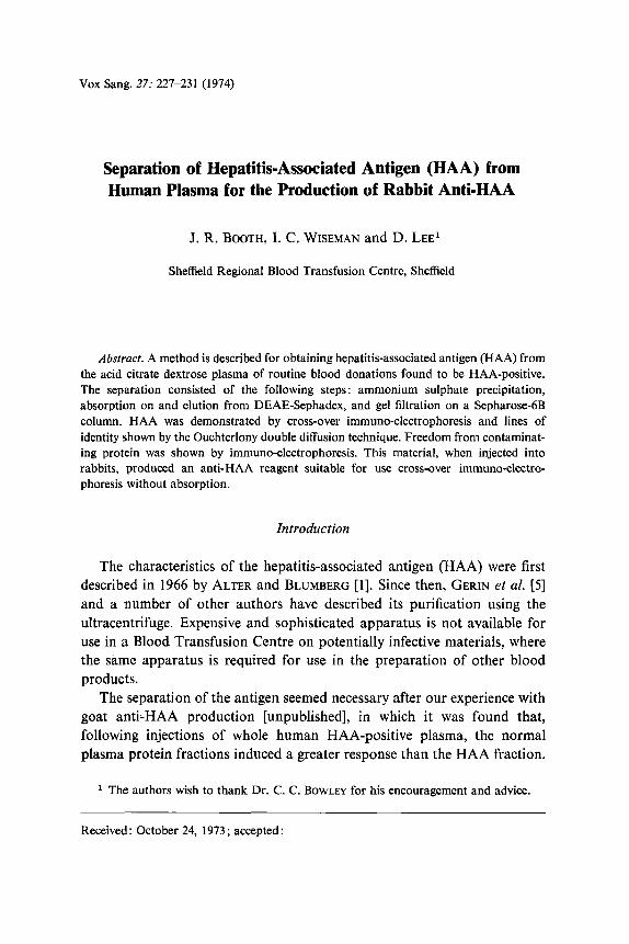
Fig. 1. Separation of HAA.
A purification which could be carried out in a restricted laboratory was described by DUIMEL et al. [4]. This is a report on an alternative method for the separation of HAA using saturated ammonium sulphate (NH,),SO,, DEAE-Sephadex and Sepharose-6B, and its use in the preparation of anti- HAA in rabbits.
Materials and Methods
Separation Acid citrate dextrose plasma from routine blood donations, which had been tested and
found HAA-positive by the cross-over immuno-electro-osmophoresis (IEOP) technique of CULLIFORD [2] modified by M ~ L N E and BARR [7], and confirmed by gel diffusion identity patterns, was removed for processing (fig. 1).
To 100 ml plasma, 40 ml saturated (NH,)$04 were added and the precipitate removed by centrifugation in an MSE Super Minor Windshield Centrifuge (MSE, London, England) at 1,800 g for 20 min, using tightly sealed 1-02 glass universal containers with screw caps. A further 40 ml saturated (NH,),SO, were added to the supernatant, and the resulting precipitate was collected by centrifugation at 1,800 g for 30 min. This precipitate was then washed with 45% saturated (NH,),SO, three times, using 160 ml (NH,),SO, per wash.
The washed precipitate was dissolved in a minimum of distilled water (15 ml) and dialysed against four changes of 4 1 distilled water, using Visking Tubing (Scientific Instruments Centre). The resulting 25 ml were centrifuged to remove the euglobulin
Separation of HAA and Anti-HAA Production 229
precipitate and then dialysed against three changes of 1-litre 0.1 M Tris-phosphate buffer pH 7.0 for 20 h.
DEAE-Sephadex was swollen and equilibrated in 0.1 M Tris-phosphate buffer pH 7.0 for a minimum of 24 h. Equal volumes of equilibrated DEAE-Sephadex and the above dialysate were mixed and left for 2 h at 25°C with occasional mixing. The supernatant was removed and divided into 5-ml aliquots and chromatographed on a Sepharose-6B column (2.5 cm x 50 cm Pharmacia, flow length 40 cm) using 0.02 M acetic acid-sodium acetate buffer, prepared from a stock solution of 0.4 M ph 5.2.
The column flow rate was 1 ml/min, and after the void volume, 9 fractions, each of 20 ml, were collected manually and concentrated to 1 ml by dialysis against Carbowax (MW : x 104).
Immunisation Programme Three rabbits, 1403, 1435 and 1457 were each given 1 ml of the preparation intra-
muscularly (i.m,) in both hind legs. After a period of 9 weeks, each was given a single hjection of 0.5 ml i.m., followed by a third i.m. injection of 0.5 ml 2 weeks later. The rabbits were bled on the loth, 11th and 13th day after the second and third injections.
Results
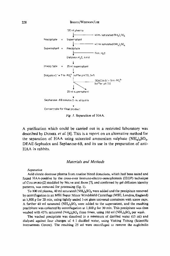
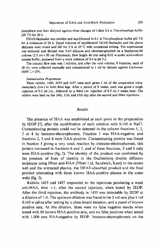
The presence of HAA was established at each point in the preparation by IEOP [7], after the equilibration of each solution with 0.145 M NaCl. Contaminating protein could not be detected in the column fractions 1, 2, 3 or 4 by immuno-electrophoresis. Fraction 1 was HAA-negative, and fractions 2, 3 and 4 were HAA-positive. Contaminating protein was found in fraction 5 giving a very weak reaction by immuno-electrophoresis, this protein increased in fractions 6 and 7, and of these fractions, 5 and 6 only were HAA-positive (fig. 2). The identity of the product was confirmed by the presence of lines of identity in the Ouchterlony double diffusion technique using Pfizer anti-HAA (Pfizer Ltd, Sandwich, Kent) in the centre well and the untreated plasma, the DEAE-absorbed product and the final product alternating with three known HAA-positive plasma in the outer wells (fig. 3).
Rabbits 1435 and 1457 responded to the injections producing a weak anti-HAA, titre <1, after the second injection, when tested by IEOP. After the third injection, the antibody in 1435 was detectable by IEOP at a dilution of 1 : 6. The optimum dilution was found to be 2 vol sera plus 1 vol 0.145 M saline after testing by a chess board titration, and a panel of known positive sera. At this dilution, there were no false negative results when tested with 80 known HAA-positive sera, and no false positives when tested with 1,000 sera HAA-negative by IEOP. Tmmuno-electrophoresis on the
230 BOOTH/WISEMAN/LEE
F1 F2 F3 F4 F5 F6
Fig. 2. Immuno-electrophoresis using anti-human serum of untreated plasma (A), (NH&SO,-washed precipitate (B) and DEAE-absorbed product (C). Fl-F7 = Column fractions 1-7, F2-F6 = HAA-positive; F1 and F7 = HAA-negative.
@ Fig. 3. Well arrangement for demonstrating lines of identity. Ab = Pfizer anti-HAA; M, G, and W = known HAA-positive plasma; 1 = untreated plasma; 2 = DEAE-absorbed product; 3 =final protein-free HAA-positive product. 0
prepared reagent revealed three weak bands with human HAA-negative serum.
The weak antibody produced by rabbit 1457 was not significantly improved by the third injection. Rabbit 1403 did not produce anti-HAA or any antibodies to human serum proteins as demonstrable by immuno- electrophoresis.
Discussion
There is a limited availability of satisfactory human anti-HAA from random blood donors, and attempts at producing anti-HAA by repeated injections of whole HAA-positive plasma into experimental animals,
Separation of HAA and Anti-HAA Production 231
suffered from difficulties due to the higher immunogenicity of some normal human plasma proteins.
Human serum proteins in fractionated human plasma, which cannot be detected by immuno-electrophoresis, when injected into rabbits can, nevertheless, raise antibodies. In the production of anti-HAA using the preparation described, some weak antibodies to serum proteins were pro- duced, but these did not interfere with the HAA-anti-HAA reaction using the IEOP method. The introduction of a pepsin digestion stage [6] in the preparation may reduce the number of these contaminating antibodies.
During the development of this method, a number of different plasma preparations were chromatographed on the Sepharose-6B column. Due to the heterogeneity of the antigen particle [3], a variation was found in the point at which the separated antigen appeared, although this point always lay within the 9 fractions collected. By selection of an appropriate flow rate (1 cm3/min), it is possible to collect all the relevant fractions in the space of a working day.
Thus, a simple method for the separation of HAA from human plasma, which can be carried out in a restricted area with a minimum of apparatus, has successfully been developed and the product used to raise a rabbit anti-HAA which does not require absorption.
References
1 ALTER, H. J. and BLUMBERG, B. S.: Further studies on a ‘new’ human isoprecipitin
2 CULLIFORD, B. J . : Precipitin reactions in forensic problems. Nature 201: 1092 (1964). 3 DANE, D. S.; CAMERON, C. H., and BRIGGS, M. : Virus-like particles in serum of patients
with Australia-antigen-associated hepatitis. Lancet i: 695 (1970). 4 DUIMEL, W. J. M.; BRUMMELHUIS, H. G. J., and KRIJNEN, H. W.: The purification of
hepatitis-associated antigen (HAA/SH/Au.) from human serum and the preparation of a sheep anti-HAA serum. Vox Sang. 23: 228 (1972).
5 GERIN, J. L.; PURCELL, R. H. ; HOGGAN, M. D.; HOLLAND, P. V., and CHARNOCK, R. M.: Biophysical properties of Australia antigen. J. Virol. 4: 763 (1969).
6 KIM, C. Y.; SPANO, J., and CLARK, G. E.: Use of pepsin treated hepatitis associated antigen in the production of precipitating antibody. J. infect. Dis. 124: 411 (1971).
7 MILNE, G. R. and BARR, A. : A rapid method for testing blood for hepatitis associated antigen and antibody using countercurrent electrophoresis in agarose gel. Sterilin techn. Bull. No. 1 (1971).
system (Australia antigen). Blood 27: 297 (1966).
Request reprints from: Mr. J. R. BOOTH, Senior Scientific Officer, National Blood Trans- fusion Service, Longley Lane, Shefield S5 7JN (England)