Embed Size (px)
Citation preview
Vol. 5: 197-204, 1988 DISEASES OF AQUATIC ORGANISMS Dis. aquat. Org.
Published December 2
Separation and in vivo analysis of two extracellular proteases and the T-hemolysin from
Aeromonas salmonicida
D. D. Rockey, J. L. Fryer, J. S. Rohovec
Department of Microbiology, Oregon State University. Corvallis, Oregon 97331, USA
ABSTRACT: A procedure using DEAE cellulose and hydroxylapetite was developed for the separation of Aeromonas salmonicida P1 protease, P2 protease, and trout cell-specific hemolysin (T-lysin) from supernatants of broth cultures. The Mferent proteases were demonstrated using protease inhibitors, substrate specificities, and polyacrylamide gel electrophoresis with protease substrates in the gels. Isolated P1 protease and T-lysin were shown to act independently in the complete lysis of rainbow trout erythrocytes in vitro. The T-lysin acted on the outer membrane and P1 protease acted on the nuclear membrane. Analysis of cell-free exudate from lesions of trout infected with A. salmonicida by injection demonstrated that more than one protease was also present in vivo. P1 protease was present in both lesions and in culture supernatants, but P2 protease was detected only in culture supernatants. No T-lysin activity was detected in cell-free exudate from lesions caused by A. salmonicida infection.
INTRODUCTION
Aeromonas salmonicida, the causative agent of fish furunculosis, inflicts serious losses in hatchery-reared salmon and trout in many parts of the world (McCarthy & Roberts 1980). Although efficacious vaccines have been developed for other, similar, gram-negative fish pathogens, commercial furunculosis vaccines have only recently been made available, and documentation of efficacy is absent in the literature (Michel 1985). Therefore, virulence factors of this organism have been the object of much research. Previous work has included investigations of cell-associated and secreted components of the bacterium. An outer membrane pro- tein matrix, the A layer, has been directly associated with virulence, and the role of Lipopolysaccharide in bacterial sunrival has also been addressed (Udey &
Fryer 1978, Munn et al. 1982). Non-cellular factors secreted by A. salmonicida have been shown to be directly responsible for many of the clinical signs associated with the disease (Ellis et al. 1981). Secreted factors which have been investigated include pro- teases, hemolysins, and a leukocidin. Several authors have reported purification of these secreted proteins, as well as their individual and interactive characteristics (Fuller et al. 1977, Sheeran & Smith 1981, Titball &
O Inter-Research/Printed in F. R. Germany
Munn 1981, 1983, 1985, Hastings & Ellis 1985, Fyfe et al. 1987).
Investigation of Aeromonas salmonicida proteases began with Dahle (1971) who reported characteristics of proteases of A. salmonicida and A. hydrophila. Sub- sequent research described a variety of proteolytic fac- tors. The major secreted protease is a serine protease of molecular weight 70000 with activity against casein and gelatin (Tajima e t al. 1983, Fyfe e t al. 1986). Sheeran & Smith (1981) reported 2 activities in culture supernatants of A. salrnonicida one of which (PI) was the major serine protease; the other was a n ethylene diamine tetraacetic acid (EDTA) sensitive protease (P2). P1 protease was active against casein and gelatin, while P2 protease had activity against gelatin but not casein. Other authors have reported a low molecular weight protease and a fibrinolytic protease with molecular weight 87 500 (Shieh & MacLean 1975, Mellergaard 1983). The presence of alternate proteases was disputed by Hastings & Ellis (1985) based on results associating all gelatinolytic activity with a single isoelectnc point in preparative isoelectric focusing. This suggested that a single component accounted for the activity.
In order to facilitate analysis of secreted virulence factors of Aeromonas salmonicida, we have developed
198 Dis. aquat. Org. 5: 197-204, 1988
a single technique for the separation of P1 protease, P2 protease, and T-lysin from culture supernatants. Using this technique, we demonstrated that multiple pro- teases are present, and have separated these activities. The relationship between the T-lysin and P1 protease in the complete lysis of trout RBC was also investigated in vitro. In addition, proteolytic and hemolytic activities were investigated in vivo. It was shown that more than one protease was present in lesions caused by injection of the microorganism, but in vivo hemolytic activity was not detected.
MATERIALS AND METHODS
Bacterial strains. Experiments were conducted with a recent isolate of Aeromonas salmonicida (RC1) from diseased spring chinook salmon Oncorhynchus tshawytscha at the Round Butte Salmon Hatchery, Madras, Oregon, USA. Comparisons for strain variation were conducted with strain SS70 (Udey & Fryer 1978). Cultures were stored on brain heart infusion (BHI) agar (Difco) at 4°C and grown for harvest in 500ml BHI broth cultures at 17 'C for 48 h with shaking.
Separation of virulence factors. Forty-eight hour broth cultures of Aeromonas salmonicida were pelleted at 2 5 0 0 ~ g for 15min and the supernatant removed and recentrifuged. The resulting supernatant was transferred to a flask on ice and powdered NH4S04 (Mallinckrodt, GenAR grade) was added over a 10min period with constant stirring to 45 O/O saturation (32 g 100ml-l supernatant). Fifty ~ ( 1 of 1 N NaOH were added for every 10 g NH4S04. The solution was stirred on ice for 20min and then centrifuged for 20min at 10000 X g. The pellet was dissolved in water, filter- sterilized, and extensively dialysed against 20 m M Tris pH7.9 at 4 "C. The supernatant from the previous cen- trifugation was concentrated with a second NH4S04 precipitation by increasing the NH4S04 concentration to 65 O/O saturation (45.8 g 100 ml-l supernatant) and repeating the pelleting and dialysis. After dialysis, ion exchange chromatography using DEAE cellulose (Whatman DE23) was performed and fractions were eluted using step-gradients of 0.1, 0.15, 0.2, and 0.35 M NaCl in 2 0 m M Tris pH7.9. Fractions containing pro- teolytic enzymes or hemolytic activity against trout red blood cells (T-lysin) were collected and dialysed into a 1 m M potassium phosphate buffer, pH 7.0. These samples were chromatographed on a hydroxylapetite column (HAP, Bio-Rad Laboratories) and step-eluted a t 0.01, 0.1, and 0.5 Mphosphate. The T-lysin was further purified on Sepharose 6 B (Pharmacia) if necessary to remove contaminating proteolytic activity as described by Titball & Munn (1981). All manipulations were per- formed at 4 to 8'C.
Protease assays. Proteolytic activity was qualitatively assayed using a 10 % gelatin overlay on 1 '30 skim m ~ l k agar in Petn dishes. Seven p1 of sample was injected through both layers with a micropipettor. Plates were incubated 3 h at 18°C before reading. Preparations with both caseinolytic and gelatinolytic activity cleared a zone in the slum milk and liquified the gelatin. Preparations with only gelatinolytic activity liquified the gelatin and left the skim milk turbid.
Quantitative protease assays were conducted using a modification of the Lowry method of protein quantifi- cation adapted for detection of digested proteins (McDonald & Chen 1965). The assays utilized gelatin as the substrate at a final concentration of 0.2% in phosphate-buffered saline (PBS, 0.85 O/O NaC1, 10 m M PO4, pH7.0). Twenty bi1 of enzyme were added, to 400 yl of the gelatin solution and incubated for 40 min at room temperature before addition of 750 p.1 of 30 % trichloro- acetic acid (TCA). After a 15min incubation on ice, precipitable material was pelleted in a microcentrifuge and 1 m1 of the supernatant transferred to another tube. This was adjusted to neutral pH and subjected to a Lowry assay. Reactions were allowed to develop for 1 h. Optical density (OD) of each sample was determined at 525 nm on a Spectronic 20 spectrophotometer (Bausch and Lomb). One unit of enzyme was defined by an increase of 0.01 OD in this assay.
Protease inhibition. Differential inhibition of the pro- teases was demonstrated by 2 methods. The first was to add a 0.1 M suspension of phenyl methyl sulfonyl fluoride (PMSF Sigma) in 50 % isopropanol to equal volumes of the protease preparations. The other was inhibition with EDTA at a final concentration of 50 m M EDTA, pH 8.0. These techniques selectively inhibit 2 different classes of proteases, serlne proteases and metalloproteases, respectively (Powers & Harper 1986a, b). In both cases, enzyme was incubated with inhibitor for 5min before the quantitative protease assay. Negative controls were made by adding enzyme and inhibitor to the reactions after addition of TCA.
Gel electrophoresis. Purity of the various prepara- tions was determined by discontinuous SDS-polyacry- lamide gel electrophoresis (SDS-PAGE) as described by Schleif & Wensink (1981). Gels were silver stained using the Bio-Rad Laboratories silver stain kit (Cat. # 161 -0443, Bio-Rad Laboratories).
Analysis of proteases by substrate gel electrophoresis followed the protocol described by Heussen & Dowdle (1980) with modifications. Gelatin or iso-casein (Difco) was added to 12 O/O resolving gels at 0.01 % from stocks made in water (G-PAGE or C-PAGE respec- tively). No substrate was added to the staclung gels. Samples were mixed with equal volumes of tracking dye (5 % SDS, 2 % sucrose, 8 big ml- ' phenol red) and were not boiled before electrophoresis. Electrophoresis
Rockey et al.: Proteases and hemolysin from Aeromonas saln~onicida 199
was conducted on a Bio-Rad mini-Protean I1 apparatus according to the manufacturer's instructions. After electrophoresis, gels were incubated in 2.5 % Triton- X 100 (Sigma) in water for 30 min to remove SDS and then in 0.1 M glycine-HC1 pH 8.3 for 2 h. Gels were stained with coomassie blue as described by Schleif & Wensink (1981). All electrophoresis and incubations were at room temperature. The effect of the protease inhibitors PMSF (10mM) and EDTA (25mM) was determined by adding the inhibitors to each incubation step in G-PAGE.
In vivo sample analysis. Lesions were induced in coho salmon with Aeromonas salrnonicida by injecting 103 colony forming units in lop1 of PBS into the body musculature adjacent to the dorsal fin. The tissue surrounding the injection site was removed aseptically from 2 fish on the 4th day post-injection, and pooled. A liquid fraction from this tissue was harvested by cen- trifugation (12 000 X g) and prepared for G-PAGE analysis. Negative controls were prepared with identi- cal methods from PBS-injected fish.
Preparation of red blood cells (RBC). Four different species of salmonids were used in this study: chinook salmon and coho salmon Oncorhynchus kisutch, and cutthroat Salrno clarki and rainbow trout Salmo gaird- neri. Blood was harvested from fish anesthetized with benzocaine by inserting a heparin-treated hypodermic needle into the caudal vein behind the anal fin. Blood was removed and kept in heparin (Sigma grade 1, 150 IU 10ml-I whole blood) for 20min on ice then centrifuged at 800 X g for 10 lnln and the plasma dis- carded. The cells were washed once in PBS and then resuspended in 2 volumes of PBS. The suspension was left on ice for 4 to 16 h for separation of cell types; the leukocyte layer was then removed with a transfer pipet.
Preparation of blood cell nuclei. Red blood cell outer membranes were lysed non-enzymatically in one of 3 ways. First, using a modification of the method described by Bayne et al. (1986), RBC were treated with 0.1 % Nonidet P 40 (NP40, Sigma) at a ratio of 50 p1 packed cells to l m1 of the NP40 solution. Cells were incubated for 5min and then diluted 1/10 in sterile PBS. The second lysis technique osmotically lysed the cells by lowering the salt concentration on a blood cell suspension. Cells were exposed for 5 min to dilutions of PBS in deionized water, centrifuged, and resuspended in PBS. Cells were examined microscopically to deter- mine the PBS dilution that would lyse the outer mem- brane but leave the nucleus intact. Nuclei were also prepared by allowing RBC suspensions to incubate for extended periods of time in unnnsed Microtiter plates (Dynatech). This facilitated spontaneous lysis of the outer membranes of a high percentage of the cells.
Hemolysin assays. One hundred p1 of ca 2.5 X 10⁶ RBC/ml PBS were placed in each well of a PBS-rinsed
Microtiter plate and 10 p1 of each column fraction were added. Wells were examined for evidence of lysis with an inverted microscope at 250x over a 90min period. Units of T-lysin present in culture supernatants were determined by log2 dilutions of supernatant in PBS. An equal volume ( 5 0 ~ 1 ) of RBC was added to each well and the plate was incubated at 18OC. Wells were examined for lysis for 20 min and the titer was reported as the reciprocal of the highest initial dilution that had 90 O/O lysed cells.
Detailed examination and photography at 1 0 0 0 ~ were accomplished with wet mounts of cells and nuclei preparations. Nuclear lysis was observed microscopi- cally by adding 10pl P1 protease (2 units p1-' PBS) 01-
bovine chymotrypsin (Sigma, l mg ml-' PBS) to the edge of a wet mount containing nuclei preparations.
RESULTS
Separation of protease and T-lysin
Fig. 1. shows a schematic representation of the pro- tocol used to isolate proteases and T-lysin from Aeromonas salrnonicida culture supernatants. Two
Culture supernarant
I HAP
Fig. 1. Flow diagram of protocol used to isolate secreted virulence factors from Aeromonas salmonjcida culture super-
natants
NH4S04 concentrations, 0 to 45% and 45 to 65%, resulted in enrichment of the proteases and T-lysin, respectively, although all activities were found in both concentrations. During separations, the P1 protease was eluted in 2 major steps from the DEAE column - 0.1 and 0.15 M NaC1. Fractions eluting at 0.15 M NaCl were the source of the P1 activity for further purifica- tion. P1 activity also eluted with the P2 activity at 0.35 M NaCl, but this contamination was removed on
200 Dis. aquat. Org. 5: 197-204, 1988
the HAP column. P1 activity eluted from the HAP column at 0.1 M PO4 and P2 activity eluted at 0.01 M PO4. Hemolysin eluted from the DEAE column at 0.35 MNaCl and subsequently from the HAP column at 0.1 M PO,. Any P1 protease contamination was removed using Sepharose 6 B (Titball & Munn 1981). This separation scheme resulted in P1 purified to near homogeneity as determined by silver-stained SDS- PAGE gels, and P2 free of contaminating P1 and T- lysin (Fig. 2). The T-lysin preparations resulted in 3
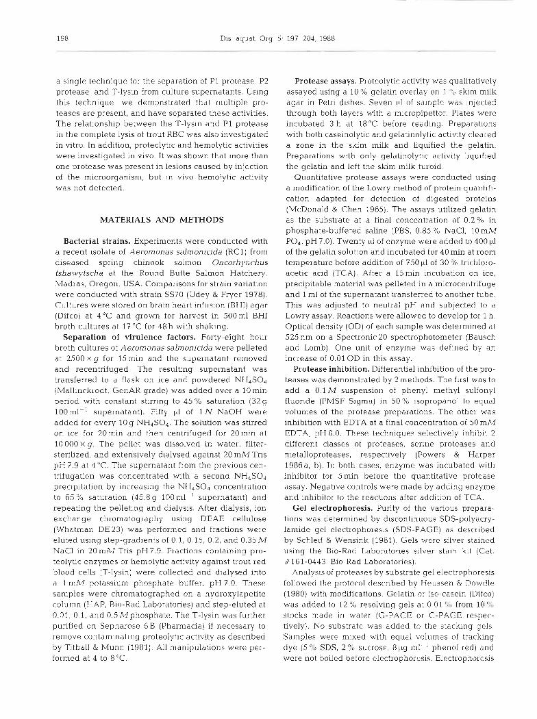
Fig. 2. Results of SDS-PAGE of Aerornonas salmonicida P1 and P2 proteases. Lane 1: Molecular weight standards, values in kilodaltons. Standards apply to Lanes 2 and 3. Lane2: Material eluted from DEAE at 0.15 M NaCl. Lane3: P1 from hydroxylapetite (HAP) Lanes4 and 5: P2 from HAP (4) elec-
trophoresed adjacent to a P1 preparation (5)
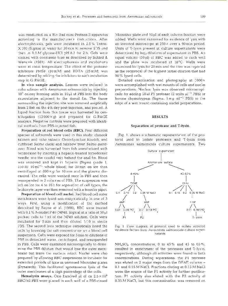
bands on SDS-PAGE gels (Fig.3) and were free of detectable contaminating proteolytic activity. No difference was observed in the column separations using A. salmonicida strain SS70.
Protease substrate specificity and sensitivity to inhibitors
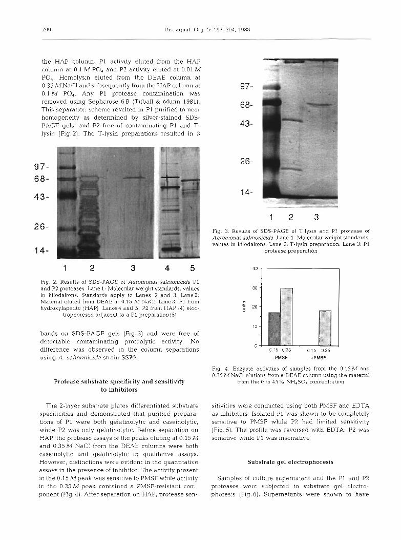
The 2-layer substrate plates differentiated substrate specificities and demonstrated that purified prepara- tions of P1 were both gelatinolytic and caseinolytic, while P2 was only gelatinolytic. Before separation on HAP, the protease assays of the peaks eluting at 0.15 M and 0.35 M NaCl from the DEAE columns were both caseinolytic and gelatinolytic in qualitative assays. However, distinctions were evident in the quantitative assays in the presence of inhibitor. The activity present in the 0.15 M p e a k was sensitive to PMSF while activity in the 0.35 M peak contained a PMSF-resistant com- ponent (Fig. 4 ) . After separation on HAP, protease sen-
Flg. 3. Results of SDS-PAGE of 7-lysin and P1 protease of Aeromonas salmonicida. Lane 1: Molecular weight standards, values in kilodaltons. Lane 2: T-lysin preparation. Lane 3: P1
protease preparation
Fig. 4. Enzyme activities of samples from the 0.15 M and 0.35 M NaCl elutions from a DEAE column using the material
from the 0 to 45 % NH,SO, concentration
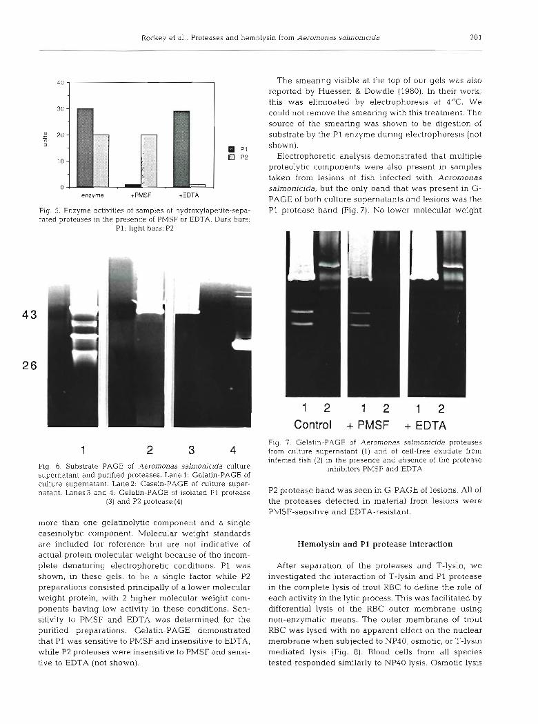
sitivities were conducted using both PMSF and EDTA as inhibitors. Isolated P1 was shown to be completely sensitive to PMSF while P2 had limited sensitivity (Fig. 5). The profile was reversed with EDTA; P2 was sensitive while P1 was insensitive.
Substrate gel electrophoresis
Samples of culture supernatant and the P1 and P2 proteases were subjected to substrate gel electro- phoresis (Fig. 6). Supernatants were shown to have
Rockey et al.: Proteases and hemolysin from Aeromonas salmonicida
enzyme +PMSF +EDTA
Fig. 5. Enzyme activities of samples of hydroxylapetite-sepa- rated proteases in the presence of PMSF or EDTA. Dark bars:
PI; light bars: P2
Fig. 6. Substrate PAGE of Aeromonas salmonicida culture supernatant and purified proteases. Lane 1: Gelatin-PAGE of culture supernatant. Lane2: Casein-PAGE of culture super- natant. Lanes3 and 4: Gelatin-PAGE of isolated P1 protease
(3) and P2 protease (4)
more than one gelatinolytic component and a single caseinolytic component. Molecular weight standards are included for reference but are not indicative of actual protein molecular weight because of the incom- plete denaturing electrophoretic conditions. P1 was shown, in these gels, to be a single factor while P2 preparations consisted principally of a lower molecular weight protein, with 2 higher molecular weight com- ponents having low activity in these conditions. Sen- sitivity to PMSF and EDTA was determined for the purified preparations. Gelatin-PAGE demonstrated that P1 was sensitive to PMSF and insensitive to EDTA, while P2 proteases were insensitive to PMSF and sensi- tive to EDTA (not shown).
The smearing visible at the top of our gels was also reported by Huessen & Dowdle (1980). In their work, this was eliminated by electrophoresis at 4OC. We could not remove the smearing with this treatment. The source of the smearing was shown to be digestion of substrate by the P1 enzyme during electrophoresis (not shown).
Electrophoretic analysis demonstrated that multiple proteolytic components were also present in samples taken from lesions of fish infected with Aerornonas salmonicida, but the only band that was present in G- PAGE of both culture supernatants and lesions was the P1 protease band (Fig. 7) . No lower molecular weight
1 2 1 2 1 2 Control + PMSF + EDTA
Fig. 7. Gelatin-PAGE of Aeromonas salrnonicida proteases from culture supernatant (1) and of cell-free exudate from infected fish (2) in the presence and absence of the protease
inhibitors PMSF and EDTA
P2 protease band was seen in G-PAGE of lesions. All of the proteases detected in material from lesions were PMSF-sensitive and EDTA-resistant.
Hemolysin and P1 protease interaction
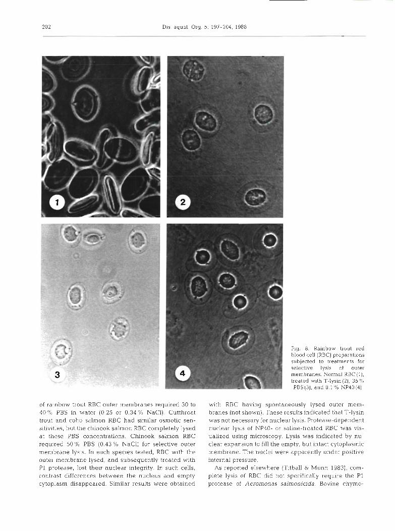
After separation of the proteases and T-lysin, we investigated the interaction of T-lysin and P1 protease in the complete lysis of trout RBC to define the role of each activity in the lytic process. This was facilitated by differential lysis of the RBC outer membrane using non-enzymatic means. The outer membrane of trout RBC was lysed with no apparent effect on the nuclear membrane when subjected to NP40, osmotic, or T-lysin mediated lysis (Fig. 8). Blood cells from all species tested responded similarly to NP40 lysis. Osmotic lysis
Dis. aquat. Org. 5: 197-204. 1988
of rainbow trout RBC outer membranes required 30 to 40 % PBS in water (0.25 or 0.34 % NaC1). Cutthroat trout and coho salmon RBC had similar osmotic sen- sitivities, but the chinook salmon RBC completely lysed at these PBS concentrations. Chinook salmon RBC required 50 % PBS (0.43 % NaC1) for selective outer membrane lysis. In each species tested, RBC with the outer membrane lysed, and subsequently treated wlth P1 protease, lost thelr nuclear integrity. In such cells, contrast differences between the nucleus and empty cytoplasm disappeared. Similar results were obtained
Fig. 8. Rainbow trout red blood cell (RBC) preparations subjected to treatments for S-4 lysis of outer membranes. Normal RBC ( l ) , treated with T-lysin (2), 3 9 PBS (3), and 0.1 '10 NP40 [4)
with RBC having spontaneously lysed outer mem- branes (not shown). These results indicated that T-lysin was not necessary for nuclear lysis. Protease-dependent nuclear lysis of NP40- or saline-treated RBC was vis- ualized using microscopy. Lysis was indicated by nu- clear expansion to fill the empty, but intact cytoplasmic membrane. The nuclei were apparently under positive internal pressure.
As reported elsewhere (Titball & Munn 1983), com- plete lysis of RBC did not specifically require the P1 protease of Aeromonas salmonicida. Bovine chyrno-
Rockey et al.: Proteases and hemolysin from Aeromonas salmonicida
trypsin also facilitated complete lysis of the nuclei. PMSF inhibited the lytic action of all nuclear lysis by P1 or chymotrypsin. PMSF had no effect on the lysis of outer membranes in the presence of P I , indicating that P1 did not contribute to outer membrane lysis. P2 preparations did not lyse RBC nuclei.
Hemolytic activity in infected tissues was compared to activity in culture supernatants. While T-lysin activ- ity in supernatants ranged from 128 to 1024 units, no hemolytic activity was observed in cell-free exudates from lesions.
DISCUSSION
There are several reports in the literature describing individual purifications and analyses of potential viru- lence factors from Aeromonas salmonicida. In this work a protocol for the separation of 3 previously identified factors from a single culture supernatant was developed. Using the described methods, 2 proteases, P1 and P2, and the T-lysin were partially purified and separated from one another for analysis. The P1 pro- tease was purified to near homogeneity and con- taminating activities were removed from both the P2 and T-lysin preparations.
The presence of P1 and P2 proteases in culture supernatants of Aeromonas salnlonicida was demon- strated by different elution profiles from hydroxylape- tite, substrate specificity, and sensitivity to inhibitors. These data support the results of Sheeran & Smith (1981) who documented 2 proteases in culture super- natants. However, by using G-PAGE and C-PAGE, w e have also demonstrated that culture supernatants of A. salmonicida contained more than 2 gelatinases. All except P1 protease are EDTA-sensitive and only P1 can digest both casein and gelatin. Investigators not detect- ing these different factors were apparently limited by the sensitivity of their assays or by substrates used in detection.
Separation of these factors also facilitated analysis of the in vitro relationship between P1 protease and T- lysin in the complete lysis of trout RBC. Selective lysis of outer membranes using NP40 and decreased salinity was also used in these experiments. We determined that T-lysin lyses the outer membranes of trout RBC and P1 protease lyses the nuclear membrane. The functions are independent of one another. This rela- tionship was first investigated by Titball & Munn (1983) and our results agree with the sequence of events they proposed. These results suggested that if enzymatic lysis of RBC is a component of pathology it is solely attributable to the T-lysin, because lysis of the outer membrane is sufficient to destroy the function of the cell.
Substrate gel electrophoresis and T-lysin activity
assays were also used to investigate activities associ- ated with these factors in coho salmon experimentally infected with Aeromonas salmonicida. Although fish infected experimentally had more than one gelatino- lytic component, P1 protease was the only protease detected in lesions which was produced by the bac- terium in culture supernatants. No low molecular weight P2 protease was detected in lesions. Gelatin- PAGE demonstrated that proteases found in lesions were PMSF-sensitive and EDTA-resistant. Non-P1 pro- teases in culture supernatants were EDTA-sensitive and PMSF-resistant. The non-P1 proteases detected in lesions were possibly host proteases activated by P1 protease or otherwise liberated during the course of infection, but their source was not determined. Pro- teases other than P1 produced by A. salmonicida may have been present in lesions, but they were not within our limits of detection. This information suggested that P1 protease is the major proteolytic factor produced by A. salmonicida that is responsible for tissue destruction and lesion formation. Other investigators using alter- nate methods have come to the same concl.usion (Sakai 1985, Fyfe et al. 1986). There is evidence, however, that A. salmonicida isolates laclung detectable caseinase activity (PI) can induce pathology and mortality in fish (Tajima et al. 1987). These infections do not involve lesion formation. Future research should address the potential role of non-P1 proteases in infection.
Attempts to detect hemolytic activity in cell-free exudate from lesions were unsuccessful. This sug- gested that released T-lysin was either not present, or was incapable of RBC lysis in vivo. A recent report by Fyfe et al. (1988) indirectly associated T-lysin with specific tissue damage after injection with P1 protease into Atlantic salmon. They did not, however, associate the T-lysin with lysis of RBC. Their work supports our conclusion that the major target tissue for T-lysin in vivo is not the erythrocyte.
Acknowledgements. Principal funding for this research was received from USDA Science and Education grant no. 85- CRSR-2-2578. We thank M. Beer for technical assistance, S. L. Kaattari for critical review of the manuscript, and T. Curry and C. Pelroy for typing the manuscript. Oregon Agriculture Experiment Station Technical Paper No. 8360.
LITERATURE CITED
Bayne, C. J., Loker, E. S., Yui, M. A. (1986). Interactions between the plasma proteins of Blomphalaria glabrata (Gastropoda) and the sporocyst tegument of Schistosoma mansoni (Trematoda). Parasitology 92: 653-664
Dahle. H. K. (1971). The purification and some properties of two Aeromonas proteinases. Acta Pathol. Microbiol. Scand. Sect. B 79: 726-738
Ellis, A. E., Hastings, T. S., Munro, A. L. S. (1981). The role of

204 Dis. aquat Org. 5 - 197-204, 1988
Aeromonas salmonicida extracellular products in the pathology of furunculosis. J Fish Dis 4 : 41-51
Fuller, D. W , Pilcher, K S., Fryer, J . L. (1977). A leukocytolytic factor isolated from cultures of Aeromonas salmonicida. J. Fish. Res. Bd Can. 34: 1118-1125
Fyfe, L., Finley, A., Coleman, G. , Munro, A. L. S (1986) A study of the pathological effect of isolated Aeromonas salmonicida extracellular protcase on Atlantic salmon, Salmo salar L. J. Fish Dis. 9. 403-409
Fyfe, L., Coleman, G., Munro, A. L. S. (1987). Identification of major common extracellular proteins secreted by Aeromonas salmonicida strains isolated from diseased fish. Appl. environ. Microbiol. 53: 722-726
Fyfe, L., Coleman, G., Munro, A. L. S. (1988). The comblned effect of isolated Aeromonas salmonicida protease and hemolysin on Atlantic salmon, Salrno salar L , compared with that of a total extracellular product. J Fish Dis 11. 101-104
Hastings, T. S . , Ellis, A. E. (1385). Diffcrenccs in the produc. tion of haemolytic and proteolytic activities by vanous isolates of Aeromonas salmonicida. In: Ellis, A. E. (ed.) Fish and shellfish pathology. Academic Press, London, p. 69-78
Heussen, C , Dowdle, E. B. (1980). Electrophoretic analysis of plasminogen activators in polyacrylamide gels contaming sodium dodecyl sulfate and copolymerized substrates. Analyt. Biochem. 102: 196-202
McCarthy, D. H., Roberts, R. J. (1980). Furunculosis of fish - the present state of our knowledge. Adv. aquat. Microblol. 2: 283-34 1
MacDonald, C. E., Chen, L. L (1965). The Lowry modification of the Folin reagent for determination of proteinase activity Analyt. Biochem. 10: 175-177
Mellergaard, S. (1983). Purification and characterization of a new proteolytic enzyme produced by Aeromonas sal- monicida. J. appl. Bacteriol. 54: 289-294
Michel, C. (1985). Failure of anti-furunculosis vaccination of rainbow trout (Salmo gairdnerii Richardson) using extra- cellular products of Aeromonas salmonicida as an immunogen. Fish Pathol. 20: 445-451
Munn, C. B., Ishiguro, E. E., Kay, W. W., Trust, T J. (1982). Role of surface components in serum resistance of virulent Aeromonas salmonicida. Infect. Immun. 36: 1069-1075
Powers, J. C., Harper, J. W (1986a). Inhibitors of serine proteases. In: Barrett, A J., Salvesen, G. (cds). Proteinase inhibitors. Elsevier Science Publishers, Amsterdam., p. 55-152
Powers, J. C.. Harper, J. W. (1986b). Inhibitors of metallo- proteases. In: Barrett, A J . , Salvesen, G (eds). Proteinase inhibitors. Elsevier Science Publishers, Amsterdam, p. 219-298
Sakai, D. K. (1985). Loss of virulence in a protease-deficient mutant of Aerornonas salrnonlcida. Infect. Immun. 48: 146-152
Schleif, R. F., Wensink, P. C. (1981). Practical methods in molecular biology. Springer-Verlag, New York
Sheeran, B., Smlth, P. R (1981). A second extracellular pro- teolytic activity associated with the fish pathogen Aeromonas salrnonicida. FEMS Microbiol. Lett. 11. 73-76
Shieh, H. S., Maclean, J. R . (1975). Purificat~on and properties of an extracellular protease of Aeromonas salmonicida, the causative agent of furunculosis. Int. J. Biochem. 6: 653-656
Tajima, K . , Takahashi, T., Ezura, Y . , Kimura, T (1983). Studies on the virulent factors produced by Aeromonas sal- monicida, a causative agent of furunculosis in salmonidae 1. Purification of the extracellular protease of Aeromonas salmonicida Ar-4 (EFDL). Bull. Fac. Fish, Hokkaido Univ. 34: 104-110
Tajirna, K., Ezura, Y., Kimura, T. (1987). Pathogenicity of a non-protease secreting strain of Aeromonas salrnonicida Bull. Fac. Fish., Hokkaido Univ. 38: 139-150
Titball, R. W., Munn, C. B. (1981). Evidence for two hemolytic activities from Aerornonas salrnonicida. FEMS Microbiol. Lett 12 : 27-30
Titball, R. C., ~Munn, C. B. (1983). Partial purification and properties of a hemolytic activity (T-Lysin) from Aerornonas salmonicida. FEMS Microbiol. Lett. 20. 207-210
Titball, R. C., Munn, C. B. (1985). Interrelationships of extracellular products from Aeromonas salmonicida. In. Ellis, A. E. (ed.) Fish and shellfish pathology. Academic Press. London, p. 61-68
Udey, L. R., Fryer, J. L. (1978). Immunization of fish with bacterins of Aerornonas salmorucida. Mar. Fish. Rev. 40. 12-17
Responsible Subject Editor: Dr T Evelyn; accepted for printing on September 6, 1988